Identification and Antibiotic-Susceptibility Profiling of
Infectious Bacterial Agents: A Review of Current and
Future Trends
Gaetano Maugeri, Iana Lychko, Rita Sobral, and Ana C. A. Roque*
Antimicrobial resistance is one of the most worrying threats to humankind with extremely high healthcare costs associated. The current technologies used in clinical microbiology to identify the bacterial agent and profile antimicrobial susceptibility are time-consuming and frequently expensive. As a result, physicians prescribe empirical antimicrobial therapies. This scenario is often the cause of therapeutic failures, causing higher mortality rates and healthcare costs, as well as the emergence and spread of antibiotic resistant bacteria. As such, new technologies for rapid identification of the pathogen and antimicrobial susceptibility testing are needed. This review summarizes the current technologies, and the promising emerging and future alternatives for the identification and profiling of antimicrobial resistance bacterial agents, which are expected to revolutionize the field of clinical diagnostics.
1. Introduction
By discovering penicillin in 1928, Sir Alexander Fleming triggered the beginning of the modern era of antibiotics, which revolutionized medicine and society, saved lives, and increased the life expectancy to what we know today. The remarkable effectiveness of antibiotics led to the euphoria mistaken belief that all infectious diseases could be successfully controlled with antibiotics. However, during the past few decades, the imprudent and excessive use (underuse, overuse, and misuse) of
antibiotics regrettably led to the rapid emergence and propagation of bacterial strains resistant to virtually all therapeuti-cally useful antibiotics.[1] The increasing
frequency of infections by antimicrobial-resistant bacteria is due to their capacity to recurrently develop new mechanisms of resistance. The lack of alternative treat-ments results in longer hospital stays, delayed recovery, long-term disability, and an increase in public healthcare costs. In the USA, the estimated healthcare cost associated to antimicrobial resistance (AMR) was $55 billion per year in 2013, and 2 million people were sick every year due to antibiotic-resistant infections, with over 23 000 deaths as a result.[2]In Europe, the 2009 report from European Centre for Disease Prevention and control (ECDC) and European Medicines Agency (EMEA)[3] estimated overall
societal costs over 1.5 billionsper year, with over 900 millions
in hospital costs. In the EU, about 25 000 patients died due to multidrug-resistant (MDR) bacteria infections.[3,4]It should be noted that the emergence and spread of AMR bacteria are prevalent in both healthcare and community settings, typically known as healthcare-associated infections and community-acquired infections.
The World Health Organization (WHO) recently published a priority list of antibiotic-resistant pathogens. Gram-negative carbapenem-resistant Acinetobacter baumannii, carbapenem-resistant Pseudomonas aeruginosa and carbape-nem-resistant and third-generation cephalosporin-resistant Enterobacteriaceae are at the top of this list, classified as critical priority agents. In the high-priority list, gram-positive bacteria for which there are treatment options likely to be successful, were included, namely the methicillin-resistant, vancomycin-intermediate and -resistant Staphylococcus au-reus and the vancomycin-resistant Enterococcus faecium.[5] The list does not includeMycobacterium tuberculosis, as it is a globally established priority, urgently needing innovative treatments, and already targeted by several dedicated programs. The WHO[6] also suggested that global research should focus on the development of new diagnostic and therapeutic tools.[7]
The current review aims at presenting the current, emerging, and future technologies implemented or in development, which target the early identification of the pathogenic agent as well as a fast antibiotic susceptibility G. Maugeri, I. Lychko, Dr. A. C. A. Roque
UCIBIO, Departamento de Química Faculdade de Ci^encias e Tecnologia Universidade NOVA de Lisboa 2819-516 Caparica, Portugal E-mail: [email protected] Prof. R. Sobral
UCIBIO, Departamento de Ci^encias da Vida Faculdade de Ci^encias e Tecnologia Universidade NOVA de Lisboa 2819-516 Caparica, Portugal
The ORCID identification number(s) for the author(s) of this article can be found under https://doi.org/10.1002/biot.201700750.
© 2018 The Authors.Biotechnology JournalPublished by Wiley-VCH Verlag GmbH & Co. KGaA, Weinheim. This is an open access article under the terms of the Creative Commons Attribution License, which permits use, distribution and reproduction in any medium, provided the original work is properly cited.
DOI: 10.1002/biot.201700750
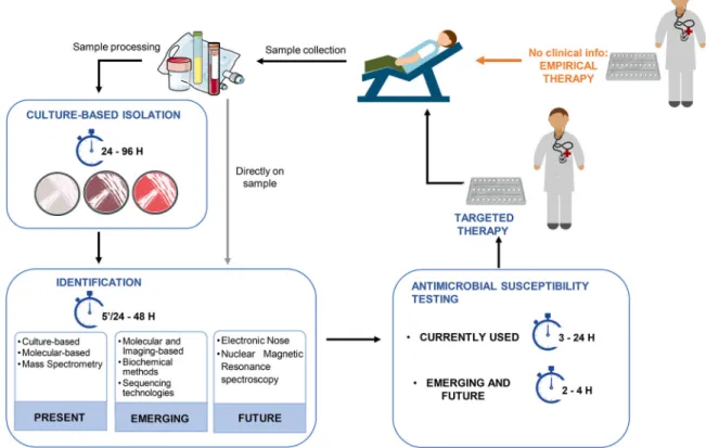
profiling. An overview of the different methodologies is summarized in Figure 1.
2. Current Technologies for Bacterial
Identification and Antibiotic Profiling
A clinical microbiologist has usually two main goals when processing clinical samples. One goal is to isolate and identify the pathogen causing the infection. A second goal is to evaluate its antibiotic susceptibility profile, providing useful information to prescribe the most efficient antibiotic treatment. The typical workflow currently in place for pathogen identification and antimicrobial susceptibility testing (AST) is presented inFigure 2, and the methodologies used are summarized inTable 1.
The isolation of pathogens from clinical samples still occurs through culture methods, using agar-based media (nutritive, differential, and/or selective). Some clinical laboratories use chromogenic agar-media harboring chromo-genic or fluorogenic substrates that are hydrolyzed in the
presence of specific enzymes. Several tests are then
performed to address genus identification, namely micros-copy cell staining, colony morphology, and rapid biochemical tests. To perform identification at the species level, the more common methods are phenotypically based, such as manual (e.g., Api bioMerieux) and automated biochemical tests, which exploit the differences in protein expression within genus (or also between genera), providing a characteristic protein expression fingerprint with a relatively high degree of certainty.[8] For example, the OmniLog ID system (Biolog) is a rapid method for the phenotypic identification of bacteria and fungi, through their ability to oxidize different carbon sources. Here, each well of the card contains one of 94 different carbon compounds and a tetrazolium-redox dye, used as aflag to indicate if the microorganism tested has or not utilized the carbon compound, providing a “metabolic
fingerprint” of the microorganism.[9] Although useful and
easy to operate, agar-based media and biochemical tests are not completely specific and occasionally fail or provide presumptive identification (percentage of possibilities). Therefore, further confirmation of species identity is often required. Different approaches, not culture-based, either current and emergent can be used, some of these are able to provide both identification and antimicrobial-susceptibility data simultaneously.
Despite the introduction of new time-saving technologies to obtain antimicrobial-susceptibility data, the current (classic) technologies are still used in many hospitals. These include culture-based, molecular-based and, more recently, spectrom-etry-based approaches or molecular and microscopy-based approaches. The first rely on the detection of phenotypic resistance by determining bacterial growth in the presence of antibiotics. These can be divided in manually performed tests (agar dilution, disk diffusion, gradient test, and broth microdilution) and automated systems. All these technologies provide qualitative data (e.g., susceptible, intermediate, resistant) for the strain tested and, except for the disk diffusion, also provide the quantitative values of the MIC (minimum inhibitory concentration), defined as the lowest
antibiotic concentration that inhibits the visible growth of an organism after overnight incubation.[10] The interpretative standards for these classifications are reviewed and updated annually by several organizations such as the Clinical and Laboratory Standards Institute (CLSI)[11]in the USA and the
Gaetano Maugeriis a microbiologist from Università degli studi di Catania in Italy. His research activities have been focusing in clinical microbiology and antibiotic-resistance in gram-negative bacteria, especially from healthcare associated infections. Currently, he is working in Roque's lab on the development of an electronic nose for the identification and resistance profiling of bacterial infections in clinical settings.
Rita Sobralholds an M.S. in biotechnology and Ph.D. in biological sciences. Since 2015, she is leading the Laboratory of Molecular Microbiology of Bacterial Pathogens at UCIBIO@REQUIMTE, and is an Assistant Professor at the Life Sciences Department
(Faculdade de Ci^encias e
Tecnologia, Universidade Nova de Lisboa). Her work was twice awarded (2011 and 2015) with a young investigator research grant by the European Society of Clinical Microbiology and Infectious diseases. Her research is focused on the molecular mechanisms of antibiotic resistance inStaphylococcus aureusand on the complex role of the cell wall on interspecies and host interaction.
European Committee on Antimicrobial Susceptibility Testing (EUCAST).[12]
2.1. Culture-Based Techniques
The dilution assay can be done in agar (agar dilution) or broth medium (macro- or micro-dilution). The agar dilution assay is performed in several Petri dishes of Mueller-Hinton agar (MHA) supplemented with the appropriate dilutions of antibiotic, each plate containing a different concentration. Antibiotic-free plates are used as control. Despite its laborious set up and the short shelf life of the plates, this method has the advantage to simultaneously test up to 36 different inocula in the same plate.[10,13,14] The micro-dilution technique has been miniatur-ized using 96-well plates to test about 12 different drugs using a wide range of eight twofold serial dilutions in a tray.[15]After overnight incubation, development of turbidity or sediment in the wells indicate growth of the organism and the MIC values can be evaluated following the CLSI or EUCAST breakpoints.[4] In the disk diffusion assay, the surface of the MHA plate is inoculated with a standardized inoculum of the test microor-ganism. Commercial filter paper disks impregnated with antibiotics at predetermined concentrations are placed on the
agar surface and the antibiotic concentrations are directly reversed to the distance from the disk.[15,16]The disk diffusion assay provides only qualitative results (e.g., susceptible, intermediate, resistant), obtained by measuring the diameter (mm) of bacterial growth inhibition around the disk. Such data can be insufficient as it does not provide the physician with the MIC value, that may be needed for an efficient prescription.[10] The gradient test meets the advantages of simple handling and the simultaneous use of several drugs as in the disk diffusion assay, while allowing MIC value determination as in dilution assays. Different commercial versions are available as
Figure 2. Typical procedures currently in place in clinical settings to provide identification of the pathogen agent and the profiling of antimicrobial susceptibility.
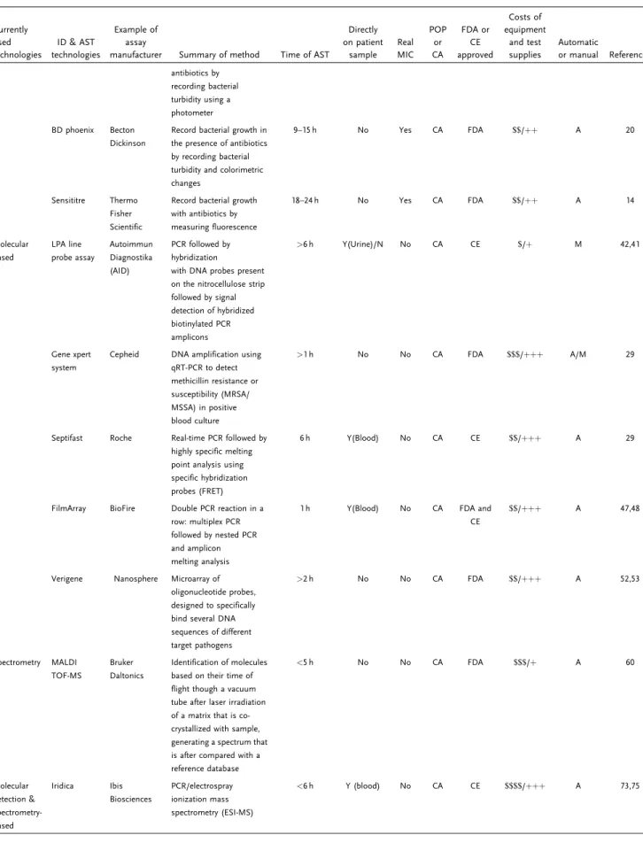
Table 1. Selected examples of current technologies for pathogen identification (ID) and antimicrobial susceptibility testing (AST).
Currently used technologies
ID & AST technologies
Example of assay
manufacturer Summary of method Time of AST
Directly on patient
sample Real MIC
POP or CA
FDA or CE approved
Costs of equipment
and test supplies
Automatic
or manual References
Culture-based
Agar diluition assay
Oxoid Bacteria inoculated on
agar plates with different concentrations of antibiotics
16–24 h No Yes/no CA FDA þ M 14
Disk diffusion
Oxoid Bacteria inoculated on
agar plates with single antibiotic disks
16–24 h No Yes/no CA FDA þ M 14,15
Gradient test bioMerieux Bacteria inoculated on
agar plates with graded antibiotic concentration strips
16–24 h No Yes CA FDA þ M 14,15
Broth dilution assay
Oxoid Bacteria inoculated in
liquid media with different antibiotics to monitor growth
12–24 h No Yes CA FDA þ M 14
MicroScan WalkAway
Beckman Coulter
Measure bacterial growth in the presence of antibiotics by recording bacterial turbidity using a photometer
4.5–18 h No Yes CA FDA $$/þþ A 4,146
Vitek
bioMerieux
bioMerieux Measure bacterial growth
in the presence of
6–11 h No Yes CA FDA $$/þþ A 20
Table 1. (Continued)
Currently used technologies
ID & AST technologies
Example of assay
manufacturer Summary of method Time of AST
Directly on patient
sample Real MIC
POP or CA
FDA or CE approved
Costs of equipment
and test supplies
Automatic
or manual References
antibiotics by recording bacterial turbidity using a photometer
BD phoenix Becton
Dickinson
Record bacterial growth in the presence of antibiotics by recording bacterial turbidity and colorimetric changes
9–15 h No Yes CA FDA $$/þþ A 20
Sensititre Thermo
Fisher Scientific
Record bacterial growth with antibiotics by measuring fluorescence
18–24 h No Yes CA FDA $$/þþ A 14
Molecular based
LPA line probe assay
Autoimmun Diagnostika (AID)
PCR followed by hybridization
with DNA probes present on the nitrocellulose strip followed by signal detection of hybridized biotinylated PCR amplicons
>6 h Y(Urine)/N No CA CE $/þ M 42,41
Gene xpert system
Cepheid DNA amplification using
qRT-PCR to detect methicillin resistance or susceptibility (MRSA/ MSSA) in positive blood culture
>1 h No No CA FDA $$$/þþþ A/M 29
Septifast Roche Real-time PCR followed by
highly specific melting point analysis using specific hybridization probes (FRET)
6 h Y(Blood) No CA CE $$/þþþ A 29
FilmArray BioFire Double PCR reaction in a
row: multiplex PCR followed by nested PCR and amplicon melting analysis
1 h Y(Blood) No CA FDA and
CE
$$/þþþ A 47,48
Verigene Nanosphere Microarray of
oligonucleotide probes, designed to specifically bind several DNA sequences of different target pathogens
>2 h No No CA FDA $$/þþþ A 52,53
Spectrometry MALDI
TOF-MS
Bruker Daltonics
Identification of molecules based on their time of flight though a vacuum tube after laser irradiation of a matrix that is co-crystallized with sample, generating a spectrum that is after compared with a reference database
<5 h No No CA FDA $$$/þ A 60
Molecular detection & spectrometry-based
Iridica Ibis
Biosciences
PCR/electrospray ionization mass spectrometry (ESI-MS)
drugs have to be tested.[14]The fractional inhibitory concentra-tion index (FICI) usually used to investigate if the antimicrobial interaction between two drugs are additive, synergistic, or antagonistic, can also be determined.[17,18]
Automated methodologies allow both identification and AST determination. These systems involve the use of cassettes (also called panels or cards) with a series of wells, each containing an individual substrate for pathogen identification. The metabolic activity of the strain such as acidification, alkalinization, and enzymatic hydrolysis of different substrates is analyzed.[19]The AST is based on the automation of the broth microdilution assay through sensitive optical detection systems, that measure bacterial growth in the presence of antibiotics, within 24 h post incubation.[20] The number and concentration of antibiotics tested is limited and their sensitivity is low, as a high number of viable cells is required to measure bacterial growth and turbidity changes. Other relevant weaknesses include the impossibility to process directly patient samples,[20] the absolute need of a pure culture of the pathogen, and the long processing time (several hours for identification and up to 18 h for AST).[21] The major advantages are their high degree of
standardization according to the international guidelines criteria of CLSI and/or EUCAST,[22] and their capacity to
manage high workloads. In fact, automated systems are commonly used in clinical microbiology laboratories which process a large number of daily tests.[22,23]
2.2. Molecular-Based Methodologies
Most of the molecular-based technologies rely on conventional polymerase chain reaction (PCR) or quantitative real-time PCR (qRT-PCR), to amplify specific sequences of nucleic acids, allowing simultaneous pathogen identification and AST.[24] However, it requires a DNA extraction step from isolated strains, a high number of cells to obtain sufficient DNA and previous knowledge on the sequences to amplify.[24]For some technolo-gies, the possibility to identify the pathogen directly from the patient sample, namely whole-blood, serum, blood culture, or urine, clearly represents an advantage.[25–27]In fact, PCR-based
techniques can be separated into culture-independent PCR assays, performed directly on raw samples, and culture-enriched assays, which instead require a previous growing step of the raw sample in an enrichment media.[28]
In the case of suspicion of bloodstream infections (BSI) or sepsis, molecular-based tests are essential for successful therapy choice, since they provide bacterial identification and/or detection of resistance traits, directly on the patient’s sample. For further details on rapid molecular diagnostics for BSI please read recent works.[25,29,30]We shortly describe the SeptiFast test (Roche), a culture-independent amplification assay, as it is the most studied and validated assay for the diagnosis of BSI. It performs bacterial identification directly on EDTA blood-whole samples (so it can be used in suspected bacteremia), and can detect 25 clinically relevant bacteria and fungi in about 6 h,[31–33]
with a sensitivity of 3–30 CFU mL 1.[34] The assay uses dual FRET probes with two differentfluorophores allowing quantifi -cation besides identification.[35]The technology using the FRET probe assay is restricted to the LightCycler instrument (Roche),
and to the High-Resolution Melting (HRM) assay, requiring highly advanced qRT-PCR instruments.[36] Several studies evaluating the SeptiFast technology, reported sensitivity values in the range of 60–95% and specificity values of 74–99%, depending on the target pathogen. However, it is labor-intensive, needs professional expertise, and has a high associated cost (about 200sper test). Moreover, except for methicillin-resistant S. aureus(MRSA), it provides no information on antimicrobial susceptibility.[29] SeptiFast can be used as a complement for traditional culture-based methods, in antibiotic-treated patients, as recently shown.[37]
Another culture-independent assay using qRT-PCR, approved by the US FDA, is the Gene Xpert system (Cepheid). It can detect MRSA and methicillin susceptibleStaphylococcus aureus(MSSA) based on sequences of spa, SCCmec, and mecA genes with a turnaround time of 1 h from positive blood cultures.[29] The reported sensitivity forS. aureusdetection is 100%, and 99.4% specificity for MRSA detection.[38–40] However,
methicillin-resistant coagulase-negativeStaphylococcusspp (CoNS) can lead to false positives in theSCCmecgene test.[29]
PCR-based technologies can also be used to identify ESβL and carbapenemase resistance genes in the Enterobacteriaceae family. The commercially available Line Probe Assay (LPA), uses conventional PCR followed by reverse hybridization of PCR amplicons, and simultaneously detects the presence of these two characteristic resistance genes in gram-negative bacteria.[41]A commercial example is the AID carbapenemase LPA (Auto-immun Diagnostika), CE-cleared, evaluated both in culture-independent assays (urine samples) and in DNA samples extracted from enriched bacterial cultures, for the detection of a wide list of carbapenem-resistant genes. The test has 100% sensitivity and 100% specificity when used in clinical isolates.[42] Although these tests detect the determinants of resistance, they have an important limitation: the presence of these resistance genes is not always correlated with phenotypic resistance.[24] This occurs in gram-negative bacteria, for which the resistance gene may be present, but at very low expression levels. Changes in the expression level occur through the insertion of mobile insertion sequences, which can act as better promoter regions, enhancing expression.[43]In gram-positive bacteria, the correla-tion between the genotype and the resistance phenotype is more reliable.[24]
Finally, qRT-PCR can be used for AST. It quantifies DNA copies and can detect bacterial growth in the presence of different antibiotic concentrations, being used to differentiate susceptible from resistant strains.[24]An example is the recently used test for antimicrobial susceptibility of clinical isolates ofA. baumanniito imipenem, ciprofloxacin, and colistin in about 6 h (from isolated colonies). The bacterial growth was indirectly established through the detection of a highly conserved region of
ompAgene.[44]Failure to limit the analysis to viable cells is the
main limitation, as the presence of non-viable bacteria can overestimate the number of cells present in the sample and lead to increased false positive rates.[45]
low volume of blood used in the amplification step and consequently, the low nucleic acid contents, may not be enough to reach clinically relevant sensitivity.[46] To provide timelier results, the FDA- and CE-cleared FilmArray (BioFire) Blood Culture Identification (BCID) panel is used directly on positive blood culture bottles with a turnaround time of about 1 h. A list of 24 etiologic agents of sepsis is screened, including 8 gram-positive and 11 gram-negative bacteria,five yeast (Candidaspp.)
and three resistance genes, mecA, vanA/B, and KPC.[47] The BCID panel also detects contaminant bacteria. The assay consists of several automated steps,first cell lysis, nucleic acid purification, multiplex PCR, nested PCR andfinally amplicon melt analysis. In the two-stage PCR, a multiplex step provides nucleic acid amplification for the subsequent nested PCR reactions. These reactions occur in an array, each well containing specific internal primers for the resistance and species marker genes. The real-time detection of multiple gene targets is achieved using afluorescent double-stranded DNA-binding dye. The combinatorial result of the signals from the different wells gives the final result.[47,48] The overall sensitivity of the test ranges between 50% and 100%, while the overall specificity is reported in the range 77–100%.[49–51]
FilmArray has recently been compared to Verigene (Nano-sphere, USA). Although these two FDA-cleared assays rely on completely different technologies, they both perform multiplex identification of individual targets.[52,53]Verigene includes two panels for bacterial identification, BC-GN that detects eight gram-negative bacterial targets and six key resistance markers, and BC-GP that detects twelve gram-positive targets and three associated resistance markers (the same as FilmArray), but lacks a panel for yeast detection. Concerning detection of resistance genes, Verigene can identify four carbapenemase (VIM, IMP, OXA, and NDM) and an ESBL gene (CTX-M).[52,53]The hands-on time is comparable for both systems, with a turnaround time of 2.5 h for Verigene. The Verigenes’s cartridges consist of a glass slide (microarray) and the associated technology is able to identify and quantify nucleic acid sequences, without an initial PCR amplification step. Several capture oligonucleotides probes, designed for a specific DNA sequence of the target pathogen, are printed over the microarray slide. Mediator oligonucleotide probes, containing a polyA tail, bind specifically to a second DNA region of the target pathogen and then to a poly T probe associated to a gold nanoparticle.[53–55] Subsequently, silver particles are deposited around the gold nanoparticles and their localization is detected by light scattering, providing accurate detection of the target sequences captured on the array.[54]The BC-GP showed values of 92.6–100% and of 95.4–100% for sensitivity and specificity of identification, respectively, and 98.6–
100% and 94.3–100% for detection of resistance markers.[56]The BC-GN showed a sensitivity of 97.1% and a specificity of 99.5% for gram-negative bacteria.[57]
2.3. Spectrometry MALDI-TOF MS
MALDI-TOF MS is based on the rapid ionization of the bacteria/ yeast ribosomal proteins using a laser pulse, directly from cultured colonies or cell pellets from the clinical sample
(Figure 3A). The calculated mass of the ions is the specific
samplefingerprint of the bacterial/yeast species. This technique is nowadays widely used in clinical microbiology laboratories, particularly at University Hospitals.[58]
MALDI-TOF MS can be used for positive, gram-negative bacteria and yeast, and does not require a specific test, in contrast to biochemical differentiation methods. However, similarly to these, it requires fresh colonies (not more than 48 h), as peaks become more difficult to distinguish, probably due to ribosomal protein degradation. The cost of each test is low, 1sor
less per sample, but a typical MALDI-TOF MS system costs
s180 000–200 000, including analysis equipment, hardware,
relevant software, and integrated databases.[58]MALDI-TOF MS workflow is high, processing 16–384 samples in a typical plate. Each sample analysis takes about 5–7 min, so the results are usually available 12–24 h after receiving the sample. The amount of cells required is low (104–106CFU), and theoretically it can be performed using a single colony, obtained in few hours, from the culture of an infected sample.[58] Even if the identification at species level is possible it does not differentiate species with similar ribosomal protein sequences (Shigella spp. and Escher-ichia coli or Streptococcus pneumoniae and members of the
Streptococcus oralis/mitisgroup); in these cases, other assays such as classical biochemical tests, antigen detection or molecular methods, are required. For the analysis of yeast with strong cell wall, a short extraction procedure may be required to provide the ribosomal proteins available for analysis.[58]
Some studies describe MALDI-TOF MS as a possible alternative technology for AST,[58]by directly analyzing the enzymatic reaction at the molecular level (Figure 3B).[59]The carbapenemase activity in gram-negative bacteria was evaluated by incubation of the bacterial samples (carbapenemase-carrying or non-carrying strains) with Ertapenem (carbapenem) and subsequent analysis through MALDI-TOF MS. Different mass peaks were obtained at the beginning of incubation and after 2.5 h, the time needed to evaluate the hydrolysis ofβ-lactam rings by the carbapenemase-carrying strain. Remarkably, the bacterial strains producers of NDM-1 or IMP-1 enzymes, were detected in just 1 h, the time to complete β-lactam hydrolysis.[60] Other carbapenems were evaluated, for instance Meropenem showed 96.67% sensitivity and 97.87% specificity values forP. aeruginosaand Enterobacter-iaceae strains,[61]and Imipenem showed 100% sensitivity and specificity forA. baumanniistrains.[62]A similar approach was used to screen otherβ-lactam substrates, to evaluate the possibility to distinguish other β-lactamases such as AmpC or TEM-1.[63] However, as for PCR-based technologies, MALDI-TOF MS may not provide a direct correlation between the presence of the hydrolytic enzymes and the phenotype of resistance[24]since some mechanisms, such as alterations in porins or upregulation of efflux pumps, are not detected.[59]In gram-positive bacteria, MALDI-TOF MS can be used to discriminate betweenE. faecium vanB
positive and negative strains.[64]However, discrimination between
MSSA and MRSA strains is contradictory, as some authors described measurable differences in the spectra,[65,66]while others
commensalflora nor host proteins, expected in some pathologi-cal conditions. In fact, pathogens were correctly identified at the species-level directly from urine samples at rates of 91.8% using a specific fast protocol.[69]However, the best results are obtained
for high bacterial counts (>105CFU mL 1) and for
gram-negative bacteria. A reliable protein profile is obtained only for bacterial counts of at least 8104CFU mL 1that corresponds to the diagnostic threshold for most UTIs. Finally, this method can under evaluate some UTIs like cystitis, which emerge with lower bacterial counts and lead to false negative results in these cases.
2.4. Spectrometry Approaches Combined with Molecular Tools
The polymerase chain reaction/electrospray-mass spectrometry (PCR/ESI-MS) is a fairly recent technology that couples a molecular method to a spectrometry approach, overcoming weaknesses in the analysis of complex samples and performing culture-independent analysis. It was originally developed by Ibis technology for biodefense and public health safety purposes, enabling rapid detection and identification of pathogens in clinical and environmental samples.[70]Later, Abbott Molecular acquired Ibis technology[71]and designed a more robust model,
PLEXID[70]that was recently improved to IRIDICA, the newest PCR/ESI-MS system.[72] IRIDICA was evaluated in complex
samples, such as whole blood,[73]bronchoalveolar lavage (BAL)
[72] and in the diagnosis of endocarditis,[74] obtaining the CE
marked designation in 2015.
The IRIDICA BAC BSI assay, for the evaluation of BSI and sepsis, includes a pre-filled 16-well PCR strip, with 18 primer pairs that target broadly conserved bacterial and fungal sequences of pathogenic species and also specific antibiotic resistance markers like mecA, vanA, vanB, and blaKPC.[73]
Subsequently, the amplicons are submitted to ESI-MS. The system can detect over 780 bacterial and candida species using a proprietary database and software that compares the DNA sample sequence with a sequence library. The limit of detection ranges between 0.25–128 CFU mL 1, depending on the target species and the estimated time to result is 5 h and 55 min. The costs of the IRIDICA system and for each test are£268 000 and £174, respectively.[75]Four studies comparing the IRIDICA BAC
BSI, with blood culture as reference method, reported an estimated summary specificity of 0.84 and sensitivity of 0.81.[75]
3. Emerging Technologies for Bacterial
Identification and Antibiotic Profiling
Some recently emerging technologies rely on the measurement of several phenotypic features such as growth, morphology, viability and metabolism, using a wide range of different approaches (sometimes concerted) for fast identification and AST such as imaging-based, non-imaging-based, molecular-based, and biochemically-based (Table 2).
3.1. Imaging-Based Technologies
The image-based assays that can provide pathogen ID and AST, represent an impressive emerging technology in thefield of clinical microbiology. Among these tools, the oCelloScope (Philips BioCell) performs AST, by relying on real-time analysis using an automated optical detection system, which analyzes up to 96 combinations of samples. The digital time-lapse microscopy scanning through the fluid sample gen-erates a series of images. The optical resolution is comparable to a 200magnification in a standard light microscope. Two algorithms, based on either pixel histogram summation or contrast segmentation and extraction of surface area, deter-mine the bacterial growth kinetics, through image stack processing. This system was evaluated in several experiments, such as monoculture infection, with results obtained in 6 min forE. coliisolates and in 30 min for complex samples such as urine collected from pigs with catheter-associated UTIs.[65]It was also evaluated in positive blood cultures and the average time to obtain susceptibility degree values ranged from 1 to 4.2 h depending on the bacteria–antibiotic combination.[66]In
contrast to competitor systems, the oCelloScope does not analyze single cells, but populations, and has lower resolution imaging. However, it has the advantage to allow bacterial growth measurement, without the need to attach the bacterial cells to an inert surface, a step that is required by other tools.[20]
In other higher resolution tools, multichannel test cassettes are used for real-time observation with high-resolution cameras, allowing the direct observation and measurement of bacterial growth. An example of such technology is the multiplexed automated digital microscopy (MADM).[76] A commercially available device using this technology is the Accelerate Pheno
System (Accelerate Diagnostics) that can perform both identifi -cation and AST of bacteria and yeast and allows the diagnosis of mono and polymicrobial infections directly from blood-cultures, dismissing the overnight sub-culturing step. This technology uses two different approaches to achieve identification and AST, thefirst is obtained within 1.5 h and the latter within 6.5 h, and both occur inside theflow cells of a multichannel test cassette. The blood culture follows a series of automated processes
(Figure 3C), starting with gel electrofiltration, that separates impurities, such as lysed blood cells and debris, from bacterial or yeast cells; the second step involves cell immobilization via electrokinetic concentration, which enables the microscopy-based single-cell analysis to achieve identification. The process of identification is performed through hybridization in situ with specificfluorescent probes (FISH) for bacterial and yeast cells, and with universal probes to obtain quantification and resolve
Table 2. Selected examples of emerging technologies for pathogen identification (ID) and antimicrobial susceptibility testing (AST).
ID & AST technologies
Example of assay manufacturer or
technology
Assay
manufacturer Summary of method
Time of AST
Direct on patient sample
Real MIC
POP or CA
FDA or CE approved
Automatic
or manual References Molecular &
imaging-based
Accelerate pheno system
Accelerate Diagnostics
Multiplexed automated digital microscopy (MADM)
<6 h Yes (positive blood-culture)
and (BAL)
Yes CA FDA A 20,67,69,70,77, 79,81,83,87
Imaging-based
oCelloscope Philips BioCell Digital time-lapse microscopy scanning population of bacterial cells
1–4 h Yes (urine) (positive blood-culture)
Yes POP No A 65,66
Bacterial cytologial profiling
Fluorescence microscopy to analyze a multitude of parameters to discriminate resistant from susceptible strains
<2 h No Yes POP No A 84
Imaging-based & microfluidics
SCMA Single-cell morphological analysis (SCMA) performed in microfluidic agarose channels (MAC) system
<4 h No Yes POP No A 82,83
Non imaging-based & microfluidics (lab on chip)
Nanodroplets/ nanoliter arrays
Measurement of the metabolically active bacteria
<6 h Yes (urine) Yes POP No A 90,91
UtiMaxTM GeneFluidics Electrochemical measurement
of bacterial 16S rRNA
<4 h Yes (urine) Yes CA CE A 87–89
pH change Microfluidic pH sensor detect metabolic products by bacteria growth
<3 h No Yes na A 92
LifeScale Affinity Biosensor
Resonant mass measurement >3 h Yes (urine) Yes CA CE A 86
Non imaging-based
BacterioScan BacterioScan, Inc.
The laser light scattering (FLLS) determines the number and size of bacterial cells suspended in a solution
3–10 h Yes (urine) Yes CA No A 85,147
Molecular and biochemical-based
Next-generation sequencing and whole generation sequencing
Sequencing entire bacterial genomes or RNA
>10 No No CA na A 99–101,148
Smarticles Roche Diagnostics
Bacteriophages carrying luciferase gene- infect bacteria producing detectable light signals
ND ND ND POP No ND 93
KeyPath MSSA/ MRSA blood culture test
Microphage Detection of phage antigens as a surrogate for the presence of bacteria in the sample
<5 h Yes (positive blood-culture)
No CA FDA M 95
the possible polymicrobial sample. The identification drives automatically the choice of antibiotics to be used. The sample is subjected to a pre-growth step to normalize growth rates, and a universal nucleic acid stain is used to quantify the number of cells in the purified inoculum. Theflow cells arefilled with an appropriate volume of purified inoculum and subsequently injected with MHA to perform the AST analysis. The susceptible cells are killed or their growth is inhibited by the treatment. Subsequently, using algorithms and mathematical regressions, based on the response of isolates with known MICs for a given antimicrobial, the growth curves are converted into MIC values.[77]
A study of BSI by gram-negative bacteria, demonstrated its capacity to correctly identify 88.7% of all episodes from blood cultures, including 10 polymicrobial BSI. However, in seven of the polymicrobial samples, cultivated gram-positive organisms were not detected.[78]This device was evaluated to successfully
discriminate MRSA/MSSA, clindamycin resistance/susceptibil-ity and VSSA/hVISA/VISA respectively using cefoxitin, erythro-mycin, and vancomycin.[79] It was also able to evaluate the processes of induction and heteroresistance, observing 10 or more growing clones per test, changing the time of exposition and the drug concentration. In only 3 h, the microscopy analysis succeeded to discriminate MRSA from MSSA, with a time gain of 15 h in comparison to the microdilution method and of 44 h to differentiate between VSSA, hVISA, and VISA in comparison with the agar-dilution method. Also, the clindamycin-resistant and susceptible phenotypes of S. aureus were successfully discriminated.[79]This tool, used to diagnose BSI, was widely described for its important role in clinical microbiology, since each hour of delay causes 7.6% decrease in survival for septic shock patients, within thefirst 6 h.[80]
Since cases of ventilator-associated pneumonia (VAP) in patients mechanically ventilated in the intensive care unit (ICU) are currently becoming recurrent, the Accelerate system was evaluated as a rapid diagnosis assay, showing capacity to detect risk of VAP in recovered patients, before the clinical signs were visible. Through a microbiological surveillance program, BAL samples were analyzed, and the automated microscopy improved the antimicrobial stewardship. The quicker diagnosis allowed to initiate an adequate antibiotic therapy, decreasing the suboptimal or inadequate use of broad-spectrum therapy instead of guided de-escalation to specific therapy. Moreover, the time reduction for identification and AST was approximately 5 h, compared to 507 h for clinical
cultures, with 100% sensitivity and 97% specificity for high-risk pneumonia organisms.[81]
Another noteworthy microfluidic image-based technology is the single-cell morphological analysis (SCMA). It uses a microfluidic agarose channel, composed of a main inlet tube which is divided in six microfluidic channels. The antibiotic diffuses to the agar-trapped cells and the bacterial growth is then monitored hourly using a microscope associated to a true-color CCD camera. Subsequently, the images are transformed into digital data and are processed using algorithms to achieve the antibiotic MIC value. This tool was evaluated for three reference strains,E. coliATCC 25922,S. aureusATCC 29213, andP. aeruginosaATCC 27853 using antibiotics like amikacin, norfloxacin, tetracycline, and gentamicin. The MIC values were
assessed in accordance with the CLSI results, obtained through the microdilution assay reference method.[82] Another study used clinical samples and a different version of the MAC chip, now in a 96-well format, to achieve high-throughput testing.
The 189 clinical samples included extended-spectrum
β-lactamase–positiveE. coli and Klebsiella pneumoniae, imipe-nem-resistant P. aeruginosa, MRSA and vancomycin-resistant enterococci (VRE); the AST results were provided in less than 4 h with 91.5% categorical agreement, 6.5% minor, 2.6% major and 1.5% very major discrepancies. Although it rapidly achieved AST results, the main weakness of this tool remains the lack of an integrated system of identification.[83]
The Bacterial cytological profiling (BCP) is another image-based tool, that measures several different cellular parameters: changes in cell length, width, permeability, chromosome number, compactness and shape, using fluorescent dyes and a microscope. The parameters are determined by the effects of the antibiotic treatment on the cells. Compared to other tools that rely only on cell lysis information, the BCP assay is able to evaluate the single effects caused by each antibiotic, allowing to estimate a fit combination for a synergic treatment.[84] This
approach can be useful in cases of infections caused by multidrug-resistant pathogens, providing rapid identification of an effective therapy.
3.2. Non-Imaging-Based Technologies
Several relevant tools using non-imaging approaches were developed for AST, which typically detect a specific physical property. Among these, the BacterioScan is an electro-optical based technology using laser light scattering (FLLS). It measures the angular variation in the intensity of light scattered by a laser beam that passes through a bacterial sample. The angular variation depends on the number and size of bacterial cells in suspension, allowing to detect very low values of optical density (OD) and to measure bacterial growth. Multiple measurements (every 3 min) are done and the signals, captured by a CMOS 2-dimensional camera sensor, are processed and a density value is generated. The instrument performs reliably down to a
minimum density of 103CFU mL 1, which is commonly
considered a diagnostic threshold for bacteriuria. For this reason, it was applied to urine analysis, although it cannot recognize a polymicrobial infection. The BacterioScan model 216 tabletop can process until 16 samples simultaneously[85]and was evaluated using strains ofS. aureus,E. coli, andP. aeruginosa
and different antimicrobials; a close agreement to the conventional tests used in clinical microbiology laboratories was demonstrated. The overall time of the assay, for 95% of the organisms tested, was approximately 10 h. The minimum time of inhibition was registered for S. aureus (32 min) and the maximum time forP. aeruginosa(16 h).[85]
3.3. Biochemical Methods
Several biosensors can identify and detect bacterial growth through biochemical flags from cells, such as quantitative changes in 16S rRNA,[87–89]NADH [90,91]and FADH, changes
in pH,[92] or emission of light caused by gene insertion in DNA.[93]
Genefluidics developed an electrochemical-based tool that performs both identification and AST. It relies on sandwich hybridization of specific capture and detector probes of bacterial 16S rRNA. The capture and detection at the sensor surface is followed by electrochemical signal amplification with an enzyme tag, which transduces the molecular hybridization events into quantitative electrical signals. It was applied directly on viable pathogens in urine samples.[87,88]The biosensor was evaluated on reference strains of E. coli, P. aeruginosa, and
Enterococcus faecalis at 20-min interval. The signal increase obtained with the biosensor was proportional to the cell number obtained through quantitative plating in MHA.[89]This tool was also evaluated directly in urine samples by measuring the levels of 16S rRNA within 3.5 h, in the presence and absence of antibiotics routinely used for UTIs. The overall agreement with the standard AST was 94%.[89]
Another interesting approach uses the “stochastic confi ne-ment” of individual bacteria in nanoliter droplets (nano-droplets), using a microfluidic system. Bacteria are placed into nanoliter plugs, which accelerate the detection of molecules,
flags of active cellular metabolism.[90] The changes in these
flags are usually registered as changes in the fluorescence intensity, which are then correlated with the efficacy of the antibiotic treatment.[90]The stationary nanoliter droplet array
(SNDA)–AST system, a tool based on the nanodroplet
modification, combines the Resazurin assay on a nanoliter well array containing lyophilized antibiotics. Briefly, Resazurin is reduced by electron acceptors of cellular metabolic activity such as NADH and FADH, forming Resofurin that emits
fluorescence. Since only viable cells produce NADH and FADH, fluorescence emission can be correlated with the efficacy of the antibiotic treatment.[90]This method performs AST directly on urine samples, using a fast-multi-step protocol. The array consists of two rows of 8-nL wells connected by a delivery channel through which the clinical sample is injected, adding 10% Resazurin and FC-40 oil, to isolate the sample inside the wells. The platform consists of 200 wells of standard dimension to allow the trapping of an average 4CFU/well, which correspond to 5105CFU mL 1, to improve the clinical translatability and interpretability, using standard breakpoints established by EUCAST and CLSI. As the clinical sampleflows inside the antibiotic-containing wells, the changes infl uores-cence are proportional to the number of bacteria in the well, allowing to measure the efficacy of the antibiotic treatment in about 6 h.[91]
A new rapid bacterial identification and AST uses bacter-iophages to recognize, bind and invade specific bacteria. Their straight-forward manipulation, production and low cost allowed their use in phage-therapy[94]and more recently as rapid tools for identification/AST tests. Recombinant bacteriophages carrying the luciferase gene were developed using synthetic biology techniques by GeneWEAVE and designated Smarticles. Once they infect the specific bacterial-host, luciferase gene expression
is triggered. The signal (light) produced by luciferase-associated enzymatic reactions is quantified and correlated to the number of cells in a sample.[20,93]Other phage-based tests are already available for diagnosis/treatment, such as the FDA-cleared KeyPath MRSA/MSSA Blood Culture Test, which detects the presence ofS. aureusdirectly on blood cultures. The test also discriminates between MRSA and MSSA using Cefoxitin in the assay; MRSA grow in the presence of cefoxitin, resulting in signal emission, in contrast to MSSA. A recent evaluation study reported 91.8% sensitivity and 98.3% specificity for detection of
S. aureus.[95]
3.4. Sequencing Technologies
High-throughput sequencing, or next-generation sequencing (NGS), is by itself the subject of several reviews, due to the widely different sequencing technologies currently available in numerous commercial platforms. Each one has its own pros and cons regarding read length (from 25 bp to 10 kb), throughput and time-per-run, dominant error type (e.g., indel, substitution and deletion), overall error rate[96,97]and of course, cost. Its successful application in the microbiologyfield is due to the ability to rapidly sequence entire bacterial genomes and analyze the large amount of data obtained with bioinformatic tools that detect previously described resistance determinants. Although being a high-resolution technique (single nucleo-tide), the associated high cost, complex workflow, need for quality control and interfering contamination events, render this technology still weak for daily use in clinical microbiology. However, several clinical microbiology laboratories already use it for rapid bacteria identification by 16S–23S rRNA sequenc-ing, for tracking the source of infection outbreaks, for surveillance of pathogens or for other applications.[98,99]
Regrettably, in the case of plasmid-mediated outbreaks, the direct repeats and insertions in the plasmids are often omitted from contigs, highlighting the need to apply alterations to the protocols.[100]Also, for the detection of novel resistance genes,
uncharacterized mechanisms of resistance or altered expres-sion of resistance genes (e.g., encoding efflux pumps or some oxacillinase genes) this technology is not useful. The possibility to guide the clinical decision, based on NGS information, is still under evaluation.[100] In one of these evaluations, the genomes of 200 bacterial isolates ofSalmonella typhimurium, E. coli, E. faecalis,and E. faecium, were compared and showed a high concordance (99.74%) with phenotypic susceptibility tests.[101]
4. Future Technologies for Bacterial
Identification and Antibiotic Profiling
Several technologies based on physical, biochemical, imaging or metabolomic approaches are emerging as rapid ID/AST alternatives, promising to revolutionize clinical diagnostics
(Table 3). Their future use, as routine assays in clinical
4.1. Electronic Nose
Increasing attention is being paid to electronic nose (E-nose) devices. E-noses do not detect single chemical components in a mixture, but recognize chemicalfingerprint patterns through an array of semi-selective sensors for volatile organic compounds (VOCs). Several types of sensors, such as conducting-polymers and metal oxide semi-conductors are frequently used.[102]
Several versions of E-noses are under development or test in pilot evaluation studies for rapid diagnostic of infectious diseases. In the context of clinical microbiology, E-noses analyze complex VOCs mixtures produced and emitted by micro-organisms. Since these complex mixtures are highly informa-tive, the assay has high potential to differentiate among bacterial species (Figure 3D).[103,104]The method also gives rapid feedback
on the samples analyzed, and is non-invasive if testing directly breath or urine.[102,105]However, the incapacity to identify and
quantify each chemical species in the usually complex VOCs mixtures, can be regarded as a weakness.[106–108]Gas
chroma-tography, followed by mass spectrometry (GC-MS), is the gold standard technology able to close this gap.[109]The reason why
this powerful technology did not emerge as routine for clinical diagnosis is its cost and slow operation, associated with expensive analytical equipment and expert operators.[110,111] More recent technologies with higher sensitivity were developed based on Ion-mobility spectrometry (IMS). An example is the hybrid technology (GC-IMS): the GC component separates the complex chemical mixture and the IMS component detects them with extreme sensitivity.[109]
In general, E-noses have been used to identify human-exhaled VOCs profiles, for the detection of respiratory infections. The
Bloodhound (Scensive Ltd) device, based on conducting polymer arrays, was used for in vitro detection ofM. tuberculosiswith an accuracy of 100%.[112]The Cyranose (Smiths Detection) was used for the detection of S. aureus, S. pneumoniae, Haemophilus influenzae,and P. aeruginosain the upper respiratory tract and was able to distinguish between control and positive samples.[113]
In vivo studies, it discriminated the VOCs of exhaled breath from patients infected by M. tuberculosis, from control VOCs, with 72% specificity and 84% sensitivity.[114]Its use was extended to diagnosis of VAP with good results[115–117]if compared with
another device, the DiagNose (C-it, Zutphen) which lacked sensitivity and specificity.[118] It also discriminated the VOCs
from patients with pneumonia, from the VOCs of healthy controls, with 100% accuracy.[119] Finally, the same device
allowed to identify the bacterial species in 72% of patients affected with sinusitis.[120]
A prospective cross sectional proof-of-concept study was performed for the GC-IMS E-nose in the analysis of VOCs from exhaled breath from patients and to distinguish bacterial from viral respiratory tract infections. The commercially available Breathspec GC-IMS (IMSPEX, UK) device was compared with traditional assays (multiplex RT-PCR, pathogen culture from sputum, bronchial washings or blood, chest X-ray and C reactive protein) results, chosen based on the patients symptoms. Tó
eliminate the VOCs background, an air sample was collected immediately after the patient’s breath sample and each result was achieved in 10 min. The GC-IMS assay showed a sensitivity of 62% and 80% specificity. Despite promising, these results should be carefully interpreted, since the study showed several limitations, such as the lack of a diagnostic algorithm validated in an external cohort and the exploratory nature of the study,
Table 3.Selected examples of future technologies for pathogen identification (ID) and antimicrobial susceptibility testing (AST).
Technologies Summary of method
Time of AST
Direct on patient sample
Real MIC
POP or CA
Automatic
or manual References E-nose Detection of VOCs as an electronic aroma signature to identify
bacteria and recently to discriminate MRSA from MSSA
NA Yes (urine, breath, positive blood
culture)
No CA A [114,119, 121–124]
Flow cytometry Follow the viability of microorganisms, after exposure to antibiotics using dyes that do not permeate the cell walls of healthy bacteria
2–3 h No Yes POP A [140,141]
IMC (isothermal microcalorimetry)
Measure the heat as signature of growing cells 3–14 h Yes (urine) Yes POP A [142,143]
Magnetic bead spin Changes in spin of magnetic beads in a magnetic field as a function of the number of bacteria bound
<5 h No Yes POP A [144]
NMR spectroscopy Analysis of the bacteria metabolome, using it to identify different bacteria and its antimicrobial susceptibility phenotype.
<6 h No Yes POP A [137,138]
fASTest Direct single-cell imaging using microfluidic chip <30 min Yes (urine) Yes POP A [125]
Impedance measurement
Measure the electrical response from target bacteria in the presence and absence of antibiotics
<90 min Yes (blood) (urine) No POP A [145]
Infrared spectroscopy Discriminate the strains on the basis of their infrared spectra ND No No POP A [149]
Surface-enhanced raman scattering (SERS)
Measure the intensity of specific bacteria biomarkers using Raman scattering (SERS) spectra
2 h No Yes POP A [139]
which analyzed a small sample size of patients with diseases that are known to affect the VOC profile.[109]
Another IMS instrument, the ChemPro 100i (Environics Inc.) was evaluated for discrimination of relevant skin and soft tissue infection (SSTI) pathogens (S. aureus, P. aeruginosa, Enterococcus, E. coliand Clostridium perfringens)from culture plates; the assay showed an accuracy of 78%, in comparison with the MALDI-TOF assay. Remarkably, it differentiated MRSA from MSSA with 83% sensitivity, 100% specificity, and 91% overall accuracy. Although the number of strains evaluated was low (12 MRSA and 11 MSSA)[121] this opens the possibility for future development as an AST technology. The same device was tested in urine samples from UTIs, and showed 95% sensitivity and 96% specificity in comparison with the reference method (urine cultures), allowing a high discriminatory power withE. coli,Klebisellaspp. and a poorer discrimination and misclassification with Staphylococcus saprophyticusand E. faecalis.[122]
Few studies exist for the analysis of sepsis samples using E-noses; reference strains E. coliATCC 35218,P. aeruginosa
ATCC 27853, S. aureus ATCC 29213 and E. faecalis ATCC
29212, were inoculated in blood culture bottles, with and without supplementation with human blood and were successfully detected.[123]The same approach was also used to discriminate gram-positive and gram-negative strains in blood cultures.[124]
4.2. Imaging-Based Technologies
Imaging-based technologies are also among future applica-tions. A miniaturized, single-cell imaging tool was recently developed, the fASTest device, that allows rapid AST. It consists of a microfluidic chip with two rows of 2000 cell traps of dimension 1.251.2550mm. One of the rows with trapped
bacteria receives culture medium without antibiotic (represent-ing the reference population) and the other row receives medium with antibiotic (treated population). By comparing the average growth rate of the treated population to the reference population, the system detects growth changes as fast as the biological response to the antibiotic. By measuring single cells dividing, it monitors the real-time response to an antibiotic.[125] This tool was evaluated directly on urine clinical samples with bacterial loads of 104CFU mL 1, the lower range for clinically relevant UTIs. The samples were loaded in 5 min and the test was performed with about 100 bacteria cells. The use of clinical samples is possible due to the small size of the bacterial traps that prevent eukaryotic cells to pass. The diagnostic tool could also detect the resistance phenotype to nine different antibiotics used for UTIs treatment, in clinical uropathogenic E. coli
(UPEC) isolates, in less than 10 min. Ciprofloxacin resistance was detected in less than 30 min, considering the sample loading time, for 24 resistant strains and 25 susceptible strains that were grouped in agreement with gold standard disk diffusion measurements. Moreover, this technology showed the cell-shape, the different division steps (growth rates) and the different phenotypes of resistance, being able to detect polymicrobial infections or to discriminate between pathogens and contaminants.[125]
4.3. Nuclear Magnetic Resonance (NMR) and Raman Spectroscopy
NMR has been used to investigate the intra and extracellular bacteria composition,[126–130] and cellular metabolic
path-ways.[131–133] Using the extracellular metabolomic approach,
NMR detects the uptake and excretion of nutrients of several bacteria. These flows represent specific metabolic footprints, applicable as bacterial identification assay as shown for patients with UTIs infections[134–136]and as AST technology.
Recently, the identification of six bacterial species (E. coli, P. aeruginosa, Proteus mirabilis, E. faecalis, S. aureus, and S. saprophyticus), frequently responsible for UTIs, was successfully achieved. Bacteria were distinguished through the production levels of several metabolites, that differed with the bacterial species. A higher amount of acetate, formate and succinate were found inE. coli and P. mirabilissamples, while higher amounts of glucose and serine were found inP. aeruginosa,S. aureus,and E. faecalis samples. These different production patterns demon-strate the usefulness of this approach as a future microbial identification method.[137]The levels of succinic acid, acetic acid,
ethanol, and threonine, were evaluated forE. coliATCC 25922 strain, in the presence of several concentrations of gentamicin. The level of threonine increased as the MIC of gentamicin was reached (this amino acid was not consumed due to the lack of bacterial growth) and decreased for metabolically active bacteria (below gentamicin MIC),[138] opening future uses for this
approach in AST.
Using Raman spectroscopy, AST was achieved in less than 2 h for MSSA and E. coli reference strains and for clinical samples ofA. baumannii,K. pneumoniae, E. coli, and MRSA, including VISA strains. The intensity of specific biomarkers in surface-enhanced Raman scattering (SERS) spectra of the bacteria was proportional to the antibiotic effectiveness, decreased for susceptible strains compared to resistant strains. The MIC values of each strain were in agreement with the ones obtained with traditional methods (agar dilution). Moreover, the MSSA reference strain was discriminated from the MRSA clinical isolates, using oxacillin treatment and, in the same way,
E. colireference strain was discriminated from the imipenem-resistant E. coli clinical isolates, using imipenem treatment, within 2 h. These results emphasize a future use for this technology in rapid AST.[139]
4.4. Other Future Technologies
Other technologies could represent valid alternatives for rapid diagnostic tools. Flow citometry assays detect viable bacteria using
cells captured inside a microchip, in the presence and absence of antibiotic treatment.[145]
5. Conclusions
One of the ways to combat the spread of antimicrobial resistance is to work towards the development of accurate diagnostic technologies, which ideally should simultaneously perform the identification of the pathogen agent and the antibiotic susceptibility profiling in a second-to-minute timeframe. Such approach would allow the virtually immediate prescription of the most adequate antibiotic therapy.
The current clinical diagnostic technologies, albeit solid, easy to use and in some cases low cost, are typically time-consuming. It is anticipated that several emerging and new technologies described herein will represent the backbone of future routine microbiology assays. Their higher resolution power, ability to directly detect infection on patient samples, and the celerity to perform identification and antimicrobial susceptibility profiling are strengths over the current protocols. The technological advances in molecular-based approaches and sequencing tools, as well as on the understanding of metabolic biomarkers or profiles with high discriminatory power between pathogens, can act together to promote the efficacy of the diagnostic tools. On the other hand, the miniaturization of sensing devices, for example through the combination of microfluidics and optical tools, can promote the development of portable, user-friendly devices to be used at the point-of-care. Some critical aspects that will need more attention in the future are the adequacy of non-invasive methodologies, and also the adaptation of protocols to include slow growing pathogens such as M. tuberculosis, or fastidious and anaerobic microorganisms. Ideally, the possibility to distinguish between resistance, tolerance, and persistence to antibiotic treatments would also represent an important breakthrough.
Abbreviations
AMR, antimicrobial resistance; AST, antimicrobial susceptibility testing; BAL, bronchoalveolar lavage; BCID, blood culture identification; BCP, bacterial cytological profiling; BSI, bloodstream infections; CFU, colony-forming unit; CLSI, Clinical and Laboratory Standards Institute; ECDC, European Centre for Disease Prevention and Control; EDTA, ethylene-diamine-tetraacetic acid; EMEA, European Medicines Agency; ESBL, extended-spectrum beta-lactamase; EUCAST, European Committee on Antimicrobial Susceptibility Testing; FADH, flavin adenine dinucleotide reduced; FDA, Food and Drug Administration; FISH, fluorescent in situ hybridized; FLLS, forward laser light scattering, FRET, fluorescent resonance energy transfer; GC, gas chromatography; HRM, high-resolution melting; hVISA, heteroresistant vancomycin-intermediate
Staphylococcus aureus; ICU, intensive care unit; IMS, ion mobility spectrometry; MAC, microfluidic agarose channel; MADM, multiplexed automated digital microscopy; MALDI-TOF MS, matrix-assisted laser desorption ionization-time of flight mass spectrometry; MDR, multidrug-resistant; MHA, Mueller-Hinton agar; MIC, minimum inhibition concentration; MRSA, methicillin-resistantStaphylococcus aureus; MSSA, methicillin susceptible Staphylococcus aureus; NADH, nicotinamide adenine dinucleotide reduced; NMR, nuclear magnetic resonance; PCR, polymerase chain reaction; PCR/ESI-MS, polymerase chain reaction/electrospray-mass spectrometry; qRT-PCR, real-time PCR; SCMA, single-cell morphological analysis; SERS, surface-enhanced
Raman scattering; UTI, urinary tract infections; VAP, ventilator-associated pneumonia; VISA, vancomycin-intermediateStaphylococcus aureus; VSSA, vancomycin- susceptible Staphylococcus aureus; WHO, World Health Organization.
Acknowledgements
This work was supported by the European Research Council through the grant reference SCENT-ERC-2014-STG-639123 (2015–2020), and by the Unidade de Ci^encias Biomoleculares Aplicadas-UCIBIO, which is financed by national funds from FCT/MEC (UID/Multi/04378/2013) and co-financed by the ERDF under the PT2020 Partnership Agreement (POCI-01-0145-FEDER-007728).
Conflict of Interest
The authors declare no commercial or financial conflict of interest.
Keywords
antimicrobial resistance, antimicrobial susceptibility profiling, bacteria identification, clinical microbiology, diagnostic methods, infectious diseases
Received: April 12, 2018 Revised: July 6, 2018 Published online: August 27, 2018
[1] S. Sengupta, M. K. Chattopadhyay, H. P. Grossart,Front. Microbiol.
2013,4, 1.
[2] CDC,Antibiotic Resistance Threats in the United States,2013,2013, p. 114.
[3] ECDC,The Bacterial Challenge: Time to React,2009,6.
[4] I. Wiegand, K. Hilpert, R. E. W. Hancock,Nat. Protoc.2008,3, 163. [5] E. Tacconelli, E. Carrara, A. Savoldi, S. Harbarth, M. Mendelson, D. L. Monnet, C. Pulcini, G. Kahlmeter, J. Kluytmans, Y. Carmeli, M. Ouellette, K. Outterson, J. Patel, M. Cavaleri, E. M. Cox, C. R. Houchens, M. L. Grayson, P. Hansen, N. Singh, U. Theuretzbacher, N. Magrini,Lancet Infect. Dis.2017,3099, 1. [6] L. J. V Piddock,Lancet Infect. Dis.2012,12, 249.
[7] World Health Organization, Global Action Plan on Antimicrobial Resistance, WHO Press, Geneva2015, pp. 1–28.
[8] L. Váradi, J. L. Luo, D. E. Hibbs, J. D. Perry, R. J. Anderson, S. Orenga, P. W. Groundwater,Chem. Soc. Rev.2017,46, 4818. [9] T. Sandle, K. Skinner, J. Sandle, B. Gebala, P. Kothandaraman,Eur. J.
Parenter. Pharm. Sci.2013,18, 44.
[10] J. M. Andrews, J. M. Andrews,J. Antimicrob. Chemother.2001,48, 5. [11] CLSI (Clinical and Laboratory Standards Institute), M100-S23
Performance Standards for Antimicrobial,2013.
[12] E. Committee, S. Testing, C. Microbiology, I. D. Escmid, Clin. Microbiol. Infect.2000,6, 509.
[13] CLSI, Methods for Dilution Antimicrobial Susceptibility Tests for Bacteria That Grow Aerobically,2015.