Licenciado
Process Engineering of Liver Cells for
Drug Testing Applications
Dissertação para obtenção do Grau de Doutor em
Bioengenharia
Orientador: Paula Marques Alves, PhD, ITQB-UNL
Co-orientador: Manuel Carrondo, Professor, FCT-UNL
Co-orientador: Daniel Wang, Professor, MIT
II
Copyright
Rui Manuel Lucas Gameiro Domingues Tostões
Faculdade de Ciências e Tecnologia
–
Universidade Nova de
Lisboa
III
I would like to acknowledge everyone who, directly or indirectly, supported and helped me during these 3 years of experimental work.
To both my supervisors at the Animal Cell Technology (ACT) unit, Doctor Paula Alves and Professor Manuel Carrondo, for giving me the opportunity to work on the hepatocyte project, in a lab with conditions much beyond the standard in our country.
To my supervisor, Doctor Paula Alves, for her pacience, support, strength and the scientific knowledge conveyed to me.
To my co-supervisor, Professor Manuel Carrondo, for helping me to disentangle some of the complex problems I have run into during this work, namely by helping me to focus on the physical meaning of my hypothesis and results.
To my co-supervisor at the MIT, Danny Wang, who will always be an example in the creation of value from scientific knowledge.
To the Fundação para a Ciência e Tecnologia, FCT, for funding my fellowship SFRH / BD / 35296 / 2007, under the scope of the MIT-Portugal programme.
To the MIT-Portugal programme, for a wonderful year during which I have been introduced to (or almost drowned in) the Bioengineering field and several concepts and tools that helped me to endure the following three years of research at the ACT. In particular, I would like to thank Professor Manuel Nunes da Ponte, Professor Isabel Rocha and Professor Eugénio Ferreira, for their scientific and personal support, and to my collegues and friends from the 2007 Bioengineering focus area.
To the ACT, namely the bioprocess (Marcos, Carina Silva and Ana Teixeira) hepatocyte (Catarina Brito, Sofia Leite and Joana Miranda) and stem cell teams (Catarina Brito, Margarida Serra, Claudia Correia). I would also like to mention the collective commitment of the entire ACT unit to bioprocess research; it is easy to work when surrounded by such committed people. Finally and again, to Catarina Brito, not only to write her name for the third time, but also to say that she has been the greatest colleague and friend along this hepatic journey. And before I forget, to Marta, for her willingness to be a part of a project born between Catarina, Gonçalo and myself; it was certainly not easy and it is my sincere hope that this project reaches its maturity.
To Cellartis AB, Sweden, namely to Doctor Petter Bjorquist, Jenny Lindquist and our most frequent partner in crime, Janne Jensen, with whom we have created the synergy essential to the work in the Chapter 4 of this thesis.
Pessoalmente, gostaria de agradecer:
Aos meus amigos de sempre (Chefe, DC, DN, Frade, Fred, Lois e Vasco) e à minha família (Mana, Sobrinhos, Cunhado, Mãe e Pai) por terem contribuído decisivamente para ser como sou.
Aos meus avós Luélia e Joaquim Serafim. Porque já não se fazem pessoas nem nomes assim.
V
Resumo
As culturas primárias de hepatócitos são indispensáveis no processo de desenvolvimento de fármacos. Esta aplicação tem duas limitações: a reduzida proliferação dos hepatócitos e a rápida perda das funcionalidades hepáticas, quando cultivados in vitro. O objectivo desta tese foi a minimização deste último problema através da cultura de hepatócitos, como esferóides multicelulares, em bioreactores totalmente controlados.
No Capítulo 1 é feita uma revisão do estado da arte em culturas primárias de hepatócitos, precedida de uma introdução à fisiologia do fígado e ao processo de desenvolvimento de fármacos.
Inicialmente, a melhoria das culturas primárias de esferóides de hepatócitos, em bioreactor, foi feita com hepatócitos de ratos; Nos Capítulos 2 e 3 foram analisados os efeitos da microencapsulação em alginato, da cultura em perfusão e das duas estratégias em simultâneo na manutenção do fenótipo hepático dos esferóides; a aplicação simultânea das duas estratégias tem um efeito sinérgico positivo neste fenótipo hepático.
No Capítulo 4 é feito o estudo da cultura, a longo prazo, de esferóides de hepatócitos isolados de 3 dadores humanos. Estes esferóides de hepatócitos responderam à administração repetida de fármacos, como esperado em hepatócitos maduros e diferenciados in vivo, até quatro semanas de cultura.
No Capítulo 5, a cultura de esferóides multicelulares foi aplicada à maturação hepática de progenitores de hepatócitos, derivados de células estaminais embrionárias humanas; esta cultura de esferóides, quando comparada com a cultura em monocamada, levou a um aumento na expressão de genes hepáticos.
No Capítulo 6 foram discutidas as melhorias na bioengenharia da cultura de hepatócitos possibilitadas por esta tese, bem como o possível trabalho futuro nesta área.
Esta tese permitiu estabelecer e validar um sistema de bioreactores de perfusão que permite testar os efeitos de um novo fármaco, a longo prazo e com administração repetida, sobre esferóides de hepatócitos humanos.
VII
The primary culture of human hepatocytes is a requirement in drug development tests. This application is currently hampered by two problems: the limited proliferation of the hepatocytes and the rapid loss of liver-specific phenotype of these cells, when cultured in vitro. This thesis aimed at minimizing this latter issue by cultivating hepatocytes, as spheroids, in fully controlled bioreactors.
The state of the art of the primary cultures of hepatocytes is reviewed in Chapter 1, after a brief introduction to the liver physiology the drug development process.
The improvement of the bioreactor cultures of hepatocyte spheroids was initially done using freshly isolated rat hepatocytes; the effects of alginate microencapsulation, perfusion culture and their synergy on the maintenance of the hepatocyte spheroids liver-specific phenotype were assessed in Chapters 2 and 3; it was concluded that the perfusion culture and alginate-encapsulation had a positive synergic effect on such hepatic phenotype.
The perfusion bioreactor developed in Chapter 3 was used in Chapter 4 for the extended culture of freshly isolated human hepatocytes, as spheroids, from three different donors. These cultures responded to repeated dose drug treatments as expected from mature and differentiated hepatocytes, in up to 4 weeks culture time.
In Chapter 5, human embryonic stem cell-derived hepatic progenitors were cultured as spheroids and further differentiated into hepatocyte-like cells; the differential expression of hepatic genes between this spheroid population and a monolayer differentiated hepatocyte-like cell population showed a more efficient differentiation under spheroid culture.
The bioengineering improvements of this thesis, as well as the future work, were discussed in Chapter 6.
This thesis has led to the establishment and validation of primary cultures of hepatocyte spheroids, in perfusion bioreactors, which can be used for long-term, repeated dose tests in drug development.
IX
CHAPTER 1 - INTRODUCTION ... 1
CHAPTER 2 - ALGINATE-ENCAPSULATED THREE-DIMENSIONAL CULTURES OF
HEPATOCYTES IN BIOREACTORS ... 21
CHAPTER 3 - BIOREACTOR PERFUSION OF 3D ENCAPSULATED HEPATOCYTES ... 39
CHAPTER 4 - HUMAN LIVER CELL SPHEROIDS IN EXTENDED PERFUSION
BIOREACTOR CULTURE FOR REPEATED DOSE DRUG TESTING ... 56
CHAPTER 5 - SPHEROID FORMATION OF HESC-DERIVED HEPATIC PROGENITORS FOR IMPROVED HEPATIC DIFFERENTIATION ... 73
XI
FIGURE 1.1:THE ARCHITECTURE OF THE LIVER ... 4
FIGURE 1.2:BILE CANALICULI PHYSIOLOGY ... 5
FIGURE 1.3:METABOLIC LIVER ZONATION ... 6
FIGURE 1.4:DRUG DEVELOPMENT WORKFLOW ... 7
FIGURE 1.5:GENERAL MECHANISM FOR DRUG METABOLISM AND DRUG-DRUG INTERACTIONS ... 9
FIGURE 1.6:SIMMETRY OF THE EPITHELIAL INTERACTIONS ... 13
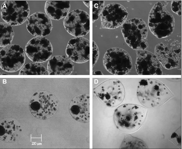
FIGURE 2.1:PHASE-CONTRAST MICROSCOPY OF ENCAPSULATED HEPATOCYTE AGGREGATES ... 29
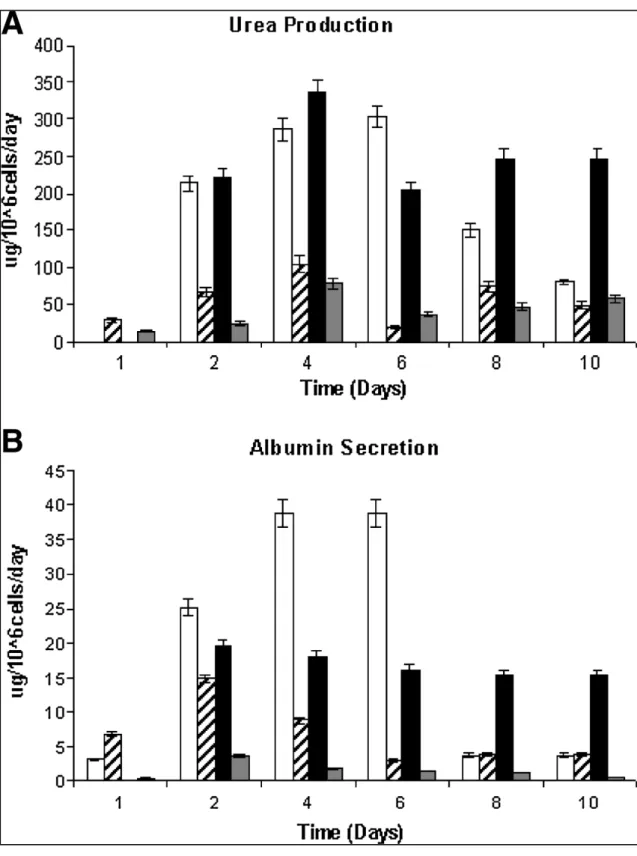
FIGURE 2.2:FUNCTIONAL CAPACITY OF HEPATOCYTES ... 30
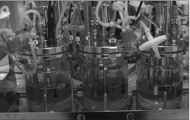
FIGURE 2.3:SARTORIUS-STEDIM BIOSTAT Q-PLUS BIOREACTOR SYSTEM ... 32
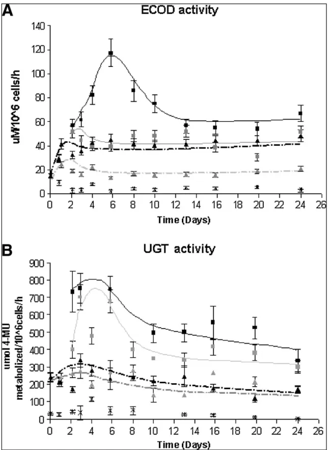
FIGURE 2.4:ECOD AND UGT ACTIVITIES IN RAT HEPATOCYTES CULTURED AS ENCAPSULATED AGGREGATES ... 33
FIGURE 2.5:3-METHYLCHOLANTHRENE INDUCTION OF CYP450-DEPENDENT ACTIVITY IN PRIMARY CULTURE OF RAT HEPATOCYTES ... 34
FIGURE 2.6:DEXAMETHASONE INDUCTION OF CYP450-DEPENDENT ACTIVITY IN PRIMARY CULTURE OF RAT HEPATOCYTES ... 35
FIGURE 3.1:PERFUSION BIOREACTOR SYSTEM WITH GRAVIMETRIC CONTROL ... 46
FIGURE 3.2:EFFECT OF PERFUSION IN THE LIVER-SPECIFIC FUNCTIONS OF PRIMARY BIOREACTOR CULTURES OF NON ENCAPSULATED HEPATOCYTE SPHEROIDS ... 47
FIGURE 3.3: CHARACTERIZATION OF ULTRA HIGH VISCOUS ALGINATE ENCAPSULATED HEPATOCYTE SPHEROIDS ... 48
FIGURE 3.4: SPECIFIC ALBUMIN AND UREA SYNTHESIS RATES IN PERFUSION AND 50% MEDIUM EXCHANGE BIOREACTOR CULTURE ... 49
FIGURE 3.5: SPECIFIC OXYGEN CONSUMPTION RATE IN PERFUSION AND 50% MEDIUM EXCHANGE BIOREACTOR CULTURE ... 50
FIGURE 3.6: CYP450 ACTIVITY IN PERFUSION AND 50% MEDIUM EXCHANGE BIOREACTOR CULTURE .. 50
FIGURE 4.1: EXPERIMENTAL DESIGN OF THE INDUCTION OF THE CYP450 ENZYMES IN PRIMARY CULTURES OF HEPATOCYTE SPHEROIDS IN THE BIOREACTOR ... 61
FIGURE 4.2: HEPATOCYTE SPHEROID DIAMETER DISTRIBUTION AND CELL VIABILITY ... 63
FIGURE 4.3: UREA AND ALBUMIN SYNTHESIS IN BIOREACTOR HEPATOCYTE SPHEROID CULTURES ... 64
FIGURE 4.4: INTER-DONOR VARIABILITY AND TIME COURSE PROFILES OF PHASE I AND II ENZYMES IN HUMAN HEPATOCYTE SPHEROIDS BIOREACTOR CULTURES ... 65
FIGURE 4.5: INDUCTION OF ECOD ACTIVITY IN BIOREACTOR ... 66
FIGURE 4.6: IMMUNOFLUORESCENCE MICROSCOPY OF LIVER-SPECIFIC ANTIGENS IN HUMAN HEPATOCYTE SPHEROIDS AFTER 2 WEEKS OF BIOREACTOR CULTURE ... 66
FIGURE 4.7: FLUORESCENCE MICROSCOPY OF STRUCTURAL AND POLARITY MARKERS AND BILE CANALICULI FUNCTION IN HUMAN HEPATOCYTE SPHEROIDS AFTER 2 WEEKS OF BIOREACTOR CULTURE ... 68
FIGURE 5.1: EFFECT OF ROCKI ON THE AGGREGATION AND VIABILITY OF HEPATIC PROGENITOR CELLS UNDER MICROGRAVITY CONDITIONS ... 79
FIGURE 5.2: HARDWARE (FIRST COLUMN) AND CULTURE PARAMETERS TESTED FOR THE OPTIMIZATION OF THE AGGREGATION PROCESS OF THE HEPATIC PROGENITORS ... 80
FIGURE 5.3: WORKFLOW FOR THE MATURATION OF HEPATIC PROGENITORS TO HEPATOCYTE-LIKE CELLS, IN 2D AND 3D ... 81
FIGURE 5.4: PHASE CONTRAST AND FLUORESCENCE MICROSCOPY IMAGES OF THE MEMBRANE INTEGRITY AND METABOLIC ACTIVITY OF ALGINATE ENCAPSULATED HEPATIC PROGENITOR SPHEROIDS ... 82
FIGURE 6.1: WORKING HYPOTHESIS FOR THE SYNERGISTIC EFFECT OF PERFUSION AND ALGINATE ENCAPSULATION IN PRIMARY CULTURES OF RAT HEPATOCYTE SPHEROIDS ... 89
XIII
List of tables
TABLE 1.1:EXPERIMENTAL MODELS FOR HEPATIC CYP450 INDUCTION STUDIES ... 10
TABLE 1.2:THE LOGISTIC AND BIOENGINEERING CHALLENGES AND CRITICAL FACTORS FOR THE APPLICATION OF PRIMARY CULTURE OF HEPATOCYTES TO LONG TERM DRUG DEVELOPMENT TESTING AND BIOARTIFICIAL LIVER DEVICES ... 12
TABLE 2.1:ACTIVITIES FOR REGION AND STEREOSPECIFIC TESTOSTERONE HYDROXYLATION OF ENCAPSULATED HEPATOCYTE AGGREGATES ... 31 TABLE 5.1:UREA AND ALBUMIN PRODUCTION, NORMALIZED TO THE EXTRACTED RNA, AT DAY 27 OF DIFFERENTIATION ... 82
XV
Abbreviation Full form
SECs Sinusoidal endothelial cells
HSCs Hepatic stellate cells
ECM Extracellular Matrix
NK Natural Killer
Km Michaelis Menten enzyme kinetics constant
ALF Acute liver failure
OLT Orthotopic liver transplant
AL or BAL artificial or bioartificial liver
NCE New chemical entity
PK Pharmacokinetics
OATP Organic anion transporting polypeptide
CYP450 Cytochrome P450
CYP1A2 Cytochrome P450, isoform 1A2
CYP2C9 Cytochrome P450, isoform 2C9
CYP3A4 Cytochrome P450, isoform 3A4
CYP7A1 Cytochrome P450, isoform 7A1
PXR Pregnane X Receptor
CAR Constitutive Androstan Receptor
AhR Aromatic Hydrocarbon Receptor
DDI Drug- drug interactions
mRNA Messenger Ribonucleic Acid
FDA Food and Drug Administration
TGF- Transforming Growth Factor Beta
ELISA enzyme- linked immunosorbent assay
3D Three-dimensional
2D Two-dimensional
RGD Arginine- Glycine - Aspartic Acid
DO Dissolved oxygen
hESC Human embryonic stem cells
UGT Uridine diphosphate glucuronosyltransferases
UGT2B7 Uridine diphosphate glucuronosyltransferase, isoform 2B7
ECOD 7- Ethoxycoumarin -O- Dealkilase
3-MC 3- Methylcholantherene
4-MU 4- Methylumbelliferone
OHT Hydroxytestosterone
Da Dämkohler number
OCR Oxygen Consumption Rate
FBS Foetal Bovine Serum
Rif Rifampicin
BNF -Naphtoflavone
dave average diameter
cDNA complementary Deoxyribonucleic acid
PCR Polymerase Chain Reaction
GAPDH Glyceraldehyde 3- phosphate dehydrogenase
PFA Paraformaldehyde
DAPI 4',6- diamidino -2- phenylindole
CDFDA 5-(and-6)-carboxy-2',7'-dichlorofluorescein diacetate
GSTA1 Glutathione S- transferase A1
HNF4 Hepatocyte Nuclear Factor 4 alfa
aPKC atypical Protein Kinase C
MRP2 associated protein 2
HLC hepatocyte-like cell
EBs embryoid bodies
DE Definitive Endoderm
HGF Hepatocyte Growth Factor
KO-DMEM knock out Dulbecco's modified Eagle medium
KO-SR knock out serum replacement
HD hanging drop
Rocki Rock inhibitor
EGF Epidermal Growth Factor
BMPs Bone Morphogenetic Proteins
1
Chapter 1
Introduction
–
Liver physiology,
drug development and state of the
art in primary cultures of
2
Table of Contents
1.
Introduction ... 3
1.1. Liver architecture and zonation ... 3
1.2. Liver pathologies ... 7
1.3. Relevance of the liver metabolism for drug development ... 7
1.4. Models for hepatic CYP450 enzyme induction ... 10
1.5. Critical factors in long-term primary cultures of hepatocytes ... 12
1.6. State of the art in long term primary cultures of hepatocytes ... 14
2. Scope of the Thesis ... 15
3
1. Introduction
The liver has a major role in maintaining physiological homeostasis and in detoxifying blood. In mammals, among other functions, it is responsible for nitrogen excretion in the form of urea, albumin biosynthesis (1), glucose storage in the form of glycogen, gluconeogenesis (2), glutamine homeostasis (3) and xenobiotic detoxification (4-7).
1.1. Liver architecture and zonation
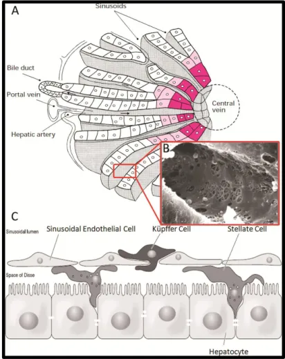
4
Figure 1.1 – The architecture of the liver: acinus (A), sinusoids (B) and the location of the different liver cell types in the sinusoid (C). Adapted from (13, 16)
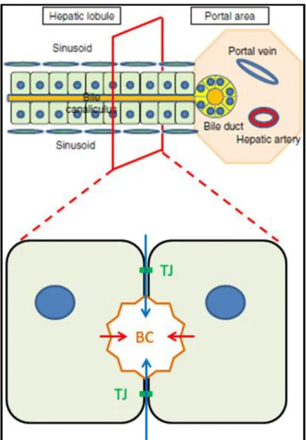
5
Figure 1.2 – Bile canaliculi physiology; a transversal section of the hepatic sinusoid (lower figure) depicts the pumping of organic anions from the hepatocytes’ cytosol to the bile canaliculi. This solute accumulation is the driving force for paracellular water osmosis, i.e., via the semi-permeable
tight junctions (green) between the hepatocytes. TJ – Tight junctions; BC – Bile canaliculus. Upper image adapted from (15).
6
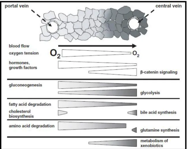
of oxidative metabolism in the oxygen-rich periportal zone (70 mmHg/90 M O2) and an enhancement of the glycolytic metabolism in the oxygen poor perivenous zone (35 mmHg/45 M O2) (18).
Figure 1.3 – Metabolic liver zonation as a function of the (variable) zonation of oxygen, hormones and growth factors and of the (stable) zonation of -catenin signalling, adapted from (22).
Interestingly, the metabolic zonation of carbohydrates has shown to be partially reversed upon retrograde liver perfusion (i.e., nutrient flow is reversed to start from the perivenous to the
periportal zone), indicating that the hepatocytes can “sense” the oxygen concentration (for a
review on the role of oxygen in liver zonation see (19))`. The fact that metabolic zonation is only partially reversed in these retrograde perfusion experiments lead to the discovery of a stable zonation mechanism based on -catenin signaling (23). In fact, stable zonation can also be seen in the non-parenchymal cell distribution and phenotype as well as in the extracellular matrix composition (17).
7
1.2. Liver pathologiesLiver tissue damage, irrespective of its etiology (viral or viral, autoimmune, alcoholic or non-alcoholic hepatitis (11, 15, 24)), leads to an increased synthesis of collagen fibers by HSC and other fibroblast-like cell types in the liver, constituting the basic liver wound-healing process. If the injury is continuous, this fibrotic response typically leads to the clogging of the liver vasculature (namely in the sinusoids) which results in necrosis, scarring of the affected area and the formation of nodules of regenerating hepatocytes; the final stage of progressive liver fibrosis is cirrhosis (see (25) for a comprehensive review), which can also lead to the onset of primary hepatocellular carcinoma. These chronic clinical conditions, as well as acute liver failure (ALF), can result in hepatic function impairment, multi-organ failure and subsequent death, unless an orthotopic liver transplant (OLT) is performed (26). However, the scarcity of liver donors creates a logistics problem for clinical situations where OLT is recommended. The need to keep patients with ALF alive from the onset of the clinical symptoms until a donated liver is available has lead to the development of artificial (27) and bioartificial liver (28) (AL or BAL, respectively) devices to perform the minimal hepatic functions which are necessary for patient survival; while the AL devices depend mainly on plasma filtration and purification operations (remnant of kidney dialysis), the BAL devices typically couple plasma filtration steps to a cell-based purification unit (i.e., a bioreactor) which can perform residual hepatic functions to sustain the patients lives. Irrespective of being AL or BAL devices, these solutions have not yet proved to be effective enough in improving ALF outcomes. In the specific case of BAL devices, the lack of an appropriate cellular component with significant volumetric hepatic activity is considered to be the major bottleneck for a clinically efficient and robust product.
1.3. Relevance of the liver metabolism for drug development
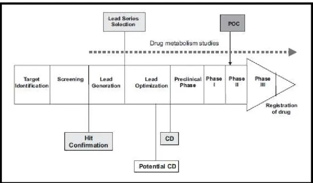
Bringing a new drug to the market costs upwards of 900 million USD and takes, on average, 12 years (29). Drug development is a multistage process which typically starts with the definition of a biological target for a given disease; in the next stages, thousands of molecules are screened for binding and efficacy towards the target and lead compounds are established and optimized. These early stages are usually followed by the pre-clinical drug development and the clinical trials from phase I to III until the registration of one new chemical entity (NCE) (Figure 1.4 (30)).
8
The progression along this process phases leads to a decrease in the compounds tested and an increase in the operation costs (i.e., clinical trials are more expensive than the pre-clinical or screening phases). The probability of a given candidate drug to progress from phase II to phase III clinical trials was about 28% from 2006 to 2007 and 18% in the period from 2008-2009; given that the attrition rate for a drug entering phase III is about 50% (31, 32), it has been suggested that the success rates of drug development are too low to cope with the increasing R&D budget (33). While more global and systemic analysis about the underlying reasons of this issue have been performed by several authors (29, 34), the concrete problems that lead to failure in phase II clinical trials are well documented (31): 51% of these failures were due to insufficient efficacy, 29% to strategic reasons, 19% to pre-clinical or clinical safety issues and 1% due to inadequate pharmacokinetics (PK). PK can be defined as a methodology to determine a drug’s (and its metabolites) concentration profile along time in the human body fluids; the critical parameters for a PK profiling are the administration, distribution, metabolization and excretion (ADME) of the drug (35). The drug concentration profile in the human body is a critical variable for its therapeutical efficiency and since pharmaceutical companies started investing in PK pre-clinical research, in the early 1990’s, the failure rate in drug development owed to inadequate PK has fallen by 10% (30). However, the effects of a drug and it’s metabolites in the patients’ physiology is more difficult to assess in pre-clinical trials and such effects can lead either to enhanced toxicology (36, 37) or to insufficient efficacy (38). Central to these two causes of attrition in drug development is the biotransformation (i.e., metabolization) of the drug itself. In fact, according to data from Brystol-Myers Squibb from 1993 to 2006, 27% of the toxicology drug attrition were metabolism-related.
9
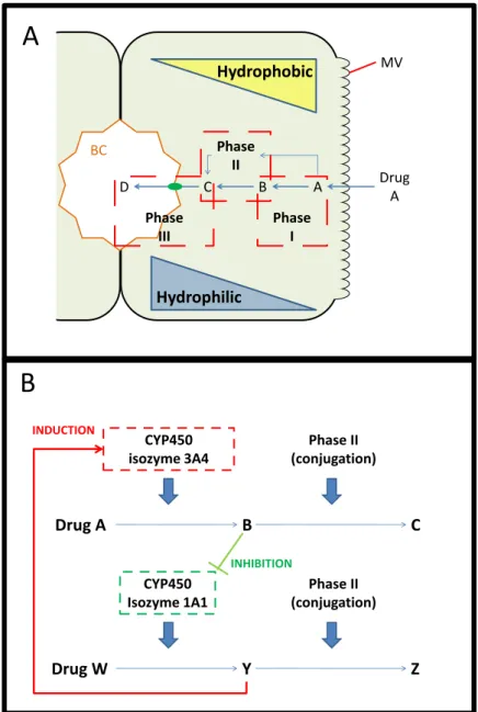
Figure 1.5 – General mechanism for drug metabolism (A) and possible drug-drug interactions (B). MV-microvilli.
The studies of the NCE’s metabolization can be performed using different approaches; the incubation of human hepatocyte microsomes (which are lipid vesicles containing bound CYP450 enzymes) with the NCE yields information about the metabolites originated from the drug; these microsome assays are also useful to determine if the NCE or any of its metabolites are inhibitors of CYP450 activity (44, 45). However, the induction of CYP450 enzymes implies de novo mRNA synthesis, which requires intact cellular machinery and the models for these studies range from cells (5) to entire organisms for in vivo studies (46). These models may be ranked according to three main categories: throughput, predictability (relative to human subjects) and long term studies.
MV
Phase III
Phase II
Phase I
A B C
D Drug
A
BC
Hydrophilic
Hydrophobic
Phase II (conjugation) CYP450
isozyme 3A4
Drug A
B
C
Phase II (conjugation) CYP450
Isozyme 1A1
Drug W
Y
Z
INDUCTION
INHIBITION
B
10
1.4. Models for hepatic CYP450 enzyme inductionIn vivo drug metabolism studies can be performed in large or small animal models; however, due to the limited throughput and high costs of larger animal models, small animal models (typically murines, such as rats and mice) have been used. These models enable long term pre-clinical studies of drug metabolism, including repeated dose effects, and show the systemic effects of drug metabolism. Such models, listed in Table 1.1, are hampered by the consistent inter-species difference in drug metabolism; in fact, this low predictability towards the human species is the reason why FDA considers that these models may not be valuable for DDI or drug metabolism studies in general (47).
Table 1.1 – Experimental models for hepatic CYP450 induction studies
Model Type Advantages Disadvantages Refere
nce
Rodents In vivo
Systemic Long term Repeated dose
Inter-species differences Low throughput
(47)
Humanized
Mice vivo In
Systemic Long term Repeated dose Human metabolism
(partial)
Endogenous murine factors crosstalk with human genes Low throughput
(48-50)
Decellularized
liver vivo
Ex- Partial maintenance of liver architecture and vascularization Human metabolism
Not
characterized for drug metabolism Complex
workflow Low Throughput
(51, 52) Primary cultures of human hepatocytes In vitro
FDA and EMA recommended All hepatic cellular
features are present Inter-donor
variability High throughput Human metabolism
Negligible proliferation in culture Liver specific
functions decay within one week Scarcity
(41, 47)
Hepatic cell lines
In vitro
Availability Some extent of
human hepatic metabolism High throughput
Clonal origin Limited hepatic
phenotype (41, 53-57) Reporter cell lines In vitro
Availability High throughput
Interference from the original cell line’s endogenous factors
Incomplete set of hepatic transcription factors
(58)
11
transgenic mice carrying a copy of the human CYP2B2 gene to phenobarbital; more recently, double transgenic mice carrying one copy of the nuclear receptor PXR and its downstream target CYP3A4 were shown to be inducible by Rifampicin, a prototypical CYP3A4 inducer in humans, unlike the wild-type animals (49). The current limitation of these models is the interaction of murine endogenous factors with the human transgenic proteins. Recently, modified Polyethilene Glycol hydrogels containing a primary culture of human hepatocytes, mouse 3T3 fibroblasts and a human liver endothelial cell line (TMNK-1) were implanted in mice for drug metabolism studies (50). Such ectopic bioartificial livers enable the host mice to have a typically human metabolism of known drugs (namely Acetaminophen cytotoxicity); with the use of the correct control animals, this approach may be useful for long term drug metabolism studies. Still, the synergy of the host’s murine metabolism with the human hepatocytes can yield confounding results in drugs with unknown metabolism.
The recellularization of previously acellularized organs is a current trend in tissue engineering (59); this technique, which consists in the removal of cells from an organ with a detergent, yields the organ scaffold with a preserved ECM and vasculature. Decellularized livers from several species have been successfully prepared and efficiently reseeded either with adult rat hepatocytes (52) or with human endothelial cells and fetal liver cells (51), respectively. Uygun and colleagues (52) have shown that this strategy allows to maintain primary cultures of rat hepatocytes for up to 2 weeks, namely drug metabolizing enzymes mRNA expression. However, these expression levels did not outperform those obtained by the corresponding collagen sandwich cultures; further improvements can be made to this system by the addition of non-parenchymal liver cells. In fact, the presence of SECs may be critical to re-organize the liver sinusoids, which are lost during the decellularization process. From a practical point of view, this technique is not hampered by the scarcity of livers, since cadaveric livers can be used.1
Cell-based assays for hepatic drug induction can be divided in 3 classes: reporter cell lines for nuclear receptor activation, immortalized hepatocytes/ hepatoma cell lines and primary cultures of human hepatocytes.
Primary cultures of human hepatocytes, either freshly isolated or cryopreserved, are FDA’s preferred human liver tissue for CYP450 induction tests (47). These cultures retain nuclear receptor, CYP450 isoforms as well as phase II and III mRNA and protein expression. This expression of all the major sensors and effectors of drug metabolism enables primary cultures of human hepatocytes to demonstrate CYP450 induction and hepatoxicity resulting from DDI. Primary cultures of hepatocytes have 3 main drawbacks: the hepatocytes have a negligible proliferation in culture, when cultured in monolayer, they lose their liver-specific activities (including CYP450) in less than one week (42) and these cells are scarce (even more since these hepatocytes do not proliferate ex vivo) and expensive, making increased throughput assays expensive.
Nuclear receptor activation is the common upstream event in CYP450 de novo mRNA synthesis. Such a reporter system has been described by Luo and colleagues (58) based on the co-transfection of a hepatoma cell line, HepG2, with a plasmid with the cDNA for PXR under the control of a constitutive promoter and another plasmid containing the CYP3A4 regulatory region which controlled the expression of a reporter luciferase gene. Upon activation by a drug, the PXR nuclear receptor binds the regulatory CYP3A4 regions and increases the expression of Luciferase. This assay, which can be translated to high throughput settings, showed a fair correlation between PXR activation in the transfected HepG2 cells and the increase of CYP3A4 activity in primary cultures of human hepatocytes, when exposed to prototypical CYP3A4 inducers. However, such reporter systems are prone to interference from the endogenous factors from the transfected cell line and lack other transcription factors which are important for CYP450 mRNA synthesis. In fact, this incomplete hepatic cellular machinery is also an issue when the use of immortalized or hepatoma cell lines such as HepG2 or Fa2N-4 is considered (41). Still, there are substantial differences between immortalized cell lines: while the HepG2 hepatoma cell line exhibits low expression levels of adult human CYP450 enzymes and high
12
levels of fetal the CYP450 (53, 54) the HepaRG cell line holds promise for hepatic drug induction tests. This latter hepatoma cell line was shown to express nuclear receptors and CYP450 transcripts at levels comparable with primary human hepatocytes (55) and its CYP450 activity induction has been proven for the CYP450 isoforms 1A2, 2C9 and 3A4, among others, as well as the presence of phase II and III drug metabolizing enzymes (57, 60); such results make this cell line an alternative to larger throughput drug metabolism studies, since it can proliferate up to several passages. The main disadvantages of using the HepaRG cell line are its clonal origin (it originated from a hepatocellular carcinoma from one single individual (55)) and the co-culture nature of this system, since HepaRG cell culture contain both hepatocytes and cholangiocytes, making it difficult to quantify the metabolic activity per hepatocyte. The former clonality issue limits the assessment of the naturally occurring inter-individual variability in CYP450 metabolism (42, 61) when using the HepaRG model. For a mid to long term perspective, the differentiation of human pluripotent stem cell lines to hepatocytes is a promising strategy which can potentially deliver an unlimited number of terminally differentiated hepatocytes. Hepatic differentiation protocols have already been established (62, 63) and adapted to large scale culture (64), but the obtained hESC derived hepatocyte-like cells have a strong fetal liver phenotype, namely the expression of Alfa-Fetoprotein and CYP7A1. Further maturation of these cells has been a subject of several publications (65-69) showing the expression of mature mRNAs and protein, such as CYP3A4 (70), thus proving the potential of pluripotent stem cell hepatic differentiation for providing hepatocytes for drug development tests.
1.5. Critical factors in long-term primary cultures of hepatocytes
Each of the models described above has its set of strengths and weaknesses. Primary cultures of hepatocytes have been the subject of several works (28, 71-77) that aim at overcoming its short lifespan, because these cultures are still FDA’s gold standard. While some of the published works aim at maintaining hepatocyte viability (78), the most pursued goal of these works is to maintain the liver specific functionality of the hepatocytes for extended time in culture (79-81).
Long term primary cultures of human hepatocytes are thus required for both drug development pre-clinical tests and as a biotransformation component in BAL devices; the scarcity and in vitro dedifferentiation of these cells are common logistic and engineering challenges for both applications, as depicted in Table 2:
Table 1.2 – The logistic and engineering challenges and critical factors for the application of primary culture of hepatocytes to long term drug development testing and bioartificial liver devices.
Logistic Engineering
Challenge Cell type/line Hepatic functionality
Critical biological factors Storage/cryopreservation Availability
Cell-cell interactions Cell-ECM interactions
Soluble factors Dimensionality (2D or 3D)
The engineering challenge for long term primary cultures of hepatocytes, i.e., the maintenance of hepatic functionality throughout culture time, is dependent upon critical biological factors which are, in fact, general barriers to obtain an in vivo-like environment when performing in vitro cultures. Thus, the design of the bioprocess system associated with these cultures must be based liver physiology principles.
13
the appropriate substrate mechanical stiffness (83) and the ligands for cell signaling; these can be part of the ECM itself, such as the Fibronectin’s RGD motif, or they can simply dock at the ECM to be presented to the cell’s surface receptors, as is the case with Fibroblast Growth Factors (84, 85). Soluble factors are typically identified with the blood-dissolved nutrients (such as glucose, aminoacids or oxygen), metabolite products (lactate or carbon dioxide) and endocrine factors, such as insulin, glucagon and the glucocorticoids dexamethasone and hydrocortisone; the design of an appropriate culture medium is thus essential for the primary culture of hepatocytes. In addition, it is also necessary to ensure an efficient mass transfer to the hepatocytes; this is a complex challenge, since the liver is one of the most irrigated organs and the fenestrated hepatic sinusoid enhances the mass transfer between the blood and the hepatocytes beyond that of most capillary delivery systems. Moreover, since some of the liver’s major homeostatic functions such as the regulation of glucose levels in the blood (2) or glutamine synthesis (86) are based on the hepatocytes’ ability to continuously sense and act upon the blood’s nutrient and hormone levels, it is essential that these concentrations are kept fairly constant throughout culture time.
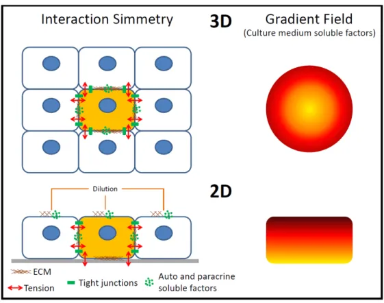
Another characteristic of the in vivo microenvironment is the three-dimensional (3D) configuration; however, most in vitro cultures are performed as cell monolayers (2D). When 2D and 3D are compared, the main difference between these systems is the lack of symmetry in the cellular interactions with other cells, ECM and the culture medium soluble factors (Figure 1.6).
Figure 1.6 – Symmetry of the epithelial cellular interactions with other cells, the ECM and the soluble factors in vitro.
14
However, if there is a gap in the cellular monolayer, the nearby cells will tend to spread on the culture surface.
1.6. State of the art in long term primary cultures of hepatocytes
One of the major breakthroughs for achieving a long term primary culture of hepatocytes has been the collagen sandwich system (87). In these cultures the hepatocyte monolayer is coated with a collagen layer and the albumin secretion of these primary cultures of hepatocytes can be kept up to 6 weeks with the formation of bile-canaliculi like structures between the cells (88). The formation of the canalicular structures was suggested to be due to a reduction in cell spreading (due to the attachment of the cells to the top collagen layer) that increased the surface area of the lateral membranes of the hepatocytes available for the formation of bile canaliculi; this hypothesis is further reinforced be the absence of stress fibers (typical of cell spreading) in the actin cytoskeleton of the collagen sandwich cultured rat hepatocytes and by the fact that matrigel (a basement membrane-like ECM) can also be used in the sandwich configuration, yielding a similar performance (89). In addition, the top collagen layer also reduces the diffusion rate of the autocrine factors and ECM to the culture medium. Nevertheless, the chemical gradient between the lower surface and the top surface of the hepatocytes is still maintained. This limitation has been recently mitigated by the development of a sandwich culture system with an oxygen permeable membrane for cell attachment; while the lack of other nutrients has not been solved, the enhancement in oxygen availability on the bottom surface of the cells has lead to increased CYP450 1A activity for 4 days, when compared to a non-permeable surface (90).
The perfusion bioreactor developed by Gerlach and co-workers (91) for BAL applications consists of three interwoven permeable capillary systems and a culture chamber; two of these capillary systems are for medium perfusion and each system flows countercurrently to the other to minimize the gradients from the fiber inlet to outlet (nutrient addition and clearance of the metabolic by-products, respectively), whereas the third is meant for gas exchange (O2 delivery and CO2 removal). This perfusion configuration is an attempt to recapitulate the intensive vascularization of the liver. The central culture chamber contains the liver cells (hepatocytes and non-parenchymal fraction), which are cultured in a compact 3D structure. This bioreactor system addresses all the critical biological factors for these cultures and has currently been scaled down from the original 800 mL working volume to 2 mL (92, 93), enabling the use of these devices for long-term drug development tests. However, this bioreactor does not allow sampling of the cells during the culture, only at its end, thus, even though it is a long term system for the primary culture of hepatocytes, these cells cannot be imaged throughout culture time nor withdrawn from the reactor to measure mRNA or protein expression. In addition, the hollow fiber system does not allow for accurate control of pH nor dissolved oxygen (DO) concentration within the fibers.
The application of micropatterning and microfluidic techniques to design miniaturized cell culture devices has also been applied to primary cultures of hepatocytes, but few of these devices span extended culture times. The Bhatia group has designed one of the most successful microscale culture devices for liver cells (80); the most important design criteria for this device were the homo and heterotypic cell-cell interactions. Such interactions were based in hepatocyte-3T3 fibroblast co-culture, which is known to improve the activity and maintenance of the hepatocytes’ liver-specific functions (94). This device was designed in a 24 well plate format to ensure it was user friendly; each well was micropatterned with 500 m collagen I islands and subsequently seeded with hepatocytes, which formed colonies on top of the collagen islands. Finally, 3T3 fibroblasts were seeded across the entire well to achieve the co-culture system with a 300 L culture volume. This system enabled the maintenance of liver-specific drug metabolizing activity for up to 2 weeks. This system also showed an impressive robustness, which may be due to the automated spotting of hepatocyte islands; nevertheless, the lack of control of pH and DO, both of them soluble factors, a static culture system and a limited 3D configuration may be the cause for the 2 week limit of these culture systems.
cell-15
cell interactions. When comparing with the Bhatia group microscale device, this one has the additional advantage of having a dynamic perfusion system and some extent of oxygen control, which, being based on Computational Fluid Dynamics calculations and oxygen sensors located at the inlet and outlet, does not act upon an actually measured oxygen concentration sensed by the hepatocytes. The dynamic perfusion minimizes the gradients of culture medium soluble factors at the surface of the cells, which improves the transport of nutrients and products of metabolism but may also wash out the cell-secreted autocrine and paracrine factors, as well as ECM components such as fibronectin. Thus, the dilution rate set by the perfusion system must balance these 2 factors. In the device herein described, the dilution rate is approximately 1.4 day-1,i.e., similar to that of the Bhatia group device (1 medium exchange
per day), with the significant difference that the former device avoids sudden changes in the concentration of these factors (i.e., full medium exchanges). Regarding the shear stress elicited by the culture medium perfusion, its effect has been shown to be negligible in this specific microfluidic device (81), but other published works on such devices typically report flow velocity optimization to minimize shear stress (96). More recently, Sonntag and colleagues (97) have described a microfluidic reactor which has connected culture chambers for liver, brain and bone marrow cells; such a device may potentially allow the assessment of a more systemic response of the human organism to xenobiotics.
The collagen sandwich cultures, hollow fiber bioreactors and microfludic devices have one limitation for long term primary culture of hepatocytes: the control of pH and DO. The main cause for this limitation is the difference between the oxygen concentrations in the inlets of fibers or microchannels (or incubator atmosphere) and the DO in the culture medium bulk. The use of stirred tank bioreactors with good mixing as well as pH and DO control will maintain steady-state concentrations of these two soluble factors in the culture medium. Our group has shown that this controlled system improves the culture of human hepatocyte spheroids when compared to stirred spinner vessels (73), which may be due to the maintenance of a physiological DO level in the bioreactor (approximately 60 M) (98). Primary cultures of hepatocyte spheroids also retain the in vivo-like 3D configuration that retains ECM and autocrine factors, while creating gradients from the surface to the center of the spheroid. These gradients have been shown not to be detrimental for the hepatocyte viability if the spheroid diameter is below 200 m (99), whereas spheroid diameters of 100 m enable the maximum specific albumin production (100). Moreover, such bioreactors have a user friendly sampling system (while the system set-up may require specialized bioengineering knowledge), which can be used for specific cell-based assays and analysis and to feed higher throughput devices (such as well plates). This flexibility raises the possibility of using these bioreactors for long term primary cultures of human hepatocytes coupled with a higher throughput analysis approach at each time point.
2. Scope of the Thesis
The main goal of this thesis was to extend primary cultures of hepatocytes for long term drug testing applications; while the current state of the art enables a 2 week culture of functional hepatocytes, their ability to synthesize CYP450 mRNA, de novo, is typically limited to the first week of culture. Thus, this hepatocyte potential to induce the CYP450 enzymes is the most demanding test used in drug development and it has been one of the most important milestones of this work.
16
increase the liver-specific functionality of hepatocyte spheroids, enabling the repeated induction of CYP1A and CYP3A isoforms during 15 and 26 days of culture, respectively.
The bioreactor system used in rat hepatocyte spheroid cultures was further optimized by adding an automated, gravimetrically controlled perfusion system, which allowed the maintenance of stable concentrations of soluble factors. In chapter 3 the effects of this perfusion system will be compared with manual medium exchange, for both non-encapsulated and alginate encapsulated rat hepatocyte spheroid cultures.
This perfusion bioreactor system has been applied to the primary cultures of human hepatocytes. In chapter 4 such cultures will be characterized and validated for long term, repeated dose, drug tests; the results show that this culture system maintains hepatocyte functionality for up to 2 weeks, and the hepatocytes CYP450 enzymes have been induced after 7, 14, 21 and 28 days using hepatocytes from 3 different donors. In addition, the 3D architecture of these human hepatocyte spheroids has been investigated by confocal immunofluorescence microscopy, revealing a liver acinus-like polarization of the hepatocytes and the presence of functional bile canaliculi.
In chapter 5 this thesis will approach the logistics problem concerning the scarcity of isolated human hepatocytes. The effects of both spheroid culture and alginate encapsulation in the hepatic differentiation of human embryonic stem cells (hESC) will be studied by comparison with a standard 2D differentiation protocol.
The results described in chapters 2-5 will be thoroughly discussed in chapter 6, as well as the future research in this area.
3. References
1. Quinlan GJ, Martin GS, Evans TW. Albumin: Biochemical properties and therapeutic potential. Hepatology 2005;41:1211-1219.
2. Pilkis SJ, Granner DK. MOLECULAR PHYSIOLOGY OF THE REGULATION OF HEPATIC GLUCONEOGENESIS AND GLYCOLYSIS. Annual Review of Physiology 1992;54:885-909.
3. Watford M, Chellaraj V, Ismat A, Brown P, Raman P. Hepatic glutamine metabolism. Nutrition 2002;18:301-303.
4. Gebhardt R, Hengstler JG, Muller D, Glockner R, Buenning P, Laube B, Schmelzer E, et al. New hepatocyte in vitro systems for drug metabolism: Metabolic capacity and recommendations for application in basic research and drug development, standard operation procedures. Drug Metabolism Reviews 2003;35:145-213.
5. Gomez-Lechon MJ, Donato MT, Castell JV, Jover R. Human hepatocytes as a tool for studying toxicity and drug metabolism. Current Drug Metabolism 2003;4:292-312.
6. LeCluyse EL. Human hepatocyte culture systems for the in vitro evaluation of cytochrome P450 expression and regulation. European Journal of Pharmaceutical Sciences 2001;13:343-368.
7. Maurel P. The use of adult human hepatocytes in primary culture and other in vitro systems to investigate drug metabolism in man. Advanced Drug Delivery Reviews 1996;22:105-132.
8. Altin JG, Bygrave FL. Non-parenchymal cells as mediators of physiological responses in liver. Mol Cell Biochem 1988;83:3-14.
9. Wisse E, Braet F, Luo DZ, DeZanger R, Jans D, Crabbe E, Vermoesen A. Structure and function of sinusoidal lining cells in the liver. Toxicologic Pathology 1996;24:100-111.
10. Bilzer M, Roggel F, Gerbes AL. Role of Kupffer cells in host defense and liver disease. Liver International 2006;26:1175-1186.
11. Kmieć Z. Cooperation of liver cells in health and disease. Adv Anat Embryol Cell Biol
2001;161:III-XIII, 1-151.
17
13. Vollmar B, Menger MD. The Hepatic Microcirculation: Mechanistic Contributions and Therapeutic Targets in Liver Injury and Repair. Physiological Reviews 2009;89:1269-1339. 14. Monga SPS. Molecular pathology of liver diseases. In: Molecular pathology library. New York: Springer,; 2011. p. xxiii, 931 p.
15. Ishibashi H, Nakamura M, Komori A, Migita K, Shimoda S. Liver architecture, cell function, and disease. Semin Immunopathol 2009;31:399-409.
16. Lindros KO. Zonation of cytochrome P450 expression, drug metabolism and toxicity in liver. Gen Pharmacol 1997;28:191-196.
17. Jungermann K, Kietzmann T. Zonation of parenchymal and nonparenchymal metabolism in liver. Annu Rev Nutr 1996;16:179-203.
18. Jungermann K, Kietzmann T. Role of oxygen in the zonation of carbohydrate metabolism and gene expression in liver. Kidney Int 1997;51:402-412.
19. Jungermann K, Kietzmann T. Oxygen: modulator of metabolic zonation and disease of the liver. Hepatology 2000;31:255-260.
20. Katz NR. Metabolic heterogeneity of hepatocytes across the liver acinus. J Nutr 1992;122:843-849.
21. Listrom CD, Morizono H, Rajagopal BS, McCann MT, Tuchman M, Allewell NM. Expression, purification, and characterization of recombinant human glutamine synthetase. Biochem J 1997;328 ( Pt 1):159-163.
22. Braeuning A, Ittrich C, Köhle C, Hailfinger S, Bonin M, Buchmann A, Schwarz M. Differential gene expression in periportal and perivenous mouse hepatocytes. FEBS J 2006;273:5051-5061.
23. Benhamouche S, Decaens T, Godard C, Chambrey R, Rickman DS, Moinard C, Vasseur-Cognet M, et al. Apc tumor suppressor gene is the "zonation-keeper" of mouse liver. Dev Cell 2006;10:759-770.
24. Hernandez-Gea V, Friedman SL. Pathogenesis of liver fibrosis. Annu Rev Pathol 2011;6:425-456.
25. Svegliati-Baroni G, De Minicis S, Marzioni M. Hepatic fibrogenesis in response to chronic liver injury: novel insights on the role of cell-to-cell interaction and transition. Liver Int 2008;28:1052-1064.
26. Gerlach JC, Zeilinger K, Patzer Ii JF. Bioartificial liver systems: why, what, whither? Regen Med 2008;3:575-595.
27. Carpentier B, Gautier A, Legallais C. Artificial and bioartificial liver devices: present and future. Gut 2009;58:1690-1702.
28. Schmelzer E, Mutig K, Schrade P, Bachmann S, Gerlach JC, Zeilinger K. Effect of Human Patient Plasma Ex Vivo Treatment on Gene Expression and Progenitor Cell Activation of Primary Human Liver Cells in Multi-Compartment 3D Perfusion Bioreactors for Extra-Corporeal Liver Support. Biotechnology and Bioengineering 2009;103:817-827.
29. Kola I, Landis J. Can the pharmaceutical industry reduce attrition rates? Nat Rev Drug Discov 2004;3:711-715.
30. Baranczewski P, Stańczak A, Sundberg K, Svensson R, Wallin A, Jansson J, Garberg
P, et al. Introduction to in vitro estimation of metabolic stability and drug interactions of new chemical entities in drug discovery and development. Pharmacol Rep 2006;58:453-472.
31. Arrowsmith J. Trial watch: Phase II failures: 2008-2010. Nat Rev Drug Discov 2011;10:328-329.
32. Arrowsmith J. Trial watch: phase III and submission failures: 2007-2010. Nat Rev Drug Discov 2011;10:87.
33. Pisano GP. Can science be a business? Lessons from biotech. Harv Bus Rev 2006;84:114-124, 150.
34. Garnier JP. Rebuilding the R&D engine in big pharma. Harv Bus Rev 2008;86:68-70, 72-66, 128.
35. Mager DE, Jusko WJ. General pharmacokinetic model for drugs exhibiting target-mediated drug disposition. J Pharmacokinet Pharmacodyn 2001;28:507-532.
36. Guengerich FP. Mechanisms of drug toxicity and relevance to pharmaceutical development. Drug Metab Pharmacokinet 2011;26:3-14.
18
38. Shuldiner AR, O'Connell JR, Bliden KP, Gandhi A, Ryan K, Horenstein RB, Damcott CM, et al. Association of cytochrome P450 2C19 genotype with the antiplatelet effect and clinical efficacy of clopidogrel therapy. JAMA 2009;302:849-857.
39. Diaz GJ. Basolateral and canalicular transport of xenobiotics in the hepatocyte: A review. Cytotechnology 2000;34:225-235.
40. Klaassen CD. Casarett and Doull's toxicology: the basic science of poisons: McGraw-Hill, 2007.
41. Sinz M, Wallace G, Sahi J. Current industrial practices in assessing CYP450 enzyme induction: Preclinical and clinical. Aaps Journal 2008;10:391-400.
42. Hewitt NJ, Lechon MJG, Houston JB, Hallifax D, Brown HS, Maurel P, Kenna JG, et al. Primary hepatocytes: Current understanding of the regulation of metabolic enzymes and transporter proteins, and pharmaceutical practice for the use of hepatocytes in metabolism, enzyme induction, transporter, clearance, and hepatotoxicity studies. In: 1st Medicon Valley Hepatocyte User Form Symposium; 2006 Jan 26-27; Copenhagen, DENMARK: Taylor & Francis Inc; 2006. p. 159-234.
43. Pritchard JF, Jurima-Romet M, Reimer ML, Mortimer E, Rolfe B, Cayen MN. Making better drugs: Decision gates in non-clinical drug development. Nat Rev Drug Discov 2003;2:542-553.
44. Mouly S, Meune C, Bergmann JF. Mini-series: I. Basic science. Uncertainty and inaccuracy of predicting CYP-mediated in vivo drug interactions in the ICU from in vitro models: focus on CYP3A4. Intensive Care Med 2009;35:417-429.
45. Hosea NA, Collard WT, Cole S, Maurer TS, Fang RX, Jones H, Kakar SM, et al. Prediction of human pharmacokinetics from preclinical information: comparative accuracy of quantitative prediction approaches. J Clin Pharmacol 2009;49:513-533.
46. Tang C, Prueksaritanont T. Use of in vivo animal models to assess pharmacokinetic drug-drug interactions. Pharm Res 2010;27:1772-1787.
47. Huang SM, Strong JM, Zhang L, Reynolds KS, Nallani S, Temple R, Abraham S, et al. New era in drug interaction evaluation: US Food and Drug Administration update on CYP enzymes, transporters, and the guidance process. J Clin Pharmacol 2008;48:662-670.
48. Ramsden R, Beck NB, Sommer KM, Omiecinski CJ. Phenobarbital responsiveness conferred by the 5'-flanking region of the rat CYP2B2 gene in transgenic mice. Gene 1999;228:169-179.
49. Ma X, Cheung C, Krausz KW, Shah YM, Wang T, Idle JR, Gonzalez FJ. A double transgenic mouse model expressing human pregnane X receptor and cytochrome P450 3A4. Drug Metab Dispos 2008;36:2506-2512.
50. Chen AA, Thomas DK, Ong LL, Schwartz RE, Golub TR, Bhatia SN. Humanized mice with ectopic artificial liver tissues. Proc Natl Acad Sci U S A 2011;108:11842-11847.
51. Baptista PM, Siddiqui MM, Lozier G, Rodriguez SR, Atala A, Soker S. The use of whole organ decellularization for the generation of a vascularized liver organoid. Hepatology 2011;53:604-617.
52. Uygun BE, Soto-Gutierrez A, Yagi H, Izamis ML, Guzzardi MA, Shulman C, Milwid J, et al. Organ reengineering through development of a transplantable recellularized liver graft using decellularized liver matrix. Nat Med 2010;16:814-820.
53. Ogino M, Nagata K, Yamazoe Y. Selective suppressions of human CYP3A forms, CYP3A5 and CYP3A7, by troglitazone in HepG2 cells. Drug Metab Pharmacokinet 2002;17:42-46.
54. Wilkening S, Stahl F, Bader A. Comparison of primary human hepatocytes and hepatoma cell line Hepg2 with regard to their biotransformation properties. Drug Metab Dispos 2003;31:1035-1042.
55. Aninat C, Piton A, Glaise D, Le Charpentier T, Langouët S, Morel F, Guguen-Guillouzo C, et al. Expression of cytochromes P450, conjugating enzymes and nuclear receptors in human hepatoma HepaRG cells. Drug Metab Dispos 2006;34:75-83.
56. Josse R, Aninat C, Glaise D, Dumont J, Fessard V, Morel F, Poul JM, et al. Long-term functional stability of human HepaRG hepatocytes and use for chronic toxicity and genotoxicity studies. Drug Metabolism and Disposition 2008;36:1111-1118.
57. Kanebratt KP, Andersson TB. HepaRG cells as an in vitro model for evaluation of cytochrome P450 induction in humans. Drug Metab Dispos 2008;36:137-145.
19
59. Gilbert TW, Sellaro TL, Badylak SF. Decellularization of tissues and organs. Biomaterials 2006;27:3675-3683.
60. Darnell M, Schreiter T, Zeilinger K, Urbaniak T, Söderdahl T, Rossberg I, Dillnér B, et al. Cytochrome P450-dependent metabolism in HepaRG cells cultured in a dynamic three-dimensional bioreactor. Drug Metab Dispos 2011;39:1131-1138.
61. Xu Y, Zhou Y, Hayashi M, Shou M, Skiles GL. Simulation of clinical drug-drug interactions from hepatocyte CYP3A4 induction data and its potential utility in trial designs. Drug Metab Dispos 2011;39:1139-1148.
62. D'Amour KA, Agulnick AD, Eliazer S, Kelly OG, Kroon E, Baetge EE. Efficient differentiation of human embryonic stem cells to definitive endoderm. Nature Biotechnology 2005;23:1534-1541.
63. Brolen G, Sivertsson L, Bjorquist P, Eriksson G, Ek M, Semb H, Johansson I, et al. Hepatocyte-like cells derived from human embryonic stem cells specifically via definitive endoderm and a progenitor stage. Journal of Biotechnology 2010;145:284-294.
64. Lock L, Tzanakakis E. Expansion and Differentiation of Human Embryonic Stem Cells to Endoderm Progeny in a Microcarrier Stirred-Suspension Culture. Tissue Engineering Part a 2009:2051-2063.
65. Agarwal S, Holton KL, Lanza R. Efficient differentiation of functional hepatocytes from human embryonic stem cells. Stem Cells 2008;26:1117-1127.
66. Baharvand H, Hashemi SM, Ashtian SK, Farrokhi A. Differentiation of human embryonic stem cells into hepatocytes in 2D and 3D culture systems in vitro. International Journal of Developmental Biology 2006;50:645-652.
67. Cai J, Zhao Y, Liu YX, Ye F, Song ZH, Qin H, Meng S, et al. Directed differentiation of human embryonic stem cells into functional hepatic cells. Hepatology 2007;45:1229-1239. 68. Duan YY, Ma XC, Zou W, Wang C, Bahbahan IS, Ahuja TP, Tolstikov V, et al. Differentiation and Characterization of Metabolically Functioning Hepatocytes from Human Embryonic Stem Cells. Stem Cells 2010;28:674-686.
69. Hay DC, Zhao D, Ross A, Mandalam R, Lebkowski J, Cui W. Direct differentiation of human embryonic stem cells to hepatocyte-like cells exhibiting functional activities. Cloning and Stem Cells 2007;9:51-62.
70. Ishii T, Yasuchika K, Fukumitsu K, Kawamoto T, Kawamura-Saitoh M, Amagai Y, Ikai I, et al. In vitro hepatic maturation of human embryonic stem cells by using a mesenchymal cell line derived from murine fetal livers. Cell and Tissue Research 2010:505-512.
71. Goral VN, Hsieh YC, Petzold ON, Clark JS, Yuen PK, Faris RA. Perfusion-based microfluidic device for three-dimensional dynamic primary human hepatocyte cell culture in the absence of biological or synthetic matrices or coagulants. Lab on a Chip 2010;10:3380-3386. 72. Kidambi S, Yarmush RS, Novik E, Chao P, Yarmush ML, Nahmias Y. Oxygen-mediated enhancement of primary hepatocyte metabolism, functional polarization, gene expression, and drug clearance. Proceedings of the National Academy of Sciences of the United States of America 2009;106:15714-15719.
73. Miranda JP, Leite SB, Muller-Vieira U, Rodrigues A, Carrondo MJT, Alves PM. Towards an Extended Functional Hepatocyte In Vitro Culture. Tissue Engineering Part C-Methods 2009;15:157-167.
74. Nishimura M, Hagi M, Ejiri Y, Kishimoto S, Horie T, Narimatsu S, Naito S. Secretion of Albumin and Induction of CYP1A2 and CYP3A4 in Novel Three-dimensional Culture System for Human Hepatocytes using Micro-space Plate. Drug Metabolism and Pharmacokinetics 2010;25:236-242.
75. Sellaro TL, Ranade A, Faulk DM, McCabe GP, Dorko K, Badylak SF, Strom SC. Maintenance of Human Hepatocyte Function In Vitro by Liver-Derived Extracellular Matrix Gels. Tissue Engineering Part A 2010;16:1075-1082.
76. Yeon JH, Na D, Park JK. Hepatotoxicity assay using human hepatocytes trapped in microholes of a microfluidic device. Electrophoresis 2010;31:3167-3174.
77. Zeisberg M, Kramer K, Sindhi N, Sarkar P, Upton M, Kalluri R. De-differentiation of primary human hepatocytes depends on the composition of specialized liver basement membrane. Mol Cell Biochem 2006;283:181-189.
20
79. Yagi H, Parekkadan B, Suganuma K, Soto-Gutierrez A, Tompkins RG, Tilles AW, Yarmush ML. Long-term superior performance of a stem cell/hepatocyte device for the treatment of acute liver failure. Tissue Eng Part A 2009;15:3377-3388.
80. Khetani SR, Bhatia SN. Microscale culture of human liver cells for drug development. Nat Biotechnol 2008;26:120-126.
81. Powers MJ, Domansky K, Kaazempur-Mofrad MR, Kalezi A, Capitano A, Upadhyaya A, Kurzawski P, et al. A microfabricated array bioreactor for perfused 3D liver culture. Biotechnology and Bioengineering 2002;78:257-269.
82. Chiang JY. Bile acids: regulation of synthesis. J Lipid Res 2009;50:1955-1966.
83. Zhao G, Cui J, Qin Q, Zhang J, Liu L, Deng S, Wu C, et al. Mechanical stiffness of liver tissues in relation to integrin β1 expression may influence the development of hepatic cirrhosis and hepatocellular carcinoma. J Surg Oncol 2010;102:482-489.
84. Gullberg D, Terracio L, Borg TK, Rubin K. Identification of integrin-like matrix receptors with affinity for interstitial collagens. J Biol Chem 1989;264:12686-12694.
85. Rahman S, Patel Y, Murray J, Patel KV, Sumathipala R, Sobel M, Wijelath ES. Novel hepatocyte growth factor (HGF) binding domains on fibronectin and vitronectin coordinate a distinct and amplified Met-integrin induced signalling pathway in endothelial cells. BMC Cell Biol 2005;6:8.
86. Haussinger D, Lamers WH, Moorman AFM. HEPATOCYTE HETEROGENEITY IN THE METABOLISM OF AMINO-ACIDS AND AMMONIA. Enzyme 1992;46:72-93.
87. Dunn JCY, Tompkins RG, Yarmush ML. Long-term in vitro function of adult hepatocytes in a collagen sandwich configuration. Biotechnology Progress 1991;7:237-245.
88. LeCluyse EL, Audus KL, Hochman JH. Formation of extensive canalicular networks by rat hepatocytes cultured in collagen-sandwich configuration. Am J Physiol 1994;266:C1764-1774.
89. Cosgrove BD, Alexopoulos LG, Hang TC, Hendriks BS, Sorger PK, Griffith LG, Lauffenburger DA. Cytokine-associated drug toxicity in human hepatocytes is associated with signaling network dysregulation. Mol Biosyst 2010;6:1195-1206.
90. Matsui H, Osada T, Moroshita Y, Sekijima M, Fujii T, Takeuchi S, Sakai Y. Rapid and enhanced repolarization in sandwich-cultured hepatocytes on an oxygen-permeable membrane. Biochemical Engineering Journal 2010;52:255-262.
91. Gerlach JC, Encke J, Hole O, Müller C, Ryan CJ, Neuhaus P. Bioreactor for a larger scale hepatocyte in vitro perfusion. Transplantation 1994;58:984-988.
92. Zeilinger K, Schreiter T, Darnell M, Söderdahl T, Lübberstedt M, Dillner B, Knobeloch D, et al. Scaling down of a clinical 3D perfusion multi-compartment hollow fiber liver bioreactor developed for extracorporeal liver support to an analytical scale device useful for hepatic pharmacological in vitro studies. Tissue Eng Part C Methods 2011.
93. Mueller D, Tascher G, Müller-Vieira U, Knobeloch D, Nuessler AK, Zeilinger K, Heinzle E, et al. In-depth physiological characterization of primary human hepatocytes in a 3D hollow-fiber bioreactor. J Tissue Eng Regen Med 2011;5:e207-218.
94. Bhatia SN, Balis UJ, Yarmush ML, Toner M. Effect of cell-cell interactions in preservation of cellular phenotype: cocultivation of hepatocytes and nonparenchymal cells. Faseb Journal 1999;13:1883-1900.
95. Domansky K, Inman W, Serdy J, Dash A, Lim MHM, Griffith LG. Perfused multiwell plate for 3D liver tissue engineering. Lab on a Chip 2010;10:51-58.
96. Korin N, Bransky A, Khoury M, Dinnar U, Levenberg S. Design of Well and Groove Microchannel Bioreactors for Cell Culture. Biotechnology and Bioengineering 2009;102:1222-1230.
97. Sonntag F, Schilling N, Mader K, Gruchow M, Klotzbach U, Lindner G, Horland R, et al. Design and prototyping of a chip-based multi-micro-organoid culture system for substance testing, predictive to human (substance) exposure. J Biotechnol 2010;148:70-75.
98. Carreau A, El Hafny-Rahbi B, Matejuk A, Grillon C, Kieda C. Why is the partial oxygen pressure of human tissues a crucial parameter? Small molecules and hypoxia. J Cell Mol Med 2011;15:1239-1253.
99. Curcio E, Salerno S, Barbieri G, De Bartolo L, Drioli E, Bader A. Mass transfer and metabolic reactions in hepatocyte spheroids cultured in rotating wall gas-permeable membrane system. Biomaterials 2007;28:5487-5497.
21
Chapter 2
Alginate-Encapsulated
Three-Dimensional Cultures of
Hepatocytes in Bioreactors
This Chapter was based on the manuscript: