Clinical, Genetic and Neuropathological
Features of Frontotemporal Dementia:
An Update and Guide
Aspetos Clínicos, Genéticos e Neuropatológicos da Demência
Frontotemporal: Atualização e Guia
1. Department of Clinical Neurosciences and Mental Health. Faculty of Medicine. University of Porto. Portugal. 2. Dementia Clinic. Department of Neurology. Centro Hospitalar São João. Porto. Portugal.
Recebido: 12 de Janeiro de 2013 - Aceite: 21 de Abril de 2013 | Copyright © Ordem dos Médicos 2013 Jorge PELICANO PAULOS1, João MASSANO1,2
Acta Med Port 2013 Jul-Aug;26(4):392-401
RESUMO
Introdução: A Degenerescência Lobar Frontotemporal engloba um conjunto de situações heterogéneas que partilham sintomas
cogni-tivos e comportamentais, bem como características patológicas macroscópicas. As bases genéticas e características histopatológicas são bastante diversas e formam a base da classificação molecular das várias doenças, sendo difícil fazer uma correlação com os achados clínicos e síndromas. A investigação científica trouxe um conjunto vasto de conhecimentos, nem sempre fáceis de acompan-har, especialmente nos últimos anos em relação à genética e histopatologia.
Material e Métodos: Os autores fizeram uma pesquisa de literatura neste tema, escolheram referências relevantes, extraíram e
sistematizaram os dados.
Resultados e Conclusão: o texto apresenta uma revisão atualizada dos aspetos clínicos, genéticos e histopatológicos da
Dege-nerescência Lobar Frontotemporal, com ênfase especial na Demência Frontotemporal, a doença mais comum. O tratamento é tam-bém revisto e são propostas pelos autores estratégias relativamente à escolha dos testes genéticos na prática clínica. Deveriam ser promovidos a atenção e conhecimento públicos sobre este grupo de doenças.
Palavras-chave: Degenerescência Lobar Frontotemporal/genética; Degenerescência Lobar Frontotemporal/patologia.
ABSTRACT
Introduction: Frontotemporal Lobar Degeneration encompasses a group of heterogeneous disorders with shared behavioural and
cognitive symptoms, as well as gross pathological features. The genetic underpinnings and histopathological aspects are quite diverse and form the basis of molecular classification, which is not easy to correlate with clinical findings and syndromes. Scientific research has brought to light an array of knowledge, often not easy to keep up with, especially in the last few years with regard to genetics and histopathology.
Material and Methods: The authors have searched the published literature on this topic, chose relevant references, and extracted and
systematized the data.
Results and Conclusion: this manuscript presents an updated review of clinical, genetic and histopathological findings in
Frontotem-poral Lobar Degeneration, with special focus on behavioural variant FrontotemFrontotem-poral Dementia, the most common disorder. Current management is also reviewed, and genetic testing strategies are proposed by the authors for use in clinical practice. Public awareness on this group of disorders should be raised.
Keywords: Frontotemporal Lobar Degeneration/genetics; Frontotemporal Lobar Degeneration/pathology.
INTRODUCTION
A 52-year-old man comes with his wife to the neuro-logy clinic referred by the psychiatrist. He does not have a clue of why he comes in and declares that, apart from mild lower back pain, nothing is wrong with him. His desper-ate wife, however, reports the changes that came about for the previous year. Her husband had always been a very sober-minded, polite and working man but had unexpect-edly become lazy and at times he did not even show up at work, putting off any questions by smiling and saying ‘why should I anyway?’. He has been on sick leave for the past 3 months now. Moreover, he now repeatedly utters unaccep-table sexual comments about other women and their way of dressing, including in their presence and his wife’s. He had gradually developed a sweet tooth, with a special prefe- rence for jelly beans. He is now very rigid, demanding for example that his wife cooks only a very limited menu. Dur-ing meals he stuffs his mouth with food almost up to the point of choking and always tries to steal some more from
other people’s plates. His father had died at the age of 56 after a few years of a behavioural disorder similar to this one. The patient has 2 children, aged 23 (single) and 28 years (married, without any children so far). After thorough clinical exploration, and analysing the blood tests and brain magnetic resonance imaging, the neurologist concludes that the patient suffers from Frontotemporal Dementia. The neurodegenerative dementias are clinically cha-racterized by cognitive and functional decline, ensuing from gradual loss of nervous cells in particular topographic loca-tions and neural systems (i.e. disease-specific neural tro-pism). The relatively selective destruction of neural popu-lations at the frontal and anterior temporal lobes, together with a relative preservation of posterior brain regions, has been associated with a heterogeneous group of disorders joined under the umbrella clinicopathological term Fron-totemporal Lobar Degeneration (FTLD), at times clinically referred to as Frontotemporal Dementia (FTD),1 however,
the latter designation should be reserved for a specific syndrome which is the main object of this text, also known as behavioural variant FTD (bvFTD). Arnold Pick first re-ported this condition in 1892, when the prototype of the current FTD concept has been forged, as he focused on a clinicopathological study that examined post-mortem the brain of patients with dementia or progressive aphasia, and described marked frontal and temporal atrophy.1 In 1911,
Alois Alzheimer identified ‘Pick bodies’ as neuronal argyro-philic inclusions in the brains of such patients.2
The prevalence of FTLD has been variably documented in several studies, estimated between 2.7-15.1 per 100000 individuals up to 65 years old.1,3 It is the third most
com-mon neurodegenerative dementia in developed countries, following Alzheimer’s disease (AD) and Dementia with Lewy bodies (DLB), with an incidence of 9.7-12% among all dementias.4 Symptoms typically emerge between 50 and
60 years old.3,5 Gender distribution is somewhat balanced,
although certain variants show higher frequency in males, particularly semantic dementia (SD) and bvFTD.
This field has seen remarkable developments in the past few years, and it is a hot topic in clinical neuroscience.6
Nevertheless, it has been challenging to keep up with the amount of research and novelties, as well as the correla-tions between phenotype, genetics and neuropathological findings. This review aims to bring a pragmatic and updated guide for clinicians and researchers, integrating the latest developments in the clinical, genetic and neuropathological fields.
METHODS
PubMed has been searched for manuscripts published until November 2012, written in English, Portuguese, Spa-nish and French. Search expressions included ‘frontotem-poral dementia’, ‘frontotem‘frontotem-poral lobar degeneration’, ‘pro-gressive nonfluent aphasia’, ‘semantic dementia’, ‘gene-tics’, ‘histopathology’, and ‘treatment’. Both authors have inspected and selected relevant references by consensus. Data have been extracted and structured in order to pro-vide the readership with an updated and practical text. The authors also provide one specific case report and brain images deriving from their own clinical practice, in order to better illustrate manuscript contents.
RESULTS AND DISCUSSION A) Clinical Features
FTLD is a unifying term for clinical, genetic and patho-logical perspectives, comprising various dementing disor-ders wherein the pathological changes mainly occur in the frontal and anterior temporal lobes. It has been classically accepted from a cognitive perspective that the right frontal lobe is involved in social cognition and emotions, whereas the left plays a nuclear role in linguistic abilities.5
Further-more, the frontal lobe may also be split into three major areas in a postero-anterior way: motor, premotor and prefrontal cortices. The latter comprises three divisions: frontomedial, mainly involved in motivational processes;
orbitofrontal, that coordinates higher-order cognitive skills with regard to social behaviour, attention and emotions; and dorsolateral, involved in cognitive control and the so-called executive functions, such as organization, planning, learning and behavioural adjustment.7 Whenever diseased
(e.g. in FTLD), neural systems will become affected, thus resulting in the disruption of behaviour and the emergence of symptoms ascribable to each of the locations/neural sys-tems involved.
Clinically, two major groups of disorders have been described:6,8 the first, largely represented by a
continu-ous but progressive impairment of personality and social behaviour, named bvFTD, correlates to prefrontal and anterior temporal lobe dysfunction, generally symmetrical or mostly right sided; and the second, with an insidious decrease of linguistic capabilities, named primary progres-sive aphasia (PPA), has been associated with essentially left-sided dysfunction and can be subdivided into three clini-cal subtypes, based on key symptoms: semantic dementia (SD), progressive nonfluent aphasia (PNFA) and logopenic variant of PPA (LPA).9 Figure 1 depicts some of the typical
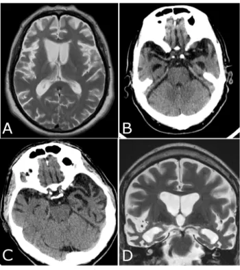
neuroimaging findings related to these patterns of atrophy. FTLD, mostly bvFTD, may also overlap with motor neuron disease in 10% of patients.10 Other conditions
pre-senting prominent symptoms related to disordered basal
Figure 1 – Neuroimaging findings in FTLD.
A (MRI, axial T2): bilateral frontal lobe atrophy, more so on the right side (left side of image) in bvFTD; B (CT scan): bilateral anterior temporal lobe atrophy in bvFTD; C (CT scan): left sided temporal lobe atrophy, around the perisylvian area, in a patient with PNFA whose symptoms had surfaced ten years before. Rostral slices (not shown here) disclose atrophy of left frontal lobe around anterior language centres; D (MRI, coronal T2): bilateral temporal lobe at-rophy, including lateral neocortex and medial structures, predomi-nantly on the right side, in a patient with SD (right-sided variant) whose symptoms had been noticed 7 years before.
ganglia circuits, such as progressive supranuclear palsy (PSP) and the corticobasal syndrome (CBS) have been as-sociated with FTLD.6,10
Behavioural variant FTD
This is the most prevalent clinical presentation within the FTLD category. Symptom onset is insidious, followed by gradually worsening course, where cognitive deficits are most recognizable with regard to executive functions, per-sonality and social decorum, with a relative maintenance of visuo-perceptive skills, at least until later stages.11
None-theless, exceptions have been described, especially in as-sociation with progranulin gene mutations.12 The diagnosis
may be hard to establish as these symptoms may also be found in other dementias, yet the most pronounced in bvFTD are personality changes along with apathy or dis-inhibition, which tend to be quite less pronounced in other disorders in early stages. Apathy is expressed by reduced motivation concerning work or prior hobbies and gradual social isolation, which can be misdiagnosed as pathologi-cal depression.13 Throughout disease course, patients may
disregard their personal hygiene and even lose sphincter control.14 Behavioural disinhibition is characterized by
im-pulsivity or misjudgement leading to overspending, inappro-priate interpersonal remarks and numerous embarrassing or antisocial attitudes, like breaking legal rules or embarking on physical threats, incongruent with premorbid personality and behaviour.14 Recurrent inappropriate sexual comments
can be noted but patients do not usually exhibit hypersexu-ality, but rather a decreased libido.1,13 The clinical picture is
not uncommonly mistaken with a psychiatric disorder. Patients with bvFTD show a variable decrease of their in-sight and commonly display stereotyped behaviours, which range from an exaggerated use of recurrent verbal sayings, to collecting and counting rituals.15 Often, they modify their
eating patterns, overeating sweets and consuming alcohol excessively and, during disease course, some may take in non-food items.16 Social emotions often become affected, as
patients show an egocentric, apathetic behaviour towards others, including close relatives who display remarkable concerns about their health status (i.e. affective blunting).17
Besides, they become inflexible when adapting to circum-stances and daily routines or distinct perspectives.18 Some
might demonstrate distractibility, perseverant attitudes, con-crete thinking, slowed speech or echolalia.19 They may also
display non-fluent aphasia, characterized by a shortage of word production and poverty of speech content.14,20
Overall cognitive decline of these patients can be less severe as compared to behavioural changes, and cognitive assessment commonly fails to recognize significant episo-dic memory deficits.14 Among the several symptoms, social
disinhibition, stereotyped actions and odd dietary changes are those most notably distinct from the clinical picture of AD.10
Imaging techniques usually disclose a pattern of frontal hypometabolism, hypoperfusion and atrophy (see Fig. 1), but topographical involvement might depend on the leading
symptoms observed: frontomedial with apathy, orbitofrontal with disinhibition and dorsolateral with executive dysfunc-tion.13,21
Primary Progressive Aphasia (PPA)
The term refers to a group of conditions typically featu-ring atrophy of the left frontal and temporal regions, bound to an insidious linguistic decline that lasts for at least two years, without compromising additional cognitive skills.22
The core presenting symptom is aphasia, and its variants are classified based on the type of specific language defi-cits.23-25 Nonetheless, the classification of PPA has been
revised recently, so that the terminology may still not be uni-formized.
SD patients display a fluent form of PPA. The hallmark of SD is a reduced efficiency on actions that rely on intact semantics. This way, besides fluent speech, they can either show anomia or comprehension decrement that end up in greater difficulties when recognizing objects and people.26
Some also suffer from variable degrees of dyslexia and dysgraphia, particularly noted while using unfamiliar or less frequent words, switching the ideal designations to broader terms or superordinate categories (e.g. ‘lion’ is identified as ‘cat’ or ‘animal’).27 Furthermore, subtle behavioural changes
similar to bvFTD are often found, especially as disease pro-gresses,28 though they do not dominate the clinical picture.
They exhibit degraded social functioning with depression, apathy or irritability, coupled with emotional coldness and loss of empathy. Behavioural rigidity, compulsive or re-petitive behaviours and peculiar food choices are also fre-quent.9,28 Imaging studies show a pattern of anterior
tempo-ral atrophy which is often more pronounced on the left side but can later spread to other temporal regions.29
In contrast, PNFA presents with an increasingly hesita-ting and less fluent speech, eventually coupled to apraxia of speech, with phonological errors, shorter and simpler phrases, agrammatism and aprosodia, and mutism might eventually ensue as disease progresses. This impairment is clearly influenced by the complexity of sentences, mean-ing that smean-ingle-word comprehension and object knowledge are usually spared, helping differentiate PNFA from other PPA variants.24,30,31 Social conduct, memory and
visuo-per-ceptive skills of these patients are usually normal, at least in early stages.32 Cortical atrophy in PNFA mainly involves
the left anterior perisylvian region, extending to the left dor-solateral prefrontal cortex as the disease progresses, which is in accordance with the regions responsible for sentence processing.24
LPA is defined by a speech output that is spontaneous but slow, but usually no discernible grammar errors or mo-tor control. However, unlike PNFA, these patients do not improve if the speech is simpler and normally show episodic memory deficits. The imaging pattern shows posterior peri-sylvian cortex atrophy, typically on the left hemisphere, and Alzheimer’s disease is usually the pathological underpin-ning, unlike the previous forms.9,24
Table 1 – Genes involved in hereditary FTLD and related disorders.
Locus Function Mutation effects (frequency) phenotypesAssociated
MAPT 17q21.31 Encodes microtubule-associated protein,
responsible for microtubule stabilization, promoting their binding with tubulin in order to enhance the protein-mediated transport
of vesicles and organelles.62, 63
Increases the number of toxic aggregates of tau protein
(< 25%)62, 64, 65 bvFTD PSP CBS bvFTD with parkinsonism bvFTD-ALS64
GRN 17q21.32 Encodes progranulin, a growth factor
involved in cell cycle and motility control, as well as on oncogenesis and inflammatory
cellular mechanisms.34, 66
Blocks progranulin translation through haploinsufficiency (5 - 25%)34, 67 bvFTD bvFTD with parkinsonism PNFA PSP CBS66, 68
C9ORF72 9p21.2 Still uncharacterized protein, with unknown function.69
Hexanucleotide expansion that leads to toxic RNA accumulation which loses its function (6 - 37%)69-71 bvFTD FTD-ALS PSP CBS71
CHMP2B 3p11.2 It is part of an endosomal complex (ESCRT) that controls endocytic pathways of protein
transport, autophagy and cytokinesis.72
Leads to the production of non-functional proteins
(< 1%)64,73
bvFTD
(later during course with parkinsonism, dystonia, myoclonus, upper motor
neuron features)73
VCP 9p13.3 Valosin containing protein is a structural
protein involved in the vesicle transport pathways and the control of cellular processes like mitosis and proteasomal
protein degradation.33,74
Decreases proteasomal
activity and increases protein aggregation
(< 1%)34,74
bvFTD
IBMPFD64,74
Legend: CHMP2B – chromatin-modifying 2B protein; C9ORF72 – chromosome 9 open reading frame 72; ESCRT – endosomal sorting complex required for transport; FTD-ALS – fron-totemporal dementia associated to amyotrophic lateral sclerosis; GRN – progranulin gene; IBMPFD – inclusion body myositis, Paget’s disease of bone and Fronfron-totemporal Dementia; MAPT – microtubule-associated protein tau gene; VCP – valosin-containing protein.
Table 2 – Neuropathological findings and phenotypical correlations in FTLD-TDP.
Type Cortical inclusions Clinico-genetic associations
NCI NII DN
A +++ + ++ (GRN and C9ORF72 mutations)bvFTD, PNFA, CBS
B ++ 0 + (C9ORF72 mutations)FTD-ALS, bvFTD
C + 0 +++ SD, bvFTD
D + +++ +++ bvFTD, IBMPFD (VCP mutations)
Legend: bvFTD – behavioural variant of frontotemporal dementia; C9ORF72 – chromosome 9 open reading frame 72; CBS – corticobasal syndrome; FTD-ALS – frontotemporal demen-tia associated to amyotrophic lateral sclerosis; GRN – progranulin gene; IBMPFD – inclusion body myositis, Paget’s disease of bone and frontotemporal demendemen-tia; PNFA – progressive nonfluent aphasia; SD – semantic dementia; VCP – valosin-containing protein.
B) Genetics
A positive family history of FTLD is present in 25-50% of cases,33,34 and the transmission is usually autosomal
domi-nant.35 A few genes have been associated with FTLD (Table
1).
C) Neuropathology
Apart from those instances where a genetic defect is re- cognized, post-mortem neuropathological brain examination is essential so that the entity underlying FTLD can be iden-tified. Also, dissimilar pathologies are often co-ideniden-tified.4
Linking phenotypical features and molecular pathology has been a huge challenge along the history of neuroscience re-search, and FTLD is probably the most paradigmatic case. The core pathological features of FTLD are the selective atrophy of frontotemporal cortex, associated with neuronal loss, gliosis and spongiosis of cortical superficial layers.1,5
Histochemically, FTLD can be categorized according to the major component of the cellular inclusions deposited in the brain (tau, TDP-43 and FUS), thus designating FTLD-tau, FTLD-TDP and FTLD-FUS, correspondingly.36
FTLD-tau
Microtubule-associated protein tau (MAPT) is a phos-phoprotein present mostly in neurons, enhancing microtu-bule polymerization, assortment and also stabilization, pri-marily in axons. This happens due to a binding interaction with the 3 or 4 microtubule-binding domains at its C-termi-nus (3R or 4R tau, respectively), relying on RNA splicing.37
Identical proportions of 3R and 4R tau can be found in the normal brain, whilst there may be preferential deposits of 3R or 4R in different tauopathies, thus offering a biochemi-cal subclassification.5 FTLD-tau cluster includes disorders
such as Pick’s disease (PiD), PSP and Corticobasal Dege-neration (CBD), as well as other entities such as Argyroph-ilic Grain Disease (AGD) or Multiple System Tauopathy with Dementia (MSTD).36,38 Fundamentally, tau mutations are
prone to considerably impair the binding to microtubules, via hyperphosphorylation mechanisms which have inhibi-tory outcomes resulting from abnormal tau aggregation.37,39
PiD is the prototype of FTLD and is a 3R tauopathy displaying Pick bodies, that are solitary, circular, argyro-philic inclusions located in neuronal cytoplasm. They can be usually seen in the dentate gyrus of the hippocampus, amygdala and frontotemporal neocortex, mainly in layers II and III.40 On the other hand, PSP and CBD are both 4R
tauopathies and they are more common than PiD.4,40 PSP
is characterized by bigger neuronal inclusions named glo-bose neurofibrillary tangles and also glial inclusions termed tufted astrocytes on the basal ganglia, subthalamic nucleus and substantia nigra.41 CBD is distinguished by astrocytic
plaques in basal ganglia, thalamus and brainstem, that are not found in any other pathology.4,40 Notwithstanding,
in both these entities significant cortical involvement might also be found.5
FTLD-TDP
TAR DNA-binding protein 43 (TDP-43) is a ubiquitously expressed RNA-binding protein most often located in the nucleus that can shuttle between the nucleus and cyto-plasm. It is a global regulator of transcription and other mul-tiple aspects of RNA processing and functioning.42 TDP-43
controls its own expression by a feedback system, ensuring that intracellular level is tightly controlled, which is impera-tive since it acts in multiprotein/RNA complexes, wherein a suitable structure needs a specified ratio between TDP-43 and its RNA partners.43 In a disease scenario (e.g.
FTLD-TDP), cellular conditions contribute to TDP-43 aggrega-tion. This may trigger a decrease in the pool of TDP-43 that could be integrated into the complexes, hence decreasing their activity and causing neurodegeneration.44 Pathological
classification is based on three types of TDP-43 immunore-active inclusions: neuronal cytoplasmatic inclusions (NCI), neuronal intranuclear inclusions (NII) and dystrophic neu-rites (DN). The first two classifications emerged in 2006 by Sampathu et al45 and Mackenzie et al46 the first using
mo-noclonal antibodies and the latter using clinicopathological correlations. Both were based on the fact that the inclusions were immunoreactive to ubiquitin but not tau, thus the desi-gnation FTLD-U.14 Later, this has been changed to
FTLD-TDP, when TDP-43 was identified as the main component, leading to a new classification centred on the relative fre-quency of four pathological subtypes summarized in Table 2, where a correlation with the main clinical phenotypes is also established.47,48
FTLD-FUS
After the FTLD-U label has been carved, about 7-20% of the patients were clinically determined to have negative in-clusions for TDP-43 pathology, thus the terminology ‘atypi-cal FTLD-U’ (aFTLD-U). Subsequently, research has been carried out in order to better characterize those inclusions which resulted in the identification of the fused in sarcoma (FUS) protein as their main component. Similarly to TDP-43, it is a ubiquitously expressed DNA/RNA binding protein that regulates numerous cellular processes like cell proli-feration, DNA repair and RNA splicing.49 For most cell types,
FUS can be found predominantly in the nucleus. Nonethe-less, once mutated, FUS becomes anomalously distributed in the cytoplasm, where it forms insoluble aggregates that feature a toxic gain-of-function.50 FUS inclusions are
mor-phologically identical to the ones containing TDP-43, with a variable amount of distinct NCI along with thick filament NII.51 These have been assigned to a few remote variants
of FTLD, such as neuronal intermediate filament disease (NIFID) and basophilic inclusion body disease (BIBD).38,51
Patients with FTD-FUS typically present with bvFTD symp-toms without associated motor neuron disease, and some with parkinsonism,49,52 yet an exclusive cognitive and
be-havioural outline can be found. Obsessions and rituals are typical, along with a social disengagement and executive impairments that present as perseveration and problematic mental shifting. Of note, cases with especially young age at
onset of symptoms have been described in association with FUS pathology.53
Clinical Management General issues
FTLD is frequently unfamiliar to the common citizen and many physicians. Therefore, FTLD caregivers can be es-pecially distressed while searching for medical advice, con-sidering that FTLD is significantly less prevalent and under-stood than AD and there is a higher frequency of upsetting behavioural symptoms in these patients.54 Medical
manage-ment should start with thorough explanation of symptoms and the condition itself to the family and caregivers, as most of the time they will be uninformed, anxious, depressed and even burned out – from our experience there is commonly the belief by the family that the patient might be faking the symptoms or acting on purpose. This might contribute to better understanding of the situation and hopefully lower stress levels. Due to the remarkable behavioural modifica-tions and compromised judgment abilities, safety tends to be problematic even in the early stages of FTLD (especially bvFTD), an issue that should be assessed and discussed with the caregiver.55
Genetic testing and counselling
Genetic counselling is a delicate issue requiring in-depth clinical and genetic knowledge with regard to disease fea-tures, allied with a degree of experience and sensitivity to-wards the subjects involved. As a rule of thumb, before em-barking on any kind of testing, patients and families should be informed in detail about the complex genetics of FTLD, the potential consequences of carrying a mutation, and the possibility of not detecting any mutations at all. The starting point is to actively obtain an extensive family history, which should include no less than three generations, in order to increase sensitivity and define the pattern of transmission, whenever possible.56 Should the classical autosomal
domi-nant pattern be uncovered, each direct family member (e.g. siblings, sons) of the index case has a 50% chance of har-bouring the genetic defect, whether it is identifiable or not through appropriate testing. If the family history is negative, the odds of finding a pathogenic mutation might decrease to about 3%.57 Clinical presentation of the index case and
affected family members should be thoroughly revised in order to define the phenotype and assist the clinician choo-sing the specific test, although phenotype-genotype corre-lation is far from perfect (Fig. 2) and phenotypic variability is common. The fact that incomplete and age-dependent
Patient diagnosed clinically with FTLD or related disorder
bvFTD with Paget’s disease of the bone and/or inclusion
body myosistis No family history of
dementia Family history of
dementia and/or ALS
Test for VCP Semantic dementia bvFTD, PNFA, CBS, PSP (without PSx) bvFTD, CBS PSP (with PSx) bvFTD, PNFA and CBS FTD-ALS If negative, test for GRN or no testing If negative, research
testing (tau, TPD, FUS) or no testing Testing unlikely to be positive Test for C9ORF72 or no testing Test for C9ORF72 Test for C9ORF72 If negative, test for GRN If negative, test for MAPT If negative Test for MAPT Test for GRN Test for C9ORF72 Family history of dementia without ALS
Figure 2 – Genetic testing algorithm: author proposal based on phenotypical correlations and estimated relative frequency of the several
genetic causes. Legend: ALS – amyotrophic lateral sclerosis; bvFTD – behavioural variant of frontotemporal dementia; C9ORF72 – chromosome 9 open reading frame 72; CBS – corticobasal syndrome; FTD-ALS – frontotemporal dementia associated with amyotrophic lateral sclerosis; FTLD – frontotemporal lobar degeneration; FUS – fused in sarcoma protein; MAPT – microtubule-associated protein tau; GRN – progranulin; PNFA – progressive nonfluent aphasia; PSP – progressive supranuclear palsy; PSx – parietal symptoms; SD – se-mantic dementia; TDP – TAR-DNA binding protein; VCP – valosin-containing protein.
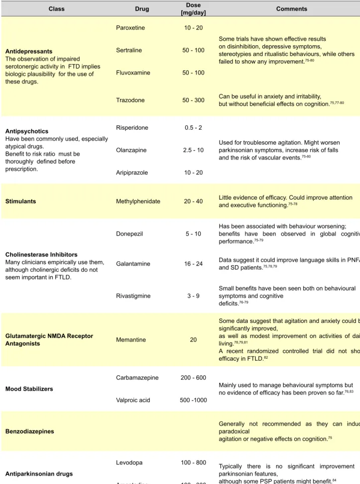
Table 3 – Current options for the pharmacological treatment of FTLD, based on published evidence and the authors’ experience.
Class Drug [mg/day]Dose Comments
Antidepressants
The observation of impaired serotonergic activity in FTD implies biologic plausibility for the use of these drugs.
Paroxetine 10 - 20
Some trials have shown effective results on disinhibition, depressive symptoms,
stereotypies and ritualistic behaviours, while others
failed to show any improvement.75-80
Sertraline 50 - 100
Fluvoxamine 50 - 100
Trazodone 50 - 300 Can be useful in anxiety and irritability, but without beneficial effects on cognition.75,77-80
Antipsychotics
Have been commonly used, especially atypical drugs.
Benefit to risk ratio must be thoroughly defined before prescription.
Risperidone 0.5 - 2
Used for troublesome agitation. Might worsen parkinsonian symptoms, increase risk of falls
and the risk of vascular events.75-80
Olanzapine 2.5 - 10
Aripiprazole 10 - 20
Stimulants Methylphenidate 20 - 40 Little evidence of efficacy. Could improve attention and executive functioning.75-78
Cholinesterase Inhibitors
Many clinicians empirically use them, although cholinergic deficits do not seem important in FTLD.
Donepezil 5 - 10 Has been associated with behaviour worsening; benefits have been observed in global cognitive
performance.75-79
Galantamine 16 - 24 Data suggest it could improve language skills in PNFA and SD patients.75,78,79
Rivastigmine 3 - 9 Small benefits have been seen both on behavioural symptoms and cognitive
deficits.76-79
Glutamatergic NMDA Receptor
Antagonists Memantine 20
Some data suggest that agitation and anxiety could be significantly improved,
as well as modest improvement on activities of daily
living.78,79,81
A recent randomized controlled trial did not show
efficacy in FTLD.82
Mood Stabilizers
Carbamazepine 200 - 600
Mainly used to manage behavioural symptoms but
no evidence of efficacy has been proven so far.76,83
Valproic acid 500 -1000
Benzodiazepines Generally not recommended as they can induce paradoxical
agitation or negative effects on cognition.75
Antiparkinsonian drugs
Levodopa 100 - 800 Typically there is no significant improvement of
parkinsonian features,
although some PSP patients might benefit.84
Amantadine 100 - 300
sedation are frequently seen, suggesting that minimal avai-lable dosage administration, gradual upward dose titration and regular clinical monitoring should be performed. Table 3 summarizes the currently available options.
CONCLUSIONS
FTLD refers to a group of disorders with heterogene-ous clinical, molecular and pathological features. Disease consequences (e.g. individual, familiar, social, economic) can be devastating, especially since it generally affects younger individuals, as compared to AD or DLB. Current therapy is purely symptomatic and efficacy modest at most. Recent advances have been seen with regard to genetic causes, thus genetic counselling and testing is an important process during clinical management. FTLD is commonly seen in Neurology and Psychiatry clinics, although it re-mains largely unknown to many clinicians and the general public. Thereby, we propose a reinforcement of the public information about FTLD, along with the implementation of interventions focused on decreasing the burden of care-givers. Intervention by patient’s associations would be most welcomed and should play a major role in this process. Fu-ture research should move the field to robust experimental designs, comprising adequate sample sizes and endpoints, as well as detailed analyses. This would maximize the amount and quality of research findings, thus contributing to enhance optimal care for these patients.
CONFLICT OF INTEREST AND FUNDING SOURCES
Jorge Pelicano Paulos reports no conflicts of interest. João Massano has acted as an advisor and received hono-raria and financial support to speak or attend meetings from Bial, Grünenthal, Lundbeck, Novartis and Tecnifar compa-nies). This work received no specific grant from any funding agency in the public, commercial or not-for-profit sectors.
REFERENCES
1. Seelaar H, Rohrer JD, Pijnenburg YA, Fox NC, van Swieten JC. Clinical, genetic and pathological heterogeneity of frontotemporal dementia: a review. J Neurol Neurosurg Psychiatry. 2011;82:476-86.
2. Brun A, Gustafson L. The birth and early evolution of the frontotemporal dementia (FTD) concept. J Mol Neurosci. 2011;45:324-9.
3. Rosso SM, Donker Kaat L, Baks T, Joosse M, de Koning I, Pijnenburg Y, et al. Frontotemporal dementia in The Netherlands: patient character-istics and prevalence estimates from a population-based study. Brain. 2003;126:2016-22.
4. Cairns NJ, Bigio EH, Mackenzie IR, Neumann M, Lee VM, Hatanpaa KJ, et al. Neuropathologic diagnostic and nosologic criteria for frontotempo-ral lobar degeneration: consensus of the Consortium for Frontotempofrontotempo-ral Lobar Degeneration. Acta Neuropathol. 2007;114:5-22.
5. Taipa R, Pinho J, Melo-Pires M. Clinico-pathological correlations of the most common neurodegenerative dementias. Front Neurol. 2012;3:68. 6. Cerami C, Scarpini E, Cappa SF, Galimberti D. Frontotemporal lobar
degeneration: current knowledge and future challenges. J Neurol. 2012;259:2278-86.
7. Miller BL, Cummings JL. The human frontal lobes : functions and disor-ders. 2nd ed. New York: Guilford Press; 2007.
8. Rascovsky K, Hodges JR, Knopman D, Mendez MF, Kramer JH, Neu-haus J, et al. Sensitivity of revised diagnostic criteria for the behavioural variant of frontotemporal dementia. Brain. 2011;134:2456-77.
9. Gorno-Tempini ML, Hillis AE, Weintraub S, Kertesz A, Mendez M, Cappa penetrance exists, especially with regard to GRN and C9orf72, brings additional difficulties to the process of ge-netic counselling.58 Next-generation genomic sequencing
techniques might render the process of genetic testing in FTLD much easier and quicker in the future.59 Lastly,
pre-dictive genetic testing can be carried out in asymptomatic individuals who manifest such a wish, but only after a clear-ly pathogenic mutation has become evident in the index case. Of note, it has been found that both symptomatic and asymptomatic GRN mutation carriers display significantly decreased serum levels, unlike non-carriers. Such testing is a viable and currently much cheaper tool as compared to genetic testing that can be used for screening both sympto-matic patients and at risk individuals.60,61 As legally defined,
predictive testing in Portugal must be performed exclusively in Medical Genetics specialized clinics, and testing is not al-lowed in asymptomatic individuals until they reach the age of 18 years.
We acknowledge that genetic testing and counselling is challenging. Clinicians ought to approach the process with exhaustive clarification of interpersonal matters, without ignoring the psychological frailty of the patients and fami-lies.
Pharmacological interventions
There is clear scarcity of evidence with regard to the pharmacological treatment of FTLD, with most data extract-ed from small trials, case series or isolatextract-ed case reports. Therefore, drugs are used off label. There is no evidence that disease-modifying interventions have been made avai-lable so far, hence treatment remains purely symptomatic. Once behavioural symptoms emerge or become trouble-some, clinicians must consider assessing pain, delirium or distress before embarking on potentially more aggressive pharmacological options. Also, adverse effects such as paradoxical responses, confusion, extrapyramidal effects or
SF, et al. Classification of primary progressive aphasia and its variants. Neurology. 2011;76:1006-14.
10. Piguet O, Hornberger M, Mioshi E, Hodges JR. Behavioural-variant frontotemporal dementia: diagnosis, clinical staging, and management. Lancet Neurol. 2011;10:162-72.
11. Huey ED, Goveia EN, Paviol S, Pardini M, Krueger F, Zamboni G, et al. Executive dysfunction in frontotemporal dementia and corticobasal syndrome. Neurology. 2009;72:453-9.
12. Taipa R, Tuna A, Damasio J, Pinto PS, Cavaco S, Pereira S, et al. Clinical, neuropathological, and genetic characteristics of the novel IVS9+1delG GRN mutation in a patient with frontotemporal dementia. J Alzheimer’s Dis. 2012;30:83-90.
13. Zamboni G, Huey ED, Krueger F, Nichelli PF, Grafman J. Apathy and disinhibition in frontotemporal dementia: Insights into their neural cor-relates. Neurology. 2008;71:736-42.
14. Rabinovici GD, Miller BL. Frontotemporal lobar degeneration: epide-miology, pathophysiology, diagnosis and management. CNS drugs. 2010;24:375-98.
15. Mendez MF, Shapira JS, Miller BL. Stereotypical movements and fronto-temporal dementia. Mov Disord. 2005;20:742-5.
16. Piguet O, Petersen A, Yin Ka Lam B, Gabery S, Murphy K, Hodges JR, et al. Eating and hypothalamus changes in behavioral-variant frontotem-poral dementia. Ann Neurol. 2011;69:312-9.
17. Kipps CM, Mioshi E, Hodges JR. Emotion, social functioning and
ties of daily living in frontotemporal dementia. Neurocase. 2009;15:182-9.
18. Rankin KP, Baldwin E, Pace-Savitsky C, Kramer JH, Miller BL. Self awareness and personality change in dementia. J Neurol Neurosurg Psychiatry. 2005;76:632-9.
19. Galariotis V, Bodi N, Janka Z, Kalman J. Frontotemporal dementia--Part I. History, prevalence, clinical forms. Ideggyogy Sz. 2005;58:164-71. 20. Neary D, Snowden J, Mann D. Frontotemporal dementia. Lancet Neurol.
2005;4:771-80.
21. Rosen HJ, Allison SC, Schauer GF, Gorno-Tempini ML, Weiner MW, Miller BL. Neuroanatomical correlates of behavioural disorders in de-mentia. Brain. 2005;128:2612-25.
22. Matias-Guiu JA, Garcia-Ramos R. Primary progressive aphasia: from syndrome to disease. Neurologia. 2013;28:366-74.
23. Mesulam MM. Primary progressive aphasia-a language-based demen-tia. N Engl J Med. 2003;349:1535-42.
24. Bonner MF, Ash S, Grossman M. The new classification of primary pro-gressive aphasia into semantic, logopenic, or nonfluent/agrammatic variants. Curr Neurol Neurosci Rep. 2010;10:484-90.
25. Mesulam M, Wieneke C, Rogalski E, Cobia D, Thompson C, Weintraub S. Quantitative template for subtyping primary progressive aphasia. Arch Neurol. 2009;66:1545-51.
26. Kertesz A, Jesso S, Harciarek M, Blair M, McMonagle P. What is seman-tic dementia?: a cohort study of diagnosseman-tic features and clinical boundar-ies. Arch Neurol. 2010;67:483-9.
27. Gainotti G. Different patterns of famous people recognition disorders in patients with right and left anterior temporal lesions: a systematic review. Neuropsychologia. 2007;45:1591-607.
28. Hodges JR, Patterson K. Semantic dementia: a unique clinicopathologi-cal syndrome. Lancet Neurol. 2007;6:1004-14.
29. Rohrer JD, Warren JD, Modat M, Ridgway GR, Douiri A, Rossor MN, et al. Patterns of cortical thinning in the language variants of frontotempo-ral lobar degeneration. Neurology. 2009;72:1562-9.
30. Josephs KA, Duffy JR, Strand EA, Whitwell JL, Layton KF, Parisi JE, et al. Clinicopathological and imaging correlates of progressive aphasia and apraxia of speech. Brain. 2006;129:1385-98.
31. Peelle JE, Troiani V, Gee J, Moore P, McMillan C, Vesely L, et al. Sen-tence comprehension and voxel-based morphometry in progressive nonfluent aphasia, semantic dementia, and nonaphasic frontotemporal dementia. J Neurolinguistics. 2008;21:418-32.
32. Knibb JA, Xuereb JH, Patterson K, Hodges JR. Clinical and pathological characterization of progressive aphasia. Ann Neurol. 2006;59:156-65. 33. Galimberti D, Scarpini E. Genetics of frontotemporal lobar degeneration.
Front Neurol. 2012;3:52.
34. Rademakers R, Neumann M, Mackenzie IR. Advances in understand-ing the molecular basis of frontotemporal dementia. Nat Rev Neurol. 2012;8:423-34.
35. Rohrer JD, Guerreiro R, Vandrovcova J, Uphill J, Reiman D, Beck J, et al. The heritability and genetics of frontotemporal lobar degeneration. Neurology. 2009;73:1451-6.
36. Mackenzie IR, Neumann M, Bigio EH, Cairns NJ, Alafuzoff I, Kril J, et al. Nomenclature and nosology for neuropathologic subtypes of frontotem-poral lobar degeneration: an update. Acta Neuropathol. 2010;119:1-4 37. Mandelkow EM, Mandelkow E. Biochemistry and cell biology of tau
protein in neurofibrillary degeneration. Cold Spring Harb Perspect Med. 2012;2:a006247.
38. Josephs KA, Hodges JR, Snowden JS, Mackenzie IR, Neumann M, Mann DM, et al. Neuropathological background of phenotypical vari-ability in frontotemporal dementia. Acta Neuropathol. 2011;122:137-53. 39. Stamer K, Vogel R, Thies E, Mandelkow E, Mandelkow EM. Tau blocks
traffic of organelles, neurofilaments, and APP vesicles in neurons and enhances oxidative stress. J Cell Biol. 2002;156:1051-63.
40. Dickson DW, Kouri N, Murray ME, Josephs KA. Neuropathology of fron-totemporal lobar degeneration-tau (FTLD-tau). J Mol Neurosci. 2011 Nov;45:384-9.
41. Boxer AL, Geschwind MD, Belfor N, Gorno-Tempini ML, Schauer GF, Miller BL, et al. Patterns of brain atrophy that differentiate corticobasal degeneration syndrome from progressive supranuclear palsy. Arch Neu-rol. 2006;63:81-6.
42. Buratti E, Baralle FE. The multiple roles of TDP-43 in pre-mRNA pro-cessing and gene expression regulation. RNA Biol. 2010;7:420-9. 43. Sephton CF, Good SK, Atkin S, Dewey CM, Mayer P 3rd, Herz J, et al.
TDP-43 is a developmentally regulated protein essential for early em-bryonic development. J Biol Chem. 2010;285:6826-34.
44. Zhang YJ, Xu YF, Cook C, Gendron TF, Roettges P, Link CD, et al. Ab-errant cleavage of TDP-43 enhances aggregation and cellular toxicity.
Proc Natl Acad Sci U S A. 2009;106:7607-12.
45. Sampathu DM, Neumann M, Kwong LK, Chou TT, Micsenyi M, Truax A, et al. Pathological heterogeneity of frontotemporal lobar degen-eration with ubiquitin-positive inclusions delineated by ubiquitin im-munohistochemistry and novel monoclonal antibodies. Am J Pathol. 2006;169:1343-52.
46. Mackenzie IR, Baborie A, Pickering-Brown S, Du Plessis D, Jaros E, Perry RH, et al. Heterogeneity of ubiquitin pathology in frontotemporal lobar degeneration: classification and relation to clinical phenotype. Acta Neuropathol. 2006;112:539-49.
47. Mackenzie IR, Neumann M, Baborie A, Sampathu DM, Du Plessis D, Jaros E, et al. A harmonized classification system for FTLD-TDP pathol-ogy. Acta Neuropathol. 2011;122:111-3.
48. Rohrer JD, Lashley T, Schott JM, Warren JE, Mead S, Isaacs AM, et al. Clinical and neuroanatomical signatures of tissue pathology in fronto-temporal lobar degeneration. Brain. 2011;134:2565-81.
49. Lagier-Tourenne C, Cleveland DW. Rethinking ALS: the FUS about TDP-43. Cell. 2009;136:1001-4.
50. Kwiatkowski TJ Jr., Bosco DA, Leclerc AL, Tamrazian E, Vanderburg CR, Russ C, et al. Mutations in the FUS/TLS gene on chromosome 16 cause familial amyotrophic lateral sclerosis. Science. 2009;323:1205-8. 51. Neumann M, Rademakers R, Roeber S, Baker M, Kretzschmar HA,
Mackenzie IR. A new subtype of frontotemporal lobar degeneration with FUS pathology. Brain. 2009;132:2922-31.
52. Roeber S, Mackenzie IR, Kretzschmar HA, Neumann M. TDP-43-neg-ative FTLD-U is a significant new clinico-pathological subtype of FTLD. Acta Neuropathol. 2008;116:147-57.
53. Snowden JS, Hu Q, Rollinson S, Halliwell N, Robinson A, Davidson YS, et al. The most common type of FTLD-FUS (aFTLD-U) is associated with a distinct clinical form of frontotemporal dementia but is not related to mutations in the FUS gene. Acta Neuropathol. 2011;122:99-110. 54. Chow TW, Pio FJ, Rockwood K. An international needs assessment of
caregivers for frontotemporal dementia. Can J Neurol Sci. 2011;38:753-7.
55. de Vugt ME, Riedijk SR, Aalten P, Tibben A, van Swieten JC, Verhey FR. Impact of behavioural problems on spousal caregivers: a compari-son between Alzheimer’s disease and frontotemporal dementia. Dement Geriatr Cogn Disord. 2006;22:35-41.
56. Quaid KA. Genetic counseling for frontotemporal dementias. J Mol Neu-rosci. 2011;45:706-9.
57. Goldman JS, Rademakers R, Huey ED, Boxer AL, Mayeux R, Miller BL, et al. An algorithm for genetic testing of frontotemporal lobar degenera-tion. Neurology. 2011;76:475-83.
58. Cohn-Hokke PE, Elting MW, Pijnenburg YA, van Swieten JC. Genetics of dementia: update and guidelines for the clinician. Am J Med Genet B Neuropsychiatr Genet. 2012;159B:628-43.
59. Bras J, Guerreiro R, Hardy J. Use of next-generation sequencing and other whole-genome strategies to dissect neurological disease. Nat Rev Neurosci. 2012;13:453-64.
60. Ghidoni R, Benussi L, Glionna M, Franzoni M, Binetti G. Low plasma progranulin levels predict progranulin mutations in frontotemporal lobar degeneration. Neurology. 2008;71:1235-9.
61. Finch N, Baker M, Crook R, Swanson K, Kuntz K, Surtees R, et al. Plasma progranulin levels predict progranulin mutation status in fronto-temporal dementia patients and asymptomatic family members. Brain. 2009 Mar;132:583-91.
62. Rossi G, Bastone A, Piccoli E, Mazzoleni G, Morbin M, Uggetti A, et al. New mutations in MAPT gene causing frontotemporal lobar degen-eration: biochemical and structural characterization. Neurobiol Aging. 2012;33:834e1-6.
63. Rovelet-Lecrux A, Hannequin D, Guillin O, Legallic S, Jurici S, Wallon D, et al. Frontotemporal dementia phenotype associated with MAPT gene duplication. J Alzheimer’s Dis 2010;21:897-902.
64. Sieben A, Van Langenhove T, Engelborghs S, Martin JJ, Boon P, Cras P, et al. The genetics and neuropathology of frontotemporal lobar degen-eration. Acta Neuropathol. 2012;124:353-72.
65. Rademakers R, Cruts M, van Broeckhoven C. The role of tau (MAPT) in frontotemporal dementia and related tauopathies. Human Mutat. 2004;24:277-95.
66. Sun L, Eriksen JL. Recent insights into the involvement of progranulin in frontotemporal dementia. Curr Neuropharmacol. 2011;9:632-42. 67. Gass J, Cannon A, Mackenzie IR, Boeve B, Baker M, Adamson J, et al.
Mutations in progranulin are a major cause of ubiquitin-positive fronto-temporal lobar degeneration. Hum Mol Genet. 2006;15:2988-3001. 68. van Swieten JC, Heutink P. Mutations in progranulin (GRN) within the
spectrum of clinical and pathological phenotypes of frontotemporal
mentia. Lancet Neurol. 2008;7:965-74.
69. DeJesus-Hernandez M, Mackenzie IR, Boeve BF, Boxer AL, Baker M, Rutherford NJ, et al. Expanded GGGGCC hexanucleotide repeat in noncoding region of C9ORF72 causes chromosome 9p-linked FTD and ALS. Neuron. 2011;72:245-56.
70. van der Zee J, Gijselinck I, Dillen L, Van Langenhove T, Theuns J, Engel-borghs S, et al. A Pan-European Study of the C9orf72 repeat associated with FTLD: geographic prevalence, genomic instability, and intermediate repeats. Hum Mutat. 2013;34:363-73.
71. Sha SJ, Takada LT, Rankin KP, Yokoyama JS, Rutherford NJ, Fong JC, et al. Frontotemporal dementia due to C9ORF72 mutations: Clinical and imaging features. Neurology. 2012;79:1002-11.
72. Urwin H, Ghazi-Noori S, Collinge J, Isaacs A. The role of CHMP2B in frontotemporal dementia. Biochem Soc Trans. 2009;37:208-12. 73. Isaacs AM, Johannsen P, Holm I, Nielsen JE, consortium FR.
Fronto-temporal dementia caused by CHMP2B mutations. Curr Alzheimer Res. 2011;8:246-51.
74. Gitcho MA, Strider J, Carter D, Taylor-Reinwald L, Forman MS, Goate AM, et al. VCP mutations causing frontotemporal lobar degeneration disrupt localization of TDP-43 and induce cell death. J Biol Chem. 2009;284:12384-98.
75. Freedman M. Frontotemporal dementia: recommendations for therapeu-tic studies, designs, and approaches. Can J Neurol Sci. 2007;34:S118-24.
76. Bei H, Ross L, Neuhaus J, Knopman D, Kramer J, Boeve B, et al. Off-label medication use in frontotemporal dementia. Am J Alzheimers Dis
Other Demen. 2010;25:128-33.
77. Huey ED, Putnam KT, Grafman J. A systematic review of neurotrans-mitter deficits and treatments in frontotemporal dementia. Neurology. 2006;66:17-22.
78. Kaye ED, Petrovic-Poljak A, Verhoeff NP, Freedman M. Frontotemporal dementia and pharmacologic interventions. J Neuropsychiatry Clin Neu-rosci. 2010;22:19-29.
79. Kerchner GA, Tartaglia MC, Boxer A. Abhorring the vacuum: use of Al-zheimer’s disease medications in frontotemporal dementia. Expert Rev Neurother. 2011;11:709-17.
80. Massimo L, Grossman M. Patient care and management of fronto-temporal lobar degeneration. Am J Alzheimers Dis Other Demen. 2008;23:125-31.
81. Boxer AL, Lipton AM, Womack K, Merrilees J, Neuhaus J, Pavlic D, et al. An open-label study of memantine treatment in 3 subtypes of frontotem-poral lobar degeneration. Alzheimer Dis Assoc Disord. 2009;23:211-7. 82. Boxer AL, Knopman DS, Kaufer DI, Grossman M, Onyike C, Graf-Rad-ford N, et al. Memantine in patients with frontotemporal lobar degenera-tion: a multicentre, randomised, double-blind, placebo-controlled trial. Lancet Neurol. 2013;12:149-56.
83. Manes FF, Torralva T, Roca M, Gleichgerrcht E, Bekinschtein TA, Hodg-es JR. Frontotemporal dementia prHodg-esenting as pathological gambling. Nat Rev Neurol. 2010;6:347-52.
84. van Balken I, Litvan I. Current and future treatments in progressive su-pranuclear palsy. Curr Treat Options Neurol. 2006;8:211-23.