1
6
Joana de Moura Gonçalves Pir
es
Influence of superparamagnetic iron o
xide nanopar
ticles on macrophages in removing att
ached bacteria
Joana de Moura Gonçalves Pires
Influence of superparamagnetic
iron oxide nanoparticles on macrophages
in removing attached bacteria
Universidade do Minho
Escola de Engenharia
Dissertação de Mestrado
Mestrado Integrado em Engenharia Biomédica
Trabalho efetuado sob a orientação do
Professora Doutora Lígia Raquel Marona Rodrigues
Universidade do Minho
Professora Doutora Henny van der Mei
University Medical Center Groningen
Joana de Moura Gonçalves Pires
Influence of superparamagnetic
iron oxide nanoparticles on macrophages
in removing attached bacteria
Universidade do Minho
Escola de Engenharia
Nome: Joana de Moura Gonçalves Pires
Endereço eletrónico: joanapires_@hotmail.com Telefone: 914405794/226183479
Número do Cartão de Cidadão: 13730489
Título da dissertação:
Influence of superparamagnetic iron oxide nanoparticles on macrophages in removing attached bacteria
Orientador/a/es:
Professora Doutora Lígia Raquel Marona Rodrigues Professora Doutora Henny van der Mei
Ano de conclusão: 2016
Mestrado Integrado em Engenharia Biomédica Ramo de Engenharia Clínica
DE ACORDO COM A LEGISLAÇÃO EM VIGOR, NÃO É PERMITIDA A REPRODUÇÃO DE QUALQUER PARTE DESTA TESE/TRABALHO.
Influence of superparamagnetic iron oxide nanoparticles on macrophages in removing attached bacteria
A
CKNOWLEDGMENTSMy first words are to my supervisor in Groningen, Henny van der Mei and Theo van Kooten. I would like to thank you so much for all the support. Henny, my words of thankfulness for the opportunity to integrate the department of biomedical engineering in UMCG. Your advices were so important for me during the time I spent in UMCG and for my research work. Theo, you were present during my research and I could not finish my research without your help. Thank you for all that you teach me.
I also would like to thank to my supervisor in Portugal, Drª. Lígia Rodrigues. My words of admiration for all the patience and time you spent helping me with this project and for all the motivation you gave to me to go in this amazing experience and country.
Betsy, Willem, Gesinda and Minnie, thank you for all the time you spent with me in the laboratory and for always finding a way to help me.
Melissa, thank you so much for the help and support during my time in UMCG and thank you also to teach me all the Dutch traditions. I know I have a friend for a life time.
Casper, Shardul and Sudi thank you for being the best office friends. You were a big support to me.
Pauline, Mathilde and Blandine, thank you for being my Erasmus family. Without you, this experience would not be the same. Thank you for all your support and patience during this amazing time. You are really important to me and I would never forget you and all the moments we spent together. You will be forever my French girls.
To all my Erasmus friends, thank you for everything. I will never forget the time that we passed together.
Another person that was all the time with me, in the best and the worst moments was my boyfriend. Thank you for being my guide and my support during the Erasmus experience. Thank you everyday for you friendship, patience, comprehension and love.
Helena, you were the best person I have ever met in this university. If it was not you, I would not be the same. Thank you for all your support and friendship. I know I can count on you until my lasts days.
Carolina, Mariana and Raquel, thank you for being there my whole life, especially when I was away from Portugal. Without you, nothing would be possible. You are the best friends that I can have.
Didi and Rui thank for all the support during high school and to let me know the existence of biomedical engineering. If it was not you, I would not be in this course and I would not go to Groningen.
My last words are to all my family. Mom and Dad, thanks for believing me all the time and to never give up on me. Thank you for giving me all the opportunities and to being there in the best and worst moments. You gave everything I have and all the words are not enough for what you did to me. You are the best parents in the world. Filipa, I would like to thank you for all the confidence, encouragement, understanding and help during my life. You were there for me every single moment I needed. Grandmom and granddad, thank you for taking care of me when I was a little child and to all the support you gave to me during my whole life. I couldn’t ask for a better family. I love you so much.
To my paternal grandparent, I know you would be proud of me.
Influence of superparamagnetic iron oxide nanoparticles on macrophages in removing attached bacteria
A
BSTRACTBiomaterials play an important role in human life to restore and support function in order to generate a better quality of life, as well as a long life time of patients. Bacterial biofilms can increase the pathogenicity of infection and constitute a major problem in modern medicine, especially on biomedical devices and implants. Biofilms are difficult to treat with antibiotics, even to eradicate by the host immune system, being the major cause of implant and device failure. Therefore, new strategies to prevent and cure bacterial infection need to be found. Nanoparticles have many special properties as small size and large surface area, surface reactivity, crystallinity, electronic properties, charge, shape, hydrophobicity / hydrophilicity, and solubility. Superparamagnetic iron oxide nanoparticles (SPIONs) represent a special class of biocompatible nanoparticles that have one specific component called magnetic particle core that can be targeted to on specific location through external magnets. These specific nanoparticles present good biocompatibility with human cells, such as macrophages, and also strong antimicrobial properties. The main goal of this thesis was to evaluate the influence of SPIONs in macrophages and consequently its impact in bacterial biofilms. Staphylococcus aureus biofilms were exposed to macrophages in the presence and absence of SPIONs and microscopically analysed. SPIONs were internalized by macrophages, yielding 10% less staphylococcal survival as compared to the macrophages alone. In conclusion, the presence of SPIONs on macrophages increases the efficacy to remove staphylococci from infectious biofilms, which can have a major impact on the prevention and cure of bacterial infections.
Keywords: Biomaterial- associated infections (BAI), bacterial biofilms, macrophages,
superparamagnetic iron oxide nanoparticles (SPIONs), fluorescent microscopy.
Influence of superparamagnetic iron oxide nanoparticles on macrophages in removing attached bacteria
R
ESUMOOs biomateriais apresentam uma enorme importância nos dias de hoje, devido às suas funções de suporte e restruturação, garantindo uma melhor qualidade de vida e melhor esperança média de vida dos pacientes. Os biofilmes bacterianos podem aumentar a fagoticidade de uma infeção, constituindo um grande problema na medicina moderna, especialmente em implantes e dispositivos biomédicos. Os biofilmes são de difícil tratamento com o uso de antibióticos, ou mesmo pela erradicação através do sistema imune do hospedeiro, sendo a maior causa de falha de implantes e dispositivos médicos. As nanopartículas apresentam diversas propriedades especiais, como o seu tamanho pequeno e larga área de superfície, bem como a superfície reativa, cristalinidade, propriedades elétricas, carga, forma, hidrofobicidade / hidrofilicidade e solubilidade. As nanopartículas superparamagnéticas de óxido de ferro (SPIONs) representam uma classe especial de nanopartículas biocompatíveis, apresentando um componente específico chamado núcleo de partículas magnéticas, que pode ser direcionado para um local específico usando magnetos externos. Estas nanopartículas apresentam uma boa biocompatibilidade com células humanas, como os macrófagos, e elevadas propriedades antimicrobianas. O principal objetivo desta tese foi observar a influência das SPIONs nos macrófagos e consequentemente o seu impacto nos biofilmes bacterianos. Os biofilmes de Staphylococcus aureus foram expostos aos macrófagos na presença e ausência das SPIONs e posteriormente analisados ao microscópio. As SPIONs foram internalizadas pelos macrófagos, tendo-se observado um decréscimo de cerca de 10% de sobrevivência de bactérias comparativamente com os ensaios em que se usou apenas macrófagos. Concluindo, a presença das SPIONs nos macrófagos aumenta a eficácia de remoção de biofilmes infeciosos de estafilococos, o que pode ter um grande impacto na prevenção e cura de infeções bacterianas.
Palavras - chave: Infeções associadas a biomateriais (BAI), biofilmes bacterianos,
macrófagos, nanopartículas superparamagnéticas de óxido de ferro (SPIONs), microscopia de fluorescência.
Influence of superparamagnetic iron oxide nanoparticles on macrophages in removing attached bacteria
T
ABLE OFC
ONTENTS Acknowledgments ... iii Abstract ... v Resumo ... vii Table of Contents ... ix List of Figures ... xiList of Tables ... xii
Abbreviations ... xiii
1. Chapter I – General Introduction ... 1
1.1. Motivation and Goals ... 3
1.2. State of Art ... 3
1.2.1 Biomaterials ... 4
1.2.2 Biomaterial- associated Infections ... 5
1.2.3 Staphylococcus aureus ... 8
1.2.4 Biofilm formation and resistance to antibiotics ... 9
1.2.4 Macrophages ... 10
1.2.4 Superparamagnetic iron oxide nanoparticles (SPIONs) ... 12
2. Chapter II – Materials and Methods ... 15
2.1. Strains and cell line ... 17
2.2. S. aureus culturing and harvesting ... 17
2.3. Macrophage culturing and harvesting ... 17
2.4. Macrophages staining ... 17
2.5. Nanoparticles characterization ... 18
2.6. Influence of the bacterial challenge concentration on phagocytosis in the presence and absence of SPIONS ... 18
2.7. Evaluation of the bacterial survival inside macrophages ... 19
3. Chapter III - Results and Discussion ... 21
3.1. Influence of the bacterial challenge concentration on phagocytosis in the presence and absence of SPIONs ... 23
3.2. Evaluation of the staphylococcal phagocytosis in the presence and absence of
SPIONs through fluorescent microscopy ... 26
3.3. Evaluation of the bacterial survival inside macrophages ... 28
4. Chapter IV - Conclusion and Future Work ... 31
Influence of superparamagnetic iron oxide nanoparticles on macrophages in removing attached bacteria
L
IST OF FIGURESFigure 1 - Risk factors associated with the development of a biomaterial- associated infection
(Taken from Busscher et al., 2012).
Figure 2 - Penetration of antibiotics and SPIONs into the biofilm (Taken from Subbiahdoss et
al., 2012).
Figure 3 – Graphic representing the number of live staphylococci after phagocytosis in the
presence or absence of SPIONs. The number of bacteria was determined by fluorescence microscopy after 2 hours of phagocytosis (J744A.1 macrophages). Error bars represent the standard deviation (SD) over different experiments, each involving images randomly localized.
Figure 4 - Graphic representing the percentage of staphylococci survival after phagocytosis in
the presence or absence of SPIONs. The number of bacteria was determined by fluorescence microscopy after 2 hours of phagocytosis (J744A.1 macrophages). Error bars represent the standard deviation (SD) over different experiments, each involving images randomly localized.
Figure 5 - Fluorescence images of green-fluorescent S.aureus ATCC 12600GFP and macrophages J774.1A after 2 hours of phagocytosis in the presence and absence of SPIONs. (A) S.aureus ATCC 12600GFP; (B) S.aureus ATCC 12600GFP and macrophages in the first contact; (C) S.aureus ATCC 12600GFP and SPIONs (after 2h); (D) S.aureus ATCC 12600GFP and macrophages (after 2h); (E) S.aureus ATCC 12600GFP and macrophages in the presence of SPIONs (after 2h).
Figure 6 – Graphic representing the number of colonies corresponding to bacterial survival
inside the macrophages. The number of colonies was evaluated by the platting method. Error bars represent the standard deviation (SD) over four experiences.
Figure 7 – Graphic representing the number of colonies corresponding to bacterial survival
inside the macrophages. The number of colonies was evaluated using the Bürker-Türk. Error bars represent the standard deviation (SD) over four experiences.
L
IST OF TABLESTable 1 - Number of colonies representing the bacterial survival inside the macrophages as
Influence of superparamagnetic iron oxide nanoparticles on macrophages in removing attached bacteria
A
BBREVIATIONSBAI: Biomaterial- associated infections
DMEM-HG: Dulbecco’s Modified Eagle’s Medium - High glucose ECM: Extracellular matrix
EPS: Extracellular polymeric substances FBS: Fetal bovine serum
GFP: Green-fluorescent protein LPS: Lipopolysaccharide PBS: Phosphate-buffered saline ROS: Reactive oxygen species
SPIONs: Superparamagnetic iron oxide nanoparticles TEM: Transmission electron microscopy
TPCS: Tissue culture polystyrene flasks TSB: Tryptone soya broth
Influence of superparamagnetic iron oxide nanoparticles on macrophages in removing attached bacteria
1. C
HAPTER
I
Influence of superparamagnetic iron oxide nanoparticles on macrophages in removing attached bacteria
1.1 Motivation and Goals
Biomaterial-associated infections (BAI) constitute a major clinical problem given the difficulty to treat them. Staphylococcus aureus are one of the most frequently isolated pathogens that affect biomaterial implants and devices. The immune cells, such as macrophages, are normally very efficient in removing pathogens. However, bacteria in their biofilm mode of growth are insensitive to the host immune system or antibiotic treatment, caused by the growing of antibiotic-resistance in many strains. Therefore, alternative treatments to prevent the infections associated with biomedical implants and devices need to be explored.
In the host, different immune cells are recruited to the infection site to promote the elimination of pathogens. Macrophages are the first cells to arrive on the infection site, remaining at that site for several weeks. During that period, macrophages play a crucial role on orchestrating the inflammatory response.
Superparamagnetic iron oxide nanoparticles (SPIONs) represent a special class of nanoparticles. SPIONs showed promising biocompatibility with human cells, while exhibiting strong antimicrobial properties. Based on the increasing antibiotic-resistance of current pathogens, a new approach based on the use of SPIONs can represent an alternative to prevent biofilm infections.
The main goal of this thesis was to study the influence of SPIONs on macrophages and consequent effect on the phagocytosis the attached bacteria.
The specific aims of this thesis were:
i) Formation of Staphylococcus aureus ATCC 12600GFP biofilm;
ii) Interaction between S. aureus ATCC 12600GFP biofilm and murine macrophages; iii) Influence of SPIONs on the phagocytosis of bacterial biofilms.
1.2 State of Art
Biofilms are especially troublesome when involved in BAI. Due to the presence of the biomaterial, the efficacy of the host immune system decreases. SPIONs together with macrophages can be combined with any biomaterial surface to assist the elimination of bacteria and replace the use of antibiotic therapy to prevent BAI.
1.2.1 Biomaterials
Nowadays, people have a long lifetime expectations, as well as a good quality of life. However, the human body reaches a state that exceeds its capacity for an effective natural repair. In this way, the discovery of new biomaterials is extremely important in the modern medicine, not only for the elderly but also for trauma patients (Gristina et al., 1987). Generally, the functional restoration is achieved by surgery, using permanent implanted biomaterials like a heart implant, or using temporary devices for transient intervention in order to promote healing, tissue regeneration and functional restoration (Busscher et al., 2012; Campoccia et al., 2006).
Many efforts have been conducted towards the creation of synthetic materials with diverse properties that allow the replacement of a tissue without an adverse response from the host (Anderson, 2004). A biomaterial can be described as a combination of substances originating from organic, inorganic or natural materials. These materials should be biocompatible in contact with the body during the healing time. Also, these materials can comprises whole or part of a living structure or biomedical device which performs, augments or replaces a natural function (Yoruç et al., 2012; Tathe et al., 2010).
Materials used in medicine are divided in three groups. Group I correspond to materials that do not enter in contact with tissues, e.g. bandages. Group II materials are those which contact occasionally with tissues, e.g. dialysis machine. Group III correspond to materials that have direct contact with tissues, e.g. joint prostheses, being commonly called biomaterials. According to the tissue-biomaterial interaction, a material can be classified as bioinert, bioactive and bioresorbable. A bioinert material refers to any material that has minimal interaction with the host tissue. On the contrary, a bioactive material corresponds to that materials able to interact with the host tissue. Bioresorbable materials correspond to those materials that are able to provide a framework for new tissue to grow while being resorbed (Ramakrishna et al., 2010).
Influence of superparamagnetic iron oxide nanoparticles on macrophages in removing attached bacteria
In summary, a biomaterial is a non-viable substance that is used in medical devices, to interact with biological systems. Their application within a physiologic context requires specific features that include reliability and efficiency. These features aspects provide a suitable combination of different chemical, biological, mechanical and physical properties (Yoruç et al., 2012). Currently, biomaterials can be used in different medical systems like drug delivery systems, tissue cultures joint replacements and contact lenses (Yoruç et al., 2012; Tathe et al., 2010).
1.2.2 Biomaterials-associated Infections (BAI)
Life expectancies grown a lot during the past few years, related with the increasing use of biomaterial implants and devices. Totally internal biomaterial implants and devices face two challenges related to their use in vivo, namely biomaterials-associated infections (BAI) and the lack of native tissue integration (Busscher et al., 2012).
Implant and devices application and composition are different, depending on which biomaterial or applications are envisaged, however it is well known that regardless of their composition and application, all biomaterials attract microorganisms, thus representing niches for in vivo infection. As mentioned, BAI are a major issue associated with the use of implants. BAI occurs in 0.5-6% of all cases, depending on the implant site, and in cases of trauma or revision (Campoccia et al., 2006). The majority of BAI are caused by the relatively non-pathogenic coagulase-negative staphylococci in 40%-75% of the cases (Boelens et al, 2000). Continued microbial presence meddles with the intended function of an implant or device, adding risks to the human body. Moreover, BAI is the number one cause of failure of biomaterial implants or devices (Gristina et al., 1987). These infections have an enormous unchanged clinical incidence, associated with morbidity and mortality, and represent significant costs (Busscher et al., 2012).
The pathogenesis of BAI constitutes a sophisticated process with different contributing factors, such as bacterial virulence, physicochemical properties of the biomaterial and modifications in the host defence (Boelens et al, 2000). BAI is difficult to treat, as the biofilm mode of growth protects the pathogenic microorganisms against the host defence system and to antibiotics (Subbiahdoss et al., 2012). Biomaterial implants or devices can become contaminated by microorganisms in different ways. In the absence of skin-penetrating trauma, the organisms have entered in the wound site by attaching to the implant during the
surgery (perioperative contamination) or during the hospitalization (postoperative contamination) (Busscher et al., 2012; Subbiahdoss et al., 2012).
The materials used in the surgery are not truly sterile, thus these represent routes of microbial contamination, which are normal in all surgeries and postoperative hospitalizations. Indeed, these comprise a huge risk of biomaterial infections, as the organisms adhere to the surface of the implant and revert to their protective biofilm phenotype, entering in senescent states to be able to survive in this environment (Busscher et al., 2012).
Perioperative contamination implies that the device or implant is contaminated before or during the surgical procedure. It is well known that during a surgery of 1 hour, the total number of bacteria that falls on a wound is around 270 bacteria/cm2. Bacterial counts during an operation are higher when personnel movement and surgical activity is bigger (RH Fitzgerald Jr, 1979). In the case of better ventilated operation room and impermeable clothing, perioperative bacterial contamination may be less than 270 bacteria/cm2 (Verkkal et al., 1998).
Another route of infection of a biomaterial may be due to postoperative contamination. The infection occurring during hospitalization may be caused by direct contamination of open wounds or by the use of invasive devices as e.g. catheters. Clinical signs of infection may not appear until many years after implantation, because a lot of bacterial strains are able to stay on the implant surface in a low metabolic state for years post-surgery, which permits the slow development of BAI (Gristina et al., 1987).
BAI can also result from haematogenous spreading of bacteria from infections elsewhere in the body or associated to an implanted biomaterial. This haematogenous spreading of bacteria can be due to surgical or dental interventions, skin infections, abscesses, pneumonia or bacteraemia, which can lead to chronic or temporal infections (Ahlberg et al., 1978). In these circumstances, effective protection is only possible by integration of the biomaterial into host tissues and establishment of a normal host immune response at the implant site. Immune cells play an important role in transporting bacteria to the implant site,
Influence of superparamagnetic iron oxide nanoparticles on macrophages in removing attached bacteria
2012). This biofilm phenotype allows the colonization of microorganisms in the biomaterials surface, avoiding antibiotics and host immune responses (Costerton, 1999). The immune responses are compromised by the trauma associated with the surgical intervention or by the presence of a foreign body in the tissue, compromising the phagocytic activity and the host immune response (Busscher et al., 2012)
The result of the host response to a foreign body consists in a broad spectrum of outcomes, ranging from complete integration with the surrounding tissues, developing minimal inflammation. This response to the material will also influence the ability of the host to phagocytose the pathogens from the surface of the implant, including macrophages (Rochford et al., 2012). Infection around the biomaterial start after sending a low concentration of bacteria to the device. Although macrophages are in the implant site, these pathogens cannot be eliminated. These kind of infections are characterized by their prolonged evolution. Therefore, bacteria will stay in the implant until surgical removal or spontaneous extrusion (Zimmerli et al., 2011).
The inflammatory response usually starts with the accumulation of wear particles at the implant/ device surface. These induce a cellular response through phagocytosis or by interactions at the cell surface. After the recognition, host cells start to produce cytokines and growth factors, such as TNF-α, IL-1α, IL-1β, IL-6, IL-8, IL-11, IL-15, TGF- α, GCSF, M-CSF, PDGF. These factors induce osteoclast formation, which stimulates osteolysis and recruit macrophages and lymphocytes. Then, these cells produce inflammatory and pro-osteoclastogenic factors, which promote the reaction (Kzhyshkowska et al., 2015).
The destiny of a biomaterial implant has been reported as a race between bacterial adhesion and biofilm growth on the implant surface and tissue integration. If the race is won by bacteria, the implant surface will be colonized by bacteria and tissue cell functions are hampered by bacterial virulence factors and excreted toxins. If the race is won by tissue cells, the surface will be covered by cellular layer, being less vulnerable to bacteria colonization (Campoccia et al., 2006)
Figure 1– Risk factors associated with the development of a biomaterial-associated infection (Taken from
Busscher et al., 2012).
1.2.3 Staphylococcus aureus
Staphylococcus are pathogens for humans and other mammals. Normally, they are
divided in two groups, depending on their ability to clot blood plasma, known as coagulase reaction. Staphylococcus aureus, together with Staphylococcus epidermidis, belongs to the coagulase-positive staphylococci (Foster, 1996).
S. aureus is a major human pathogen causing significant morbidity and mortality due
to hospital acquired infections, being present in approximately 30% of the population. This pathogen causes a variety of diseases, such as necrotizing pneumonia, food poisoning, endocarditis and infections of surgical wound or prosthetic materials (Koziel et al., 2009; Thammavongsa et al., 2015). Localized S.aureus infections are frequently followed by bacterial invasion of the vascular system, which may cause bacteraemia and sepsis (Koziel et al., 2009). Biofilms infections are one of the problems associated with this staphylococci
Influence of superparamagnetic iron oxide nanoparticles on macrophages in removing attached bacteria
proteins (Scherr et al., 2013). The ability of this pathogen to form biofilms presents an enormous concern for the diagnosis and therapeutic treatment of these infections.
S. aureus, known as an extracellular host pathogen, stimulates the inflammatory
response and undergoes an intracellular phase within phagocytes, which contains cells from macrophage/monocyte lineage. The only effective treatment for the infections caused by intracellular forms is the use of an antibiotic that can concentrate sufficiently at the site of microbial residence and maintain in the intracellular environment. Nevertheless, it has been observed that some microbial pathogens are protected from antibiotics, inside the immune cells. Some studies found that S. aureus express a wide array of secreted and cell-surface-associated virulence factors to help evade immune responses. The treatment of these types of infections has become problematic due to the high prevalence of multi-antibiotic-resistant-strains, such as methicillin-resistant S. aureus (MRSA) (Dey et al., 2015; Thurlow et al., 2011).
Investigations made in the past showed that S. aureus is able to invade and survive inside mammalians cells, including immune cells that are responsible for bacterial clearance (Lehar et al., 2015). S. aureus are vigorously phagocytosed by macrophages using different mechanisms but are not efficiently killed. S. aureus, as well as the macrophage cell type may promotes the bacterial evasion from the macrophages and persist in these cells for a prolonged time (Münzenmayer et al., 2016). It is unclear if the mechanisms are employed by S. aureus to escape from different cell types or if different toxins/factors act in concert to help the bacteria escaping from phagocytosis. Also, it is not clear how the intracellular expression of the relevant genes is regulated. The newest identified S. aureus leukotoxin is LukAB, leukotoxin that kills primary human macrophages (Melehani et al., 2015). LukAB is the most recently identified member of the bicomponent leukocidin family. This toxin contributes to the cytotoxicity of clinical isolates toward innate immune cells and has been shown to play an important role in the success of S. aureus to adhere and form biofilms. (DuMont et al., 2013). LukAB binds to CD11b, to target and kill human neutrophils.
1.2.4 Biofilm formation and resistance to antibiotics
Antibiotics have revolutionized the treatment of common bacterial infections, being widely used since their appearance. The frequent use of antibiotics in small doses constitutes a constant selective pressure on pathogens and results in antibiotic-resistant strains (Larrson et
al., 2000). Antibiotics are ineffective when biofilms form due to their non- permeability, subpopulations of persistent strains, the variable physiological status of microorganisms and the different phenotypes present (Taylor et al., 2009).
A biofilm can be defined as an agglomerations of microbial cells adherent to a living or nonliving surface embedded in a matrix of extracellular polymeric substances (EPS) of microbial origin, representing a considerable therapeutic challenge because organisms within these matrices are recalcitrant to antibiotics treatment (Hall-Stoodley et al., 2012; Thurlow et al., 2011). Bacterial biofilms present a high resistance to mechanical interference, mechanisms of innate and acquired host defences and antibiotic treatment (Periasamy et al., 2012).
The formation of bacterial biofilms is mediated by three different steps. Initial adhesion can occurs in any biotic or abiotic surface. S. aureus has an enormous capacity to attach to indwelling medical devices, interacting with the device’s polymer surface or establishing connections with the human matrix proteins when the proteins have covered the device.
Secondly, proliferation proceeds through the production of an ECM that contributes to intracellular aggregation. The matrix produced in the presence of staphylococci consists in several secreted polymers and specific proteins as well as DNA originating by lysed cells.
The last step is the detachment. A viable biofilm requires channels that allow the penetration of nutrients into deeper biofilm layers, promoting the disruption of cell-cell interactions. These factors lead to detachment of cells and cell clusters from the biofilm, which controls the thickness and expansion of the biofilm. Biofilm detachment plays an important role during BAI due to the capacity of enabling cells to spread through the blood and other body fluids to new infection sites (Periasamy et al., 2012).
1.2.5 Macrophages
Influence of superparamagnetic iron oxide nanoparticles on macrophages in removing attached bacteria
responding to damage by activating. Once activated, macrophages are excellent phagocytes, as they can remove unwanted materials including apoptosis cells (Dunster, 2016).
These immune cells are one of the most predominant immune cells that arrive in a short period of time at an implant site and can remain for several days in the biomaterial surface, providing the inflammatory process and foreign body reactions (Anderson JM, 2004). After recognition and phagocytosis, macrophages activate cellular functions such as cell proliferation and secretion of enzymes, reactive oxygen and nitrogen species, cytokines, chemokines and growth factors, in order to destroy the phagocytised bacteria (Thurlow et al., 2011; Mantovani et al., 2009) However, in the presence of a biomaterial, the normal host response is damaged, contributing to the virulence of BAI (Boelens et al., 2000).
Macrophages play an important role in the cascade of immunologic responses towards medical devices and implants. Macrophages are evolutionarily designed to initiate, orchestrate, and resolve inflammation by modulating their own phenotype, as well as that of surrounding cells. They are versatile biochemical factories with an arsenal of molecules to contain invading microorganisms or foreign bodies at the risk of collateral damage to surrounding tissue (Kzhyshkowska et al., 2015).
As the resistance to antibiotics increased in the last years, new strategies are needed to prevent bacterial infections. Activated macrophages can rapidly recognize and clear pathogens. This fact encouraged the approach to macrophage-specific targeting by modified nanoparticles. Lysosomal enzymes promote the degradation of nanocarriers, realising the drugs into a phagolysosomal vesicle by diffusion or transport. Infected macrophages together with nanoparticles can direct the drug agent carrier to lysosomes where the pathogens reside, promoting an approach to effective microbial killing (Chuang et al., 2016). Several studies found that macrophages together with SPIONs are an interesting tool for plaque imaging and also for experimental settings like kidney allograft chronic rejection, lymph- node metastases and brain ischemia. However, the mechanisms that promote the interaction between macrophages and SPIONs are not understood and are also greatly dependent on the size of the nanoparticles (Von zur Muhlen et al., 2007).
1.2.6 Superparamagnetic iron oxide nanoparticles (SPIONs)
Nanoscale science and engineering are providing us the knowledge of the atomic and molecular scales. These particles have taken a lot of attention given their magnetic, electronic and optical properties. Nanoparticles are simple particles in the nanosize range (10-9 m), normally with less than 100 nm. Their dimensions make them an ideal candidate for nanoengineering of surfaces, the production of functional nanostructures and biomedical applications, such as drug delivery systems (Mahmoudi et al., 2010; Wahajuddin et al., 2012).
Metal nanoparticles have been used to solve bacterial infections. The antibacterial efficacy of metal nanoparticles has been suggested to be due to their high surface area to volume ratio rather than the sole effect of metal ion release. A high surface area to volume ratio promotes the production of reactive oxygen species (ROS). These properties allow the interaction between nanoparticles and microbial membranes, damaging their structures and inactivating bacteria (Subbiahdoss et al., 2012).
Superparamagnetic iron oxide nanoparticles (SPIONs) represent a special class of biocompatibility nanoparticles, consisting of cores made of iron oxide that can be targeted to a specific area through external magnets (Subbiahdoss et al., 2012; Mahmoudi et al., 2010). Consequently, SPIONs have a lot of potential in a vast variety of biomedical applications such as magnetic resonance imaging (MRI), targeted delivery of drugs or genes, targeted destruction of tumour tissue through hyperthermia, magnetic transfections, iron detection, chelation therapy and tissue engineering (Singh et al., 2010).
SPIONs can be divided in three categories, namely oral SPION, 300-nm-3.5 µm, standard SPION (SSPION), 50-150 nm, and ultrasmall SPION (USPIO), <50 nm. SPIONs with 10-100 nm size are considered optimal for intravenous administration whereas particles with more than 200 nm and less than 10 nm are sequestered by the spleen or removed through renal clearance (Singh et al., 2010).These category of nanoparticles are small synthetic ϒ-Fe2O3 (maghemite), Fe3O4 (magnetite) or α- Fe2O3 (hermatite) particles with a core range between 10 and 100 nm in diameter. Mixed oxides of iron with transition metal ions such as
Influence of superparamagnetic iron oxide nanoparticles on macrophages in removing attached bacteria
catalysts in Fenton-type reactions. ROS may also be generated from altered mitochondrial function due to nanoparticles uptake into mitochondria, which can probably damage the mitochondrial membrane and contribute to oxidative stress (Buyukhatipoglu et al., 2010)
The scientific community has devoted a lot of attention to SPIONs not only because of their superparamagnetic properties, but also to of their low toxicity against human cells. A study comparing different metal oxide nanoparticles in vitro showed that iron oxide nanoparticles are safe and non-cytotoxic below a concentration of 100 µg/ml (Subbiahdoss et al., 2012). There are several reports in the literature showing that a range of SPIONs with varying physico-chemical characteristics exhibit low toxicity or cytotoxicity at doses of 100 µg/ml [28].
The application of metals in their nanoparticulate form is being considered to solve bacterial infections. Penetration of a colloid to any depth in a biofilm is related with an inverse relationship to their size due to steric and mobility factors while plasma has an important role in decreasing the nanoparticles local concentration. Nanoparticles are small enough to penetrate the biofilm, large enough to have a long plasma half-life and offer a surface to volume ratio optimized for mass loading of targeting drugs and antibiotics (Subbiahdoss et al., 2012; Taylor et al., 2009).
The biofilm mode of growth on a biomaterial surface prevents the penetration of antibiotics into the biofilm, however SPIONs are able to penetrate into the biofilm. An external magnetic field can facilitate the deep penetration of SPIONs into the biofilm, and magnetic concentration in a region can enhance antibacterial efficacy (Figure 2) (Subbiahdoss et al., 2012).
Influence of superparamagnetic iron oxide nanoparticles on macrophages in removing attached bacteria
2. C
HAPTER
II
Influence of superparamagnetic iron oxide nanoparticles on macrophages in removing attached bacteria
2.1 Strain and cell line
The S. aureus strain used in this work was the S. aureus ATCC 12600GFP.
The murine macrophages cell line used in this work was the J774A.1 (ATCC TIB-67; obtained from LCG, Wesel, Germany).
2.2 S. aureus culturing and harvesting
S. aureus ATCC 12600 GFP was grown in tryptone soya broth (TSB; OXOID, Basingstoke, England) agar plate with 1 % of tetracycline from a frozen stock, overnight, aerobically at 37°C. The strain was cultured by inoculating one colony in 10 ml TSB, incubated for 24h at 37°C. This culture was used to inoculate second culture in 200 ml TSB, and was grown for 24 h at 37°C. Bacteria were then harvested by centrifugation (5 min at 6500 rpm at 10 °C) (Avanti J-E Centrifuge, Beckman Coulter) and washed three times with sterile phosphate-buffer saline (PBS, 10mM potassium phosphate, 0.15 M NaCl, pH 7.0). Then, the harvested bacteria were sonicated on ice (3 × 10 s) in PBS in order to break bacterial aggregates. Bacteria were then resuspended in sterile PBS to a concentration of 1 × 10⁸ bacteria/ml, determined with a Bürker-Türk counting chamber.
2.3 Macrophage culturing and harvesting
Macrophages were cultured in Dulbecco’s Modified Eagle’s Medium supplement with 4.5 g/l D-glucose, pyruvate and 10% fetal bovine Serum (DMEM-HG + 10% FBS) in tissue culture polystyrene flasks (TCPS). TCPS flasks were maintained at 37ºC in humidified atmosphere with 5% CO2 and cells were separated between 70 and 80% of confluence by scraping. Macrophages were counted using a Bürker-Türk counting chamber and were diluted to a concentration of 1 × 10⁵ macrophages/ml in DMEM-HG + 10% FBS.
2.4 Macrophages Staining
CellTrackerTM CM-DiI (C7001) was used to stain the murine macrophage cell line used in this work. To prepare the staining solution, 1 ml of DMSO sterile was added to 1 mg of CM-DiI (C7001). The macrophages were grown TCPS. Afterwards, the old medium was
was added to the TCPS flask. After 1 h, 10 ml of fresh medium was added and the macrophages were stained and ready to use.
2.5 Nanoparticles characterization
Transmission electron microscopy (TEM; CM100; FEI Company, Eindhoven, the Netherlands) was used to obtained detailed morphological information on the samples and was carried out using a Fei Tecnai 10 microscope (Oregon, USA) operating at an accelerating voltage of 80 kV. The samples were produced by putting a drop of diluted suspension of iron oxide nanoparticles on a copper-grid (300 mesh), allowing the liquid to dry at room temperature. The statistical analysis of TEM images were executed by iTEM (Germany) on multiple images for each samples. The mean diameter, standard deviation, and polydispersity index (PDI) were calculated by measuring the diameter of the particles. The number of nanoparticles counted ranged between 500 and 700. Measurements of the size distribution and zeta potential of the suspended nanoparticles in aqueous medium were conducted on a Zetasizer nano zs (Malvern Instruments, United Kingdom) using laser He-Ne (633 nm). The zeta potential was determined in the solution containing NaCl (0.01 nm). In order to adjust the pH of the aqueous suspensions containing the particles, 0.1-0.001 nm HNO3 or NaOH solution was added.
2.6 Influence of the bacterial challenge concentration on phagocytosis in the presence and absence of SPIONs
In order to determine the influence of SPIONs on the phagocytosis of bacteria, experiments were done in 12 well plates with S. aureus ATCC 12600 GFP at a concentration of 1 × 10⁸ bacteria/ml. Afterwards, 1 ml of the bacteria and 1 ml of TSB with 1 % of tetracycline was added to each well and the bacteria was attaching the surface for 1h. Subsequently, the
Influence of superparamagnetic iron oxide nanoparticles on macrophages in removing attached bacteria
incubation, images were taken at 3 different locations with a fluorescence microscope (Leica DM4000B, Heidelberg, Germany) using the 40X water immersion objective.
2.7 Evaluation of the bacterial survival inside macrophages
Experiments were made in tryptone soya broth (TSB; OXOID, Basingstoke, England) agar plate with 1 % of tetracycline to determine the number of bacteria that were alive inside the macrophages. Bacteria were harvested by centrifugation (5 min at 2000 rpm at room temperature). Then, 100 µL of suspension was plated in the agar plate. Then, 3 different dilutions were made (no dilution; 1:10; 1:100). In the end, 100 µL of suspension was plated in the agar plate and split through the plate with a Drigalski spatula. The lyses of the macrophages was done by introducing 1 ml of ultrapure water and left in contact for 5 minutes. Afterwards, the suspension was vigorously ressupended to promote the disruption of the macrophage wall. Then, three different dilutions were made (no dilution; 1:10; 1:100). In the end, 100 µL of suspension was plated in the agar plate and split through the plate with a Drigalski spatula.
Influence of superparamagnetic iron oxide nanoparticles on macrophages in removing attached bacteria
3. C
HAPTER
III
Influence of superparamagnetic iron oxide nanoparticles on macrophages in removing attached bacteria
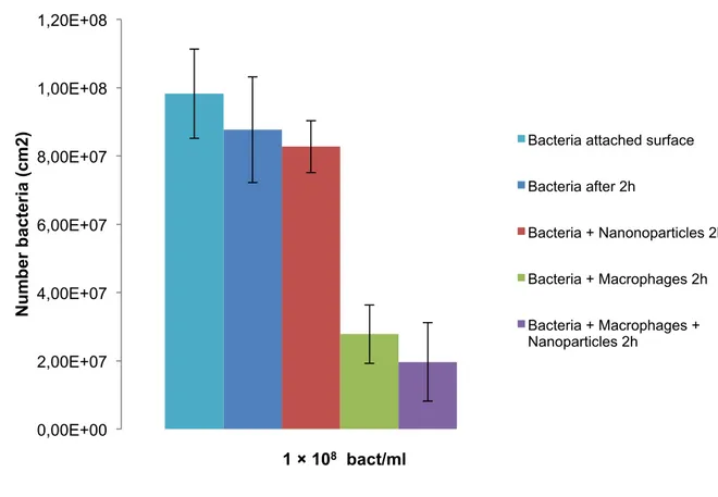
3.1 Influence of the bacterial challenge concentration on phagocytosis in the presence and absence of SPIONs
The influence of the bacterial concentration on the intracellular inactivation of bacteria by SPIONs was quantified by fluorescent microscope (Figure 3). The number of bacteria without macrophages and SPIONs, after 2 h was 9.83 × 10⁸ ± 1.31 × 107 cm2. SPIONs alone were found to cause no significant reduction in the bacterial survival after 2h of interaction with the staphylococci biofilm. The presence of macrophages generates a strong reduction in the bacterial survival to (2.78 × 10⁷ ± 7.60 × 106 cm2 of their initial number), depending on the initial number of staphylococci present in the surface. The staphylococci survival decrease significantly in the presence of both macrophages and SPIONs (1.97 × 10⁷ ± 1.15 × 107 cm2), also depending on the initial bacterial numbers.
Figure 3 – Number of live staphylococci after phagocytosis in the presence or absence of SPIONs. The number
of bacteria was determined by fluorescence microscopy after 2 hours of phagocytosis (J744A.1 macrophages). Error bars represent the standard deviation (SD) over different experiments, each involving images randomly localized. 0,00E+00 2,00E+07 4,00E+07 6,00E+07 8,00E+07 1,00E+08 1,20E+08 N u m b e r b a c te ri a (c m 2 ) 1 × 10⁸ bact/ml
Bacteria attached surface
Bacteria after 2h
Bacteria + Nanonoparticles 2h
Bacteria + Macrophages 2h
Bacteria + Macrophages + Nanoparticles 2h
From the results obtained, it is possible to conclude that the number of bacteria decreases with the use of macrophages alone or with the use of macrophages together with SPIONs (Figure 3). The use of SPIONs can promote a more efficient elimination of bacteria by macrophages. Although, the results obtained in this study showed not to be significant.
Analyzing the graphic from Figure 3, it was found that the difference of staphylococci survival in the presence and absence of SPIONs is around 8.10 × 106 cm2. ANOVA was applied to demonstrate statistically differences and p< 0.05 were considered significant. The p number was higher than 0.05 (p=0.803), showing that these results are not statistically significant. Further studies need to be executed to obtain better results.
However, it is possible to notice a small difference between bacterial survival in the presence and absence of SPIONs. SPIONs can be taken up from different cell types, such as macrophages, neutrophilic granulocytes and monocytes. Shanhua et al., (2013) reported that LPS-activated neutrophilic granulocytes increase the uptake of mannan-coated SPIONs. Grosse et al., (2016) expressed that SPIONs were taken up by primary human monocytes. SPIONs can reach sites with increased macrophage activity. Bierry et al., (2010) showed that SPIONs can detect the presence of bacterially induced arthritis by showing activated infiltration macrophages inside infected synovium.
In terms of percentage of bacterial survival (Figure 4), SPIONs alone in contact with bacteria caused no significant reduction, being the bacterial survival around 84%. On the other hand, the presence of macrophages themselves yields a reduction of the staphylococcal survival in about 30% of their initial number. The combination of macrophages and SPIONs promotes the reduction of the S. aureus to 20% of their initial bacterial number.
Influence of superparamagnetic iron oxide nanoparticles on macrophages in removing attached bacteria 0 20 40 60 80 100 120 Pe rc e n ta g e b a c te ri a l s u rv iv a l (% ) 1 × 10⁸ bact/ml
Bacteria attached surface
Bacteria after 2h
Bacteria + Nanoparticles 2h
Bacteria + Macrophages 2h
Bacteria + Macrophages + Nanoparticles 2h
Figure 4 - Percentage of staphylococci survival after phagocytosis in the presence or absence of SPIONs. The
number of bacteria was determined by fluorescence microscopy after 2 hours of phagocytosis (J744A.1 macrophages). Error bars represent the standard deviation (SD) over different experiments, each involving images randomly localized.
Through analysis of Figure 4, it is possible to see that bacteria in contact with macrophages in the absence and presence of SPIONs showed a huge decrease when compared to the control experiment (S. aureus attached to the surface). Comparing the values of S.
aureus elimination in the presence or absence of SPIONs, it was possible to conclude that this
difference is not significant (10%). ANOVA was applied to verify if the results were statistically significant. The p number obtained was higher than 0.05, which confirms the non-significant difference. However, it is important to bear in mind that nanoparticles are capable of reaching sites with increased macrophages activity (Von sur Muhlen et al. 2007), which in turn might represent a great advantage. Macrophages play an important role in mediating a wide range of inflammatory diseases, making them a good target for nanoparticle mediated therapies (Chellat et al., 2005).
In summary, it has been found that the percentage of staphylococci survival decreases in contact with macrophages either in the presence or absence of SPIONs. In order to observe
the phagocytosis of bacteria by macrophages in the presence or absence of the nanoparticles, fluorescence microscopy images were taken.
3.2 Evaluation of the staphylococcal phagocytosis in the presence and absence of SPIONs through fluorescent microscopy
Fluorescence images of staphylococcal attached to the surface showed a high number of green-fluorescent organisms in the absence of SPIONs and macrophages (Figure 5). Also, it was found that the fluorescence was not affected by the presence of SPIONs (Figure 5). The green- fluorescence corresponds to S. aureus ATCC 12600GFP, while macrophages appear with a red color (red-fluorescence).
Influence of superparamagnetic iron oxide nanoparticles on macrophages in removing attached bacteria
Figure 5– Fluorescence images of green-fluorescent S.aureus ATCC 12600GFP and macrophages J774.1A after 2 hours of phagocytosis in the presence and absence of SPIONs. (A) S.aureus ATCC 12600GFP; (B) S.aureus ATCC 12600GFP and macrophages in the first contact; (C) S.aureus ATCC 12600GFP and SPIONs (after 2h); (D)
S.aureus ATCC 12600GFP and macrophages (after 2h); (E) S.aureus ATCC 12600GFP and macrophages in the presence of SPIONs (after 2h).
400,00 500,00 600,00 700,00 800,00 f c o lo n ie s / m l Centrifugation
Analyzing the images from Figure 5, it was found that the percentage of S. aureus ATCC 12600GFP (Figure5A) and the bacteria with SPIONs (Figure 5C) is similar. In the presence of macrophages (Figure 5D), S. aureus was phagocytosed and it is visible the fluorescence of S. aureus inside and outside macrophages. On the other hand, macrophages and SPIONs (Figure 5E) were found to promote more elimination of bacteria, although this difference is not significant.
According to Figure 5D and Figure 5E, the shape of macrophages did not remain the same as the ones in Figure 5B. This fact may be related with some disintegration of the macrophage. It is not known what caused this alteration. One fact that can be associated with disintegration is the staining solution used to stain macrophages. The other possibility is the possible formation of ROS. Buyukhatipoglu et al. (2010) showed that SPIONs induce the formation of ROS that can be toxic to cells, which cannot be balanced by the host cells and can induce the apoptosis of the cells containing SPIONs. Further studies need to be conducted to explore this topic.
3.3 Evaluation of the bacterial survival inside macrophages
Macrophages engulfed bacteria during 2 h. In order to know the bacterial survival inside macrophages, two techniques were used, namely centrifugation and lyses of macrophages. Firstly, we plated 100 µL of bacteria, without any dilution, in petri dishes with TSB + 1% of tetracycline. The number of bacteria counted in the agar plates was 377.50 colonies/ml with centrifugation and 650 colonies/ml with lysis (Figure 6).
Influence of superparamagnetic iron oxide nanoparticles on macrophages in removing attached bacteria 0 0,5 1 1,5 2 2,5 3 3,5 4 4,5 5 1 N u m b e r o f b a c te ri a E+0 7 b a c te ri a / ml Centrifugation Lysis
Figure 6 – Number of colonies corresponding to bacterial survival inside the macrophages. The number of
colonies was evaluated by the platting method. Error bars represent the standard deviation (SD) over four experiences.
The number of bacteria counted with the Bürker-Türk chamber was 2.06 × 10⁷ bacteria/ml with centrifugation and 3.45 × 10⁷ bacteria/ml with lysis (Figure 7).
Figure 7 – Number of colonies corresponding to bacterial survival inside the macrophages. The number of
colonies was evaluated using the Bürker-Türk. Error bars represent the standard deviation (SD) over four experiences.
The number of bacteria alive inside the macrophages using the two techniques has been gathered in Table 1 for comparison purposes.
Table 1 – Number of colonies representing the bacterial survival inside the macrophages as determined
by two methods. Centrifugation Lysis Platting (colonies/ml) 337.50 650 Bürker-Türk chamber (bacteria/ml) 2.06 × 10 7 3.45 × 10⁷
Thesis results were not conclusive. ANOVA was applied to demonstrate statistically significant differences. The ANOVA analysis showed a p > 0.05 in both situation, platting (p=0.0964) and Bürker-Türk chamber (p=0.141). As p > 0.05, these results were not statistically significant. Future studies need to be done in order to understand these differences. The calculation of bacterial concentration in the Bürker-Türk chamber is not adequate. The formula to calculate the number of bacteria prevailing in 1 ml of solution has a standard factor. As the counting of bacteria was really low, this measurement may be ambiguous.
Although it is not possible to accurately know the number of bacteria alive inside the macrophages, one can conclude that there are bacteria still alive inside the macrophages.
S. aureus subverts the host immune response by numerous mechanisms, including increased
resistance to cationic antimicrobial peptides, impairment of phagocyte recruitment, interference with Ab-mediated opsonization and complement activation, and resistance to intracellular killing (Scherr et al., 2015). Hamza et al. (2014) reposted that S. aureus could survive up to 5 and 7 days within macrophages, after their internalization. Intracellular S.
aureus can escape the intracellular confinement, proliferated in the conditioned medium and
killed cells (Kubica et al., 2008). In summary, new strategies need to be explored to allow the counting of bacterial survival inside macrophages.
Influence of superparamagnetic iron oxide nanoparticles on macrophages in removing attached bacteria
4. C
HAPTER
IV
Influence of superparamagnetic iron oxide nanoparticles on macrophages in removing attached bacteria
Nowadays, BAI are the number one cause of implant failure, being one of the major problems emerging from the use of implants and medical devices. Pathogens can be introduced on an implant in a perioperative or postoperative contamination, competing with the host cells to integrate the implant. Scientific communities have long recognized that biomaterial devices and implants provide foreign surfaces, alien to the human body, where bacteria can adhere and start the biofilm formation.
S. aureus are the most frequent isolated pathogens from infected biomaterial implants
or devices. S. aureus stimulates the inflammatory response, promoting the high influx of macrophages to the infected site. Macrophages derive from blood monocytes, playing an important role on recognizing engulfing and killing invading pathogens, such as S. aureus. However, this pathogen has the capacity of surviving and infecting these immune cells, which promotes the chronic and recurrent infections.
The only effective treatment of S. aureus infection is the use of antibiotics. However, one big problem associated with this pathogen is the growing antibiotic-resistance. Bacteria in their biofilm mode of growth are relatively to antibiotic treatment across many pathogens. So, new strategies needed to emerge in order to prevent or cure bacterial infections.
Superparamagnetic iron oxide nanoparticles are one type of biocompatibility nanoparticles, that can achieved a specific area. SPIONS can be combined with any biomaterial surface to promote the elimination of bacteria, replacing the antibiotic therapy used in these days. These type of nanoparticles can change cell function by formation of ROS, which can be produced by degradation of nanoparticles or by the cells.
In this thesis, it was possible to visualize the influence of SPIONs with macrophages in the removal of bacterial biofilms. A low concentration of SPIONs was found to be sufficient to assist macrophages in the phagocytosis of S. aureus. However, comparing the results obtained in the presence and absence of SPIONs no significant differences could be observed, as the ANOVA analyses demonstrate not statistically significant differences. To promote a bigger phagocytosed concentration, the number of SPIONs used should be higher,
Influence of superparamagnetic iron oxide nanoparticles on macrophages in removing attached bacteria
In the future, it would be important to find the minimal concentration of SPIONs that can help macrophages in the phagocytosis of bacteria without causing any toxic effects to the host immune cells. Also, it would be important to study the effect of ROS formation, as they provide means through which macrophages remove bacteria from host cells.
To further validate the results of this thesis, in vivo models could be used to link the clinical outcome to the in vitro studies.
Herein, despite the high number of phagocytosed bacteria, macrophages were found to disintegrate. This fact should be further studied to understand if the staining solution affects the macrophages shape, which may affect the whole outcome of the study.
Additionally, new experiments must be performed to understand if the bacteria are still alive when they are phagocytosed. In the future, new techniques lyse of macrophages should be used.
The cell line used in this thesis was a murine line of macrophages (J774.1). By taking human line of macrophages, maybe experiments could be closer to the clinical reality. Also, the bacterial biofilm could be removed from a truly BAI site. In this way, the whole study could be more efficient and realistic.
In summary, bacteria can adhere and grow on any biomaterial implant surface. The antibiotic treatment used to prevent BAI is insufficient, and therefore, new strategies ought to be developed to decrease the number of bacterial infections associated with the use of implants and medical devices. The use of SPIONs together with macrophages has showed to be promising as a future therapy to prevent or eliminate BAI.
5. C
HAPTER
V
Influence of superparamagnetic iron oxide nanoparticles on macrophages in removing attached bacteria
Ahlberg, A., Carlsson, A. S., Lindberg, L. (1978). Hematogenous infection in total joint replacement. Clin Ortthop Relat Res, (137), 69-75.
Anderson, J. M. (2004). Inflammation, wound healing, and the foreign- body response.
Biomaterials Science. An introduction to Materials in Medicine, Elsevier, San Diego,
CA. pp 296-304.
Aribi, M., Meziane, W., Habi, S., Boulatika, Y., Marchandin, H., Aymeric, J. L. (2015). Macrophage Bactericidal Activities against Staphylococcus aureus Are Enhanced In
Vivo by Selenium Supplementation in a Dose-Dependent Manner. PLoS ONE, 10(9),
e0135515.
Bandyopadhyay, A., Bose, S. (2013). Introduction to Biomaterials .Characterization of
Biomaterials. Elsevier, pp 1-6.
Bierry, G., Jehl, F., Neuville, A., Lefevre, S., Robert, P., Kremer, S., Dietemann, J. L. (2010). MRI of macrophages in infectous knee synovitis. Am J Roentgenol, 194(6), W521-W526.
Busscher, H. J., Van der Mei, H. C., Subbiahdoss, G., Jutte, P. C., Van den Dunges, J. J. A. M., Zaat, S. A. J., Schultz, M. J., Grainger, D. W. (2012). Biomaterial - Associated Infection: Locating the finish line in the race for the surface. Sci Transl Med, 4(153), 153rv10.
Buyukhatipoglu, K., Clyne, A. M. (2010). Superparamagnetic iron oxide nanoparticles change endothelial cell morphology and mechanics via reactive oxygen species formation. J biomed Res, 96A, 186-195
Campoccia, D., Montanaro, L., Arciola, C. R. (2006). The significance of infection related to orthopaedic devices and issues of antibiotic treatment. Biomaterials, 27(11), 2331-2339. Chellat, F., Merhi, Y., Moreau, A., Yahia, L. (2005). Therapeutic potential of nanoparticulate
systems for macrophage targeting. Biomaterials, 26(35), 7260-7275.
Chuang, S.-Y., Lin, C. -F., Aljuffali, I. A., Fang, J.-Y. (2016). Specific targeting of engineered nanoparticles to activated macrophages. Current Nanoscience, 12(1), 63-69. Costerton, J. W. (1999). Bacterial biofilms: A common cause of persistent infections. Science,
Influence of superparamagnetic iron oxide nanoparticles on macrophages in removing attached bacteria
Dunster, J. L. (2016). The macrophage and its role in inflammation and tissue repair: mathematical and systems biology approaches.WIREs Syst Biol Med, 8(1), 87–99.
Fitzgerald, R. M. (1979). Microbiologic environment of the conventional operating room.
Arch Surg, 114(7), 772-775.
Foster T. (1996). Staphylococcus. Medical Microbiology. 4th edition. Chapter 12.
Gristina, A. G. (1987). Biomaterial-centered infection: microbial adhesion versus tissue integration. Science, 237(4822), 1588-1595.
Grosse, S., Stenvik, J., Nilsen, A. M. (2016). Iron oxide nanoparticles modulate lipopolysaccharide-induce inflammatory responses in primary human monocytes. Int J
Nnanomedicine, 11, 4625-4642.
Guo, W., Andersson, R., Ljungh, A., Wang, X. D., Bengmark, S. (1993). Enteric bacterial translocation after intraperitoneal implantation of rubber drain pieces. Scand J
Gastroenter, 28(5), 393-400.
Hall-Stoodley, L., Stoodley, P., Kathju, S., Høiby, N., Moser, C., Costerton, J. W., Moter, A., Bjarnsholt, T. (2012). Towards diagnostic guidelines for biofilm-associated infections.
FEMS Immunol Med Microbiol, 65(2), 127–145
.
Hamza, T., Li, B. (2014). Differential responses of osteoblasts and macrophages upon Staphylococcus aureus infection. BMC Microbiology, 14, 207.
Karlsson, H. L., Cronholm, P., Gustafsson, J., Moller, L. (2008). Copper oxide nanoparticles are highly toxic: a comparison between metal oxide nanoparticles and carbon nanotubes. Chem Res Toxicol, 21(9), 1726-1732.
Koziel, J., Maciag-Gudowska, A., Mikolajczyk, T., Bzowska, M., Sturdevant, D. E., Whitney, A. R., Shaw, L. N., DeLeo, F. R. Potempa, J. (2009). Phagocytosis of Staphylococcus
aureus by macrophages exerts cytoprotective effects manifested by the upregulation of
antiapoptotic factors. PLoS ONE, 4(4), e5210.
Kubica, M., Guzik, K., Koziel, J., Zarebski, M., Richter, W., Gajkowska, B., et al. (2008). A potential new pathway for Staphylococcus aureus dissemination: the silent survival of
S. aureus phagocytosed by human monocyte-derived macrophages. PLoS ONE, 3(1),
e1409.
Kzhyshkowsha, J., Gudima, A., Riabov, V., Dollinger, C., Lavalle, P., Vrana, N. E. (2015). Macrophage responses to implants: prospects for personalized medicine. J Leukoc Biol, 98(6), 953-962.
Larsson, M., Kronvall, G., Chuc, N. T., Lager, F., Tomson, G., Falkenberg, T. (2000). Antibiotic medication and bacterial resistance to antibiotics: a survey of children in a Vietnamese community. Trop Med Int Health, 5(10), 711-721.