Corresponding author:Dr.Henrique Silveira Costa. e-mail: [email protected]
Received 9 May 2018 Accepted 31 July 2018
Review Article
Reduced functional capacity in patients with Chagas disease:
a systematic review with meta-analysis
Henrique Silveira Costa
[1], Márcia Maria Oliveira Lima
[2], Fábio Silva Martins da Costa
[1],
Ana Thereza Chaves
[1], Maria Carmo Pereira Nunes
[1],
Pedro Henrique Scheidt Figueiredo
[2]and Manoel Otávio da Costa Rocha
[1][1]. Curso de Pós-Graduação em Infectologia e Medicina Tropical, Departamento de Clínica Médica, Faculdade de Medicina, Universidade Federal de Minas Gerais, Belo Horizonte, MG, Brasil.
[2]. Escola de Fisioterapia, Faculdade de Ciências Biológicas e da Saúde, Universidade Federal dos Vales do Jequitinhonha e Mucuri, Diamantina, MG, Brasil.
Abstract
Reduced peak oxygen uptake (VO2peak) is a common clinical finding in progressive Chagas disease. However, the disease stage
in which functional impairment is detectable remains uncertain. The present study compared functional capacity between healthy controls and patients with different clinical forms of Chagas disease. A systematic review and meta-analysis (PROSPERO database CRD42017058353) was conducted following a search of the MEDLINE, Web of Science, CINAHL, and LILACS databases from September to December 2017 for articles published in English, Spanish, or Portuguese, with no date restrictions. We included studies that compared the VO2peak between healthy and Chagas disease patients, stratified according to 3 clinical
forms [no apparent cardiac disease, non-dilated Chagas heart disease (CHD), and dilated CHD]. Seven cross-sectional studies
were included. Chagas disease patients without apparent cardiac disease (n=208) had VO2peak values [mean difference, -1.55ml/
kg/min; 95% confidence interval (CI), -4.98ml/kg/min to 1.88ml/kg/min] similar to those of healthy controls (n=105; p=0.38,
I2=52%). In non-dilated CHD (n=159), VO
2peak was 8.71ml/kg/min lower (95% CI, -13.99 to -3.42ml/kg/min) than in healthy
controls (n=59; p=0.001, I2=75%). VO
2peak was also significantly lower (mean difference, -9.30ml/kg/min; 95% CI, -11.34 to
-7.25ml/kg/min) in dilated CHD patients (n=131) than in healthy controls (n=53; p<0.001, I2=0%). Exercise capacity in Chagas
disease patients without apparent cardiac disease is similar to that in healthy controls. Functional impairment in Chagas disease
is detectable in the early stages of cardiac involvement, even in the absence of systolic dysfunction and signs of heart failure.
Keywords: Chagas disease. Functional capacity. Exercise testing. Systematic review.
INTRODUCTION
Chagas disease remains a serious public health problem in
Latin American countries. Approximately 6-7 million people worldwide are estimated to be infected with Trypanosoma cruzi1,
which is endemic in 21 countries1, and 80% of the patients do not
have access to diagnosis or appropriate treatment2, emphasizing
the lack of attention to this disease3.
Recently, the Benznidazole Evaluation for Interrupting
Trypanosomiasis trial (BENEFIT)4 showed that parasiticidal
treatment does not seem to reduce cardiac damage in Chagas heart disease (CHD), the most severe clinical form of the infection5. In contrast, exercise training has shown positive
effects on the overall patient health6. The benefits include a
significant increase in functional capacity7-9, respiratory muscle
strength9, quality of life7, and left ventricular ejection fraction
(LVEF)9.
Nevertheless, the stage of disease in which reduced
functional capacity can be observed remains unclear. Mady et al.10 reported significant reduction in peak oxygen uptake
(VO2peak), even in the absence of signs and symptoms of
heart failure. In contrast, other studies11,12 reported that reduced
functional capacity can be detected only in the advanced stage of
the disease, with fatigue and dyspnea during ordinary physical activity as a well-defined marker of poor prognosis13,14.
As the stage of disease in which reduced functional capacity
can be observed may be important in the adoption of cardiac rehabilitation therapies, the present study compared functional
capacity between healthy individuals and patients with different
METHODS
Study design
This systematic review aimed to determine the clinical form of Chagas disease in which reduced functional capacity is detectable. The study was registered in the PROSPERO database CRD42017058353 and edited following the guidelines of the Preferred Reporting Items for Systematic Reviews and Meta-Analyses (PRISMA) statement15.
Search strategy and study selection
Potential studies were identified through a systematic search. The databases used were MEDLINE, Web of Science, the Cumulative Index to Nursing and Allied Health Literature (CINAHL) and Latin American & Caribbean Health Sciences Literature (LILACS) for relevant studies, without date restrictions from inception until December 2017. The search was conducted independently by 2 authors (HSC and MMOL) from September to December 2017. Disagreements were resolved by a third reviewer (PHSF). Search terms included words related to Chagas heart
disease, Chagas cardiomyopathy and functional capacity.
The following strategy was used for the PubMed search—
["Chagas Disease"(Mesh) OR "Chagas heart disease" OR
"Chagas Cardiomyopathy"(Mesh)] AND ["Exercise"(Mesh)
OR "Exercise Test"(Mesh) OR "functional capacity" OR "oxygen consumption"(Mesh) OR "oxygen uptake" OR "VO2peak"]—and was modified for each database.
Eligibility criteria
Eligibility criteria included studies a) that evaluated patients
diagnosed with Chagas disease and defined the criteria; b) that
assessed the VO2peak, expressed in ml/kg/min; c) with at least
1 group of patients with Chagas disease and 1 control group; d) written in English, Spanish, or Portuguese.
Those with positive serology for T. cruzi but without signs,
symptoms, or evidence of changes on the electrocardiogram
or chest radiogram were considered patients with Chagas disease without apparent cardiac disease. Non-dilated CHD patients were defined according to the presence of
arrhythmias and intraventricular and atrioventricular conduction
disorders with normal ventricular function16. Patients with
ventricular dysfunction (LVEF<50%) and cardiac dilation on echocardiography were considered to have dilated CHD.
Exclusion criteria were review studies, articles in duplicate,
animal studies, and those that did not match the objective of this
review. There were no restrictions on the year of publication
until December 2017. Studies of authors using the same
population were excluded, as well as those without adequate
statistical analysis.
Quality assessment
The quality assessment was evaluated using the adapted form of the Newcastle Ottawa cohort scale for cross-sectional
studies17. The scale consists of 8 items grouped under 3 topics,
namely, selection, comparability and confounders, and outcome.
Outcomes and data analysis
The following data were extracted from the included articles:
author, publication year, characteristics of the sample, and
VO2peak value and method of measurement.
Chagas disease patients were stratified according to the clinical form of the disease, that is, Chagas disease without apparent cardiac disease, patients with normal left ventricular dimensions and function (non-dilated CHD), and patients with a dilated left ventricle with impaired ventricular systolic function (dilated CHD).
The primary outcome measure was the difference in
VO2peak, expressed in ml/kg/min, between healthy individuals
and Chagas disease patients (Chagas disease without apparent
cardiac disease, non-dilated CHD, and dilated CHD).
The software Review Manager 5.3 (The Nordic Cochrane Centre, Copenhagen, Denmark) was used to conduct
meta-analyses for the outcome measures, reported as the mean
difference with 95% confidence interval (CI). Heterogeneity was defined as low, moderate, or high according to I2 values
(25, 50, or 75%, respectively). A forest plot was used to display the results of the meta-analysis. Data analysis was performed using the fixed-effects model when the results showed low
heterogeneity (I2 ≤25%) and the random-effects model when
the results showed moderate or high heterogeneity (I2 >25%).
RESULTS
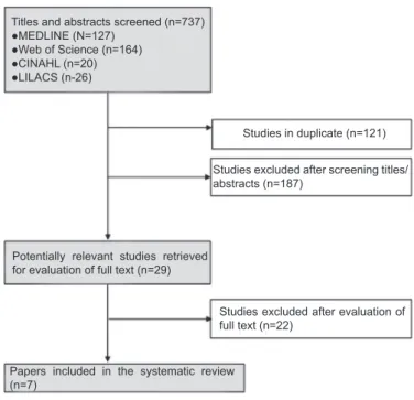
Flow of papers through the review
The electronic search strategy identified 363 studies but 147 (40.5%) were duplicates. After screening titles and abstracts, 187 (51.5%) papers were excluded. Twenty-five articles were review studies, 58 did not perform maximal exercise testing, 26 consisted of a sample without Chagas disease, 24 did not have
a healthy control group, 40 conducted animal studies, and 14
articles were not found.
After reading the full text, 16 articles were excluded for failing to meet the objective of the present review, 1 did not define the clinical form of patients with Chagas disease, 1 did not report the sample size, and 3 were by authors who evaluated
the same group of study patients in already included studies.
Another article did not report VO2peak in ml/kg/min. A total of
7 articles were included in the present review. Figure 1 outlines
the flow of papers through the review.
Participants
Four studies12,18-20 compared the functional capacity of healthy
individuals to patients with Chagas disease without apparent
cardiac disease. Three studies12,20,21 reported the difference in
VO2peak between healthy individuals and patients with non-dilated CHD. Finally, 4 articles12,20,22,23 demonstrated the difference
in functional capacity between healthy subjects and patients with dilated CHD. Two studies12,20 compared the VO
2peak of healthy
subjects with more than 1 group with Chagas disease.
Outcomes
The treadmill was used to evaluate VO2peak in 6 studies18-23
TABLE 1: Characteristics of included studies that compared the VO2peak between healthy individuals and Chagas disease patients.
Study Sample characteristics VO2peak (ml/kg/
min)
VO2peak
evaluation p-value Quality
Comparison between healthy subjects and Chagas disease patients without apparent cardiac disease
Costa et al. (2015)
Healthy Control=38 (42% females, 44±9.1 years, LVEF 69.7±5.2%) ChD without cardiac disease=75 (52% females, 44.7±8.7 years,
LVEF 68.2±5.6%)
40.4±7.6 40.6±10
Treadmill Estimated VO2
No difference 9
De Oliveira, Pedrosa, Giannella-Neto (2000)
Healthy Control=15 (All males, 36±9 years)
ChD without cardiac disease=17 (All males, 47±11 years, LVEF 68±4%)
31±12 24±5
Bicycle Direct VO2
No difference 6
Rabelo et al. (2013)
Healthy Control=28 (57% females, 52±8 years, LVEF 63±3%) ChD without cardiac disease=64 (63% females, 50±12 years,
LVEF 65±5%)
26±11 28±11
Treadmill
Estimated VO2 No difference 6
Rocha et al. (2006)
Healthy Control=24 (29% females, 35.6±9.3 years, LVEF 64% [61-66]) ChD without cardiac disease=52 (40% females, 39.8±9 years,
LVEF 62% [60-65])
49.4±10.1 45.8±10.6
Treadmill Estimated VO2
No difference 7
Comparison between healthy subjects and non-dilated Chagas heart disease patients
De Oliveira, Pedrosa, Gianella-Neto (2000)
Healthy Control=15 (36±9 years) Non-dilated CHD=23 (49.6±11 years)
31±12 23.6±8.1
Bicycle Direct VO2
No difference 6
Mady et al. (1997) Healthy Control=20 (All males, 32.9±6.1 years) Non-dilated CHD=18 (All males, 29.2±5.6 years)
36.98±5.45 24.32±4.25
Treadmill Direct VO2
<0.001 8
Rocha et al. (2006) Healthy Control=24 (29% females, 35.6±9.3 years, LVEF 64% [61-66]) Non-dilated CHD=118 (44% females, 41.8±9.2 years, LVEF 60% [50-64])
49.4±10.1 44.2±8.6
Treadmill Estimated VO2
<0.05 7
Comparison between healthy subjects and dilated Chagas heart disease patients
Baiao et al. (2013) Healthy Control=15 (47% females, 47.3±9.1 years) Dilated CHD=15 (47% females, 50.3±5.7 years, mean LVEF 36%)
38.53±7.81 28.46±6.58
Treadmill Estimated VO2
0.004 6
De Oliveira, Pedrosa, Gianella-Neto (2000)
Healthy Control=15 (36±9 years) Dilated CHD=12 (55±9 years, LVEF 28±8%)
31±12 19±5
Bicycle Direct VO2
<0.05 6
Mady et al. (1996) Healthy Control=23 (All males, 35±8.7 years, LVEF 75.1±3.2%) Dilated CHD=104 (All males, 24.5±2.9 years, LVEF 42±11.7%)
33.3±5.6 24.5±2.9
Treadmill Direct VO2
<0.001 9
VO2peak: peak oxygen uptake; ChD: Chagas disease; CHD: Chagas heart disease; LVEF: left ventricular ejection fraction. Values highlighted in bold
were statistically significant (p<0.05).
Titles and abstracts screened (n=737)
●MEDLINE (N=127) ●Web of Science (n=164) ●CINAHL (n=20) ●LILACS (n-26)
Studies in duplicate (n=121)
Studies excluded after screening titles/
abstracts (n=187)
Potentially relevant studies retrieved for evaluation of full text (n=29)
Studies excluded after evaluation of
full text (n=22)
Papers included in the systematic review (n=7)
FIGURE 1: Flow of studies through the review. MEDLINE: Medical
Literature Analysis and Retrieval System Online; CINAHL: Cumulative
Index to Nursing and Allied Health Literature; LILACS: Latin American &
Caribbean Health Sciences Literature.
was directly assessed using gas analysis in 3 articles12,21,23 and
estimated by formulas in 4 studies18-20,22.
Quality of papers
The mean Newcastle Ottawa scale score adapted for cross-sectional studies was 7.3 points (range from 6.0 to 9.0). All studies adequately verified the primary outcome and statistically demonstrated the difference between the groups. However, only
1 study reported the sample calculation. The scores according
to the Newcastle Ottawa scale and the characteristics of the
included studies are summarized in Table 1.
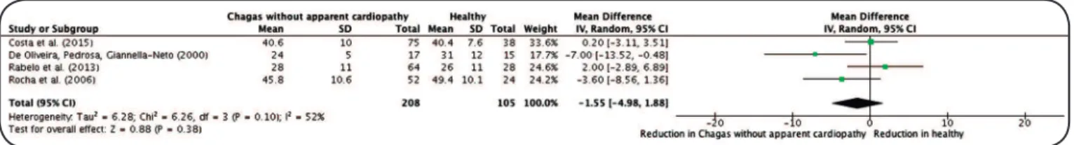
VO2peak in patients with Chagas disease without apparent cardiac disease
The mean score for quality was 7.0 points (ranging from 6.0
to 9.0). Only 1 (25%) study evaluated VO2peak directly using gas analysis. One paper used the cycle ergometer and 3 used the treadmill in exercise testing.
No study found significant differences in the VO2peak
between healthy individuals and patients with Chagas disease without apparent cardiac disease. The forest plot (Figure 2)
-1.55ml/kg/min; 95% CI from -4.98ml/kg/min to 1.88ml/kg/
min) similar to those of healthy subjects (n=105) (p=0.38). The heterogeneity of the comparison was moderate (I2 = 52%).
VO2peak in non-dilated CHD patients
The mean score for quality was 7.0 points (ranging from 6.0 to 8.0). Two (66.6%) studies evaluated VO2peak directly using
gas analysis. One paper used the cycle ergometer and the other
2 performed exercise testing with a treadmill.
Of the 3 studies included, only 1 (33.3%) reported no
significant difference in VO2peak between healthy subjects and dilated CHD patients and 2 (66.6%) showed that the VO2peak
was significantly lower in non-dilated CHD patients than in
healthy subjects.
The forest plot (Figure 3) showed that VO2peak was
8.71ml/kg/min lower (95% CI -13.99 to -3.42ml/kg/min) in non-dilated CHD patients (n=159) than in healthy subjects (n=59) (p=0.001). The heterogeneity of the comparison was
high (I2 = 75%).
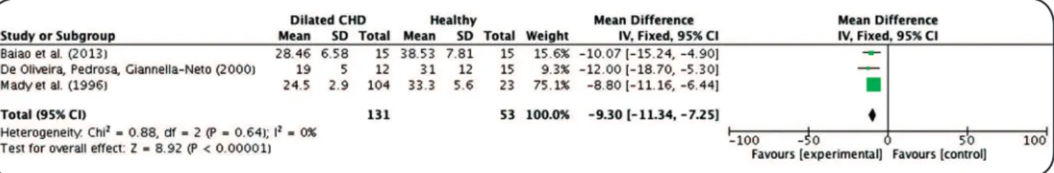
VO
2peak in dilated CHD patients
The mean score for quality was 7.0 points (ranging from 6.0 to 9.0). Two (33.3%) studies evaluated VO2peak directly using gas analysis. One (33.3%) paper used the cycle ergometer and 2
(66.6%) performed maximum exercise testing with a treadmill. All 3 included studies showed that the VO2peak was
significantly lower in dilated CHD patients than in healthy
subjects. The forest plot (Figure 4) showed that VO2peak was
significantly lower (mean difference 9.30ml/kg/min; 95% CI -11.34 to -7.25ml/kg/min) in dilated CHD patients (n=131) than in healthy subjects (n=53) (p<0.001). The analysis was
homogeneous (I2 = 0%).
DISCUSSION
The VO2peak has been widely used in assessing the
functional capacity of patients with Chagas disease and is
FIGURE 3: Mean difference in VO2peak between non-dilated CHD patients and healthy controls. CHD: Chagas heart disease; SD: standard deviation;
95% CI: 95% confidence interval; VO2peak: peak oxygen uptake.
FIGURE 2: Mean difference in VO2peak between patients with Chagas disease without apparent cardiac disease and healthy controls. SD: standard
deviation; 95% CI: 95% confidence interval; VO2peak: peak oxygen uptake.
considered a potential prognostic marker24. However, the
association between functional impairment and the stage of disease in which it is detectable has been unclear. To our knowledge, this is the first study to systematically determine the stage of Chagas disease at which reduced exercise capacity can be observed. The main findings of the present study were: 1) patients with Chagas disease without apparent cardiac disease
have VO2peak values similar to those of healthy individuals and
2) patients with CHD, even with preserved systolic function, showed a significant reduction in functional capacity when
compared to healthy individuals.
For patients with Chagas disease without apparent cardiac disease, no study included in this review reported reduced
functional capacity in this group of patients compared to that
in the healthy subjects. In fact, patients with Chagas disease without apparent cardiac disease have excellent prognosis, similar to that of healthy individuals. However, although
VO2peak values are within the normal range, exercise tests in these patients are highly recommended since Chagas patients present a greater number of exercise-induced arrhythmias, both during exercise and the recovery phase18.
In patients with non-dilated CHD, 2 studies found a significant decrease in VO2peak compared to that in the healthy
subjects, and 1 reported no difference between these groups. The forest plot showed a significant difference in functional capacity between the non-dilated CHD and healthy control groups. The
initial phase of Chagas cardiac disease is usually characterized by impairment in electrical stimulus formation and conduction.
As shown by electrocardiography, the changes in ventricular
function are discrete and segmental, but may indicate subclinical heart disease capable of affecting the functional status.
According to Mady et al.10, electrocardiographic changes present
FIGURE 4: Mean difference in VO2peak between dilated CHD patients and healthy controls. CHD: Chagas heart disease; SD: standard deviation;
95% CI: 95% confidence interval; VO2peak: peak oxygen uptake.
characterized by great heterogeneity in clinical presentation.
With regard to functional capacity, for example, the included studies showed a standard deviation of up to 12ml/kg/min.
Furthermore, heterogeneity in the observational studies may be frequent and more common than in interventional studies25.
In the analysis of patients with dilated CHD, all studies showed significantly lower VO2peak values compared to those
in healthy subjects. The forest plot confirmed this result, with homogeneity among the studies. Briefly, patients with dilated
CHD have VO2peak values 9.30ml/kg/min lower than healthy
patients, equivalent to almost 3 metabolic equivalents (METs).
Some findings may suggest the functional impairment at this
stage of the disease. The presence of symptoms of heart failure,
such as fatigue and dyspnea, may be the most significant. These
symptoms limit the patient's ability to exercise26, especially
in association with abnormalities in the structure of skeletal
muscle27. In addition, reduced cardiac output, one of the
hallmarks of heart failure28, implies lower blood flow to the
muscles used during exercise and early lactate accumulation.
Respiratory muscle weakness may also reduce functional capacity, as previously reported in patients with heart failure29.
Other factors, such as progressive deterioration of ventilatory response30 and chronotropic incompetence20, can also account
for functional impairment. Finally, it may also be possible that the fear of exercise and the discomfort caused by exercise-induced arrhythmias may lead to progressive physical inactivity.
This review had some strengths and limitations. The mean quality score of 7.3 for the included studies was good31.
However, there was a major difference in the mean age among the groups. In addition, few studies were included in each
meta-analysis. The heterogeneity among the studies included in the
meta-analysis was high for the comparison between non-dilated CHD and healthy individuals. In addition, since there was no
restriction on the date of publication, some studies could not be found, especially the oldest ones. Finally, publication bias
inherent to systematic reviews was avoided by including studies
published in languages other than English.
CONCLUSION
Patients with Chagas disease without apparent cardiac
disease have functional capacity similar to that of healthy
individuals. In patients with the cardiac form of the disease, even
in the absence of systolic dysfunction, functional capacity is markedly decreased, and exercise training should be mandatory in this population.
Conflict of interest
All authors should disclose any type of conflict of interest during the
development of the study.
Financial support
Conselho Nacional de Desenvolvimento Científico e Tecnológico (CNPq).
REFERENCES
1. World Health Organization (WHO). Chagas disease (American
trypanosomiasis). Geneva: WHO; 2017. Updated 2017 March; cited 2018 March 13]. Available from: http://www.who.int/mediacentre/
factsheets/fs340/en/
2. Dias JCP, Ramos Jr AN, Gontijo ED, Luquetti A, Shikanai-Yasuda
MA, Coura JR, et al. II Consenso Brasileiro em Doença de Chagas, 2015. Epidemiol Serv Saúde. 2016;25(n.spe.):7-86.
3. Pinheiro E, Brum-Soares L, Reis R, Cubides J-C. Chagas disease:
review of needs, neglect, and obstacles to treatment access in Latin America. Rev Soc Bras Med Trop. 2017;50(3):296-300.
4. Morillo CA, Marin-Neto JA, Avezum A, Sosa-Estani S, Rassi Jr
A, Rosas F, et al. Randomized Trial of Benznidazole for Chronic Chagas' Cardiomyopathy. N Engl J Med. 2015;373(14):1295-306.
5. Rocha MO, Teixeira MM, Ribeiro AL. An update on the
management of Chagas cardiomyopathy. Expert Rev Anti Infect
Ther. 2007;5(4):727-43.
6. Borghi-Silva A, Trimer R, Mendes RG, Arena RA, Schwartzmann PV. Rehabilitation Practice Patterns for Patients with Heart Failure The South American Perspective. Heart Fail Clin. 2015;11(1):73-82.
7. Lima MM, Rocha MO, Nunes MC, Sousa L, Costa HS, Alencar
MC, et al. A randomized trial of the effects of exercise training in Chagas cardiomyopathy. Eur J Heart Fail. 2010;12(8):866-73.
8. Fialho PH, Tura BR, Sousa AS, Oliveira CR, Soares CC, Oliveira JR, et al. Effects of an exercise program on the functional capacity
of patients with chronic Chagas' heart disease, evaluated by
cardiopulmonary testing. Rev Soc Bras Med Trop. 2012;45(2):220-4. 9. Mediano MFF, Mendes FSNS, Pinto VLM, Silva GMS, Silva PS,
Carneiro FM, et al. Cardiac rehabilitation program in patients with
Chagas heart failure: a single-arm pilot study. Rev Soc Bras Med
Trop. 2016;49(3):319-28.
10. Mady C, Ianni BM, Arteaga E, Salemi VM, de Carvalho
Frimm C. Maximal functional capacity in patients with Chagas' cardiomyopathy without congestive heart failure. J Card Fail. 2000;6(3):220-4.
11. Molina A, Carrasco H, Milanes J, Molina C, Pacheco JA, Fuenmayor
A. Exercise test in chronic Chagas' cardiomyopathy. Its value in
conduction disorders due to exercise in the more advanced stages of
the disease. Arq Bras Cardiol. 1981;36(2):95-100.
12. Oliveira FP, Pedrosa RC, Giannella-Neto A. Gas exchange during exercise in different evolutional stages of chronic Chagas' heart
disease. Arq Bras Cardiol. 2000;75(6):481-98.
13. Nunes MC, Carmo AA, Rocha MO, Ribeiro AL. Mortality prediction in Chagas heart disease. Expert Rev Cardiovasc Ther. 2012;10(9):1173-84.
14. Ribeiro AL, Nunes MP, Teixeira MM, Rocha MO. Diagnosis and
management of Chagas disease and cardiomyopathy. Nat Rev Cardiol. 2012;9(10):576-89.
15. Stewart LA, Clarke M, Rovers M, Riley RD, Simmonds M, Stewart
G, et al. Preferred reporting items for systematic review and meta-analyses of individual participant data: the PRISMA-IPD Statement. JAMA. 2015;313(16):1657-65.
16. Andrade JP, Marin Neto JA, Paola AA, Vilas-Boas F, Oliveira GM, Bacal F, et al. I Latin American Guidelines for the diagnosis and treatment of Chagas' heart disease: executive summary. Arq Bras Cardiol. 2011;96(6):434-42.
17. Herzog R, Alvarez-Pasquin MJ, Diaz C, Del Barrio JL, Estrada JM,
Gil A. Are healthcare workers' intentions to vaccinate related to their knowledge, beliefs and attitudes? A systematic review. BMC
Public Health. 2013;13:154.
18. Costa HS, Nunes MC, Souza AC, Lima MM, Carneiro RB, Sousa GR, et al. Exercise-induced ventricular arrhythmias and vagal
dysfunction in Chagas disease patients with no apparent cardiac
involvement. Rev Soc Bras Med Trop. 2015;48(2):175-80.
19. Rabelo DR, Rocha MO, de Barros MV, Silva JL, Tan TC, Nunes
MC. Impaired coronary flow reserve in patients with indeterminate form of Chagas' disease. Echocardiography. 2014;31(1):67-73.
20. Rocha AL, Lombardi F, da Costa Rocha MO, Barros MV, Val Barros
VC, Reis AM, et al. Chronotropic incompetence and abnormal
autonomic modulation in ambulatory Chagas disease patients.
Ann Noninvasive Electrocardiol. 2006;11(1):3-11.
21. Mady C, Ianni BM, Arteaga E, Salemi VMC, Silva PRS, Cardoso
RHA, et al. Maximal functional capacity and diastolic function in patients with cardiomyopathy due to Chagas' disease without congestive heart failure. Arq Bras Cardiol. 1997;69(4):237-41.
22. Baião EA, Costa Rocha MO, Lima MM, Beloti FR, Pereira DA, Parreira VF, et al. Respiratory function and functional capacity in
Chagas cardiomyopathy. Int J Cardiol. 2013;168(5):5059-61.
23. Mady C, Cardosa RH, Ianni BM, Arteaga E, Koide NS, Silva PR, et
al. Normal maximal functional capacity in patients with congestive heart failure due to Chagas' cardiomyopathy. Arq Bras Cardiol. 1996;67(1):1-4.
24. Mady C, Cardoso RH, Barretto AC, da Luz PL, Bellotti G, Pileggi
F. Survival and predictors of survival in patients with congestive
heart failure due to Chagas' cardiomyopathy. Circulation.
1994;90(6):3098-102.
25. Stroup DF, Berlin JA, Morton SC, Olkin l, Williamson GD, Rennie D, et al. Meta-analysis of observational studies in epidemiology: a proposal for reporting. Meta-analysis Of Observational Studies
in Epidemiology (MOOSE) group. JAMA. 2000;283(15):2008-12. 26. Yancy CW, Jessup M, Bozkurt B, Butler J, Casey Jr DE, Drazner
MH, et al. 2013 ACCF/AHA Guideline for the management of heart failure: a report of the American College of Cardiology Foundation/ American Heart Association Task Force on practice guidelines. J Am Coll Cardiol. 2013;62(16):1495-1539.
27. de Oca MM, Torres SH, Loyo JG, Vazquez F, Hernández N,
Anchustegui B, et al. Exercise performance and skeletal muscles in patients with advanced Chagas disease. Chest. 2004;125(4):1306-14.
28. Bocchi EA, Braga FGM, Ferreira SMA, Rohde LEP, Oliveira WA,
Almeida DR, et al. III Diretriz Brasileira de Insuficiência Cardíaca Crônica. Arq Bras Cardiol. 2009;93(supl 1):3-70.
29. Ribeiro JP, Chiappa GR, Callegaro CC. The contribution of inspiratory muscles function to exercise limitation in heart failure:
pathophysiological mechanisms. Rev Bras Fisioter. 2012;16(4): 261-7.
30. Oliveira FP, Pedrosa RC. Ventilatory response during exercise among chronic Chagas cardiopathy patients. Sao Paulo Med J.
2006;124(5):280-4.
31. McPheeters ML, Kripalani S, Peterson NB, Idowu RT, Jerome RN,
Potter SA, et al. Quality Improvement Interventions To Address
Health Disparities closing the quality gap - revisiting the state of the