Acknowledgments
A special acknowledgment to my parents and my sister. You are the best. Thank you for
all.
To my supervisor, Professor Miguel Coimbra, and my cosupervisor, Daniel Pereira, for
the guidance, patience and knowledge that was very helpful to my continuous learning
process.
To Ana Castro, Cristina Oliveira, Pedro Gomes and all colleagues from the Vision Group,
for the friendship and support given to me throughout this year of work.
To Professora Cristina Santos, for clarifying my doubts in statistical analysis.
To Dr. João Coimbra, for helping me recording the sounds.
To Rui Guedes, for the untiring help to improve the thesis.
To all the participants in this research study. Without their participation this study would
not have been possible.
At last, to my incredible friends and rest of my family by their constant support
throughout my life and for always believing in me.
To all those who were with me, in some way, during this process, either directly or
Abstract
Introduction: Electronic stethoscopes are medical devices that can collect, store and transmit physiologic sounds, as heart beats, of acoustic auscultation in a digital format. These can then be replayed, sent to a colleague for a second opinion, studied in detail after an auscultation, used for training or, as we envision it, can be used as a cheap powerful tool for screening cardiac pathologies. Despite having several electronic stethoscopes available on the market nowadays, there are no published studies about the recording quality of auscultation.
Objectives:
• Develop a ranking in order to the quality of the sound, based on experts opinion, of the 6 most relevant electronic stethoscopes on the market, according to its popularity, attendance at scientific studies and market penetration - Littmann, Cardionics, Jabes, Welch Allyn, Wise, Thinklabs;
• Check if the sound recording has enough quality to be reproduced for education and telemedicine.
Methodology: Based on similar studies, was created two study groups made up of different observers: a first group consisting of 10 cardiologists and cardiology house officers, our gold standard, and a second consisting of 15 medical students or general practitioners. Using a database of sounds recorded in hospitals, questionnaires were made to observers from each group, through Moodle. The questionnaire of the first group consisted of a sound of a particular stethoscope and the observer to infer the condition of the patient. The questionnaire of the second group consisted of the presentation of two sounds from stethoscopes, where the observer gave his opinion about the best.
differences between Thinklabs [55; 76] and Cardioncs [33; 55]; Thinklabs [55; 76] and Littmann [31; 53]; Thinklabs [55; 76] and Welch Allyn [25; 47]. Also show a statistically significant difference between Jabes [53; 75] and Littmann [31; 53] and Jabes [53; 75] and Welch Allyn [25; 47].
Conclusions: The main conclusion of this study is that in fact the Jabes is the best stethoscope to record cardiac auscultation, according to Group 2. Furthermore, we find that the electronic stethoscopes is a good instrument to record heart sounds in order to use in teaching or telemedicine.
Resumo
Introdução: Os estetoscópios eletrónicos são aparelhos médicos que podem coletar, armazenar e transmitir sons fisiológicos, tal como os batimentos cardíacos, de uma auscultação acústica num formato digital. O som coletado pode ser reproduzido, enviado para um colega para uma segunda opinião, estudado ao pormenor depois de uma auscultação, usado para ensino ou, tal como encaramos isso, pode ser utilizado como uma ferramenta barata e poderosa para triagem de patologias cardíacas. Hoje em dia, existem vários estetoscópios eletrónicos disponíveis no mercado, contudo, não há estudos publicados sobre a qualidade da gravação da auscultação destes.
Objetivos:
Elaborar um ranking, quanto à qualidade do som, segundo a opinião de peritos, dos 6 estetoscópios eletrónicos mais relevantes no mercado, baseado na sua popularidade, presença em estudos científicos e penetração no mercado – Littmann, Cardionics, Jabes, Welch Allyn, Wise, Thinklabs;
Averiguar se a gravação do som tem qualidade suficiente para ser reproduzida para fins de educação e telemedicina.
Metodologia: Tendo por base estudos semelhantes, foram criados dois grupos de estudos constituídos por observadores distintos: um primeiro grupo constituído por 10 cardiologistas ou internos de cardiologia, o nosso gold standard, e um segundo constituído por 15 estudantes de medicina ou médicos de medicina geral e familiar. Recorrendo a uma base de dados de sons gravados em ambiente hospitalar, foram feitos questionários aos observadores de cada grupo, através do Moodle. O questionário do primeiro grupo consistiu num som de um determinado estetoscópio e o observador inferir sobre a patologia do paciente. O questionário do segundo grupo consistiu na apresentação de dois sons provenientes de estetoscópios diferentes, em que o observador deu a sua opinião sobre o melhor.
Os resultados do Grupo 1 indicam que não existem diferenças estatisticamente significativas entre os estetoscópios. Contudo, o estetoscópio com maior percentagem de acertos foi o Wise, enquanto que o Cardionics foi o estetoscópio que obteve a percentagem de acertos mais baixa. Os resultados do Grupo 2, indicam que o Thinklabs foi o estetoscópio mais escolhido como sendo o melhor (66%), seguido pelo Jabes (65%), Wise (48%), Cardionics (44%), Littmann (41%) e por fim o Welch Allyn (35%). Os resultados revelaram diferenças estatisticamente significativas entre Thinklabs [55; 76] e Cardionics [33; 55]; Thinklabs [55; 76] e Littmann [31; 53]; Thinklabs [55; 76] e Welch Allyn [25; 47]. Também mostraram uma diferença estatisticamente significativa entre o Jabes [53; 75] e Littmann [31; 53] e entre o Jabes [53; 75] e Welch Allyn [25; 47].
Conclusões: A principal conclusão deste estudo assenta no facto de o Thinklabs assumir-se como o melhor estetoscópio para gravação de auscultação cardíaca. Para além disso, obtemos confirmação do estetoscópio eletrónico como bom método de gravação de som cardíaco para ensino ou telemedicina.
Abbreviations and Acronyms
ECO Echocardiography
G1 Group 1
G2 Group 2
Hz Hertz
WHO World Human Health
Contents
1. Introduction ... 11
1.1. Research problem ... 12
1.2. Objetives ... 13
1.3. Thesis structure ... 13
2. Context... 15
2.1. The history of stethoscope ... 16
2.1.1. The invention ... 16
2.1.2. Evolution of stethoscope... 17
2.1.3. Anatomy of the stethoscope ... 21
2.1.4. Electronic stethoscope ... 23
2.2. Cardiac auscultation ... 24
2.2.1. Heart Sounds ... 25
2.2.2. Auscultation ... 27
2.2.3. Heart diseases ... 28
3. State of the art ... 31
3.1. Research methodology ... 32
3.2. Results of the article selection process ... 32
3.3. Analysis of comparative studies ... 33
3.4. Analysis of types of stethoscopes compared... 38
4. Study Design ... 51
4.1. Dataset ... 52
4.3. Evaluation Methodology ... 55
4.4. Sample size ... 55
5. Methodology ... 59
5.1. Sounds Treatment ... 60
5.2. Listening Platform: Moodle ... 61
5.3. Headphones ... 63
5.4. Study Procedures ... 64
6. Results ... 65
6.1. Group 1 ... 66
6.2. Group 2 ... 67
7. Discussion ... 69
8. Conclusion ... 71
8.1. Main Findings ... 72
8.2. Main Recommendations ... 72
9. References ... 73
10. Appendix ... 78
Appendix I: Sounds distribution by stethoscope ... 79
Appendix II: Environment classroom conditions of Group 1 (Expert Group) ... 81
Appendix III: Environment classroom conditions of Group 2 (Normal Group) ... 82
Appendix IV: Answers of observers of Group 1 (Expert Group) ... 83
List of tables
Table 1: Analysis of comparative studies ... 34
Table 2: The stethoscopes evaluated in analyzed studies ... 39
Table 3: The stethoscopes evaluated in analyzed studies (cont.) ... 40
Table 4: Technical characteristics of the most relevant electronic stethoscopes available on the market ... 42
Table 5: Resume of sounds division by stethoscope ... 54
Table 6: Various hypothesis obtained through calculator ... 56
Table 7: The layout of study design of G1. The shaded column represents an outline of possible observer 1 answers ... 56
Table 8: Headphones selection ... 63
Table 9: Final results of Group 1 ... 66
List of figures
Figure 1: The evolution of the acoustic stethoscope (Med, 2015): ... 20 Figure 2: Anatomy of acoustic stethoscope (Standris, 2015) ... 22 Figure 3: Anatomy of heart. (Image, 2014) ... 25 Figure 4: Main four auscultation areas: blue - aortic; yellow- pulmonic; red - tricuspid; light blue – erb point; green – mitral (EstudMed, 2015) ... 28
Figure 5: Six of the most relevant electronic stethoscopes available on the market... 49 Figure 6: The auscultation areas by pathology ... 52 Figure 7: Digiscope - Software used to record sounds with the Littmann stethoscope. 53 Figure 8: An example of sound before split: The break in the middle represents the moment of noise caused by a change of the auscultation spot ... 60
Figure 9: An example of sound after being split by spot: The break was eliminated (remained in first track, which won’t be saved) and individual files created for the first and second spot (the first and second track, respectively) ... 61
Figure 10: Normalizing the sounds ... 61 Figure 11: Questionnaire on Moodle for G1: red points represents the various auscultation spots, which were previously split. ... 62
n recent years, Portugal has been observing an increase in the number of sick people as well as in the European Union. Factors such as physical inactivity, obesity and poor dietary practices lead to an increase in diseases in general. Cardiovascular diseases are among the most common. According to the PORDATA, in 1960 the proportion of deaths due to cardiovascular diseases in Portugal was 29, 5%, while in 2012 was 30, 4% (PORDATA, 2015). The same happens around the world. In 2008, an estimated 17.3 million people died from cardiovascular disease, and is expected that over years the number of deaths continues increasing. By 2030 more than 23 million of people will die annually from this type of disease (WHO, 2013).
By the other hand, the ageing population has been steadily growing over the past few years. According to World Health Organization,” the proportion of population over 60 years in the WHO European Region is growing faster than any age, as a result of both longer life expectancy and declining fertility rates” (WHO, 2011).
Therefore, with a high number of deaths caused by cardiovascular diseases, with an ageing population with increasing health problems and with healthcare costs augmenting around the world, there is the need for inexpensive healthcare solutions. Cardiac auscultation is a very old technique used by healthcare professionals on their daily routines. Auscultatory findings provides a cheap and quick initial assessment of a patient’s clinical condition and allows health care professionals to choose better treatments and possible complementary exams, and despite is becoming a lost art because there is a shortage of available experienced clinician teachers skilled in the art of auscultation and more sophisticated tests are available, is still one of the most powerful cost-effective solutions available.
In 1816, Laennec invented the first stethoscope, which depends solely on acoustics to amplify and transmit the heart sounds to the physicians (Fayssoil, 2009). Since it was invented, the stethoscope has been constantly in evolution. The concept of electronic stethoscopes arrived in the late of XX century when electronic components were first used essentially to amplify, filter and record the sound (Durand & Pibarot, 1995). The use of a digital stethoscope, adequate for training inexperienced physicians, or as a tool for worldwide screening of specifics cardiac diseases, are just some examples where advanced technology can be used to benefit society.
1.1.
Research problem
There are several types of electronic stethoscopes available on the market today, however, no published studies have previously investigated their effectiveness, as a recorder of heart sounds that may be used for education or telemedicine.
The motivation for this work arose from the need for an urgent response to these issues, raised by either biomedical engineering or by healthcare professionals.
1.2.
Objetives
The main objectives of this thesis are as follows:
Evaluate whether the electronic stethoscopes have enough quality to record cardiac sounds and that this recording can be reproduced for education and telemedicine;
Define the ranking of stethoscopes, within a set of six, at the level of their performance in the recording of cardiac auscultation.
1.3.
Thesis structure
2.1.
The history of stethoscope
2.1.1.
The invention
The stethoscope as we know it, is the one instrument common to all doctors. No other symbol identifies so strongly a doctor than a stethoscope dangling around his neck.
The stethoscope, from two Greek words “stethos”, meaning chest, and “skopein”, meaning to see (Roguin, 2006), is used to listen to the internal sounds of the body, from the cardiovascular, pulmonary, and gastrointestinal systems. The act of listening to the sounds produced by organs within these systems is called auscultation (Dolan, Oliver, & Maurer, 1816). Auscultation is a technique used by physicians in their daily routines since auscultation is a key diagnostic tool providing a cheap and quick initial assessment of a patient’s clinical condition.
In 1816, René Théophile Hyacinthe Laënnec, a French physician, invented the stethoscope. Laënnec was born at Quimper, in Brittany, on 17 February of 1781. When he was 5 years old, his mother died with tuberculosis, and René was sent to live with his grand-uncle Guillaume, dean of the Faculty of Medicine at Nantes. At the age of 20, he studied at the École de Médecine in Paris. At the time, Paris was considered the leading center of medicine in the world with such great clinicians, teachers and researchers as Xavier Bichat, Gaspard Laurent Bayle, Guillaume Dupuytren, and Jean-Nicholas Corvisart. Laennec received his medical degree in 1804, having already published papers on peritonitis, amenorrhea, and establishing that phthisis was due to pulmonary tuberculosis (Roguin, 2006) .He received his medical degree in 1804 (Fayssoil, 2009). Laënnec influenced by his teachers, mainly Corvisart, had interest in autopsy studies and sounds, however thought that immediate auscultation, the method of auscultation used by physicians at that time, pressing the ear to the chest wall “…was as uncomfortable for the doctor as it was for the patient, disgust in itself making it impracticable in hospitals. It was hardly suitable where most women were concerned and, with some, the very size of their breasts was an obstacle to the employment of this method” (Sakula, 1981), refers Laënnec.
roll to the precordium; then inclining my ear to the other end, I was surprised and pleased to hear the beating of the heart much more clearly than if I had applied my ear directly to the chest” (Cheng, 2007).
The French physician had, therefore, the instrument to stand between him and the patient: the stethoscope. He used the first primitive stethoscope between September 1816 and August 1819, and has investigated the sounds made by heart and lungs with his new tool. In the same year, 1819, Laennec found that his diagnoses were supported by observations made in autopsies and he published the first seminal work on the use of listening to body sounds entitled “De l’auscultation mediate ou traité du diagnostic des maladies du poumons et du Coeur” (Sakula, 1981), through which he achieved widespread recognition. At the same time, he carried out extensive investigations in order to test various types of materials to make tubes and perfecting the design. The first real stethoscope, after the version constructed from paper journal, consisted in a monaural stethoscope, made in wood with a cylinder 25 cm in length and a channel full of air, with a 3.5 of diameter, which transmitted sound (Roguin, 2006) - Figure 1(A). The stethoscope allowed Laennec make extensively studies about chest diseases, especially tuberculosis. In 1826, he published the second edition of his book and died after a shortly time. This new instrument was not accepted immediately by medical community but, over time, began to understand that it was a valuable tool for physical diagnoses (“Monaural stethoscope,” n.d.).
2.1.2.
Evolution of stethoscope
In 1852, the stethoscope had its next major improvement – Figure 1 (E). George Cammann of New York designed a stethoscope that used the both ears, what would be the chosen design. However, the first commercially available binaural stethoscope was created by Marsh and patented in 1851 - Figure 1 (D). His model was made of India rubber with a long stem to which a flaring bell made of wood could be attached, but it proved cumbersome, very fragile and quickly faded. The Cammann’s model was made with ivory earpieces, a wooden chest piece and woven tubing held together by a broad rubber band (“The binaural stethoscope,” n.d.) – Figure 1 (F). The models proposed by Cammann usually came in a carrying case and were designed with different types of tension mechanisms in order to hold the binaural ear pieces together so they would be firm against the listener's ears. The original tension mechanism designed by Dr. Cammann was an elastic band stretched between the two earpieces. As was the case with Laennec's model, Cammann's models were initially met with some skepticism. Doctors worried about hearing imbalances caused by using both ears instead of one. However, there was a doctor named Austin Flint, who had previously spoken against the binaural in 1856, but finally endorsed it in 1866, and helped this model become more accepted by the scientific community. Nevertheless, many doctors continued to use monaural stethoscopes into the early 1900’s.
the binaural ear piece with a very long tube and wide bell, enabled the patient to read or speak to himself stimulating the dormant auditory center by natural means of the voice in order to treat middle ear deafness – Figure 1(G). Between 1945 and 1946 Rappaport, Sprague and Groom experimented various designs to determine ideal properties for the modern binaural stethoscope (“The binaural stethoscope,” n.d.)– Figure 1 (H).
Figure 1: The evolution of the acoustic stethoscope (Med, 2015):
A – Original version of Laennec’s stethoscope, 1816; B – Piorry’s stethoscope, 1830; C – Piorry’s stethoscope with flexible tube, 1835; D – The first original binaural stethoscope by Marsh, 1851; E –
Cammann’s stethoscope, 1852; F – Stethoscope used in 1880 with ivory earpieces; G – Stethoscope used in 1931 to enable the patient to read or speak to himself in order to treat ear deafness; H – The stethoscope present by Rappaport – Sprague, in 1960; I –The Littmann’s stethoscope, 1961.
stethoscopes. We can thus think of digital stethoscopes as an evolution of the later, since we exploit the advantages of converting the audio signal to the digital domain, whether these are storage, transmission, analysis or simply visualization.
The electronic stethoscope was first described as a ‘magnoscope’ by Sato e Nukuyama, in 1926 (Bishop, 1980). In 1995, Durand & Pibarot described an electronic stethoscope, which allowed users to amplify, filter and transmit the sound (Durand & Pibarot, 1995). Bredesen and Schmerle, in 1993, have patented an intelligent stethoscope designed for performing auscultation and for automatically diagnosing abnormalities by comparing digitized sounds to reference templates using a signature analysis technique (Mark S. Bredesen, 1993). Several other electronic stethoscopes have been developed and described in the literature: In 1994 a study proposed by Tavel et al. described a portable system with a new graphic display (Tavel, Brown, & Shander, 1994); In 2006, Brusco & Nazeran, presented a system which has been able to record and display the heart sounds, but also apply signal processing and statistical techniques (Brusco & Nazeran, 2005); In 2007 Hedayioglu, Mattos, Moser, & de Lima, developed a tele-stethoscope to be applied on pediatric cardiology (Hedayioglu, Mattos, Moser, & de Lima, 2007).
Beyond these articles, there is very little published data about the evolution and validation of electronic stethoscopes. Over time, physicians had knowledge of electronic stethoscopes through essentially of Littmann. Littmann was always the benchmark of acoustic stethoscopes, and, the main inventor of electronic stethoscopes. The doctors knew the brand and the divulgation to scientific community of this new technology, was more accessible. Furthermore, at the time, was very common physicians exchanged ideas with colleagues about new discoveries, and it may have contributed to the acceptance of electronic stethoscope. Until today, the electronic stethoscopes have been constantly in evolution, improving not only their usability but also the technical features.
2.1.3.
Anatomy of the stethoscope
Figure 2: Anatomy of acoustic stethoscope (Standris, 2015)
Ear tips
The eartips and the ear tube form the headset. The ear tips, usually made in rubber, are the pieces that fit on ear listener canal and allows us to hear the vibrations of the body as sounds. The seal created by earpieces is very important and allows improving the acoustic function of the stethoscope. Furthermore, the insertion pressure, and ear tip insertion angle are variables that can affect the connection between earpieces and human ear. Some manufactures offer various sizes of ear tips, which give the user the ability to accommodate on ear canal size, adjust the insertion pressure and angle by manually manipulating the head set (T. T. Center, 2015)
Tubing
When a clinician places the stethoscope chest piece on a patient, they are closing a circuit that allows sound energy to travel internally from the patient, through the tubes and to the listener ear’s. These tubes are air-filled hollow and can vary both in material, length and diameter. The flexible tubes can be neoprene, plastic or latex. As with the material, the length, normally about 25 to 30 cm, and the internal width of the tubing will affect the frequency response of acoustic transmission from the body surface to the ears too (Northrop, 2001). An increase in the length of the tubing will decrease the pressure at the end of the tubing as a result of frictional and other internal forces. In other words, resonant frequency decreases and sounds have greater potential to be attenuated. Thin tubing have worse insulation than thicker tubing, which enable sound to be transmitted with smaller distortion and noise from external sources (Linehan & Jaques, 2011).
two tubes attached to the chest piece, and each tube or lumen goes directly into one of the listener's ears. Double lumen stethoscopes are more sensitive and create more ambient noise, so the single lumen stethoscopes are more common.
Chest piece
The chest piece is the part of the stethoscope that when placed on the skin directs sound energy generated from the body into the tubes. The chest piece comprises a diaphragm and a bell, which can be combined. This piece is available in a couple different designs and can be made from different materials. To selectively pick up certain frequency ranges, the appropriate bell size and diaphragm tension must be chosen. The diaphragm side (plastic disk) is larger and is used to listen the higher frequency sounds and the physician should press more firmly the skin of the patient. The bell side (hollow cup) is the smaller part and is for listening to medium and lower frequency sounds and the user should press lightly on the skin of the patient. When the bell is used, the vibrations of the skin directly produce acoustic pressure waves, instead of the diaphragm. Some stethoscopes use only one of these designs, but some two-sided stethoscopes incorporate both designs so that listeners can reverse the stethoscope to listen to either high frequency sounds or low frequency sounds. Despite the sound with bell or diaphragm side be different and involve new training about that, the majority of physicians learns auscultation with diaphragm side because it adapts to all circumstances and, therefore, they use this side in their clinical routines. The bell is only used by cardiologists to detect very specific pathologies.
2.1.4.
Electronic stethoscope
Electronic stethoscopes offer potential advantages compared to conventional stethoscopes and several of these unique features could influence the performance of cardiac auscultation. These new stethoscopes utilize advanced technology to amplify the body sounds, filter sound frequencies and eliminate background noise. Each electronic chest piece contains a broadband microphone, amplifier power supply, and frequency bandwidth and volume controls (T. T. Center, 2015). Additionally, electronic stethoscopes have dry cells or a battery.
They convert acoustic sound waves into electronic signals which are then transmitted through uniquely designed circuitry and processed for optimal listening. The circuitry allows the energy to be digitized, encoded and decoded, to have the ambient noise reduced or eliminated, and sent through speakers or headphones.
Wireless transmission: Some electronic stethoscopes allow transmission of data through a wireless or Bluetooth interface to other devices, which must have Bluetooth too. Other stethoscopes allow transmission with a cable provided by the manufacturer;
Sound recording: These devices should be able to store audio recordings in a non-volatile memory, which permits to create a record of the exam;
Visual data presentation: Some electronic models provide visual information to be shown to the physician.
Introducing an electronic stethoscope that allows the replay and the transmission of sounds in clinical practice can bring several advantages. With these stethoscopes it is possible to make a medical consultation at a distance, sending the sound to a colleague for a second opinion. According to WHO, this process is called Telemedicine (WHO-Global Observatory for eHealth, 2010). On the other hand, auscultation is a hard skill to master and, like most clinical skills, requires repetition. The recorded sounds can be used as a teaching tool. Listening to real sounds, the medicine students or even physicians can train the art of auscultation, by associating signs and pathologies to sounds. Furthermore, researchers have recently explored the information contained in the sounds in order to create reproducible systems that may aid in the detection of alarming events in the signals, and in the quantification of these events in terms of severity.
More technical characteristics about electronic stethoscopes will be presented on Chapter 3.
2.2.
Cardiac auscultation
We now live in the digital era, and despite having much more sophisticated and reliable methods like the ultrasonic imaging and Doppler techniques, cardiac auscultation is still taught and used in modern cardiology, because its simplicity, quickness, efficiency and its excellent relation between cost and benefit.
According to Diário da República (Agroambientais, Integradas, Rural, & Dom, 2014), an echocardiography can cost between 110 and 278€, and the money can be wasted with false positives, while a routine consultation, which cardiac auscultation is included, can cost around the 30€ and establish the same previous diagnosis.
2.2.1.
Heart Sounds
It is possible, that the existence of heart sounds was known to Hippocrates (460 to 370 BC), the father of medicine. He used his knowledge for diagnostic purposes, described a noise heard when a body cavity containing air and water is shaken briskly. However, it was only in 1628 that William Harvey, an English physician, made the first specific reference to them «... with each movement of the heart, when there is the delivery of a quantity of blood from the veins to the arteries, a pulse take place and can be heard within the chest» (Hanna & Silverman, 2002). Two centuries later, Laënnec, with his own invention, gave the first precise description of the character of sounds in normal and pathologic conditions and founded the art of auscultation, which developed considerably during the nineteenth century. Henceforth and over the years, various works have studied the source of heart sounds, a better understanding of the physiology of the heart and the knowledge about some less frequent sounds like some types of murmurs correlated with heart disease.
The heart, as we know it today, is one of the most important organs of human body. It is a muscular organ, located slightly to the left of the chest, between lungs, which pumps blood through the blood vessels of the circulatory system to every part of the human body (see Figure 3).
Figure 3: Anatomy of heart. (Image, 2014)
through the tricuspid valve into the right ventricle and then to the lungs where carbon dioxide is exchanged for oxygen. The pulmonary valve controls blood flow from the right ventricle into the pulmonary arteries, which carry blood to the lungs to pick up oxygen. The mitral valve, separates left ventricle and left atrium, and opens the way for oxygen-rich blood from the lungs to pass from the left atrium into the left ventricle. The aortic valve lets oxygen-rich blood to pass from the left ventricle into the aorta (T. H. I. H. I. Center, 2015)
This process produces sounds. The generation of heart sounds is essentially related to cardiac muscle contraction, the closing of the valves and turbulence generated by blood flow. Listening with a stethoscope to a normal heart, one hears two types of sounds: the “lub” is the first sound (S1) and the “dub” represents the second heart sound (S2). The time period between S1 and S2 are due to the contraction phase, called systole. S1 is related with the closing of the mitral and tricuspid valves (called atrioventricular valves) that causes vibration of the adjacent walls of the heart and major vessels around the heart. The time period between S2 and S1 is due to the expansion of the heart, named as the diastole. S2 results from the sudden closure of the aortic valve with the pulmonic valve at the end of systole. As S1, the vibrations travel through the adjacent tissues to the chest wall, where they can be heard as sounds by using a stethoscope. In some heart disease scenarios, there are some extra sounds like the S3 and S4. The third heart sound (S3) and the fourth heart sound (S4) correspond to the cessation of ventricular filling and the atrial contraction, respectively. They appear at very low amplitudes with low frequency components and are difficult to be caught in usual auscultation (Willacy, 2011)
normally identify two sounds separated by between 0.02 and 0.03 seconds as two distinct sounds. When, this difference is smaller, the human ear typically interprets them as a single sound. In turn, the perception of sound, which improves with training, is influenced by the sensory and integrative mechanisms involved in listener’s hearing (Heart, 2015)[21
When a human listens to a sound, he engages statistical learning mechanisms to passively and tacitly get rules and regularities about musical and linguistic structures. Studies have revealed that there is evidence that musical training changes brain structure, brain function, and performance of auditory tasks. For example, the brains of non-musicians compared with brains of musicians with a lifetime of intensively musical training are substantially different. They have, between other curious capabilities, enhanced sensitivity to acoustic stimuli presented within a noisy background.(Ellis, 2015)
2.2.2.
Auscultation
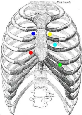
Cardiac auscultation is a physical examination that allows the listening of cardiovascular sounds, in order to evaluate the frequency, intensity, duration, number and quality of them to deduce possible diseases. As seen in the subchapter presented above, these sound characteristics are affected by their location of origin and the relative amplitudes in the different auscultation areas have important clinical value for the diagnosis of heart disease. Thus, there is a need of listening to different auscultation areas.
Figure 4: Main four auscultation areas: blue - aortic; yellow- pulmonic; red - tricuspid; light blue – erb point; green – mitral (EstudMed, 2015)
Heart auscultation is initiated with the patient seated or in the supine position, in a quiet room. During auscultation, the chest piece is repositioned several times as the auscultator moves it slowly between the different areas.
2.2.3.
Heart diseases
Heart sound characteristics are linked to blood pressure, and its interpretation is important for the detection of some heart diseases. According to WHO, in 2008 about 17.3 million died people from cardiovascular diseases, particularly heart attacks and strokes (WHO, 2013).
There are many different conditions that affect the normal function of the heart. The most common are:
Coronary artery disease, results from a buildup of plaque on the inside of the arteries, which reduces blood flow to the heart;
Congenital heart diseases, which are structural heart or intrathoracic great vessels defects present at birth, that are potentially of functional significance;
Abnormal heart rhythms called arrhythmias mean heart beating too fast or too slow;
Heart infections are caused by virus which attacks the heart muscles and causes the disruption of the electrical pathways that signal the heart to beat properly;
Cardiovascular disease related with blood vessels;
Heart valve disease occurs when the heart valves do not work the way they should because they are damaged.
Almost all of these can be detected by heart auscultation, which allows a quicker diagnosis avoiding bigger problems. Although coronary disease, heart failure and hypertension are more frequent when compared with heart valve disease, this last contributes to the wide spectrum of arterial diseases such as aortic aneurysms, intramural hematoma, atherosclerotic, among others, and therefore requires attention (Erbel et al., 2014).
Heart valve disease occurs when the heart valves do not work the way they should. The valves are situated at the exit of each of the four heart chambers and their function is to keep blood flowing through the heart in the right direction and that there is no backward leakage.
However, a variety of conditions can lead to valve damage. The two main problems that we can found in heart’s valves called stenosis or regurgitation (also called insufficiency).
The stenosis can occur in all four valves (if aortic valve developed a stenosis, the condition is called aortic stenosis; and so on) and it’s related with the obstruction of blood flow across the aortic valve. The narrowed opening may make the heart work very hard to pump blood through it and can lead, for example, to heart failure. Aortic stenosis typically results in a heart murmur and, nowadays, is the most common valve heart disease in the developed world (Czarny & Resar, 2014).
The regurgitation is the name for non-sealed valves. The valve does not close tightly and some blood will leak backwards across the valve into the upper heart chamber from the lower chamber or leaks through the leaflets when they should be completely closed. This leads to less blood flowing to the rest of the body. The heart tries to compensate and pumps harder, which can lead to congestive heart failure. Similarly to stenosis, it can occur in all four valves and the condition’s name depends in which valves it occurs and results in a heart murmur. (Encyclopedia, 2014)
3.1.
Research methodology
In order to understand how previous studies have compared the performance of acoustic and electronic stethoscopes, the following research methodology was used. Articles were initially identified by searching PubMed and Google Scholar. The final search included the terms: stethoscopes, comparison, evaluation and randomized trial.
The query used was: "stethoscopes"[All Fields] AND ("comparison"[All Fields] OR "evaluation"[All Fields] OR "randomized trial"). Efforts were made to gather all full-text papers, including contact with authors.
Afterwards, these were further selected during two phases. The first phase was based on the analysis of the title and abstract of the articles found in the initial search. Articles were classified as included or not, according to its relevance for the research question. The second phase was the analysis of the full-text paper of study. Again, articles were classified as included or not.
3.2.
Results of the article selection process
We did not found any published articles about comparisons between different electronic stethoscopes. However, there are many between electronic and conventional stethoscopes. This initial search using the previously described query was made on the 2nd of October of 2014.
Excluded: 51 No comparison between stethoscopes: 49
Duplicates: 2 Title / Abstract: 10
References found: 61
Excluded: 2 Not available in English: 1
PDF not available: 1
Full text: 8
Nine studies were included in this analysis. The work carried out by (Philip & Raemer, 1986) was not found during our search, but it was present in the references of the study proposed by (Grenier, Gagnon, Genest, Durand, & Durand, 1998). Given its relevance, we decided to include it in our final list of articles.
3.3.
Analysis of comparative studies
Table 1: Analysis of comparative studies
Paper, Year, Number of citations
Number of evaluators Participants
Variables of
comparison Methodology Statistical analysis
Effect of teaching and type of stethoscope on
cardiac auscultatory performance, 2006,
11
72 house officers, with 0-18 months of postgraduate clinical
experience
20 patients, 16 with disease and 4 without
disease
Teaching; type of stethoscope
Division in 4 groups associated with the possible 2 variables combination;
Multiple-choice questionnaire
Accuracy, agreement (k values); t tests,
unpaired t tests, general linear models
A randomized trial comparing electronic and
conventional Stethoscopes,
2005, 12
24 physicians: 4 specialists in cardiology, 4 specialists in general internal medicine, 4 specialist registrars, 4 senior house officers, 4 house officers
and 4 medical students.
42 patients, one third of the patients without disease Agreement between clinicians; type of stethoscope
Each group with 4, was divided in 2 groups associated with the type of
stethoscope; Multiple-choice questionnaire
K values for all 12 pairs of observers
An electronic stethoscope is judged better than conventional stethoscopes for anesthesia monitoring, 1986, 13
21 members: 11 residents, 4 certified registered nurse anesthetists and 6 staff
anesthesiologists _____ Clarity of sounds; efficacy of monitoring; other qualities; type of stethoscope Questionnaire;
Rating system: +3 - much better to -3 - much worse
Wilcoxon signed rank test for differences
Paper, Year, Number of citations
Number of evaluators Participants
Variables of
comparison Methodology Statistical analysis
Auscultation in Flight: Comparison of Conventional and Electronic Stethoscopes, 2010, 8
9 physicians: 7 anesthetist, 2 intensivist
36 evaluationsa Assess heart and lung sounds;
type of stethoscope
Questionnaire;
Visual rating scale: 0 - hear nothing, to 100 - hear perfectly
Paired t-tests
Cardiac auscultation training of medical
students: a comparison of electronic sensor-based and acoustic stethoscopes, 2005,
12
48 third year medical students; (2 cardiologists defined the
correct answers) 10 patients, one twice Auscultation skills, type of stethoscope
Division in 2 groups associated with the type of stethoscope;
Train the students with the type of stethoscope respective; Teaching of auscultation; Multiple-choice questionnaire; Rating system: number of points from 1 to
6 for each question and total score for the questionnaire.
Paper, Year, Number of citations
Number of evaluators Participants
Variables of
comparison Methodology Statistical analysis
Clinical Comparison of
Acoustic and Electronic Stethoscopes and
Design of a New Electronic Stethoscope, 1997,
42
9 cardiologists, 10 general practitioners, 11 nurses
378 auscultationsa Clinical performance; Type of stethoscope
6 stethoscopes, 3 of each type; Each patient was auscultated 3 successive
times by using 3 randomized different stethoscopes;
Evaluation grid: 1 – excellent to 5 – not acceptable; The questions depended of
clinical position; Frequency of appreciation Comparison of conventional and sensor-based electronic stethoscopes in detecting cardiac murmurs of dogs,
2012, 3
2 investigators: 1 final year veterinary student, 1 an expert in physical examination of the canine cardiorespiratory system
21 dogs, with disease
Diagnostic capabilities; type
of stethoscope
Questionnaire;
Arbitrary classification system: 0 - no difference between stethoscopes to 3 - advantage for the electronic stethoscope
Sensitivity , k -values,
aUnknown number of patient Paper, Year,
Number of citations
Number of evaluators Participants
Variables of
comparison Methodology Statistical analysis
Clinical evaluation of the 3M Littmann Electronic Stethoscope Model
3200 in 150 cats, 2013, 0
Acoustic stethoscope 2 observers: 1 board certified cardiologist with more than 20 years of experience with cardiac
auscultation, 1 cardiology resident with 3 years of clinical
experience Electronic stethoscope 8 observers: 3 cardiology
diplomats, 3 cardiology residents, 3 small animal
rotating interns
150 cats Clinical
performance; Type of stethoscope
Auscultation with traditional stethoscope by 2 observers;
30 seconds of heart sounds were recorded using an electronic stethoscope; The sounds were compared, off line, with
each other by the 8 observers;
k - Cohen’s , k –
Fleiss’s, McNemar’s
test
Pulmonary Auscultation in the
Operating Room: A Prospective Randomized Blinded Trial Comparing Electronic and Conventional Stethoscopes, 2013, 1
Anesthesiologists 100 patients,
who had general anesthesia for various surgeries Quality of pulmonary auscultation; type of stethoscope
3 stethoscopes (2 acoustic, 1 electronic); Questionnaire;
Numeric scale: 0 - hear nothing to 10 - hear perfectly
As shown in the Table 1, all the studies compare the type of stethoscope with other variables such as the clinical performance, by using questionnaires. About the methodology used in the comparisons, there are two types of studies: some compare the answers given by observers about the patient’s disease with the echocardiography or opinions of experts, which establish the correct diagnoses; others compare only the quality of sound between the stethoscopes evaluated.
K values are the statistical method more used in the studies because it allows us to calculate the agreement between observers, which is a crucial factor in this type of comparisons. Another important point, is the fact that the majority of analyzed studies ensure the homogeneity of analysis, making groups with observers which have identical level of experience. This detail, allows us to make comparisons between levels of experience, regarding the performance with a particular type of stethoscope. For example, it’s not expected that students have greater auscultatory proficiency than the most experienced physicians. (Høyte, Jensen, & Gjesdal, 2005)
The number of evaluators used varies and decided by the authors. While in studies carried out by Høyte et al., Iversen et al., and Philip & Raemer (Høyte et al., 2005; Iversen et al., 2005, 2006; Philip & Raemer, 1986) have a relatively high number, the others studies have a small number of evaluators. This can compromise the results, because the studies with a higher number of observers, theoretically, would be able to find a significant difference regarding the analyzed variable between groups of observers. According to Iversen et al., (Iversen et al., 2006) a high number of observers gives their study the largest discriminative power. Furthermore, when the number of observers included in each group is small and the comparisons among levels of experience depend on groups with only few observers, the observational skills of a single observer could have a marked impact on the average agreement. Also the low number of patients participating could of course be a problem regarding the representation of diseases, as mentioned in the study carried out by Iversen et al.
An important comment drawn by some authors (Iversen et al., 2006) is the need for studies on how the type of stethoscope affects a student’s ability to his auscultatory skills. These should measure the effect of teaching among the most inexperienced doctors for whom it seems reasonable to assume that the reliability of their cardiac auscultation would give room for most improvement and not among experienced doctors because already have a lots of experience.
All these findings were deemed relevant, and shaped the decisions made for our comparison methodology, as explained in the following chapters of this thesis.
3.4.
Analysis of types of stethoscopes compared
Table 2: The stethoscopes evaluated in analyzed studies (Iversen et al., 2005) (Vörös, Bonnevie, & Reiczigel, 2012) (Iversen et al., 2006) (Hoffmann et al., 2013)
(Blass et al., 2013) (Philip & Raemer, 1986) (Tourtier et al., 2010) (Høyte et al., 2005) (Grenier et al., 1998) Total Acoustic
Rappaport- Sprague X X 2
Holtex Ideal X 1
Littmann Cardiology III
X X 2
Welch Allyn - Elite X X 2
3M Littmann Master Cardiology
X 1
3M Littmann Classic II SE Stethoscope
X X X X 4
Electronic 3M Health Care - Littmann 4000
X 1
3M Health Care - Littmann 3200
X X 2
Labtron- Graham Field
X 1
EST40 Bosch X 1
3M Health Care - Littmann 3000
X 1
ST3 Starkey Laboratories
Table 3: The stethoscopes evaluated in analyzed studies (cont.)
(Iversen et al., 2005)
(Vörös, Bonnevie,
& Reiczigel,
2012)
(Iversen et al., 2006)
(Hoffmann et al., 2013)
(Blass et al., 2013)
(Philip & Raemer,
1986)
(Tourtier et al., 2010)
(Høyte et al., 2005)
(Grenier et al., 1998)
Total
Welch Allyn - Elite Electronic
Stethoscope
X 1
The stethoscope- Meditron
It is clear from Table 2 that not only there are a lot of electronic stethoscopes present in these studies, but more importantly they are spread among such studies, limiting our ability to understand the role and true potential of electronic stethoscopes. If we look at the market, this is even clearer. After a search in sites of commercialization and reviews of stethoscopes, the electronic stethoscopes that to the best of our knowledge are available on the market are the following:
3M Health Care - Littmann 3200;
Cardionics - E-Scope II Electronic Stethoscope;
Welch, Allyn - Elite Electronic Stethoscope;
GS Technology - JABES Electronic Stethoscope;
Thinklabs - ds32a Digital Electronic Stethoscope;
Contec Medical Systems – CMS VE;
Sun Meditec – WISE (Wireless Digital Stethoscope);
Dong Jin Medical – i-scope 200;
HB Meditech - SP-S2 Electronic Stethoscope;
Mabis healthcare – Signature Series Electronic Stethoscope;
Koratek – AUSCO ES-3100;
ADC - ADSCOPE 657;
ThinkLabs One.
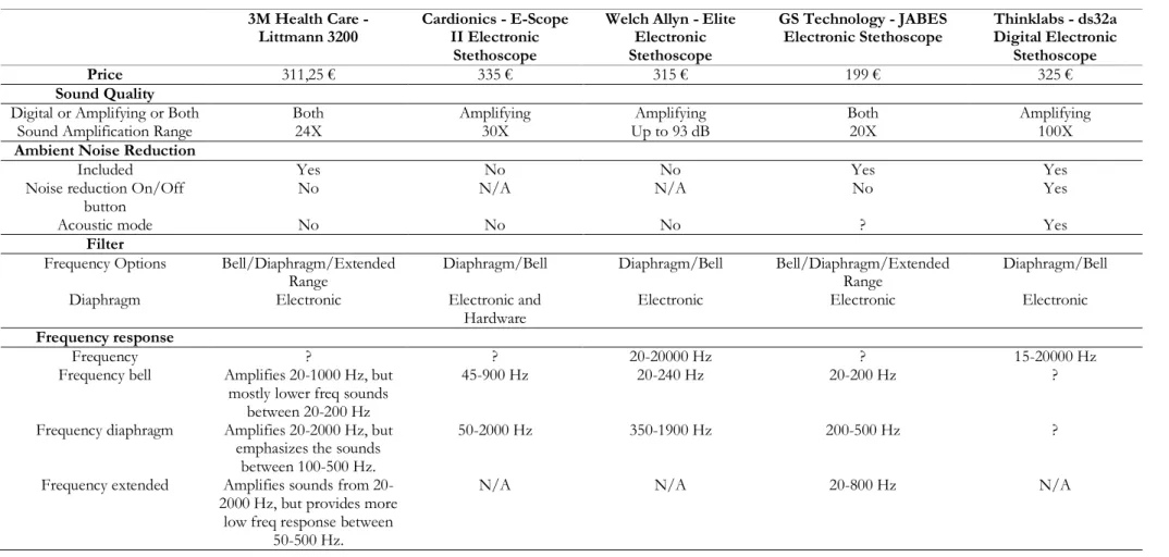
Table 4: Technical characteristics of the most relevant electronic stethoscopes available on the market
3M Health Care - Littmann 3200
Cardionics - E-Scope II Electronic
Stethoscope
Welch Allyn - Elite Electronic Stethoscope
GS Technology - JABES Electronic Stethoscope
Thinklabs - ds32a Digital Electronic
Stethoscope
Price 311,25 € 335 € 315 € 199 € 325 €
Sound Quality
Digital or Amplifying or Both Both Amplifying Amplifying Both Amplifying
Sound Amplification Range 24X 30X Up to 93 dB 20X 100X
Ambient Noise Reduction
Included Yes No No Yes Yes
Noise reduction On/Off button
No N/A N/A No Yes
Acoustic mode No No No ? Yes
Filter
Frequency Options Bell/Diaphragm/Extended
Range
Diaphragm/Bell Diaphragm/Bell Bell/Diaphragm/Extended
Range
Diaphragm/Bell
Diaphragm Electronic Electronic and
Hardware
Electronic Electronic Electronic
Frequency response
Frequency ? ? 20-20000 Hz ? 15-20000 Hz
Frequency bell Amplifies 20-1000 Hz, but
mostly lower freq sounds between 20-200 Hz
45-900 Hz 20-240 Hz 20-200 Hz
?
Frequency diaphragm Amplifies 20-2000 Hz, but
emphasizes the sounds between 100-500 Hz.
50-2000 Hz 350-1900 Hz 200-500 Hz ?
Frequency extended Amplifies sounds from
20-2000 Hz, but provides more low freq response between
50-500 Hz.
User Interface
Dedicated On/Off Button Yes Only Off Button Only Off Button Yes Yes
Power Stay-On Time 10-30 seconds before
entering standby. 30 minutes to 5 hours before
power off
2 minutes (possibility to 8 and 60 minutes shut
off)
3 minutes 3 minutes 2-5 minutes
Sleep mode indicador Yes No No No No
Display LCD No LED LED LED
Frequency Mode Indicator LCD No No LED No
Volume Level Indicator Yes No Yes No Yes
Patient Heart Rate Display Yes No No No No
Volume Control Yes (9 levels) Yes (64 levels) Yes Yes (7 levels) Yes (10 levels)
Mute function No No No No Yes
Advanced Features
On-Board Recording Yes (29 seconds) No No No No
Bluetooth Capability Yes No No No No
Communication mode Infrared Wired (jack) Wired connection Wired connection No
Wireless Capacity No No No No No
Connector No Square 4 pin connector Audio 2.5 mm (stereo) Audio Audio 2.5 mm
Retained
settings/Programming Preset Modes
Retains settings in standby mode. Restores factory setting when fully powered
off (these settings can be modified).
Retains audio. No Retains volume and mode. Retains bell, diaphragm
and amplification setting. Power-on mode
can be preset.
Adjustments
Eartips angle No No Yes ? Yes
Headphone tension Yes ? No Yes Yes
Cleaning
Alcohol cleaning Yes Yes Yes No. Use mild detergent. Yes
Maintenance
Eartips replacement Yes Yes Yes Yes Yes
Battery
Type 1 X AA 1 X AAA 1 X CR123A 2 X AAA 2 X AAA
Low battery indicator LCD level No No LED LED
Specifications
Weight 186 grams 176 grams 170 grams 170 grams 180 grams
Length 69 cm 96.5 cm 82.5 cm 73.6 cm 73.7 cm
Contec Medical Systems –
CMS VE
Sun Meditec – WISE
(Wireless Digital Stethoscope)
Dong Jin Medical – i-scope 200
HB Meditech - SP-S2 Electronic Stethoscope
Mabis healthcare –
Signature Series Electronic Stethoscope
Price 133,01 € 226 € 180,86 € N/A N/A
Sound Quality Digital or Amplifying or
Both
Amplifying Amplifying Amplifying ? Amplifying
Sound Amplification Range
32X Up to 100 dB 20X ? Up to 24 dB
Ambient Noise
Reduction
Included ? Yes No ? Yes
Noise reduction On/Off button
? No N/A ? No
Acoustic mode ? ? ? ? ?
Filter
Frequency Options Bell/Diaphragm/Extended
Range Bell/Diaphragm/Extended Range Bell/Diaphragm/Extended Range Bell/Diaphragm/Extended Range ?
Contec Medical Systems – CMS VE
Sun Meditec – WISE
(Wireless Digital Stethoscope)
Dong Jin Medical – i-scope 200
HB Meditech - SP-S2 Electronic Stethoscope
Mabis healthcare –
Signature Series Electronic Stethoscope Frequency response
Frequency 20-20000 Hz ? ? 5-5000 Hz ?
Frequency bell 20-230 Hz 20-350 Hz 20-500 Hz 20-200 Hz ?
Frequency diaphragm 100-800 Hz 350-1200 Hz 100-1500 Hz 200-500 Hz ?
Frequency extended 20-800 Hz 20-2000 Hz 20-1500 Hz 20-2000 Hz ?
User Interface Dedicated On/Off
Button
Yes No Yes ? No
Power Stay-On Time 3 minutes 3 minutes 20 seconds – 3 minutes 3 minutes 2 minutes
Sleep mode indicador No No No ? No
Display LCD LED LED ? LED
Frequency Mode Indicator
LCD LED LED ? LED
Volume Level Indicator Yes No Yes ? No
Patient Heart Rate Display
Yes No No No No
Volume Control Yes (16 levels) Yes (10 levels) Yes (5 levels) Yes (16 levels) Yes (8 levels)
Mute function No No No No No
Advanced Features
On-Board Recording No No No No No
Bluetooth Capacity No No No No No
Wireless Capacity No Yes No No No
Connector Audio 3.5 mm Audio 3.5 mm Audio 3.5 mm Analog output port No
Retained settings/Programming
Preset Modes
Retains volume and mode. No Retains volume and mode. Retains volume and mode. Retains volume and
Contec Medical Systems – CMS VE
Sun Meditec – WISE
(Wireless Digital Stethoscope)
Dong Jin Medical – i-scope 200
HB Meditech - SP-S2 Electronic Stethoscope
Mabis healthcare –
Signature Series Electronic Stethoscope Adjustments
Eartip angle N/A N/A N/A ? ?
Headphone tension N/A N/A N/A ? ?
Cleaning
Alcohol cleaning Yes ? ? ? Yes
Maintenance Diaphragm remplacement
Yes ? ? ? Yes
Eartips remplacement N/A N/A N/A ? Yes
Battery
Type Mini USB, 4.2V rechargeable
battery
Battery (Transmitter-DC 3V, Receiver-DC 5V)
2 X AAA 1 X CR123A 3 X LR44
Low battery indicador LCD level No LED ? LED
Specifications
Weight 50 grams 270 grams 79 grams 175 grams ?
Koratek – AUSCO ES-3100 ADC - ADSCOPE 657 ThinkLabs One
Price N/A 180.86 € 704.54 €
Sound Quality
Digital or Amplifying or Both Amplifying Amplifying Amplifying
Sound Amplification Range 15X 16X 100x
Ambient Noise Reduction
Included Yes ? Yes
Noise reduction On/Off button
? ? No
Acoustic mode ? ? ?
Filter
Frequency Options Bell/Diaphragm Bell/Diaphragm/Extended
Range
Bell/Diaphragm
Diaphragm ? ? ?
Frequency response
Frequency ? ? 20-2000 Hz (five filters mode)
Frequency bell ? 15-200 Hz 30-500 Hz
Frequency diaphragm ? 100-500 Hz 80-500 Hz
Frequency extended ? 15-1000 Hz ?
User Interface
Dedicated On/Off Button Yes Yes Yes
Power Stay-On Time 2 minutes 3 minutes 1-10 minutes
Sleep mode indicador No ? ?
Display LED LED LED
Frequency Mode Indicator LED LED LED
Volume Level Indicator ? ? Yes
Patient Heart Rate Display No No No
Volume Control Yes (8 levels) Yes (8 levels) Yes (10 levels)
Mute function ? ?
Advanced Features
On-Board Recording No No ?
Bluetooth Capability ? No ?
Wireless Capacity ? No ?
Stereo Jack ? Audio 3.5 mm
Retained
settings/Programming Preset Modes
Retains volume Retains volume and mode ?
Adjustments
Eartip angle ? Yes Yes
Headphone tension ? ? ?
Cleaning
Alcohol cleaning ? ? Yes
Maintenance
Diaphragm remplacement ? ? Yes
Eartips remplacement ? ? Yes
Battery
Type 2 X AAA 2 X AAA Battery
Low battery indicador LED LED LED
Specifications
Weight ? 175 grams ?
Length ? 74 cm ?
Given all this, it is safe to assume that the quality of all of these can vary significantly, reinforcing the need for our comparative study of electronic stethoscopes. Given that it is unrealistic to compare all of them, a selection of the 6 most relevant stethoscopes was made, based on their popularity, presence in scientific studies, and market penetration. Figure 5 depicts these, which are now listed:
GS Technology - JABES– Figure 5 (A);
3M™ Health Care - Littmann 3200 – Figure 5 (B);
Cardionics - E-Scope II – Figure 5 (C);
Welch, Allyn - Elite™– Figure 5 (D) ;
Thinklabs - ds32a – Figure 5 (E);
Sunmeditec - WISE – Figure 5 (F);
A - GS Technology - JABES B - 3M Health Care -
D - Welch, Allyn - Elite E - Thinklabs - ds32a F – Sun Meditec - WISE
4.1.
Dataset
The dataset used for this comparative study of electronic stethoscopes is composed by sounds recorded from 89 patients. Each patient’s cardiac sound was recorded with the six different types of stethoscopes (more details on these in section 3.4 of this thesis), in five cardiac spots if the patient’s disease is mitral regurgitation and six if the patient’s disease is aortic stenosis – See Figure 6. The area excluded is the Erb Point represented on light blue because it is an accessory spot.
The recording of 4 of the 6 stethoscopes – Cardionics, Jabes, Welch Allyn, Thinklabs – was conducted using a cable, 1 by WiFi transmission – WISE - and 1 by Bluetooth transmission – Littmann. The sounds with this last stethoscope were recorded with a specific system that is capable of collecting heart sounds by spot, using a tablet, called Digiscope Collector, and presented in Figure 7. The others were recorded with the Audacity software1
, on a laptop computer. All of these sounds were recorded with a 8kHz sampling frequency, excepted Littmann which was recorded at 4kHz and after, converted to 8kHz with Audacity software.
1 http://audacityteam.org
Mitral regurgitation: blue - aortic; yellow- pulmonic; red - tricuspid; light blue – erb point;
green – mitral
–
Aortic stenosis: blue - aortic; yellow- pulmonic; red - tricuspid; green – mitral
The volume of 5 stethoscopes was adjusted: Jabes and Thinklabs to 0,3; Welch Allyn and Wise to 1,0 and Cardionics to 0,7. All of these were empirically obtained with previous tests, in order to maximize signal strength and, at the same time, avoid distortion and sound clipping. All the stethoscopes were configured with diaphragm option active.
Whenever the physician changed the stethoscope position to a different spot, he knocked his fingers on diaphragm, to mark the passage with an easy to distinguish strong sound. This simplified the process of splitting the full audio recording into individual sound files for each spot. The exception was the Littmann, since we could use the DigiScope Collector application that conveniently simplifies all this process.
Heart sounds were collected at Centro Hospitalar do Alto Ave (Guimarães, Portugal) between September and December of 2013, by the same cardiologist. The patients of interest were called for a consultation, based on echocardiographic findings, and during the consultation was explained to the patients, the objectives to the study, and in followed, the recording of sounds was conducted. All data were collected with a formal patient consent, assigned by them. Furthermore, was asked the Ethics Committee in order to obtain Authorization to proceed the study. The number of authorization is 274-12.
Recordings were conducted in a real hospital environment, always in the same room of the cardiology department, which during this period was used exclusively for this purpose. The ambient noise level was measured using the Smart Tools application (version 1.5.8) installed in a Samsung S4 smartphone and its typical value was about 65 decibels.
Due to technical problems and usability issues some of this data was either lost or did not have enough quality to integrate this study. In order to make this study viable, an extra 4 normal patients were recruited with a mean age of 71 years, and their heart sounds were
recorded in a home environment by an experienced physician. Also, an extra 16 patients with aortic stenosis and mitral regurgitation were recruited and auscultated with the Littmann and the Cardionics stethoscopes at Hospital São João by a cardiology house officer, at the Internal Medicine and Cardiothoracic Department. All data was again collected with formal patient consent and all efforts were made to replicate as well as possible the environmental conditions of the main dataset. As explained later in section 4.4, this was enough to successfully complete the proposed study.
4.2.
Dataset Analysis
The created dataset also includes data relative to the patients, like gender, age, weight, height, type of person, hairiness, measure sight, systolic and diastolic pressure, pathology, cardiac frequency and date relative to the situation like date, hour, local, position and noise level. A basic analysis using the SPSS2 software was conducted to characterize the dataset: 53
of patients are female and 36 are male; the mean age is 71 years and 6 months; 30 patients without disease, 29 with mitral regurgitation and 30 with aortic stenosis; all the auscultations were collected with the patient sitting down.
As Attachment I shows, 89 patients recorded with Jabes, Thinklabs and Welch Allyn; 88 with Wise; 84 with Cardionics and 43 with Littmann (see Table 5). As explained in section 4.2, the data of Littmann and Cardionics were collected in two distinct moments and situations. Firstly, at Centro Hospitalar do Alto Ave, were recorded 18 cases with Littmann and 73 with Cardionics and later were recorded more 23 with Littmann and 16 with Cardionics at Hospital São João and patient’s home. This is explained by technical difficulties that happened during the data collection process, which led to both the loss and the creation of data that was unusable for comparison purposes. Although this is far from ideal, we consider it to also be a relevant result, hinting that this technology is interesting but sometimes not robust enough for unobtrusive use, especially when wireless transmission is involved. Fortunately, it was still possible to obtain a dataset that was good enough for the comparison experiments presented in this thesis.
Table 5: Resume of sounds division by stethoscope
Without disease Mitral
regurgitation
Aortic stenosis Total
Littmann 8 15 20 43
Cardionics 30 21 33 84
Wise 30 29 29 88
Welch Allyn 30 29 30 89
Thinklabs 30 29 30 89
Jabes 30 29 30 89
4.3.
Evaluation Methodology
Inspired by previous literature in which evaluators were divided into groups of different levels of experience, we decided to select two different groups of evaluators for our studies:
G1 - Expert group - Elements of this group perform heart auscultations many times on their daily routines and have experience in identifying pathologies
G2 - Normal group - Elements of this group have received some form of training in the art of auscultation, but have a more limited field experience making it difficult to identify pathologies.
The first group (G1) included cardiologists and cardiology house officers. The second group (G2) included medical students and general practitioners.
The chosen evaluation tool was the questionnaire, as with previous studies described in literature. For the Expert Group (G1), a sound was presented and a question asked about which pathology was present, while for Normal Group G2 two sounds were given and a question asked to choose which one is better. The evaluation was a blind test with no other information about each patient’s condition given, besides the heart sounds.
For our study purposes we only use one question related with sound quality, and eliminated other possible questions such as: compare the given sound with the sound of stethoscope in use in daily routines; evaluate background noise and evaluate if the given sound has enough quality to issue a diagnostic. This elimination is a pragmatic decision in order to obtain statistical relevance of the chosen question. Asking more required an unrealistic number of evaluators, which was not possible to obtain during the timeframe of this thesis.
4.4.
Sample size
In order to obtain statistical significance of comparison results, the number of evaluators and observations necessary was calculated for each group. The term “observation” is often used in this thesis and represents the listening of a case with one or more sounds.
Group 1
Statistically, the best stethoscope will be considered the one which allows the evaluator to identify a pathology better. In other words, we will measure the proportion of successful classifications using each stethoscope and find a difference between the six. The proportion of success is given by following equation: