of the Tungsten-Containing Formate Dehydrogenase
from Desulfovibrio gigas
dehydrogenase isolated from D. desulfuricans ATCC 27774 is homologous to DvMo-FDH and was character-ized by EPR and Mo¨ssbauer spectroscopies [4]. A monoheme cytochrome, c553, was identified as the
physi-ological partner for DvMo-FDH [3, 5]. The physiphysi-ological Hans Raaijmakers,1,4,5Sofia Macieira,1,2,4
Joa˜o M. Dias,1Susana Teixeira,1
Sergey Bursakov,1Robert Huber,2
Jose´ J.G. Moura,1Isabel Moura,1
and Maria J. Roma˜o1,3
1REQUIMTE/CQFB donor of DgW-FDH is also a monoheme cytochrome
that was recently purified (our unpublished data). Departamento de Quı´mica
FCT DgW-FDH consists of two subunits of 977 and 214
amino acids, and it belongs to the DMSO reductase Universidade Nova de Lisboa
2829-516 Caparica (DMSOR) family of enzymes, one of the four classes into which molybdopterin-containing enzymes have been Portugal
2Max-Planck-Institut fu¨r Biochemie classified [6–9]. This family is considerably broad, and,
besides DMSOR [10, 11], it includes enzymes such as Am Klopferspitz 18a
D-82152 Martinsried dissimilatory nitrate reductases (NAP) [12] and formate dehydrogenases. The members of this family have two Germany
molybdopterins (MGD cofactor) in the coordination sphere of Mo (or W), which is also bound to an amino acid side chain. This amino acid changes with the enzy-Summary
matic functionality and is a serine residue in DMSOR, a cysteine in NAP, and a selenocysteine in FDHs [2, 13]. Desulfovibrio gigas formate dehydrogenase is the first
representative of a tungsten-containing enzyme from At the buried active site of DgW-FDH, the tungsten atom is bound to two molybdopterin cofactors, to a seleno-a mesophile thseleno-at hseleno-as been structurseleno-ally chseleno-arseleno-acterized.
It is a heterodimer of 110 and 24 kDa subunits. The cysteine, and to either an inorganic sulfur atom or a hydroxyl ligand. It is accessible via a positively charged large subunit, homologous to E. coli FDH-H and to D.
desulfuricans nitrate reductase, harbors the W site tunnel, and product release may be facilitated for H⫹by buried waters and protonable amino acid side chains and one [4Fe-4S] center. No small subunit ortholog
containing three [4Fe-4S] clusters has been reported. and for CO2through a hydrophobic channel conserved
in other FDHs. The structural homology with E. coli FDH-H shows
that the essential residues (SeCys158, His159, and
Arg407) at the active site are conserved. The active Results and Discussion site is accessible via a positively charged tunnel, while
product release may be facilitated, for Hⴙby buried
Primary Sequence Determination
waters and protonable amino acids and for CO2 Since no primary sequence was initially available, gene
through a hydrophobic channel. isolation, sequencing, and model building were carried out simultaneously and in an interactive manner. Introduction
For isolating the gene coding for the formate dehydro-genase large (␣) subunit (fdhA), two degenerated oligo-Formate dehydrogenases (FDHs) are a heterogeneous
nucleotides, CPKGASTWQ and VGDPNTIPrev, were group of enzymes found in prokaryotes and eukaryotes.
synthesized and used to amplify, by PCR, a DNA frag-They catalyze the reversible two-electron oxidation of ment of about 2700 bp. This PCR product was cloned formate to carbon dioxide. In aerobic organisms FDHs
and sequenced by primer walking (see Experimental are mainly NAD⫹-dependent enzymes, while, in
anaero-Procedures) and gave information about the protein se-bic bacteria, they comprise a variety of proteins that
quence, 100 amino acids away from the N terminus. contain a complex inventory of redox centers and are
Furthermore, an in-frame TGA was detected in its DNA very sensitive to oxygen. This is not the case for
Desul-sequence, suggesting the possible presence of a fovibrio gigas formate dehydrogenase (DgW-FDH),
selenocysteine in the DgW-FDH large subunit, initially which, unlike other FDHs, loses activity in the presence not expected [1], but revealed by crystallography [2]. of air, but this activity can be restored [1, 2]. Formate
Information on the flanking upstream DNA sequence dehydrogenases have been isolated from other sulfate
was acquired by inverse PCR with primers FDH-reducers, such as Desulfovibrio vulgaris Hildenborough EEKSWDWrev and FDH-NAKGQVV. The downstream (DvFDH), which contains three subunits (␣, , and ␥).
DNA sequence was obtained by PCR with primers FDH-Subunits␣ and  are similar to those from DgW-FDH,
IEHPFSKT and small-CV. Assembly of the overlapping but, instead of W, the enzyme contains Mo [3]. The ␥ sequence fragments allowed the reconstitution of an subunit is a multiheme c-type cytochrome. The formate
open reading frame of 3036 bp (fdhA) coding for a 1012-amino acid polypeptide chain and the identification of
3Correspondence: mromao@dq.fct.unl.pt 4These authors contributed equally to this work.
5Present address: Crystal and Structural Chemistry, University of Key words: tungsten; selenium; formate dehydrogenase;
selenocys-teine; molybdopterin; iron-sulfur cluster Utrecht, Padualaan 8, 3584 CH Utrecht, Netherlands.
Structure 1262
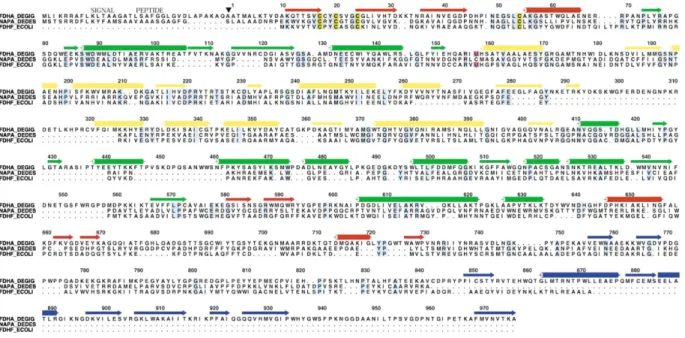
Figure 1. Amino Acid Sequence Alignment of the DgW-FDH Large Subunit with Its Structural Neighbors
FDHA_DEGIG, DgW-FDH␣ subunit, SWALL accession number Q934F5; NAPA_DEDES, NAP, SWALL accession number P88186; FDHF_ECOLI, FDH-H, SWISS-PROT accession number P07658. Conserved residues, light blue; cysteines involved in iron-sulfur cluster binding, dark yellow; selenocysteine, magenta. The secondary structural elements, strands (→), and ␣ helices (䊏) are colored according to the domains to which they belong. Domain I, red; domain II, green; domain III, yellow; domain IV, blue.䉲, signal peptide cleavage site. The residue numbering refers to the FDHA_DEGIG sequence and starts at the first residue of the mature protein. This figure was prepared with the programs PILEUP, in the Wisconsin Package Version 10.0 (Genetics Computer Group [GCG], Madison, WI), and ALSCRIPT [39].
an open reading frame coding for the formate dehydro- garis Hildenborough Genome Sequencing Project, cur-rently under processing at The Institute for Genomic genase small () subunit (fdhB).
After the inclusion of the DgW-FDH large subunit poly- Research (http://www.tigr.org), and detected two ho-mologous FDHs in this genome. One is the already de-peptide sequence in the structural model available at
that time, it was also possible to start tracing the poly- scribed three-subunit FDH [3], and the other is an FDH, like DgW-FDH, composed only of two subunits. peptide chain of the small subunit in the electron density
map and to complete the sequence determined by PCR,
by giving information on the C terminus. Structure Solution
The initial absence of sequence information for DgW-The fdhA and fdhB sequences have been submitted
to the DDBJ/EMBL/GenBank under accession numbers FDH was a major hurdle in solving its crystal structure. Yet a molecular replacement solution was obtained [2] AJ318781 and AJ427412, respectively. The C-terminal
sequence data for the formate dehydrogenase subunit with the structure from an enzyme of the same family, the periplasmic nitrate reductase (NAP) from D. desul-has been submitted to the SWISS-PROT Protein Data
Bank under accession number P83237. furicans [12], as a search model. This molecular replace-ment solution provided a model for half of the protein Analysis of the gene sequence revealed the features
of the preprocessed DgW-FDH (Figure 1). The large sub- content, but the electron density map was of rather poor quality outside the region defined by the search model. unit precursor (FDHA) carries a signal peptide (the first
35 amino acid residues) with the twin-arginine motif Therefore, since the MR method did not prove to be enough to solve the structure, additional phasing infor-RR-x-F-L-K [14] characteristic of Sec-independent
ex-ports to the periplasm and harbored by periplasmic mo- mation was necessary and could be obtained from a four-wavelength data set (MAD) collected at ESRF (the lybdoproteins. The mature formate dehydrogenase
large subunit is then composed of 977 residues and has Fe K edge is 1.73649 A˚ and the inflection point is 1.74149 A˚ ; the W LIIIinflection point is 1.21417 A˚ and
a deduced molecular weight of 109,712.8 Da.
The small subunit (214 residues) has a deduced mo- the remote is 0.992 A˚). Both solutions (MR and MAD) were used to solve the structure, since the electron lecular weight of 23,852.0 Da and is devoid of an export
signal. This strongly suggests the formation of the com- density maps calculated with combined MAD and model phases were of superior quality when compared to those plex between both large and small subunits in the
cy-toplasm before translocation and DgW-FDH export to calculated with MAD phases alone.
The model was built in several cycles of model build-the periplasm as an oligomeric structure, as it was
demonstrated for bacterial periplasmic [Ni-Fe] hydro- ing, phase combination with experimental phases, den-sity modification, and tracing with ARP/wARP [16]. At genases [15].
We ran a FASTA search of our sequence data against first, automated tracing was unsuccessful, but it worked after strategies were used to improve both the model the preliminary sequence data of the Desulfovibrio
vul-amino acids) harbors the W[(MGD)2,-SH,-SeCys]
cofac-Table 1. Refinement Statistics
tor as well as one of the [4Fe-4S] clusters and is an␣ Data resolution range (A˚ ) 35–1.8 structure, similar to the structures of FDH-H from E. coli Number of reflections 208,782
[13] and to the periplasmic D. desulfuricans ATCC 27774 Number of free reflections 10,979
nitrate reductase (NAP) [12]. The superposition of the Number of nonhydrogen protein
large subunit of DgW-FDH with NAP shows an rmsd of
atoms 17,783 (plus 298 double
2.0 A˚ for the C␣ atoms of 636 out of 723 amino acid conformations)
2,382 amino acids residues. The comparison with E. coli FDH-H gives a Number of water molecules 1,707 corresponding rmsd of 2.1 A˚ for 659 out of 715 amino
R factor 0.173
acids (Figure 2).
Free R factor 0.213
The large subunit can be divided into four domains, Rmsd bonds (A˚ ) 0.019
two of which correspond to noncontiguous stretches of Rmsd angles (⬚) 1.792
the polypeptide chain, following the same classification Mean B factor (A˚2) (main chain/
side chain/solvent) (20.7/23.0/31.1) used for FDH-H and NAP. However, DgW-FDH is
consid-Ramachandran plot (%) erably larger than these homologous proteins (977
Favored regions 89.3 amino acid residues versus 723 and 715 for NAP and
Allowed regions 10.5
FDH-H, respectively), and the extraⵑ250 amino acids Generously allowed regions 0.2
are distributed over the surface of the molecule and Rmsd NCS-related atoms (A˚ )a
correspond to insertions throughout all four domains.
C␣ 0.197 (without
less-ordered parts) These insertions are represented in Figure 3 as gray
All 0.391 regions of the structure.
The N terminus (residues 1–8) of the large subunit
aThe rmsd values correspond to the exclusion of residues 1, 2,
protrudes like an arm holding the small subunit, inter-837–843, and 956–966 of the large subunit; these residues are poorly
defined in the electron density. acting with residues Gln23, Trp24, Thr145, and Asn146 (from the small subunit) and strengthening the interac-tion between both subunits. Domain I (in red) is defined and the data. The model was significantly enhanced by residues 9–68, 575–603, and 647 –734 and, as in NAP once the DNA corresponding to the amino acids resi- and in FDH-H, carries the characteristic cysteine motif dues 54–365 had been sequenced. Regarding the data, that binds the first [4Fe-4S] center (-CXXCX
nCXmC-).
care was taken to include all data, not only the weak When compared to the structures of NAP and FDH-H, reflections (between 2.0–1.8 A˚ ), but also those selected it has a shortcut 732–733 in domain I, which replaces for the Rfreetest. Only at later stages of the refinement the hydrophilic loop 530–539 in FDH-H. Flanking this
were these reflections omitted (Rfree) to monitor the prog- location are two inserts of domain I: residues 658–689 ress of the refinement. Additional primary sequence data and 709–726 and an insert of domain IVa (740–770). were incorporated as soon as they became available. These inserts, 709–726 and 740–770 on one side and For some loops, the electron density stayed rather poor insertion 658–689 on the other side, seem to define a in both NCS-related molecules. Though omit maps show clamp tailored to bind an␣ helix of the physiological some density for residues 1, 2, 837–843, and 956–966 partner. The proposed interface is an elongated cleft, of the large subunit, these particular regions seem to with a diameter of 12–15 A˚ and a length of 30–40 A˚, be partially disordered. The two heterodimers in the lined with hydrophobic and basic residues. Domain II asymmetric unit are essentially identical, as they can be (in green) contains 205 amino acid residues and is de-superimposed to an RMSD of 0.39 A˚ for all protein atoms fined by three stretches of polypeptides, 69–157, 410– or 0.20 A˚ for all C␣atoms when the less-ordered parts 573, and 604–646. In relation to the structures of NAP are omitted (see Table 1). and FDH-H, it contains four insertions corresponding to 71 amino acid residues (106–122, 431–466, 552–566, and Quality and Topology of the Model 641–647). The polypeptide 431–466 defines an extended
Large Subunit loop, which protrudes over the cleft that gives access to
The DgW-FDH molecule consists of two subunits. The the buried active site. The loop 641–646 on the molecular surface makes important crystal contacts.
three-dimensional structure of the large subunit (977
Figure 2. Superposition of the Large Subunit of DgW-FDH in Blue with the Structure of the Periplasmic Nitrate Reductase from D.
desul-furicans ATCC 27774 Represented in Green
The rmsd for the superposition of 636 amino acid residues is 2.0 A˚ . The structure of FDH-H from E. coli [13] (red) and the corresponding rmsd for 659 amino acids of DgW-FDH is 2.1 A˚ .
Structure 1264
Figure 4. Schematic Representation of the Two Pyranopterin Guanosine Dinucleotide (MGD) Cofactors and the Hydrogen Bonding Interaction with Surrounding Residues and Buried Water Molecules
Hydrogen bonds were identified using CON-TACT from the CCP4 package [36] and TURBO-FRODO [37].
Domain III (in yellow) is defined by a continuous poly- and Asp395 OD1 (2.84 A˚ ) and a water (2.79 A˚), which, in turn, is hydrogen-bonded to Pro361 O (3.12 A˚ ) and peptide (residues 157–409) and, when compared to the
structures of FDH-H and NAP, shows one long insertion Gly394 O (2.78 A˚ ). The calcium ion is too far from other active parts of the protein to propose an active role of 51 amino acid residues (269–320) and a shorter one
of 5 amino acids (359–363). The long insertion has an for it. However, without this insertion, the interaction between Phe259 and Phe750 (absent in FDH-H and irregular secondary structure and covers the linear linker
between domain IVa and IVb (795–784) with hydrophobic NAP) would be solvent exposed.
The C-terminal domain IV (in blue) is defined by the side chains (see below). Since it sits above the formate
entrance to the active site, this tunnel isⵑ10 A˚ longer last 241 amino acid residues and is ca. 70 residues longer than the corresponding domain of FDH-H and than that in FDH-H.
The insertion of residues 359–363 supplies a Ca2⫹ NAP. Because of the large insertion 740–770, this
do-main can be subdivided into two subdodo-mains connected binding site (Figure 3A, pink sphere) that involves the
backbone oxygen atoms of residues Thr358 O (2.62 A˚ ), by a linker (795–784): IVa (residues 735–795) and IVb (residues 796–977). The latter has two insertions spa-Lys360 O (2.92 A˚ ), Lys363 O (2.67 A˚), Leu393 O (2.68 A˚),
Figure 3. Stereo Representation of the Overall Structure of DgW-FDH from D. gigas
The small domain (214 amino acid residues) is represented in purple, and the large domain (977 amino acid residues) is colored after its domain classification. Domain I (red) corresponds to residues 9–68, 575–603, and 647–734; domain II (green) contains residues 69–157, 410–574, and 604–646; domain III (yellow) is defined by residues 157–409; domain IV (blue) is defined by residues 735–977. Domain IV can be divided into two subdomains, IVa (residues 735–795) and IVb (residues 796–977). The insertions (relative to the homologous NAP and FDH-H structures) present in several domains are shown in gray.
(B) Representation of the four “exploded” domains of the large 977 aa subunit.
(C) Comparison of the domain organization of DgW-FDH, D. desulfuricans NAP, and E. coli FDH-H. The insertions in DgW-FDH are depicted as dashed areas.
(D) Stereo C␣ trace numbered every 20 residues for domains II and III (top).
Structure 1266
Figure 5. Small Subunit of DgW-FDH
(A) Overall structure of the small subunit, as cartoon and stereo C␣ trace. Domain A (red), 1–72 and 137–165; domain B (green), 73–136 (71–72 and 137 can be assigned to both domains A and B); domain C (yellow), 166–214.
(B) The ferredoxin-like domains A (red) and B (green) can be superimposed to each other with an rmsd of 1.0 A˚ for 31 C␣ atoms plus 8 atoms from the [4Fe-4S] cluster or an rmsd of 1.1 A˚ for 132 main chain atoms. The domains are related by a proper rotation.
(C) Ferredoxin (Protein Data Bank entry 1blu; blue) has been superimposed on domain A (red) with an rmsd of 0.99 A˚ for 35 C␣ atoms plus 16 [4Fe-4S] atoms. Domain B, green.
tially close together (818–843 and 921–930), while the residues 818–843 can be seen as an insertion, where the related chain goes through the disulfide bridge. In less-ordered loop 957–966, near the active site, adopts
a different orientation in FDH-H. DgW-FDH, cysteines 817 and 844 correspond topologi-cally to FDH-H 567 and 569.
The only disulfide bridge, C817–C844, is located in a
hydrophobic patch on the surface, close to the guani- As DgW-FDH needs to be activated with 2-mercapto-ethanol in an anaerobic environment in order to function, dine of MGD 1001 and is flanked by the solvent-exposed
side chains of Val819, Val843, Pro846, Pro849, Phe850, it is likely that this disulphide bridge will be broken in the active form. Release of the disulfide bridge would and Leu891. The disulfide bridge is far away from the
proposed electron transfer pathway, so it is unlikely to allow wider opening and movement of the formate entry cleft and would alter the position of residues 818–848 be actively involved in catalysis. Compared with FDH-H,
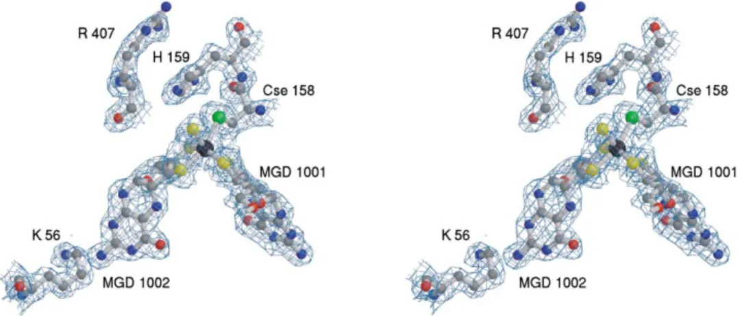
Figure 6. Representation of the Tungsten Catalytic Site of DgW-FDH, Superimposed with the Final 2Fo⫺ FcElectron Density Map Contoured
at 1.0
Surrounding residues important for catalysis are also included: SeCys158 (bound to the W atom [black sphere]), His159, and Arg407. To the left is Lys56, which makes a hydrogen bond to the exocyclic NH2of MGD 1002.
(with unknown function). Maybe the disulfide bridge stability of the DgW-FDH complex, which allowed copur-evolved as a protection or regulation of the enzyme to ification.
mildly oxidized (toxic) environments, contrary to FDH-H, The fold of the small subunit is of the␣ type and which is permanently inactivated by oxygen [18, 19]. contains three domains (Figure 5A): domain A (residues The C-terminal helix from FDH-H is absent, but the 1–72 and 137–165), domain B (residues 71–137), and corresponding binding cleft in DgW-FDH (ca. 40 A˚ long) domain C (residues 166–214). Domain A, which holds is lined with basic and aromatic residues and might two iron-sulfur clusters, and domain B, which binds an-accommodate a seven-turn␣ helix. It might be provided other cluster, are related by a proper 2-fold rotation. by another protein to form a complex in vivo. Without They can be superimposed to an rmsd of 0.87 A˚ for 26 such a helix, Cys156 from the small subunit, which binds C␣ atoms and the 8 atoms of one [4Fe-4S] cluster. They [4Fe-4S] cluster 2 (the second in its relative distance to are cunningly intertwined, such that some residues were W), is solvent exposed (the most exposed from all four). assigned to both domains. The fold of domains A and This would be a suitable interaction site for the artificial B resemble 2 ⫻ [4Fe-4S] ferredoxins. With about 52 electron acceptor benzylviologen, used in in vitro activ- structurally conserved residues, these ferredoxins share ity studies or for the physiological electron acceptor. two [4Fe-4S] clusters sandwiched between two helices As in other enzymes from the same family, the on one side and a sheet on the other side. The 2 ⫻ W(MGD)2cofactor is buried at the interior of the protein [4Fe-4S] ferredoxin from Chromatium vinosum (Protein
and is stabilized by an extensive network of hydrogen Data Bank entry 1blu; [20]), for example, can be superim-bonding interactions to residues of mainly domains II,
posed to an rmsd of 0.99 A˚ for 35 C␣ atoms (out of 82) III, and IV (Figure 4). MGD1002 interacts with domain III
and 16 atoms of the metal clusters (2⫻ [4Fe-4S]) (Figure and IV and with N of Lys56 of domain I. This lysine
5C). The two helices and the [4Fe-4S] clusters fit particu-makes a bridge between the pterin and the first
[4Fe-larly well. Similar results are obtained with other ferre-4S] cluster. The other MGD (1001) interacts with domains
doxins. Even the Fe-only hydrogenase from Desulfovi-II and IV.
brio desulfuricans (Protein Data Bank entry 1hfe [21]) Small Subunit
can be superimposed to 0.68 A˚ for 29 C␣ atoms (the The small subunit (214 amino acid residues) holds three
region of Hase around Fe-S clusters 422 and 423) and of the iron sulfur clusters of DgW-FDH (Figure 5). It is
atoms from these 2⫻ [4Fe-4S] clusters. located opposite from the cleft leading to the active site
During the latest stages in the preparation of this and interacts with the large subunit covering a surface
manuscript, the E. coli FDH-N structure was published of ca. 2362 A˚2. Several hydrogen bonds, salt bridges,
[22]. In this structure the subunit harbors four [4Fe-tightly bound waters, and hydrophobic interactions
sta-4S] clusters. Interestingly, as already advanced by us bilize the interface between the subunits of the
hetero-(our unpublished data), there is space and the correct fold dimer. This involves over 40 residues of domains I and
for an additional [4Fe-4S] cluster in the DgW-FDH small IV in the large subunit and 30 amino acids of the small
subunit, which would be present if the residues Ala84-subunit (residues 1–6, 39–41, 51–61, 229, 858, 859, 871–
xn-Leu110-xx-Tyr113-xx-Val116 were mutated to Cys. 879, 881–883, 896, 897, and 912–918 from the large
The FeS-3 cluster present in FDH-N [22] is the experi-subunit and residues 11, 14, 15, 22–30, 32–38, 56, 58,
mental evidence that corroborates our former predic-65, 141–146 and 154–157 from the small subunit). The
tion. Furthermore, the same motif found in the E. coli N terminus of the large subunit wraps around the smaller
FDH-N is conserved in all sequences of FDHs currently subunit and contributes to the stability of the functional
Structure 1268
Tungsten Site and Coordinating Ligands atom, the Mo-S distance refined to 2.39 A˚ , and the posi-tive residual density vanished. In this case the B factors In the crystallized form of the enzyme, the W atom is
coordinated to the four dithiolate sulfur atoms from the were 37 A˚2/30 A˚2for S/Mo (our unpublished data).
As has been demonstrated by activity experiments of two MGDs, by the selenium atom of SeCys158 and by
one hydroxyl or sulfide ligand (Figure 6). In the vicinity of E. coli FDH-H using13C-labeled formate in18O-enriched
water [18], the reaction catalyzed does not involve an the buried W site, similarly to E. coli FDH-H, are His159,
probably directly involved in the enzymatic mechanism oxygen transfer. Therefore, a putative reaction mecha-nism could make more sense with a sulfur ligand to W. (as proposed by EPR for FDH-H [18]), and Arg 407,
which points toward the entry of the tunnel and may be Although EXAFS data seem to favor the OH ligand for FDH-H [19], these data are not conclusive, probably responsible for orienting the substrate molecule.
The dithiolene function of both MGD cofactors is ex- because it is difficult to distinguish similar ligands with different distances in EXAFS [G. George, personal com-pected to be flat (double bonded), as found in other
molybdopterin-containing enzymes [10–13, 17]. This munication]. Our crystallographic data favor a sulfur atom for the sixth ligand, although the resolution of the seems to be true for MGD1002, but, in MGD1001, it
refines better when restraints for the reduced form data does not permit us to discriminate unambiguously between O and S until higher resolution data are avail-[C12⬘(R)-C13⬘(R)] are applied.
The sixth tungsten ligand, supposedly an oxygen li- able or until decisive biochemical assays settle the ar-gument.
gand, as proposed also for FDH-H [10, 14], behaved poorly in refinement, giving rise to a peak (⬎5 ) in an
mFo ⫺ DFcdifference electron density map. Besides, How DgW-FDH Works
The Active Site and Substrate Access the B factor for an OH ligand was only half that from
the tungsten atom: 11.6/20.6 A˚2 and 10.8/20.6 A˚2 for The active site of DgW-FDH is buried and ca. 25 A˚ away
from the molecular surface. However, as in other molyb-both NCS monomers. If refined as a sulfur atom, the
corresponding temperature factors were of the same dopterin-containing enzymes, there is a clear funnel-shaped entrance that leads access to the internal active magnitude as those from surrounding atoms, with B
factors for S/W of 25.1/20.8 A˚2and 24.7/20.6 A˚2for both site. From a careful analysis of the DgW-FDH structure,
one is tempted to assign functions to structural peculiar-independent molecules. In this case, the bond distance
W-SH (unrestrained) refined to 2.41 A˚ , a typical value ities. The formate molecule could easily reach the buried active site through the tunnel (Figure 7A), which is posi-for this type of bond and too long posi-for a W-OH bond
distance, and the spurious difference electron density tively lined with His159, Arg156, His423 His963, His376, Lys791, Lys444, Lys445, Arg587, and Arg407. At the peak disappeared. Further anisotropic refinement of the
sixth tungsten ligand showed that it possesses a large bottom of this channel is the W active site (Figure 6), which resembles the Mo site of the homologous FDH-H anisotropy, i.e., more angular freedom than bond length
variations. Taking this anisotropy (assuming an OH li- from E. coli [13]. His159 (His141 in FDH-H) is an impor-tant active site residue and establishes a interaction gand) into account lead to B factors of 14.0 A˚2, instead
of 11 A˚2, compared to 15.6–19.0 A˚2for the MGD-sulfur with the Se atom of the metal ligand Se-Cys158.
Be-sides, there is a clear pH dependency in the catalytic atoms and 20.7 A˚2for the W atom. This seems
sugges-tively low, though it might be attributed to experimental activity, for FDH-H [13,18] as well as for DgW-FDH (our unpublished data), and protonation of this histidine resi-error. Such anisotropic refinement of the O ligand has
not been carried out for related structures with low B due by the ␣ proton of formate has been proposed for FDH-H, supported by EPR studies [18, 19]. Arg407 factors for their OH ligands (e.g., FDH-H [13]). Also
sug-gestive are the bond distance W-OH, which stays at (Arg333 in FDH-H) provides a positive charge and a hydrogen bond to orient the substrate molecule for ca-2.41 A˚ , and the distance from the hydrogen bond to the
backbone N of Glu409 at 3.6 A˚ , which is too long for a talysis.
Putative Proton Channel hydroxide, but would be acceptable for a sulfur ligand.
In the case of the NAP structure [12], although the differ- The enzymatic reaction involves the release of H⫹, and there is a putative proton channel, which connects the ence in the relative B factors between Mo and the OH
ligand was not significant (27 A˚2/31 A˚2for OH/Mo), there active site to the exterior of the FDH molecule. It is
located almost perpendicular to the “formate cleft”, as was always a residual electron density ofⵑ6 , close
to the OH ligand. When the latter was refined as a sulfur depicted on the surface electrostatic potential
represen-Figure 7. How DgW-FDH Works
(A) Diagram of DgW-FDH cofactors and GRASP [38] electrostatic-potential surface plot contoured for positive charges (blue) and negative charges (red). The top cleft is lined with basic amino acids and provides an excellent entry point for the formate. At the W/Se center, the formate is split into CO2, H⫹, and two electrons. To the left are the four [4Fe-4S] clusters. Counting at increasing distance from W: [4Fe-4S]
1 (large subunit) Cys17-xx-20-xxx-24-xn-54; [4Fe-4S] 2 (small subunit) Cys11-xx-Cys14-xx-Cys17-xn-156; [4Fe-4S] 3 Cys21-xn
-Cys137-xx-Cys140-xn-152; [4Fe-4S] 4 Cys72-xx-Cys75-xxxx-Cys80-xn-Cys120. There would be space and the correct fold for a fifth [4Fe-4S] if the
residues Ala84-xn-Leu110-xx-Tyr113-xx-Val116 were mutated to Cys.
(B) A chain of water and protonable residues provides a channel from the reaction center to the surface of the FDH. The orientation of the picture is similar to that of Figure 7A, middle and left.
(C) More or less perpendicular to the formate entry and the proton channel is channel lined with aromatic and hydrogen bond-donating residues. Putative CO2binding sites are indicated with sticks between observed water molecules (red). The picture has been rotatedⵑ20⬚
Structure 1270
tations (Figure 7B), and is coated with protonable amino one [4Fe-4S] center (in an arrangement resembling the related monomeric enzymes FDH-H [13] and NAP [12]), acid side chains (Glu409, Asp132, Glu134, and Glu579)
and a chain of water molecules. and a small subunit that harbors a series of three [4Fe-4S] clusters as well as a putative vacant binding site for Not all of these residues are conserved in FDH-H. An
alternative pathway in FDH-H may involve phosphates a fourth cluster.
Recently, the sequences of two other tungsten for-and the guanine of MGD-801. These differences may
be due to the different cell location of both enzymes mate dehydrogenases became available in the data-bases. FDH-I and FDH-II from Eubacterium acidami-(different pH environment), since, while DgW-FDH is
present in the periplasmic space, the E. coli FDH-H is nophilum are also heterodimers (SWALL accession numbers for the respective subunits are Q93V06 and located in the cytoplasm.
Putative CO2Channel Q93SF5 and Q93V05 and Q93SF1), but no significant
sequence similarity with DgW-FDH was found. A spe-The release of the product molecule CO2from the buried
active site may be facilitated by a hydrophobic channel, cific tungsten ABC transporter, discriminating molybde-num, was reported for this mesophile last year [24], and, which is coated by several H bond-donating amino acid
residues (Figure 7C). The residues Val412, His159, so far, no molybdenum-dependent enzymes have been detected. On the contrary, molybdenum enzymes, such Trp730, and Tyr428 are conserved in FDH-H (Val338,
His141, Trp528, and Tyr354). Other amino acids from as the aldehyde oxidoreductase (MOP) [17], and tung-sten enzymes, such as the DgW-FDH [1], have been this channel (Arg172, Arg734, Trp458, Trp693, Trp730,
and Trp459), which are closer to the molecular surface, isolated from cultures of D. gigas grown under identical conditions. Since Mo is a more versatile element than W correspond to one of the insertions in DgW-FDH, being
absent in the smaller FDH-H, and, consequently, the and Mo-containing centers have higher redox potentials [25], the sulfate reducer D. gigas can be regarded as a equivalent channel is much shorter. Both Arg172 and
Arg734 donate bidentate hydrogen bonds to what could mesophilic organism representative of a transition from a “W world” to a “Mo world”, which would correspond be a CO2binding site, perpendicular to the plane of the
tryptophan side chain, clamping the CO2between them. to the adaptation to more oxidized environments.
(Figure 7C).
Experimental Procedures In respect to the differences between FDH-H and
DgW-FDH, the CO2channel is longer and more selective
Gene Sequence Determination in DgW-FDH as well as in the formate entry, which is
Unless otherwise mentioned, standard molecular biology protocols
ⵑ10 A˚ deeper. One possible explanation may be that were used [27]. Oligonucleotides CPKGASTWQ, 5⬘-TGYCCIAARG more specific and longer channels are needed to protect GIGCIWSIACITGGCA-3⬘, and VGDPNTGIrev, 5⬘-GGIATIGTRTTIG GRTCICCIAC-3⬘, were synthesized on the basis of on the formate the active site of DgW-FDH because of its location in
dehydrogenase large (␣) subunit internal peptides CPKGASTW-the periplasmic space, where more undefined molecules
QLAENERRPANPLYRAPGSDQ and TPSVGDPNTGIPETK and used may interact with (and eventually block) the enzyme.
to amplify, by PCR, a 2700 bp DNA fragment of the corresponding Electron Transfer Pathway
gene (fdhA). The resulting product was cloned in pPCR-Script Amp As found in other enzymes from the same family, the SK (⫹) (Stratagene) and sequenced with primers T3 and T7 (New electron transfer involves just one of the pterins (MGD England Biolabs) and by primer walking, with an automated DNA sequencer (Model 373; Applied Biosystems, Forster City, CA) and 1002). The closest pathway between the pteridine
moi-the PRISM ready reaction dye deoxy terminator cycle sequencing ety and the nearest [4Fe-4S] center is mediated by the
kit (Applied Biosystems). From the obtained DNA sequence, we N of Lys56 (Lys44 in FDH-H and Lys49 in NAP), which
designed the “inside-out” primers FDH-EEKSWDWrev, 5⬘-AGTCC makes a favorable hydrogen bond with the exocyclic
CAGCTTTTTTCTTCCC-3⬘, and FDH-NAKGQVV, 5⬘-ATGGACAAC NH2of the pyranopterin (Figures 4 and 6). This electron GAAGAGTGCTG-3⬘, for inverse PCR DNA amplification of the miss-transfer pathway via the exocyclic NH2is found not only ing flanking regions [26–29]. Attempts to amplify the downstream region with this technique were not successful, so it was just used in the enzymes from the DMSOR family (NAP and
to acquire information on the upstream region. On the basis of a FDH-H), but also in the enzymes from the xanthine
oxi-restriction enzyme map of the known fdhA sequence, the oxi-restriction dase family (MOP [17] and xanthine oxidase [23]),
al-enzyme AvaI (New England Biolabs) was chosen for hydrolysis of though, in the latter case, the closest contact is to the
1g genomic DNA in a volume of 200 l. After incubation for 1 hr S␥ of one cysteine residue from the nearest [2Fe-2S] 30 min, the enzyme was heat inactivated for 20 min at 70⬚C. The center. All four [4Fe-4S] clusters are 9 to 10 A˚ apart [2] restriction fragments were then diluted 100-fold, and 530l were ligated with 28 U of T4 DNA ligase (New England Biolabs) for 16 hr (Figure 7A), providing an easy electron pathway to the
at 4⬚C in a total volume of 600 l. Using 14.5 l of these DNA physiological electron donor (monoheme cytochrome)
circles as templates, 50 pmol of each primer, and 2.5 U Herculase of DgW-FDH.
(Stratagene) in 25l reaction mixture, we obtained a PCR product of about 1500 bp. The PCR product was directly sequenced.
The downstream sequence was obtained by PCR with primers Biological Implications FDH-IEHPFSKT, 5⬘-CATCGAGCATCCCTTCTCCAAG-3⬘ (internal to
fdhA), and small CV, 5⬘-ACCGGTGGGGCAGGTCTTCACGCA-3⬘, on
the basis of the amino acid sequence CVKTCPTG, conserved in the In contrast to the related molybdoenzyme FDH-H [13],
formate dehydrogenases iron-sulfur () subunits currently available which is composed of a single polypeptide chain, and
in the databases and predicted by us from the preliminary sequence to molybdenum-containing formate dehydrogenases of
data of the Desulfovibrio vulgaris Hildenborough Genome Sequenc-the Desulfovibrio species [3, 4], which are complexes
ing Project, accessible at The Institute for Genomic Research Web-of three different subunits, the tungsten enzyme from site (http://www.tigr.org). D. gigas codon usage was taken into ac-D. gigas, DgW-FDH, is a heterodimer. The complex is count when we used this approach. A 1000 bp product was obtained
and directly sequenced. composed of a large subunit carrying the W active site,
Assembly of the overlapping sequence fragments allowed the distance was refined without restraints, and the final bond distances were 2.54 A˚ and 2.55 A˚ for the two DgW-FDH molecules. reconstitution of an open reading frame of 3036 bp (fdhA), coding
for a 1012-amino acid polypeptide chain and the identification of Initial restraints on the [4Fe-4S] clusters proved to be too tight and were loosened considerably. In the final refinement, only loose an open reading frame coding for the formate dehydrogenase small
() subunit (fdhB). A signal peptide harboring the twin-arginine motif restraints on the Fe-S distances and Fe-S-Fe and S-Fe-S angles were applied (d⫽ 2.4 A˚, ⫽ 0.08 A˚, Fe-S-Fe ⫽ 72.5⬚, ⫽ 5⬚, S-Fe-was detected in the large subunit, and its cleavage site S-Fe-was
pre-dicted with Signal P (http://www.cbs.dtu.dk) [30]. The molecular S⫽ 106⬚, and ⫽ 5⬚). Anisotropic refinement did not improve the quality of the corresponding electron density maps or the Rfreeand
weights of the mature polypeptide chains were calculated with the
ProtParam tool (http://www.expasy.org/tools/protparam.html). was not applied in the final refinement.
The dithiolene function of the MGD cofactor was restrained to be flat in MGD 1002, but, in MGD 1001, restraints for the corresponding Protein Purification
reduced form (dithiolate in alternate conformation) described the Purification of D. gigas formate dehydrogenase was performed
ac-electron density better. The difference can be attributed to the prop-cording to the procedure described in previous publications [1, 2].
agation of angle and bond length restraints, since the differences The purification scheme included two steps of ion exchange
chro-in the MGD C12 and C13 positions are relatively small (0.35 A˚ ) when matography (DEAE-52 and Source 15), a gel filtration step (Superdex
compared to the resolution of the data. The fact that 2mFo⫺ DFc
200), and another step on an ion exchange column (Source 15). The
and mFo⫺ Fcmaps improved locally was decisive.
remaining impurities precipitated with the addition of isopropanol to a final concentration of 30%.
Acknowledgments Crystallization, Data Collection, and Reprocessing
This work was supported by EC-TMR/FMRX-CT980204, PhD grant The crystallization conditions and data collection of DgW-FDH are
PRAXISXXI/BD/16009/98 (S.M.), and PhD grant PRAXISXXI/BD/ described in reference [2]. DgW-FDH (ⵑ10 g/l) was crystallized in
13530/97 (J.M.D.). We thank the EMBL Grenoble Outstation, beam-sitting drops. Red-brown needle-shaped crystals grew in 2–7 days
line BM-14, for MAD measurements at the ESRF under the European at 4⬚C in drops containing 0.1 M HEPES (pH 7.5), 20% (w/v) PEG
Union TMR/LSF Program. Hans Bartunik and Gleb Bourenkov are 3350, 10% (v/v) isopropanol, and 1%-octylglucoside. Only very
acknowledged for help at the BW6 beamline of the MPG-ASMB in pure protein batches crystallized, and they were further improved
DESY, Hamburg, for collecting the first FDH native data set. The through macroseeding techniques. All data were collected at 100 K
Institute for Genomic Research (TIGR) is kindly acknowledged for using 10% PEG 400 as a cryoprotectant.
making the preliminary sequence data on the Desulfovibrio vulgaris A four-wavelength MAD data set, collected at the BM-14 beam
Hildenborough genome available to the public at their Website line at ESRF, was used to obtain phases: the Fe K edge, 1.73649 A˚ ,
(http://www.tigr.org). and inflection point, 1.74149 A˚ , and the W LIII inflection point,
1.21417 A˚ , and remote wavelength, 0.992 A˚, which were used for
refinement. The data were processed with Denzo and Scalepack Received: March 28, 2002 Revised: May 2, 2002 [31] in space group P21, with cell dimensions a⫽ 73.4 A˚, b ⫽ 110.4 A˚,
c⫽ 156.2 A˚, and  ⫽ 93.5⬚, which allows for two molecules in the Accepted: July 1, 2002 asymmetric unit (VM⫽ 2.65 A˚3/Da). The crystals diffracted up to
2.7 A˚ at the iron K edge, to 2.15 A˚ at the tungsten LIIIedge, and to References
2.0 A˚ near the tungsten LIedge ( ⫽ 0.992 A˚), defined by a 2 I/ cutoff.
Data collected at the latter wavelength were used for refinement but
1. Almendra, M.J., Brondino, C.D., Gavel, O., Pereira, A.S., Ta-were reprocessed to include all reflections between 2.0 and 1.8 A˚ .
vares, P., Bursakov, S., Duarte, R., Caldeira, J., Moura, J.J.G., Even though the statistics of the extra data were rather poor, they
and Moura, I. (1999). Purification and characterization of a tung-were essential for ARP/wARP [16], in order to build parts of the
sten-containing formate dehydrogenase from Desulfovibrio gi-model automatically.
gas. Biochemistry 38, 16366–16372.
2. Raaijmakers, H., Teixeira, S., Dias, J.M., Almendra, M.J., Bron-dino, C.D., Moura, I., Moura, J.J.G., and Roma˜o, M.J. (2001). Phasing and Model Building
Both tungsten atom positions were located with Patterson methods, Tungsten-containing formate dehydrogenase from
Desulfovi-brio gigas: metal identification and preliminary structural data
while iron and selenium positions were identified in phased
disper-sive difference maps. Multiple-wavelength (MAD) phases (SHARP by multi-wavelength crystallography. J. Biol. Inorg. Chem. 6, 398–404.
[32]) were combined with the molecular replacement (MR) solution
that was obtained with the nitrate reductase from D. desulfuricans 3. Sebban, C., Blanchard, L., Bruschi, M., and Guerslequin, F. (1995). Purification and characterization of the formate dehydro-[12] as a search model. The resulting phases were further improved,
as described elsewhere in detail [2]. After manual rebuilding, ARP/ genase from Desulfovibrio vulgaris Hildenborough. FEMS Mi-crobiol. Lett. 133, 143–149.
wARP was able to build parts of the structure automatically when
all data up to 1.8 A˚ were used, i.e., it was essential not to exclude 4. Costa, C., Teixeira, M., LeGall, J., Moura, J.J.G., and Moura, I. (1997). Formate dehydrogenase from Desulfovibrio desulfuri-reflections for Rfree. About 700 out of 2⫻ (977 ⫹ 214) amino acids
were found initially and used to rebuild the model with the program cans ATCC 27774: isolation and spectroscopic characterization
of the active sites (heme, iron-sulfur centres and molybdenum). O [33, 34]. Amino acid side chains were first built in as alanine,
glycine, or serine residues, except in those regions where the elec- J. Biol. Inorg. Chem. 2, 198–208.
5. Sebban-Kreuzer, C., Dola, A., and Guerlesquin, F. (1998). The tron density was beyond any doubt or for which the DNA sequence
had already been determined. The two NCS monomers were as- formate dehydrogenase-cytochrome c553complex from Desul-fovibrio vulgaris Hildenborough. Eur. J. Biochem. 253, 645–652.
sumed to be identical, and parts that were clear in one molecule
were also used in the other. The resulting model was used for phase 6. Roma˜o, M.J., Moura, J.J.G., Kna¨blein, J., and Huber, R. (1997). Structure and function of molybdopterin containing enzymes. combination with the experimental MAD phases, and it was used
as an improved input for ARP/wARP. After several rounds of such Prog. Biophys. Mol. Biol. 68, 121–144.
7. Roma˜o, M.J., and Huber, R. (1998). Structure and function of cycles, the model was completed manually. Further refinement was
carried out with Refmac5 [35], while ARP was employed for updating the xanthine-oxidase family of molybdenum enzymes. In Metal Sites in Proteins and Models—Redox Centers, Structure and the water molecules. NCS restraints were maintained throughout
refinement, except for 47 amino acids that differed clearly. Bonding, H.A.O. Hill, A.J. Sadler, and A.J. Thomson, eds. (Berlin: Springer-Verlag), pp. 69–96
Initially, the mFo⫺ DFcmaps showed large difference peaks at
the W-Se site. This was alleviated at the later stages of the refine- 8. Hille, R. (1996). The mononuclear molybdenum enzymes. Chem. Rev. 96, 2757–2816.
ment when all distance restraints to the W were released, and W,
Molybdenum-Structure 1272
cofactor containing enzymes: structure and mechanism. Annu. tides and prediction of their cleavage sites. Protein Eng. 10, 1–6.
Rev. Biochem. 66, 233–267.
10. Schneider, F., Lo¨we, J., Huber, R., Schindelin, H., Kisker, C., 31. Otwinowski, Z., and Minor, W. (1997). Processing of X-ray dif-fraction data collected in oscillation mode. In Methods in Enzy-and Kna¨blein, J. (1996). Crystal structure of dimethyl sulfoxide
reductase from Rhodobacter capsulatus at 1.88 A˚ resolution. mology, Volume 276: Macromolecular Crystallography, Part A, C.W. Carter, Jr., and R.M. Sweet, eds. (New York: Academic J. Mol. Biol. 263, 53–69.
11. Schindelin, H., Kisker, C., Hilton, J., Rajagopalan, K.V., and Press), pp. 307–326.
32. de La Fortelle, E., and Bricogne, G. (1997). Maximum-likelihood Rees, D.C. (1996). Crystal structure of DMSO reductase:
redox-linked changes in molybdopterin coordination. Science 272, heavy-atom parameter refinement for multiple isomorphous re-placement and multiwavelength anomalous diffraction meth-1615–1621.
12. Dias, J.M., Than, M.E., Humm, A., Huber, R., Bourenkov, G.P., ods. Methods Enzymol. 276, 472–494.
33. Jones, T.A., Zou, J.-Y., Cowan, S.W., and Kjeldgaard, M. (1991). Bartunik, H.D., Bursakov, S., Calvete, J., Caldeira, J., Carneiro,
C., et al. (1999). Crystal structure of the first dissimilatory nitrate Improved methods for building protein models in electron den-sity maps and the location of errors in these models. Acta Crys-reductase at 1.9 A˚ solved by MAD methods. Structure 7, 65–79.
13. Boyington, J.C., Gladyshev, V.N., Khangulov, S.V., Stadtman, tallogr. A 47, 110–119.
T.C., and Sun, P.D. (1997). Crystal structure of formate dehydro- 34. Kleywegt, G.J., and Jones, T.A. (1996). OOPS—efficient rebuild-genase H: catalysis involving Mo, molybdopterin, selenocys- ing of protein structures. Acta Crystallogr. D Biol. Crystallogr. teine, and a [4Fe-4S] cluster. Science 275, 1305–1308. 52, 829–832.
14. Berks, B. (1996). A common export pathway for proteins binding 35. Murshudov, G.N., Vagin, A.A., and Dodson, E.J. (1997). Refine-complex redox cofactors? Mol. Microbiol. 22, 393–404. ment of macromolecular structures by the maximum-likelihood 15. Rodrigue, A., Chanal, A., Beck, K., Mu¨ller, M., and Wu, L.-F. method. Acta Crystallogr. D Biol. Crystallogr. 53, 240–255.
(1999). Co-translocation of a periplasmic enzyme complex by 36. Collaborative Computational Project, Number 4. (1994). The a hitchhiker mechanism through the bacterial Tat Pathway. J. CCP4 suite: programs for protein crystallography. Acta Crys-Biol. Chem. 274, 13223–13228. tallogr. D Biol. Crystallogr. 50, 760–763.
16. Perrakis, A., Morris, R.J., and Lamzin, V.S. (1999). Automated 37. Roussel, A., and Cambilau, C. (1989). TURBO-FRODO, molecu-protein model building combined with iterative structure refine- lar modeling package. In Silicon Graphics Geometry Partner ment. Nat. Struct. Biol. 6, 458–463. Directory (Mountain View, CA: Silicon Graphics), pp. 77–78. 17. Roma˜o, M.J., Archer, M., Moura, I., Moura, J.J.G., LeGall, J., 38. Nicholls, A., Sharp, K., and Honig, B. (1991). Protein folding and
Engh, R., Schneider, M., Hof, P., and Huber, R. (1995). Crystal association: insights from the interfacial and thermodynamic Structure of the xanthine oxidase-related aldehyde oxido- properties of hydrocarbons. Proteins 11, 134–138.
reductase from D. gigas. Science 167, 1170–1176. 39. Barton, G.J. (1993). ALSCRIPT—a tool to format multiple se-18. Khangulov, S.V., Gladyshev, V.N., Dismukes, G.C., and Stadt- quence alignments. Protein Eng. 6, 37–40.
man, T.C. (1998). Selenium-containing formate dehydrogenase
H from Escherichia coli: a molybdopterin enzyme that catalyses Accession Numbers formate oxidation without oxygen transfer. Biochemistry 37,
3518–3528. The atomic coordinates and structure factors have been deposited 19. George, G.N., Colangelo, C.M., Dong, J., Scott, R.A., Khangulov, in the Protein Data Bank with the entry codes 1h0h and r1h0hsf,
S.V., Gladyshev, V.N., and Stadtman, T.C. (1998). X-ray absorp- respectively. tion spectroscopy of the molybdenum site of Escherichia coli
formate dehydrogenase. J. Am. Chem. Soc. 120, 1267–1273. 20. Moulis, J.M., Sieker, L.C., Wilson, K.S., and Dauter, Z. (1996).
Crystal structure of the 2[4Fe-4S] ferredoxin from Chromatium
vinosum: evolutionary and mechanistic inferences for
[3/4Fe-4S] ferredoxins. Protein Sci. 5, 1765–1775.
21. Nicolet, Y., Piras, C., Legrand, P., Hatchikian, E.C., and Fontec-illa-Camps, J.C. (1999). Desulfovibrio desulfuricans iron hydro-genase: the structure shows unusual coordination to an active site Fe binuclear center. Structure 7, 13–23.
22. Jormakka, M., To¨rnroth, S., Byrne, B., and Iwata, S. (2002). Molecular basis of proton motive force generation: structure of formate dehydrogenase-N. Science 295, 1863–1868. 23. Enroth, C., Eger, B.T., Okamoto, K., Nishino, T., Nishino, T.,
and Pai, E.F. (2000). Crystal structures of bovine milk xanthine dehydrogenase and xanthine oxidase: structure-based mecha-nism of conversion. Proc. Natl. Acad. Sci. USA 97, 10723–10728. 24. Makdessi, K., Andreesen, J.R., and Pich, A. (2001). Tungstate uptake by a highly specific ABC transporter in Eubacterium
acidaminophilum. J. Biol. Chem. 276, 24557–24564.
25. Kletzin, A., and Adams, M.W. (1996). Tungsten in biological systems. FEMS Microbiol. Rev. 18, 5–63.
26. Sambrook, J., Fritsch, E.F., and Maniatis, T. (1989). Molecular Cloning—A Laboratory Manual, Second Edition (Cold Spring Harbor, New York: Cold Spring Harbor Laboratory Press). 27. Ochman, H., Gerber, A.S., and Hartl, D.L. (1988). Genetic
appli-cations of an inverse polymerase chain reaction. Genetics 120, 621–623.
28. Triglia, T., Peterson, M.G., and Kemp, D.J. (1988). A procedure for in vitro amplification of DNA segments that lie outside the boundaries of known sequences. Nucleic Acids Res. 16, 8186. 29. Silver, J., and Keerikatte, V. (1989). Novel use of the polymerase chain reaction to amplify cellular DNA adjacent to an integrated provirus. J. Virol. 63, 1924–1928.
30. Nielsen, H., Engelbrecht, J., Brunak, S., and von Heijne, G. (1997). Identification of prokaryotic and eukaryotic signal