Association of nutritional status indicators with hospital length of stay: usefulness of components from undernutrition screening and diagnostic tools
Ciências da Nutrição Faculdade de Ciências da Saúde
Universidade Fernando Pessoa Porto, 2019
Cátia Cristina Magalhães Pinheiro
Association of nutritional status indicators with hospital length of stay: usefulness of components from undernutrition screening and diagnostic tools
Ciências da Nutrição Faculdade de Ciências da Saúde
Universidade Fernando Pessoa Porto, 2019
Cátia Cristina Magalhães Pinheiro
Association of nutritional status indicators with hospital length of stay: usefulness of components from undernutrition screening and diagnostic tools
Declaro para os devidos efeitos ter atuado com integridade na elaboração deste Trabalho de Projeto, atesto a originalidade do trabalho, confirmo que não incorri em plágio e que
todas as frases que retirei de textos de outros autores foram devidamente citadas ou redigidas com outras palavras e devidamente referenciadas na bibliografia.
________________________________________________
(Cátia Cristina Magalhães Pinheiro)
Trabalho apresentado à Universidade Fernando Pessoa como parte dos requisitos para obtenção do grau de licenciado em Ciências da Nutrição
Orientadora: Prof.ª Doutora Rita Guerra
I
II
Gostaria de agradecer aos meus pais, Amâncio e Ana Cristina, por me terem providenciado o apoio financeiro e moral necessários, assim como todo o encorajamento essencial durante toda a minha vida, mas em especial, durante estes últimos quatro anos; à minha irmã Joana, que testemunhou várias horas do meu trabalho árduo e dedicação: que tenha servido para te inspirar a seguires sempre os teus sonhos.
Ao meu grande amigo Diogo Freitas, que esteve sempre ao meu lado, nos bons e maus momentos durante os últimos quatro anos – não tenho palavras para te agradecer.
Aos meus amigos e familiares, que sempre esperaram pacientemente que terminasse as minhas horas de estudo para poder encontrar-me com eles por breves horas, mas sempre me deixaram com palavras de incentivo.
Ainda, gostaria de agradecer à Prof.ª Doutora Rita Guerra pela sua disponibilidade e paciência e ainda à Professora Cláudia Silva por me permitir alcançar os meus objetivos. A todos os que, de alguma forma, estiveram presentes na minha vida, durante o meu percurso académico.
III
Resumo ………. VII Abstract ………..…. VIII
Introduction ………...……….……….. 1
Methods ……….……….………..……… 3
Study Population and Design ……….……….…………..…… 3
Ethical Disclosure ……….……… 4 Data Collection ……….……… 4 Statistical Analysis ……….……….. 6 Results ……….……….………… 8 Discussion ………..……….. 9 Conclusions ………..……….………. 13 Acknowledgements ……….………... 14 References …………..………...………. 15 Table 1……….………...………. 20 Table 2 ……….……….………. 21 Table 3 ……….………...………… 25 Figure 1. ……….……… 27 Appendix I ……..……….……….……….. 32
IV
Table 1: Individual nutritional indicators from the six undernutrition screening and
diagnostic tools used in the present study ………..…………...………. 20
Table 2: Baseline sociodemographic, functional, clinical and nutritional status
characteristics of 633 Portuguese inpatients from a prospective observational study according to short (<7 days) and long (≥7 days) hospital length of stay (LOS) ………. 21
Table 3: Hazard ratios (HR) and 95% confidence intervals (CI) for Cox proportional
regression models, for being discharged home or to usual residence in a sample of 633 Portuguese inpatients participating in a prospective observational study ……..……… 25
Figure 1. Kaplan-Meier curves for being discharged-free over time for 633 Portuguese
inpatients enrolled in a prospective observational study, according to (A) Weight loss, (B) Gastrointestinal symptoms, (C) Food intake in the preceding month, (D) Food intake in the preceding week, (E) Physical exam, (F) Handgrip Strength, (G) BMI, (H) FFMI and (I) Inflammation ...………....………… 27
V
List of abbreviations and acronyms
AIC - Akaike’s information criterion
AND-ASPEN – Academy of Nutrition and Dietetics and the American Society for Parenteral and Enteral Nutrition
BMI – Body mass index CI – Confidence intervals
ESPEN - European Society for Clinical Nutrition and Metabolism
ESPEN-DC – European Society for Clinical Nutrition and Metabolism Diagnostic Criteria of Malnutrition
FFMI – Free Fat Mass Index GI – Gastrointestinal
GLIM – Global Leadership Initiative on Malnutrition HGS – Handgrip strength
HR – Hazard ratios IQR – Interquartile range LOS – Length of stay
MUST – Malnutrition Universal Screening Tool NRS-2002 – Nutritional Risk Screening
PG-SGA – Patient-Generated Subjective Global Assessment SD – Standard deviation
VI
Association of nutritional status indicators with hospital length of stay: usefulness of components from undernutrition screening and diagnostic tools
Pinheiro C1, Sousa AS1,2, Amaral TF3,4, Guerra RS1,4
1 - Faculdade de Ciências da Saúde (Ciências da Nutrição), Universidade Fernando Pessoa
2 - ciTechCare - Center for Innovative Care and Health Technology
3 - Faculdade de Ciências da Nutrição e Alimentação, Universidade do Porto
4 - Unidade de Integração de Sistemas e Processos Automatizados, Instituto de Ciência e Inovação em Engenharia Mecânica e Industrial (UISPA-INEGI/LAETA)
Corresponding author:
Cátia Cristina Magalhães Pinheiro
Faculdade de Ciências da Saúde, Universidade Fernando Pessoa Rua Carlos da Maia, 269 --- 4200-150 Porto
E-mail: [email protected]
Short title: Association of nutritional status indicators with hospital length of stay Word count: 4363
Number of tables and figures: 4 Conflict of interest: Nothing to declare
VII
Resumo
Objetivos: Investigar quais os componentes das ferramentas de rastreio e diagnóstico da
desnutrição mais usadas, validadas e mais recentes, que se associam com o tempo de internamento (TI) e avaliar quais desses indicadores têm maior poder em prever o TI.
Métodos: 633 doentes portugueses hospitalizados foram incluídos num estudo
prospetivo. Foram recolhidos dados sociodemográficos, clínicos, funcionais e nutricionais. Os indicadores do estado nutricional avaliados foram a perda de peso, sintomas gastrointestinais, ingestão alimentar na última semana e no último mês, força preensora da mão (FPM), exame físico, índice de massa corporal, índice de massa livre de gordura e inflamação relacionada com doença ou lesão. O tempo de internamento foi determinado como sendo o tempo decorrido desde a admissão hospitalar, até à alta para o domicílio ou residência habitual. Foram conduzidos modelos de regressão de Cox para calcular os respetivos hazard ratios (HR) e intervalos de confiança (IC) a 95%, que foram ajustados para a idade, índice de Charlson, atividade profissional e índice de Katz.
Resultados: Os fatores independentemente associados com o TI foram: perda de peso de
5.1-10.0% (HR=0.64, IC 95%=0.50-0.82) e >10.0% (HR=0.58, IC 95%=0.44-0.76), presença e presença grave de sintomas gastrointestinais (HR=0.74, IC 95%=0.60-0.91), ingestão alimentar moderada e gravemente reduzida no último mês (HR=0.65, IC 95%=0.53-0.79), FPM reduzida (HR=0.72, IC 95%=0.57-0.90), alterações leves a moderadas (HR=0.72, IC 95%=0.60-0.87) e graves no exame físico (HR=0.65, IC 95%=0.52-0.82) e inflamação relacionada com doença ou lesão aguda (HR=0.74, IC 95%=0.62-0.88).
Conclusões: De todos os componentes estudados, perda de peso, presença de sintomas
gastrointestinais, diminuição da ingestão alimentar no último mês, FPM reduzida, alterações detetadas no exame físico e inflamação relacionada com doença ou lesão aguda mostraram associação com o TI, uma vez que para estes indicadores, uma menor probabilidade de alta para o domicílio ou residência habitual foi encontrada. Este estudo fornece evidência científica que pode ser usada para melhorar as ferramentas de rastreio e diagnóstico da desnutrição.
Palavras-chave: Análises de sobrevivência; Desnutrição; Ferramentas de rastreio e
VIII
Abstract
Aims: To investigate the association of the nutritional indicators from the most used,
validated and recent undernutrition screening and diagnostic tools, with LOS and to evaluate which of these have the greatest power in predicting LOS.
Methods: 633 Portuguese inpatients were included in a prospective study. Data collection
incorporated sociodemographic, clinical, functional and nutritional characteristics. The nutritional status indicators studied were weight loss, gastrointestinal symptoms, food intake in the preceding week and month, handgrip strength, physical exam, body mass index, fat-free mass index and disease-related inflammation. LOS was determined as the time from the date of hospital admission until discharge home or to usual residence. Cox regression analysis was conducted and Cox proportional hazard ratios (HR) and 95% confidence intervals (95% CI) models were adjusted for age, Charlson Index, professional activity and Katz Index.
Results: Factors independently associated with LOS were: weight loss of 5.1-10.0%
(HR=0.64, 95% confidence interval (CI)=0.50-0.82) and >10.0% (HR=0.58, 95% CI=0.44-0.76), presence or severe presence of gastrointestinal symptoms (HR=0.74, 95% CI=0.60-0.91), moderate and severe decreased food intake in the preceding month (HR=0.65, 95% CI=0.53-0.79), reduced handgrip strength (HR=0.72, 95% CI=0.57-0.90), mild to moderate (HR=0.72, 95% CI=0.60-0.87) and severe changes in the physical exam (HR=0.65, 95% CI=0.52-0.82) and acute disease-related inflammation (HR=0.74, 95% CI=0.62-0.88).
Conclusions: From all the components analysed, weight loss >5.1%, gastrointestinal
symptoms, decreased food intake, reduced handgrip strength, changes detected in the physical exam, and disease-related inflammation showed to be independently associated with longer LOS, since a lower probability of being discharged home or to usual residence was found. This study provides scientific evidence that can be used to revise and improve undernutrition screening and diagnostic tools.
Keywords: Hospital length of stay; Nutritional indicators; Survival analysis;
1
INTRODUCTION
Undernutrition corresponds to the lack of adequate energy, protein or other nutrients required for tissue maintenance and repair. This condition typically occurs as a result of inadequate food intake and/or increased nutritional requirements, as well as impaired absorption, modified transport and/or nutrient utilization (1).
Undernutrition is still highly prevalent all over the world, even in developed countries and especially in the hospital setting (2). In fact, at least one in three patients in developed countries is diagnosed with undernutrition at hospital admission (3, 4). This picture can get even worse since one third of the patients that are not undernourished at the admission may become undernourished during the hospital stay (5). Hospital undernutrition is related to deleterious health outcomes such as increased infection risk caused by impaired immune response, flawed hound healing, increased recovery time, higher hospital readmission rates, longer hospital length of stay (LOS), higher morbidity index and mortality rates, increased hospitalization costs, among other complications (6). Despite its high frequency and negative consequences, undernutrition is often under-recognized and undertreated and so, besides the urgent need to increase professionals' awareness, the use of simple strategies to correctly identify patients at risk is recommended (7-9). Increasing awareness and simplifying methods will possibly broad undernutrition screening, diagnosis and treatment to all patients admitted to hospital setting. Besides the positive outcomes to patients' health, undernutrition treatment can lower LOS and hospitalization costs (10-13).
In 2012, the British Dietetic Association defined Nutritional Assessment as the process carried out to make decisions about the cause and nature of nutrition related health issues that affect an individual (14).
It is recommended to evaluate nutritional status recurring to information or indicators of the patient’s clinical or medical history, anthropometric measurements, biochemical and functional parameters, physical exam and food intake. Indicators from the different dimensions of nutritional status evaluation can be combined in the form of tools that once validated shall be used to assess nutritional risk and to determine patient’s undernutrition status (15). Examples of undernutrition screening tools include Malnutrition Universal Screening Tool (MUST) (16) and Nutritional Risk Screening 2002 (NRS-2002) (17), whereas to diagnose undernutrition, Patient Generated Subjective Global Assessment
2
(PG-SGA) (18), Academy of Nutrition and Dietetics and the American Society for Parenteral and Enteral Nutrition (AND-ASPEN) clinical characteristics of undernutrition tool (1), European Society for Clinical Nutrition and Metabolism (ESPEN) diagnostic criteria of malnutrition (ESPEN-DC) (19) and Global Leadership Initiative on Malnutrition (GLIM) tool (20), can be used.
ESPEN recommends NRS-2002 for undernutrition screening (21) whereas the British Association for Parenteral and Enteral Nutrition (BAPEN) recommends MUST (9). In 2015, a consensus was published where a new diagnosis tool was presented by ESPEN: ESPEN-DC (19). Moreover, and for the diagnosis of undernutrition as well, ASPEN recommends the use of Patient-Generated Subjective Global Assessment (PG-SGA) (22), although ASPEN in association with the Academy of Nutrition and Dietetics had created, in 2012, a tool for undernutrition diagnosis: AND-ASPEN clinical characteristics of undernutrition (1). This tool has shown to be a useful instrument for hospitalized patients since it presented good agreement with PG-SGA (15, 23, 24). ESPEN-DC and GLIM tools are very recent (19, 20), however, ESPEN-DC has already been validated, confirming undernutrition diagnosis suggested by other tools (25). For the GLIM tool, although in agenda, validation needs to be further conducted (20).
It has been previously demonstrated that undernutrition evaluated by different undernutrition screening tools, such as NRS-2002 and MUST, and by undernutrition diagnostic tools, such as PG-SGA, AND-ASPEN clinical characteristics of undernutrition and ESPEN-DC is associated with longer LOS (1, 16-19).
Moreover, previous research has shown that some nutritional indicators such as weight loss, food intake, body weight, midarm and calf circumference, serum albumin and handgrip strength (HGS) are associated with longer LOS (26, 27) but beyond the indicators used in those studies, undernutrition screening and diagnosis tools are composed of more and different nutritional indicators. Thereby, to identify which indicators have the greatest power in predicting LOS would be a very significant contribution for the improvement and further validation of undernutrition tools.
The purposes of this study are to study the association of the nutritional indicators from the most used, validated and recent undernutrition screening and diagnostic tools, with LOS and to evaluate which of these have the greatest power in predicting LOS. This
3
information is aimed to produce a significant contribution for revision, improvement, and further validation of the undernutrition screening and diagnostic tools.
METHODS
Study Population and Design
A prospective observational study was conducted in a University Hospital from the northern region of Portugal between July 2011 and June 2013. The study’s population was selected using a consecutive sampling approach from the daily list of patients admitted to the several hospital wards. Eligibility criteria were being an adult (aged ≥18 years old), Caucasian, being given an expected length of hospital stay >24 hours, being conscious and cooperative and being able to provide written informed consent. Patients who met inclusion criteria were invited to participate in the study.
Accordingly, inpatients from vascular and angiology surgery, digestive, nondigestive and hepatobiliary surgeries were recruited as well as patients from the endocrinology, cardiology, gastroenterology, internal medicine, nephrology, orthopaedics, otolaryngology and urology wards.
Patients excluded from the study were pregnant women, inpatients in isolation, individuals who were admitted for procedures that implied strict bed rest and in which the study protocol evaluation could put them clinically at risk, as well as patients with hemodynamic instability at the time of evaluation. Likewise, those who were unable to perform HGS technique, meaning patients unable to understand verbal instructions or having a condition that would inhibit them to correctly perform HGS technique, as well as critically ill patients, meaning those with failure of at least one vital organ (28) and also those admitted to intensive care units were excluded from the study.
From the 1053 subjects invited to participate in this research, 249 refused to participate. Moreover, 21 were screened positively for cognitive impairment, 9 were discharged in the first 24 hours after hospital admission and 141 had missing crucial data for the present study analysis. Therefore, present paper final sample is composed of 633 inpatients (64.1% of the original sample). The period of follow-up for LOS was 30 days.
4
Sample size for multiple regression was calculated assuming an effect size of 0.15, a statistical power of 80% and a level of significance of 0.05. The minimum required sample size to detect a difference in the probability of experiencing the event of interest: being discharged home or to usual residence, was 91 individuals. Thus, our study sample (n=633) had enough statistical power to estimate hazard ratios of discharge-free, associated with undernutrition and with adjustment for confounders.
Ethical Disclosure
The current research was approved by the Institutional Review Board and the Ethics Committee of Centro Hospitalar do Porto and was conducted accordingly to the standards established in the Declaration of Helsinki. All the study participants understood the objectives, methods and procedures of the study and gave their written informed consent.
Data Collection
Data on age, sex, inpatient’s hospital ward, date of hospital admission, clinical history and diagnosis were obtained from patients’ medical records at the time of hospital admission. Date of hospital discharge, discharge’s destination (home, another ward, another hospital, continuing care unit, discharge against medical advice or death) and discharge’s medical diagnosis were collected from patients’ medical records after subjects’ hospital discharge. Additional information such as educational level, marital status, smocking status and professional occupation was accessed by two well trained registered nutritionists, using a structured questionnaire.
All subjects were screened for potential cognitive impairment by the Abbreviated Mental Test (29). This tool consists of a questionnaire with 10 questions, each scored one point if answered correctly. Despite Abbreviated Mental Test being designed to detect cognitive impairment in older adults, it has also been applied in adults younger than 65 years (30). In this study, a score <6 was considered an indicator of cognitive impairment because it has shown the best combination of sensitivity and specificity in a mixed sample of adults and older adults (31). Education level was determined by the number of completed school years. Katz index was used to evaluate patients’ capability of performing activities of daily living (32). Participants were considered independent if
5
they could perform six activities - bathing, toileting, dressing, transferring, continence and feeding, moderately dependent if capable of performing three to five activities, and severely dependent if capable of performing up to two activities (33). Charlson disease severity index (34) was obtained by the trained interviewers, using medical discharge diagnoses from the patient’s clinical record.
Detailed information about each participant’s nutritional status was collected by one of the two interviewers using AND-ASPEN clinical characteristics of undernutrition (1), NRS-2002 (17), MUST (16) and PG-SGA (18). Both GLIM (20) and ESPEN-DC (19) tools were later used retrospectively in this study and its components were considered for answering the study’s purpose.
Weight loss was assessed and studied as weight loss in the previous six months. For creating weight loss categories, PG-SGA (18), AND-ASPEN clinical characteristics of undernutrition(1), MUST (16) and NRS-2002 (17) were considered and the following categories were created: without weight loss, weight loss from 0.1 to 5.0%, from 5.1 to 10.0% and >10.0%.
Regarding GI symptoms, all participants were sort by three categories: none, presence and presence of severe GI symptoms (18). Concerning food intake in the preceding month, all subjects were classified as having no changes in their intake, as presenting a decrease or a severe decrease in their intake (18). Food intake was also assessed in the preceding week and participants were categorized as having normal food intake, an intake of 50 to 75% of their normal requirement, of 25 to 50% of their normal requirement or of 0 to 25% of the normal requirement (17). Patients were also dichotomized as having normal or measurably reduced HGS (1). Concerning physical exam three classes were considered: without changes, mild to moderate changes and severe changes (1, 18). Participants were categorized into one of the three body mass index (BMI) categories: <18.5 kg/m2, 18.5 to 20.5 kg/m2 and >20.5 kg/m2 (17, 21). For the free fat mass index (FFMI), patients were dichotomized as presenting normal FFMI values if ≥15 kg/m2 for females and ≥17 kg/m2 for males or as having low FFMI values, <15 kg/m2 for females and <17 kg/m2 for males (1, 19). Concerning inflammation, two categories were also created, and participants were categorized as having acute disease or injury-related inflammation and as presenting chronic disease-related inflammation (20).
6
Regarding GLIM tool (20), all its indicators were included in the previously mentioned tools (but with different cut-off points) thus we decided not to include them except for the “Inflammation” and its categories: acute disease or injury and chronic disease-related, that was not included in none of the previous tools.
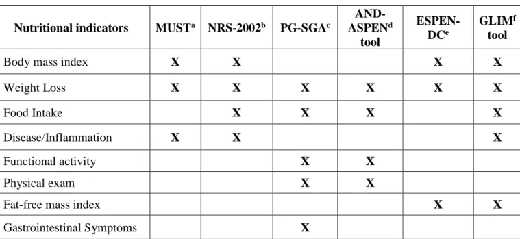
Nutritional indicators of each tool used, MUST, NRS-2002, PG-SGA, AND-ASPEN clinical characteristics of undernutrition, ESPEN-DC and GLIM criteria for the diagnosis of malnutrition, are summarized in Table 1.
Statistical Analysis
All categorical variables were described as frequencies. Kolmogorov-Smirnov test was used to assess the variables’ distribution and continuous variables were described as median and interquartile range (IQR) or as mean and standard deviation (SD).
Regarding age, participants were classified as <65 years or ≥ 65 years. For education level, the following categories were created: 0-4, 5-12, and >12 school years. Participants were also categorized as married/common-law marriage or as single/divorced/widowed. Smocking status and professional occupation were dichotomized as yes or no.
For bivariate analyses, all patients were dichotomized considering LOS as short (<7 days) and long (≥7 days) and were compared for several sociodemographic, functional, clinical and nutritional characteristics. Statistical differences were tested using Chi-squared test for categorical variables and the Mann-Whitney test or t-Test for independent samples for the continuous variables, according to its distribution. LOS was determined in days, as the time from the date of hospital admission until discharge.
Survival analysis were used to estimate the cumulative probability of being discharged from the hospital over time, considering the follow-up for LOS as 30 days and discharge home or to usual residence as the event. Kaplan-Meier curves were obtained for each nutritional indicator studied.
Cox regression analysis was applied to estimate hazard ratios (HR) and corresponding 95% confidence intervals (CI). A model was created for each nutritional component studied: weight loss (categories: 0%, 0.1-5.0%, 5.1-10.0% and >10.0%; reference: 0% weight loss), gastrointestinal symptoms (categories: none and presence / severe presence;
7
reference: none), food intake in the preceding month (categories: without changes and decrease / severe decrease; reference: without changes), food intake in the preceding week (categories: normal, 50-75% of normal requirements, 25-50% of normal requirements and 0-25% of normal requirements; reference: normal), HGS (categories: normal and measurably reduced; reference: normal), physical exam (categories: without changes, mild to moderate changes and severe changes; reference: without changes), BMI (categories: <18.5 kg/m2, 18.5-20.5 kg/m2 and >20.5 kg/m2; reference: >20.5 kg/m2), FFMI (categories: normal and low; reference: normal) and disease-related inflammation (categories: chronic disease-related inflammation and acute disease or injury-related inflammation; reference: chronic disease-related inflammation). Furthermore, adjusted models were created for each nutritional indicator considering the following variables: age (categories <65 years and ≥65 years; reference: <65 years), comorbidities (Charlson index, continuous), independence in activities of daily living (Katz index, categories: independence, moderate dependence and severe dependence; reference: independence) and professional activity (categories: yes and no; reference: yes).
Due to the small sample number of patients with presence of severe gastrointestinal symptoms, when applying the Cox Regression Models and in the survival analysis, the categories “presence" and "severe presence [of gastrointestinal symptoms]” were merged into one category. Also, due to the small sample number of patients with “severe decrease” of “food intake in the preceding month”, participants in this category were grouped with participants presenting “decrease” of “food intake in the preceding month”. Akaike’s information criterion (AIC) was applied to compare the quality of each one of the Cox regression models used. This analysis’ purpose is to conclude which statistical model better explains the dependent variable, LOS. For comparing models based on their AICs, the model with lower AIC is the preferred one (35, 36).
Data were analysed using the statistical package IBM® SPSS® Statistics 2017, version 25 and p values <0.05 were considered as statistically significant.
8
RESULTS
A total of 633 inpatients were included in this study, 288 (45.5%) females and 345 (54.5%) males. Ages ranged from 18 to 90 years old. The median age was 56 years (IQR=22 years).
The sociodemographic, functional, clinical and nutritional status characteristics of the study sample are presented in the Table 2. Participants in the long LOS group were older, presented a higher level of comorbidity evaluated by Charlson index, and both men and women presented lower HGS median values, compared with participants in the short LOS group. Moreover, men in the long LOS group presented lower median BMI values (Table 2).
Higher proportions of patients in the long LOS group were aged 65 years old or older, presented moderate and severe dependence according to the Katz index, were transferred to another hospital ward or to a different hospital, were discharged against medical advice or deceased; additionally, a lower proportion of patients in the long LOS group had a professional occupation (Table 2).
Moreover, concerning the nutritional status indicators, a higher number of participants from the long LOS group showed weight loss from 0.1% to 5.0%, from 5.1% to 10% or more than 10% and a higher proportion of individuals from the long LOS group presented gastrointestinal symptoms. In addition, a higher number of participants in the same group reported decreased or severely decreased food intake in the preceding month, as well as decreased food intake between 50 and 75% of their normal requirements in the preceding week. A higher proportion of participants from the long LOS group exhibited mild to moderate and severe changes in the physical exam (Table 2).
Kaplan-Meier curves were obtained for each singular component of the undernutrition screening and diagnostic tools. These curves show that inpatients that presented weight loss of 5.1-10.0% and >10.0% have a higher probability of remaining in hospital and thus have longer LOS (p<0.001). The same findings were accomplished for presence and severe presence of GI symptoms (p=0.002), for decrease and severe decrease of food intake in the preceding month (p<0.001), for mild to moderate and severe changes in the physical exam (p<0.001), for decreased HGS (p=0.006) and for acute disease or injury related inflammation (p=0.001) (Figure 1).
9
A percentage of weight loss of 5.1-10.0% and higher than 10.0%, the presence or severe presence of gastrointestinal symptoms, decreased and severe decreased food intake in the preceding month, measurably reduced HGS, mild to moderate and severe changes in the physical exam and acute disease or injury related inflammation were associated with a lower probability of being discharged home or to usual residence in both crude and adjusted models, whereas percentage of weight loss of 0.1-5.0%, food intake in the preceding week, BMI and FFMI showed no association with being discharged home or to usual residence over time, again for crude and adjusted models (Table 3).
For the crude Cox regression models, AIC values varied between 6 352.2 and 6 376.9, whereas for adjusted models, AIC values varied between 6 353.4 and 6 375.7. Comparing crude model with adjusted model for each nutritional indicator, the differences were very small. Also, AICs values obtained for the different nutritional indicators are similar, for both crude and adjusted models (Table 3).
DISCUSSION
The results of the present study show that weight loss higher than 5.1%, presence of gastrointestinal symptoms, decreased food intake in the preceding month, reduced HGS as well as changes in the physical exam and acute disease or injury can be used to predict inpatients’ LOS. Among all the nutritional indicators studied, food intake in the previous week, BMI and FFMI showed no association with hospital LOS.
Cox regression models were adjusted for the potential confounding variables age, Charlson index (participants’ comorbidities), professional activity and Katz index (participants’ ability to perform activities of daily living) and small changes were observed in the HR and respective 95% CI and in the AICs between crude (unadjusted) and adjusted Cox proportional hazard models for all indicators. These findings show that, except for the food intake in the preceding week, BMI and FFMI, for which no association was found, the association between all the undernutrition screening and diagnostic tools’ indicators studied and LOS was not weakened by the adjustment.
AIC values were determined for comparing the different Cox proportional hazards regression models. For both unadjusted and adjusted models, the AIC values are lower for the percentage of weight loss, making it possible to deduce that this is the model that
10
better explains the dependent variable LOS. However, differences in AICs between indicators were so small that present study results do not allow us to conclude that some models, and therefore that some indicators are better than others in predicting LOS. Apart from food intake in the previous week, BMI and FFMI, all studied indicators can be used to predict LOS. Nonetheless, they are different in what concerns expertise needed for the health professional, time they take as well as resources needed to be evaluated and thus since their validity is similar, the most “efficient” should be considered and the information obtained from this study should be used to revise and improve undernutrition screening and diagnostic tools.
In a recent study conducted in 699 patients from acute care hospitals, Subjective Global Assessment (SGA) and NRS-2002 were applied and the following nutritional indicators were assessed: body weight, midarm and calf circumference, serum albumin, HGS and patient-self assessment of food intake (26). In this study, logistic regression models were conducted for each single nutritional indicator for predicting length of stay ≥7 days. All models were adjusted for age, sex and number of diagnosis. Results showed that each kg of increase in HGS was associated with a lower probability of LOS ≥7 days (Odds ratio (OR), (95% CI) = 0.97 (0.96-0.99), p=0.002). In the same study, food intake <50% in the first week of hospitalization was associated with LOS ≥7 days: OR (95% CI) = 1.56 (1.12-2.18), p=0.009).
Our study results are in accordance with this study’s conclusions concerning HGS, since our findings show that measurably reduced HGS was independently associated to a lower probability of being discharged home or to usual residence and hence to prolonged hospital LOS. However, for food intake in the previous week, the different findings can be due to a different evaluation of food intake, as we evaluated consumption of all food items in the previous week and Jeejeebhoy et al. (26) evaluated mean plate consumption at lunch.
In a study conducted in 396 hospitalized patients from internal medicine wards, decreased food intake prior to hospital admission, assessed by Subjective Global Assessment, was correlated to longer LOS. Patients with decreased food intake presented longer hospital LOS compared with patients without decrease in food intake: median LOS (95% CI) = 15.0 (2-59) days vs. median LOS (95% CI) = 12.0 (1-76) days, p=0.001 (37). These findings are in accordance with our results.
11
Findings from another study on 1312 inpatients from intensive care units (38) revealed that patients who had simultaneously two or more GI symptoms in the first 24 hours after hospital admission had significantly higher LOS than inpatients with a maximum of one GI symptom: mean (SD) = 11.6 (14.3) days vs. mean (SD) = 8.5 (13.1) days, p<0.001), even though the presence of one GI symptom prolonged LOS compared to presenting no GI symptoms: mean (SD) = 8.5 (13.1) days vs. mean (SD) = 5.7 (8.3) days, p<0.001. Moreover, the same study showed that during the whole hospital stay, patients’ LOS was higher according to the severity of their GI symptoms at discharge, since patients without GI symptoms had mean (SD) LOS = 2.9 (4.3) days, whereas patients with one GI symptom had LOS = 4.2 (6.0) days, patients with three GI symptoms had LOS = 11.6 (10.6) days, patients with four GI symptoms has LOS = 16.8 (16.1) days and patients with five or six GI symptoms had LOS > 30 days (no p values presented). Present study findings are in accordance with these results since we concluded that the presence or severe presence of GI symptoms were independently associated with lower probability of being discharged to usual residence and so to longer LOS.
A 2014 research about prognostic indices of poor nutritional status and their impact on prolonged LOS in 295 hospitalized patients from a University Hospital (27) indicates, similarly to our results, that in-hospital recent weight loss is a major contributor for prolonged LOS: OR (95% CI) = 2.950 (1.1797-4.850), using as reference no weight loss. In a study published in the present year, that aimed to investigate the impact of preoperative cachexia on postoperative LOS in 98 elderly Japanese patients with GI cancer, preoperative cachexia was associated with prolonged postoperative LOS (39). Cachexia is characterized by weight loss, reduced BMI, muscle mass and function (several findings), combined with an underlying disease that exhibits an ongoing substantial inflammatory activity (40). The findings of this study demonstrate that underlying presence of inflammation has a negative impact on hospital stay, since mean (SD) LOS was 17.1 (8.7) days in the non-cachexia group and 20.6 (10.8) days in the cachexia group (no p values presented). These results show an association of chronic inflammation with longer LOS, while our findings indicate that when compared to chronic disease related-inflammation, acute disease or injury-related inflammation prolongs LOS in hospitalized patients. Moreover, we found no association between BMI with LOS. However, differences concerning study sample and design impair direct comparisons.
12
The present study has multiple strengths. Undernutrition in hospitalized patients has far back been related to worse patient outcomes including increased hospital LOS (6, 41-44), but no other study had compared all the nutritional components from the six tools used here: AND-ASPEN clinical characteristics of undernutrition (1), NRS-2002 (17), MUST (16) and PG-SGA (18), ESPEN-DC (19) and GLIM criteria for the diagnosis of malnutrition (20). The indicators from the most used and more extensible validated tools, as well as the indicators from the most recent tools recommended by ESPEN and by GLIM were used. The statistical analysis used allowed to treat LOS as a continuous variable and also allows to censor data. By doing that, it was possible to include inpatients that otherwise would be excluded from data analysis. Moreover, the sample characteristics are another study strength since a wide and representative number of hospitalized patients (n=633) with ages between 18 and 90 years old, with several diseases and diagnosis from different medical and surgical wards were included.
Nutritional and medical procedures were not recorded hence HR values were not adjusted for these potential confounders. Furthermore, patients’ nutritional status was not monitored or evaluated at the time of their hospital discharge.
For LOS, 30 days were arbitrarily chosen. Despite this follow-up interval has previously been used in other studies (21, 45), in our study only 15 patients (2.4%) remained hospitalized for more than 30 days so this probably did not affect our study findings.
13
CONCLUSIONS
It is possible to conclude that weight loss >5.1%, presence and severe presence of gastrointestinal symptoms, decrease and severe decrease in food intake in the preceding month, measurably reduced HGS, mild to moderate and severe changes detected in the physical exam, and acute disease or injury-related inflammation were independently associated with a lower probability of being discharged home or to usual residence and thus to longer LOS.
These indicators can be used in the hospital setting for predicting the patient’s LOS. They are different in what concerns expertise needed for the health professional, time they take as well as resources needed as this should be considered when choosing which indicator to use.
Since all these indicators have similar validity in predicting LOS, nutritional indicators that are less time consuming, require less trained health professionals or less resources must be preferred to predict hospital length of stay.
Furthermore, this study provides new scientific evidence on the association of nutritional status indicators with LOS that can be used to improve undernutrition screening and diagnostic tools in a near future.
14
Acknowledgments
I would like to express my gratitude to Fundação para a Ciência e Tecnologia, to Centro
15
REFERENCES
1. White JV, Guenter P, Jensen G, Malone A, Schofield M. Consensus statement: Academy of Nutrition and Dietetics and American Society for Parenteral and Enteral Nutrition: characteristics recommended for the identification and documentation of adult malnutrition (undernutrition). JPEN J Parenter Enteral Nutr. 2012;36(3):275-283. 2. Peter C. Konturek et al. Malnutrition in Hospitals: It Was, Is Now, and Must Not Remain a Problem!. Med Sci Monit. 2015; 21: 2969–2975.
3. Barker L, Gout B, Crowe T. Hospital Malnutrition: Prevalence, Identification and Impact on Patients and the Healthcare System. Int J Environ Res Public Health. 2011;8(2):514-527.
4. Somanchi M, Tao X, Mullin G. The Facilitated Early Enteral and Dietary Management Effectiveness Trial in Hospitalized Patients With Malnutrition. J Parenter Enteral Nutr. 2011;35(2):209-216.
5. Braunschweig C, Gomez S, Sheean P. Impact of Declines in Nutritional Status on Outcomes in Adult Patients Hospitalized for More Than 7 days. J Am Diet Assoc. 2000;100(11):1316-1322.
6. Hickson M, Smith S. Advanced nutrition and dietetics in nutrition support. Wiley Blackwell; 2018.
7. McWhirter JP, Pennington CR. Incidence and recognition of malnutrition in hospital. Br Med J 1994;308:945-948.
8. Lamb CA, Parr J, Lamb EI, et al. Adult malnutrition screening, prevalence and management in a United Kingdom hospital: cross-sectional study. Br J Nutr 2009;102:571-575.
9. Elia M. The 'MUST' report. Nutritional screening for adults: a multidisciplinary responsibility. Development and use of the 'Malnutrition Universal Screening Tool' (MUST) for adults [Monograph]. Redditch, UK: British Association for Parenteral and Enteral Nutrition (BAPEN); 2003.
10. Philipson T, Snider J, Lakdawalla D, Stryckman B, Goldman D. Impact of oral nutritional supplementation on hospital outcomes. Clin Nutr. 2013;32:6-7.
16
11. Müller-Richter U, Betz C, Hartmann S, Brands R. Nutrition management for head and neck cancer patients improves clinical outcome and survival. Nutr Res. 2017;48:1-8. 12. Amaral T, Matos L, Tavares M, Subtil A, Martins R, Nazaré M et al. The economic impact of disease-related malnutrition at hospital admission. Clin Nutr. 2007;26(6):778-784.
13. Lim S, Ong K, Chan Y, Loke W, Ferguson M, Daniels L. Malnutrition and its impact on cost of hospitalization, length of stay, readmission and 3-year mortality. Clin Nutr. 2012;31(3):345-350.
14. Model and Process for Nutrition and Dietetic Practice. British Dietetic Association; 2012.
15. Mahan L, Raymond J. Krause's Food & the Nutrition Care Process. 14th ed. St. Louis, Missouri: Elsevier; 2017.
16. Elia M. The 'MUST' report. Nutritional screening for adults: a multidisciplinary responsibility. Development and use of the 'Malnutrition Universal Screening Tool' (MUST) for adults [Monograph]. Redditch, UK: British Association for Parenteral and Enteral Nutrition (BAPEN); 2003.
17. Kondrup J, Rasmussen HH, Hamberg O, Stanga Z. Nutritional risk screening (NRS 2002): A new method based on an analysis of controlled clinical trials. Clin Nutr. 2003;22(3):321-336.
18. Ottery F. Definition of standardized nutritional assessment and interventional pathways in oncology. Nutrition. 1996;12(1):15-19.
19. Cederholm T, Bosaeus I, Barazzoni R, Bauer J, Van Gossum A, Klek S et al. Diagnostic criteria for malnutrition – An ESPEN Consensus Statement. Clin Nutr. 2015;34(3):335-340.
20. Cederholm T, Jensen G, Correia M, Gonzalez M, Fukushima R, Higashiguchi T et al. GLIM criteria for the diagnosis of malnutrition – A consensus report from the global clinical nutrition community. Clin Nutr. 2019;38(1):1-9.
21. Kondrup J. ESPEN Guidelines for Nutrition Screening 2002. Clin Nutr. 2003;22(4):415-421.
17
22. August D, Huhmann M. A.S.P.E.N. Clinical Guidelines: Nutrition Support Therapy During Adult Anticancer Treatment and in Hematopoietic Cell Transplantation. J
Parenter Enteral Nutr. 2009;33(5):472-500.
23. Mulasi U, Vock D, Kuchnia A, Jha G, Fujioka N, Rudrapatna V et al. Malnutrition Identified by the Academy of Nutrition and Dietetics and American Society for Parenteral and Enteral Nutrition Consensus Criteria and Other Bedside Tools Is Highly Prevalent in a Sample of Individuals Undergoing Treatment for Head and Neck Cancer. J Parenter
Enteral Nutr. 2016.
24. Guerra RS, Fonseca I, Pichel F et al. Usefulness of Six Diagnostic and Screening Measures for Undernutrition in Predicting Length of Hospital Stay: A Comparative Analysis. J Acad Nutr Diet. 2015 Jun;115(6):927-38.
25. Guerra R, Fonseca I, Sousa A, Jesus A, Pichel F, Amaral T. ESPEN diagnostic criteria for malnutrition – A validation study in hospitalized patients. Clin Nutr. 2017;36(5):1326-1332.
26. Jeejeebhoy, K et al. Nutritional assessment: comparison of clinical assessment and objective variables for the prediction of length of hospital stay and readmission. Am J
Clin Nutr. 2015;101:956–65.
27. Tsaousi G, Panidis S, Stavrou G, Tsouskas J, Panagiotou D, Kotzampassi K. Prognostic Indices of Poor Nutritional Status and Their Impact on Prolonged Hospital Stay in a Greek University Hospital. Biomed Res Int. 2014;2014:1-8.
28. Guidelines for the use of parenteral and enteral nutrition in adult and pediatric patients. JPEN J Parenter Enteral Nutr. 2002;26(1 suppl): 1SA-138SA.
29. Hodkinson HM. Evaluation of a mental test score for assessment of mental impairment in the elderly. Age Ageing. 1972;1(4):233-238.
30. Brodaty H, Pond D, Kemp NM, et al. The GPCOG: A new screening test for dementia designed for general practice. J Am Geriatr Soc. 2002;50(3):530-534.
31. Bonaiuto S, Rocca WA, Lippi A, et al. Study on the validity of the Hodkinson Abbreviated Mental Test Score (AMTS) in detecting dementia of elderly subjects in appignano (Macerata province), Italy. Arch Gerontol Geriatr. 1992;15(suppl 1):75-85.
18
32. Katz S, Downs TD, Cash HR, Grotz RC. Progress in development of the index of ADL. Gerontologist. 1970;10(1):20-30.
33. Katz S. Assessing self-maintenance: Activities of daily living, mobility, and instrumental activities of daily living. J Am Geriatr Soc. 1983;31(12):721-727.
34. Charlson ME, Pompei P, Ales KL, MacKenzie CR. A new method of classifying prognostic comorbidity in longitudinal studies: Development and validation. J Chronic
Dis. 1987;40(5):373-383.
35. Akaike H. A new look at the statistical model identification. IEEE Trans Autom
Control. 1974;19(6):716-722
36. Burnham K, Anderson D. Model selection and multimodel inference - 2nd ed. New York, NY: Springer-Verlag New York Inc.; 2002.
37. Ordoñez A, Schieferdecker M, Cestonaro T, Neto J, Campos A. Nutritional status influences the length of stay and clinical outcomes in hospitalized patients in internal medicine wards. Nutr Hosp. 2013;28(4):1313-1320.
38. Gastrointestinal symptoms in intensive care patients. A. Reintam, P. Parm, R. Kitus, H. Kern And J. Starkopf. Acta Anaesthesiol Scand 2009; 53: 318–324.
39. Fukuta A, Saito T, Murata S, Makiura D, Inoue J, Okumura M et al. Impact of preoperative cachexia on postoperative length of stay in elderly patients with gastrointestinal cancer. Nutrition. 2019;58:65-68.
40. Cederholm T, Barazzoni R, Austin P, Ballmer P, Biolo G, Bischoff S et al. ESPEN guidelines on definitions and terminology of clinical nutrition. Clin Nutr. 2017;36(1):49-64.
41. Allison S P. Malnutrion, disease, and outcome. Nutrition 2000; 16:590–591.
42. Chima C S, Barco K, Dewitt J L A, Maeda M, Teran J C, Mullen K D. Relationship of nutritional status to length of stay, hospital costs, and discharge status of patients hospitalized in the medicine service. Aliment Pharmacol Ther 1997; 11: 975–978. 43. Robinson G, Goldstein M, Levin G M. Impact of nutritional status on DRG length of stay. J Parenter Enteral Nutr 1987; 11: 49–51.
19
44. Ruiz A, Buitrago G, Rodríguez N, Gómez G, Sulo S, Gómez C et al. Clinical and economic outcomes associated with malnutrition in hospitalized patients. Clin Nutr. 2019;38(3):1310-1316.
45. Mendes J, Alves P, Amaral TF. Comparison of nutritional status assessment parameters in predicting length of hospital stay in cancer patients. Clin Nutr. 2014;33(3):466-470.
20
Table 1. Individual nutritional indicators from the six undernutrition screening and
diagnostic tools used in the present study.
Nutritional indicators MUSTa NRS-2002b PG-SGAc
AND-ASPENd tool ESPEN-DCe GLIMf tool
Body mass index X X X X
Weight Loss X X X X X X
Food Intake X X X X
Disease/Inflammation X X X
Functional activity X X
Physical exam X X
Fat-free mass index X X
Gastrointestinal Symptoms X
a MUST=Malnutrition Universal Screening Tool. b NRS-2002=Nutritional Risk Screening Tool.
c PG-SGA=Patient-generated Subjective Global Assessment.
d AND-ASPEN tool = Academy of Nutrition and Dietetics and the American Society for Parenteral and Enteral Nutrition recommended clinical characteristics of malnutrition tool e ESPEN-DC=ESPEN Diagnostic Criteria.
21
Table 2. Baseline sociodemographic, functional, clinical and nutritional status
characteristics of 633 Portuguese inpatients from a prospective observational study according to short (<7 days) and long (≥7 days) hospital length of stay (LOS).
Characteristics Short LOS (<7 days)
(n=294)
Long LOS (≥7 days)
(n=339) p
Gender, n (%)
Female 136 (47.2) 152 (52.8)
0.720a
Male 158 (45.8) 187 (54.2)
Age, median (IQR) 53 (24) 59 (21) <0.001b
Age categories, n (%) <65 y 226 (51.4) 214 (48.6) <0.001a ≥65 y 68 (35.2) 125 (64.8) Education (years), n (%) 0-4 116 (45.7) 138 (54.3) 0.075a 5-12 145 (44.6) 180 (55.4) >12 33 (61.1) 21 (38.9) Marital Status, n (%) Married/common-law marriage 192 (47.5) 212 (52.5) 0.470a Single/Divorced/Widowed 102 (44.5) 127 (55.5) Smocking status, n (%) Yes 67 (50.8) 65 (49.2) 0.272a No 227 (45.4) 273 (54.6) Professional occupation, n (%) Yes 120 (57.1) 90 (42.9) <0.001a No 174 (41.1) 249 (58.9)
Charlson index, median (IQR) 1.00 (2) 2.00 (3) 0.004b
Katz index, n (%)
Independence 276 (48.6) 292 (51.4)
0.002a
Moderate dependence 13 (35.1) 24 (64.9)
22
Table 2. Baseline sociodemographic, functional, clinical and nutritional status
characteristics of 633 Portuguese inpatients from a prospective observational study according to short (<7 days) and long (≥7 days) hospital length of stay (LOS). (cont.)
Characteristics Short LOS (<7 days)
(n=294)
Long LOS (≥7 days)
(n=339) p
Discharge destination, n (%)
Home/Usual residence 278 (48.8) 292 (51.2) <0.001a
Transfer or discharge against medical
advice or death 16 (25.4) 47 (74.6) <0.001 a Weight loss, n (%) None 155 (54.8) 128 (45.2) <0.001a 0.1 - 5.0% 84 (48.0) 91 (52.0) 5.1 – 10.0% 33 (35.1) 61 (64.9) > 10.0% 22 (27.2) 59 (72.8) Gastrointestinal Symptoms, n (%) None 259 (51.0) 249 (49.0) <0.001a Presence 32 (26.9) 87 (73.1) Severe presence 3 (50.0) 3 (50.0)
Food Intake in the preceding month, n (%)
Without changes 246 (52.7) 221 (47.3)
<0.001a
Decrease 46 (30.1) 107 (69.9)
Severe decrease 2 (15.4) 11 (84.6)
Food Intake in the preceding week, n (%)
Normal 213 (52.3) 194 (47.7)
<0.001a
50-75% of normal requirement 60 (39.7) 91 (60.3)
25-50% of normal requirement 12 (32.4) 25 (67.6)
0-25% of normal requirement 9 (23.7) 29 (76.3)
Handgrip Strength, median (IQR)
Male 34.0 (12.2) 31.0 (11.0) 0.001b
23
Table 2. Baseline sociodemographic, functional, clinical and nutritional status
characteristics of 633 Portuguese inpatients from a prospective observational study according to short (<7 days) and long (≥7 days) hospital length of stay (LOS). (cont.)
Characteristics Short LOS (<7 days)
(n=294)
Long LOS (≥7 days)
(n=339) p Handgrip Strength, n (%) Normal 57 (50.9) 55 (49.1) 0.298a Measurably reduced 237 (45.5) 284 (54.5) Physical Exam, n (%) Without changes 155 (54.6) 129 (45.4) <0.001a
Mild to moderate changes 98 (42.4) 133 (57.6)
Severe changes 41 (34.7) 77 (65.3)
Body Mass Index (kg/m2), median (IQR)
Male 25.7 (5.3) 24.5 (6.5) 0.041b
Female 26.1 (7.8) 27.1 (7.8) 0.107b
Body Mass Index (kg/m2), n (%)
< 18.5 5 (33.3) 10 (66.7)
0.522a
18.5 – 20.5 24 (43.6) 31 (56.4)
> 20.5 265 (47.1) 298 (52.9)
Free Fat Mass Index (kg/m2), mean (SD)
Male 20.1 (2.3) 19.7 (2.5) 0.072c
Female 17.3 (2.5) 17.7 (2.8) 0.224c
Free Fat Mass Index (kg/m2), n (%)
Normald 259 (47.1) 291 (52.9) 0.402a Lowe 35 (42.2) 48 (57.8) Inflammation, n (%) Acute disease/injury 139 (44.6) 173 (55.4) 0.346a Chronic disease-related 155 (48.3) 166 (51.7)
24
Table 2. Baseline sociodemographic, functional, clinical and nutritional status
characteristics of 633 Portuguese inpatients from a prospective observational study according to short (<7 days) and long (≥7 days) hospital length of stay (LOS). (cont.)
IQR=Interquartile range. SD=Standard Deviation.
aChi-squared test was used for categorical variables. bMann–Whitney test.
ct-Test for independent samples.
dNormal values for Free Fat Mass Index are ≥15 kg/m2 for females and ≥17 kg/m2 for males.
25
Table 3: Hazard ratios (HR) and 95% confidence intervals (C.I.) for Cox proportional
regression models, for being discharged home or to usual residence in a sample of 633 Portuguese inpatients participating in a prospective observational studya.
Model 1b Model 2c
Characteristics HR 95% C.I. AICd HR 95% C.I. AICd
Percentage of weight loss
0% 1 6352.2 1 6353.4 0.1-5.0% 1.007 0.825 – 1.230 0.986 0.807 – 1.206 5.1-10.0% 0.644 0.503 – 0.825 0.638 0.498 – 0.818 >10.0% 0.573 0.439 – 0.749 0.580 0.440 – 0.764 Gastrointestinal Symptoms None 1 6367.3 1 6367.4
Presence and Severe presence 0.723 0.587 – 0.889 0.736 0.597 – 0.907
Food Intake in the preceding month
Without changes 1
6358.2 1 6355.3
Decrease and severe decrease 0.663 0.548 – 0.802 0.648 0.534 – 0.785
Food Intake in the preceding week
Normal 1 6375.6 1 6375.5 50-75% of normal requirement 0.843 0.69 – 1.027 0.862 0.707 – 1.052 25-50% of normal requirement 0.721 0.507 – 1.026 0.738 0.515 – 1.057 0-25% of normal requirement 0.877 0.625 – 1.231 0.859 0.611 – 1.208 Handgrip Strength Normal 1 6371.1 1 6368.7 Measurably reduced 0.750 0.602 – 0.934 0.718 0.571 – 0.903 Physical Exam Without changes 1 6361.0 1 6360.1 Mild to moderate changes 0.719 0.598 – 0.864 0.723 0.601 – 0.870
26
Table 3: Hazard ratios (HR) and 95% confidence intervals (C.I.) for Cox proportional
regression models, for being discharged home or to usual residence in a sample of 633 Portuguese inpatients participating in a prospective observational studya.
a Lower Hazard ratio values represent a lower probability of hospital discharge; participants exhibiting the occurrence of an event different (inpatients’ deaths, discharges against medical advice and transfers to a different ward/hospital) from the outcome of interest (discharge home/usual residence) were considered until the time of occurrence of those events; the cut-off point for hospital length of stay was 30 days. b Model 1= crude Cox proportional regression models.
c Model 2 = adjusted Cox proportional regression models for age (dichotomic; reference: <65 years), Charlson Index (continuous), professional activity (dichotomic; reference: having a professional activity), Katz Index (categoric; reference: independence). d AIC=Akaike’s information criterion.
eNormal values for Free Fat Mass Index are ≥15 kg/m2 for females and ≥17 kg/m2 for males.
fLow values for Free Fat Mass Index are <15 kg/m2 for females and <17 kg/m2 for males.
Model 1b Model 2c
Characteristics HR 95% C.I. AICd HR 95% C.I. AICd
Body mass index
>20.5 1 6376.9 1 6375.7 18.5-20.5 0.670 0.386-1.162 0.680 0.390-1.186 <18.5 1.033 0.766-1.393 1.098 0.809-1.491
Fat-free mass index
Normale 1 6376.6 1 6376.0 Lowf 0.898 0.740-1.145 0.942 0.736-1.206 Inflammation Chronic disease-related 1 6367.5 1 6363.5 Acute disease/injury 0.768 0.651 – 0.906 0.734 0.619 – 0.870
27
Figure 1. Kaplan-Meier curves for being discharge-free over timea for 633 Portuguese inpatients enrolled in a prospective observational study, according to (A) Weight loss, (B) Gastrointestinal symptoms, (C) Food intake in the preceding month, (D) Food intake in the preceding week, (E) Physical exam, (F) Handgrip Strength, (G) BMI, (H) FFMI and (I) Inflammation.
28
Figure 1. Kaplan-Meier curves for being discharged-free over timea for 633 Portuguese inpatients enrolled in a prospective observational study, according to (A) Weight loss, (B) Gastrointestinal symptoms, (C) Food intake in the preceding month, (D) Food intake in the preceding week, (E) Physical exam, (F) Handgrip Strength, (G) BMI, (H) FFMI and (I) Inflammation (cont.)
29
Figure 1. Kaplan-Meier curves for being discharged-free over timea for 633 Portuguese inpatients enrolled in a prospective observational study, according to (A) Weight loss, (B) Gastrointestinal symptoms, (C) Food intake in the preceding month, (D) Food intake in the preceding week, (E) Physical exam, (F) Handgrip Strength, (G) BMI, (H) FFMI and (I) Inflammation. (cont.)
30
Figure 1. Kaplan-Meier curves for being discharged-free over timea for 633 Portuguese inpatients enrolled in a prospective observational study, according to (A) Weight loss, (B) Gastrointestinal symptoms, (C) Food intake in the preceding month, (D) Food intake in the preceding week, (E) Physical exam, (F) Handgrip Strength, (G) BMI, (H) FFMI and (I) Inflammation. (cont.)
31
Figure 1. Kaplan-Meier curves for being discharged-free over timea for 633 Portuguese inpatients enrolled in a prospective observational study, according to (A) Weight loss, (B) Gastrointestinal symptoms, (C) Food intake in the preceding month, (D) Food intake in the preceding week, (E) Physical exam, (F) Handgrip Strength, (G) BMI, (H) FFMI and (I) Inflammation. (cont.)
aHigher values of discharge-free over time (not experiencing the event of interest=hospital discharge for home/usual residence) represent a higher probability of remaining at the hospital, at a certain point in time; inpatients’ deaths, discharges against medical advice and transfers to a different ward or hospital were censored at time of occurrence of these events. The cut-off point for hospital length of stay was 30 days.
32
33
34
35
36