1. Beni Messous University Hospital, Department of Immunology and Algiers Faculty of Medicine, University of Algiers 1, Algiers, Algeria 2. Specialized Medical Center of Ben Aknoun, Department of Rheumatology, Algiers, Algeria
3 Beni Messous University Hospital, Department of Epidemiology and Preventive Medicine, Algiers, Algeria
for the A allele), and with interstitial lung disease (p = 0.025 for the AG genotype) have also been found. Conclusion: The study revealed that IL-17F and IL-21 genes were associated with systemic sclerosis (SSc) sus-ceptibility and that IL-17A, IL-17F, IL-21 and IL-23R genes influence the clinical and immunological fea-tures, which suggest the implication of Th17 cells in SSc pathogenesis.
Keywords: Systemic sclerosis; Th17 cell; Polymorphism.
IntroductIon
Systemic sclerosis (SSc) is an autoimmune connective tissue disease characterized by excessive collagen deposition in the skin and internal organs with associa -ted vasculopathy and autoantibody production1.
SSc is a multifactorial disease in which genetic factors play a crucial role, in fact the genome-wide and the can-didate gene studies allowed to associate HLA (human leukocyte antigen) and non HLA genes to the onset of the SSc, to subsets of the disease, or to autoantibodies production2–7. Among the susceptibility genes to SSc, many encode proteins of immune response, moreover several studies showed the presence of immune cells in inflammatory skin infiltrate of sclerodermic patients (monocytes/macrophages, mast cells and T cells), gene -rally in perivascular localization8–10, the majority of T cells infiltrating the lesions express activation mar kers and has a limited T cell receptor (TCR) repertoire thus indicating their antigen induced expansion8,10–12.
The T cells can cause the activation of fibroblasts either by direct contact or by the action of secreted cytokines and chemokines13. In addition, autoreactive T cells can in-teract with B cells and lead to the autoantibodies pro-duction13,14. Several studies showed that the predominate cells in the lesions and the blood of scleroderma patients was the T Helper (Th)2 cells15–17, these cells are largely in-criminated in the SSc pathogenesis because of the potent
Th17 pathway genes polymorphisms in
Algerian patients with systemic sclerosis
Mellal Y1, Allam I1, Tahiat A1, Abessemed A2, Nebbab R3, Ladjouze A2, Djidjik R1
ACTA REUMATOL PORT. 2018;43:269-278
AbstrAct
Objective: Th17 cells have involved in the pathoge -nesis of several autoimmune diseases including sys-temic sclerosis (SSc). The aim of our study was to in-vestigate an association of IL-17A, IL-17F, IL-21, IL-23R and STAT3 genes with SSc susceptibility, and clinical and immunological phenotypes.
Patients and methods: The case-control study in-cluded 136 patients suffering from SSc and 317 healthy controls of the Algerian population. Eight single nu-cleotide polymorphisms (SNP) of genes encoding Th17 pathway were genotyped using TaqMan allelic discrimination assays. These SNPs are: IL-17A (rs2275913), IL17F (rs2397084 and rs763780), IL-21 (rs6822844), IL-23R (rs10489629, rs11209026 and rs1343151) and STAT3 (rs2293152).
Results: The current study showed a significant associ-ation of rs2397084 SNP (p = 0.049 and p = 0.036 for the TT genotype and the T allele, respectively) and rs6822844 SNP (p = 6.6 10-4 for the G allele) with sys-temic sclerosis (SSc) susceptibility. Also, we found an association of rs2275913 SNP (pc= 0.015 and p = 0.005 for the GG genotype and the G allele, respe ctively) and rs6822844 SNP (pc= 0.024 for the TT genotype) with digestive involvement. Also an association with anti--RNAPIII antibodies production have been found with rs6822844 SNP (pc= 0.012 and pc = 0.029 for the GT genotype and the T allele, res pectively). Association of rs10489629 SNP with digital infarcts (p = 0.043 for the C allele), interstitial lung disease (p = 0.045 for the CT genotype) and anti-SSA antibodies production (p = 0.001 and p = 0.008 for the CT genotype and the T allele, respectively) have been showed. Finally, an associa -tion of rs1343151 SNP with digital infarcts (p = 0.028
profibro tic action of their secreted cytokine interleukin (IL)-4 which induces the fibroblasts proliferation, and increases the production of collagen and tumor growth factor (TGF)-b, furthermore, IL-4 contributes to mononuclear cells infiltration18–20.
As to Th1 cells, minority during the SSc, they have an anti- fibrotic effect exerted essentially via IFN-g which has antagonist effects to those of IL-4. Howe ver, these cells may be involved in the inflammatory pro-cess that occurs early in the course of the disease13.
Constituting the third population of T helper after Th1 and Th2 populations, the Th17 cells have been as-signed a pivotal role in the pathophysiology of various autoimmune diseases such as the Crohn's disease, the rheumatoid arthritis and the multiple sclerosis21,22. Even if their role in SSc has not been established se veral studies have found that Th17 cells at higher frequen-cy in the peripheral blood and in the bronchoalveolar lavage (BAL) fluid of patients with scleroderma com-pared to healthy subjects13,23–30. It was also found elevated levels of IL-17 in serum29,31and IL-17A mRNA29,32, with an increase of IL-17A po sitivity in skin biopsies of patients with SSc28,33. IL-23, crucial cy-tokine in Th17 differentiation, was also found at high levels in SSc34.
Furthermore, IL-1b, IL-6 and TGF-b, necessary cy-tokines for the differentiation of Th17 and for pro-fi-brotic processes promotion are found at higher levels in the serum and tissues of patients with SSc24,35–37.
All evidences are in favor of Th17 cells involvement in the pathogenesis of SSc. This is why our research work focused on studying polymorphisms affecting the genes encoding key cytokines, their receptor and transcription factor involved in Th17 pathway: IL-17A (rs2275913), IL-17F (rs2397084 and rs763780), IL-21(rs6822844), IL-23R (rs10489629, rs11209026 and rs1343151) and STAT3 (rs2293152). Our aim was to highlight potential associations of single nucleotide polymorphisms (SNPs) with the occurrence of SSc, disease subsets, clinical features, and produced au-toantibodies.
pAtIents And methods
systemIc sclerosIs pAtIents And controls
A total of 136 patients divided in 14 men and 122 women (sex ratio W/M: 8.71; mean age: 45.8 ± 13.2) fulfilled the American Rheumatism Association's pre-liminary criteria of SSc diagnosis, were recruited from
the Rheumatology department of the specialized cen-ter of Ben Aknoun and Beni Messous university hos-pital in Algiers, Algeria8. Table I records their demo-graphic and clinical features. 317 healthy controls divided in 35 men and 282 women (sex ratio W/M: 8.06; mean age: 36.65 ± 12.07) and without any fa-milial history of autoimmune diseases were also in-cluded in the study.
AutoAntIbody AnAlysIs
All patients were tested for antinuclear antibodies (ANA) by indirect immunofluorescence (IIF) test using HEp-2 substrate. They were also tested for anti-topoisomerase antibodies (ATA), anti-centromere an-tibodies (ACA), anti-RNA polymerase III anan-tibodies (RNAPIII), RNP, SS-A, SS-B, anti--Sm, anti-dsDNA, anti-cardiolipin antibodies (aCL), anti-citrullinated peptide antibodies (ACPA), anti-neu-trophil cytoplasmic antibodies (ANCA), anti-pyruvate deshydrogenase (anti-PDH) and anti-gp210 by en-zyme-linked immunosorbent assay (Elisa). Positive
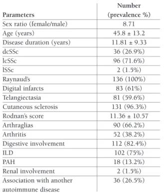
pa-tAble I. demogrAphIc And clInIcAl feAtures of ssc pAtIents
Number Parameters (prevalence %)
Sex ratio (female/male) 8.71
Age (years) 45.8 ± 13.2
Disease duration (years) 11.81 ± 9.33
dcSSc 36 (26.9%) lcSSc 96 (71.6%) lSSc 2 (1.5%) Raynaud’s 136 (100%) Digital infarcts 83 (61%) Telangiectasia 81 (59.6%) Cutaneous sclerosis 131 (96.3%) Rodnan’s score 11.36 ± 10.57 Arthraglias 90 (66.2%) Arthritis 52 (38.2%) Digestive involvement 112 (82.4%) ILD 102 (75%) PAH 18 (13.2%) Renal involvement 2 (1.5%)
Association with another 36 (26.5%) autoimmune disease
dcSSc: diffuse cutaneous systemic sclerosis; lcSSc: limited cutaneous systemic sclerosis; lSSc: limited systemic sclerosis; ILD: interstitial lung disease; PAH: pulmonary arterial hypertension.
tients for anti-nucleolar antibodies were also tested for PM/Scl, fibrillarine, Th/To and anti--NOR90 using immunodot test. Table II summarizes the patients’s autoimmune profile.
genetIc AnAlysIs
Genomic DNA of controls and patients was extracted from peripheral blood by salting out method and the genotyping of the eight single nucleotide poly -morphisms (rs2275913, rs2397084, rs763780, rs6822844, rs10489629, rs11209026, rs1343151 and rs2293152) was realized by real time polymerase chain reaction (PCR) using TaqMan technology according to the manufacturer's instructions (Applied biosystems, Foster City, CA, USA).
stAtIstIcAl AnAlysIs
The comparison of allelic and genotypic frequencies was evaluated by the Pearson’s Chi-square (c2) test using the Compare 2 test and p values lower than 0.05 were considered as statistically significant. For the small groups the p values were corrected by Yates or Fisher tests.
results
Only statistically significant results are shown in the
tAble III. genotypIc And AllelIc frequencIes of the rs2397084 snp And the rs6822844 snp in ssc pAtIents And controls
SNP SSc OR SSc OR
Genotype patients Controls (95% CI) p Allele patients Controls (95% CI) p
N=106 N=306 rs2397084 TT 98 (92.5%) 260 (85%) 2,17 0,049 T 204 563 (92%) 2,22 0,036 [0,97-5,50] pc=0,573 C (96.2%) 49 (8%) [1,02-5,52] 0,036 CC 0 (0%) 3 (1%) / 0,080 8 (3.8%) 0,45 CT 8 (7.5%) 43 (14%) / [0,18-0,98] N=123 N=296 rs6822844 GG 101 (82.1%) 213 (72%) 1,79 0,029 G 222 (90,2%) 504 1,62 0,048 [1,03-3,18] (85,1%) [0,99-2,73] TT 2 (1.6%) 5 (1.7%) / pC=1 T 24 (9.8%) 88 (14,9%) 0,62 0,048 [0,37-1,01] GT 20 (16.3%) 78 0,54 0,026 (26.3%) [0,30-0,96] OR: odds ratio; SNP: single nucleotide polymorphism; SSc: systemic sclerosis
tAble II. AutoAntIbodIes profIle of ssc pAtIents. Number Autoantibodies (prevalence %) ANA 128 (94,1%) ATA 74 (54,4%) ACA 20 (14,7%) anti-RNAPIII 10 (7,3%) Anti-nucleolar antibodies 15 (11%) • Anti-PM/Scl: 66,7% • Anti-fibrillarine: 26,7% • Anti-Th/To: 20% • Anti-NOR90: 6,7% Anti-U1RNP 15 (11%) Anti-SSA 41 (30,1%) Anti-SSB 10 (7,4%) Anti-Sm 4 (2,9%) aCL 16 (11,7%) ACPA 20 (14,7%) ANCA 7 (5,1%) Anti-PDH 2 (1,8%)
ACA: anti-centromere antibodies; aCL: anti-cardiolipin antibodies; ACPA: anti-citrullinated peptide antibodies; ANA: anti-nuclear antibodies; ANCA: anti-neutrophil cytoplasmic antibodies; anti-PDH: anti-pyruvate deshydrogenase antibodies; anti-RNAP III: anti-ARN polymerase III; ATA: anti-toposimerase I.
Table III for the genotypic and allelic frequencies of studied single nucleotide polymorphisms in patients and controls, and in the Table IV for the stratification of patients according to clinical features and autoanti-bodies production.
Il-17A polymorphIsm (rs2275913)
Genotypic and allelic analysis of this single nucleotide polymorphism showed no difference between patients and controls. However, the stratification of patients ac-cording to the presence or absence of digestive in-volvement showed a significant difference in patients with digestive involvement versus patients without di-gestive involvement for the GG genotype (72% vs 30.8%, pc= 0.015, OR= 5.79 [1.31-29.15]), the AG genotype (28% vs 61.5 %, p = 0,024, OR= 0,24 [0,05--1,04]), the G allele was found more frequently in the SSc patients with digestive involvement (86% vs 61.5%, p = 0.005, OR= 3.84 [1.27-11.17]), whereas the A allele was found less frequently in the SSc pa-tients with digestive involvement (14% vs 38.5%, p = 0.005, OR= 0.26 [0.09-0.79]) (Table IV).
Stratification according to the form, other clinical manifestations and autoantibodies profile shows no sig-nificant difference.
Il-17f polymorphIsms (rs2397084 And rs763780)
For the rs2397084 SNP, analysis of the distribution of different genotypes in both patient and control groups showed that the TT genotype was significantly more frequent in patients than in control group: 92.5% vs 85%; p = 0.049, OR = 2.17 [ 0.97 to 5.50 ]. Analysis of allelic frequencies indicated that the T allele was signi -ficantly more frequent in patients than in controls: 96.2% vs 92 %, p = 0.036, OR = 2.22 [ 1.02 to 5.52 ] (Table III).
As for the rs763780 SNP, there is no association with SSc susceptibility or with clinical and immunological phenotypes.
Il-21 polymorphIsm (rs6822844)
The study of this SNP showed that the GG genotype was significantly more frequent in patients than in con-trols: 82.1% vs 72%, p = 0.029, OR = 1.79 [ 1.03 to 3.18 ], conversely, the GT genotype was significantly less frequent in patients than in controls: 16.3% vs 26.3%, p = 0.026, OR = 0.54 [ 0.30 to 0.96 ]. Analysis also found that the G allele was significantly more frequent in patients than in controls: 90.2% vs 85.1%,
p = 0.048, OR = 1.62 [ 0.99 to 2.73 ] and, conversely, the T allele was less frequent: 9.8% vs 14.9%, p = 0.048, OR = 0.62 [ 0.37 to 1.01 ] (Table III).
Stratification of patients according to the presence or absence of digestive involvement showed a significant difference in patients with digestive involvement vs pa-tients without digestive involvement for the TT geno-type: 0% vs 10.5%, pc = 0.024, OR = 0.00 [ 0.0000 to 0.9652 ] (Table IV). Also, stratification according to the presence or absence of anti-RNA polymerase III (antiRNAPIII) antibodies showed a significant diffe -rence between RNAPIII(+) patients versus anti--RNAPIII(-) patients for the GT genotype (57.1% vs 13.5%, pc = 0.012, OR = 8.58 [ 1.27 to 62.30 ]), the GG genotype (42.9% vs 85.5%, pc = 0.016, OR = 0.13 [ 0,02 to 0.84 ]), the T allele found more frequent T in patients with antibodies to RNAPIII (28.6% vs 7.7%, pc = 0.029, OR = 4.82 [0, 99 to 18.91 ] ), and for the G allele found less frequent in patients with anti--RNAPIII (71.4% vs 92.3%, pc = 0.029, OR = 0.21 [ 0.05 to 1.01 ] ) (Table IV).
Il-23r polymorphIsms (rs10489629, rs11209026 And rs1343151)
For the rs10489629 SNP, allelic and genotypic analy-sis showed no significant difference between patients and controls. However, stratification according to the presence or absence of digital infarcts showed a hi gher frequency of the C allele in patients with digital infarcts: 58% vs 43.9%, p = 0.043, OR = 1.76 [0.98 to 3.18] (Table IV). Similarly, stratified by the presence or absen -ce of interstitial lung disease (ILD) showed a hi gher frequency of the CT genotype: 55.7% vs 31.8%, p = 0.045, OR = 2.69 [0.91 to 8.54] (Table IV). As for research association in autoantibody profile, stratifica-tion according to the presence or absence of anti-SSA antibody showed a significant difference for the CT genotype (55.9% vs 48.7%, p = 0.001, OR = 4.22 [1.64 to 10.87]), the CC genotype (11.8% vs 34.6%, pc = 0.024, OR = 0.25 [0.06 to 0.83]), the T allele found significantly more frequent in patients with anti--SSA antibodies (60.3% vs. 41%, p= 0.008, OR = 2.18 [1.17 to 4.08]) , and for the C allele found less frequent in patients with SSA antibodies (39.7% vs. 59%, p= 0.008, OR = 0.46 [0.25 to 0.85]) (Table IV).
Finally, for the rs1343151 SNP, genotypic and alle -lic analysis showed no difference between patients and controls. However, stratification according to the pre -sence or ab-sence of digital infarcts showed a higher fre-quency of the A allele: 53.3 % vs. 38.6%, p = 0.028,
tA b le IV . s tr A tI fI ed A n A ly sI s fo r t h e rs 2 2 7 5 9 1 3 , r s6 8 2 2 8 4 4 , r s1 0 4 8 9 6 2 9 A n d r s1 3 4 3 1 5 1 s n p s S N P G en o ty p e S tr at if ie d p ar am et er O R ( 9 5 % C I) p A ll el e S tr at if ie d p ar am et er O R ( 9 5 % C I) p D ig es ti v e D ig es ti v e D ig es ti v e D ig es ti v e in v o lv em en t in v o lv em en t in v o lv em en t in v o lv em en t + N = 5 0 – N = 1 3 + – A A 0 ( 0 % ) 1 ( 7 .7 % ) / pC = 0 .2 0 6 A 1 4 ( 1 4 % ) 1 0 ( 3 8 .5 % ) 0 .2 6 0 .0 0 5 [0 .0 9 -0 .7 9 ] 0 .0 0 5 G G 3 6 ( 7 2 % ) 4 ( 3 0 .8 % ) 5 .7 9 pC = 0 .0 1 5 G 8 6 ( 8 6 % ) 1 6 ( 6 1 .5 % ) 3 .8 4 [1 .3 1 -2 9 .1 5 ] [1 .2 7 -1 1 .1 7 ] A G 1 4 ( 2 8 % ) 8 ( 6 1 .5 % ) 0 .2 4 0 .0 2 4 [0 .0 5 -1 .0 4 ] D ig es ti v e D ig es ti v e in v o lv em en t in v o lv em en t + N = 1 0 2 – N = 1 9 T T 0 ( 0 % ) 2 ( 1 0 ,5 % ) 0 ,0 0 pC = 0 ,0 2 4 T 1 8 ( 8 ,8 % ) 5 ( 1 3 ,2 % ) / 0 ,4 0 3 [0 ,0 0 0 0 -0 ,9 6 5 2 ] pC = 1 G 1 8 6 ( 9 1 ,2 % ) 3 3 ( 8 6 ,8 % ) / 0 ,4 0 3 G G 8 4 ( 8 2 ,4 % ) 1 6 ( 8 4 ,2 % ) / pC = 0 ,3 0 2 G T 1 8 ( 1 7 ,6 % ) 1 ( 5 ,3 % ) / A n ti -R N A P + A n ti -R N A P – A n ti -R N A P + A n ti -R N A P – N = 7 N = 1 1 1 T T 0 ( 0 % ) 1 ( 1 % ) / pC = 1 T 4 ( 2 8 ,6 % ) 1 7 ( 7 ,7 % ) 4 ,8 2 [ 0 ,9 9 -1 8 ,9 1 ] pC = 0 ,0 2 9 G G 3 ( 4 2 ,9 % ) 9 5 ( 8 5 ,5 % ) 0 ,1 3 [ 0 ,0 2 -0 ,8 4 ] pC = 0 ,0 1 6 G 1 0 ( 7 1 , 4 % ) 2 0 5 ( 9 2 ,3 % ) 0 ,2 1 [ 0 ,0 5 -1 ,0 1 ] pC = 0 ,0 2 9 G T 4 ( 5 7 ,1 % ) 1 5 ( 1 3 ,5 % ) 8 ,5 3 [ 1 ,2 7 -6 2 ,3 0 ] pC = 0 ,0 1 2 D ig it al i n fa rc ts D ig it al i n fa rc ts D ig it al i n fa rc ts D ig it al i n fa rc ts + N = 6 9 – N = 4 1 + – T T 1 2 ( 1 7 ,4 % ) 1 2 ( 2 9 ,3 % ) / 0 ,1 4 5 T 5 8 ( 4 2 % ) 4 6 ( 5 6 ,1 % ) 0 ,5 7 [ 0 ,3 1 -1 ,0 2 ] 0 ,0 4 3 C C 2 3 ( 3 3 ,3 % ) 7 ( 1 7 % ) / 0 ,0 6 4 C 8 0 ( 5 8 % ) 3 6 ( 4 3 ,9 % ) 1 ,7 6 [ 0 ,9 8 -3 ,1 8 ] 0 ,0 4 3 C T 3 4 ( 4 9 ,3 % ) 2 2 ( 5 3 ,7 % ) / 0 ,6 5 7 IL D + N = 8 8 IL D – N = 2 2 IL D + IL D – T T 1 6 ( 1 8 ,2 % ) 8 ( 3 6 ,4 % ) / 0 ,0 6 5 T 8 1 ( 4 6 % ) 2 3 ( 5 2 ,3 % ) / 0 ,4 5 8 C C 2 3 ( 2 6 ,1 % ) 7 ( 3 1 ,8 % ) / 0 ,5 9 2 C 9 5 ( 5 4 % ) 2 1 ( 4 7 ,7 % ) / 0 ,4 5 8 C T 4 9 ( 5 5 ,7 % ) 7 ( 3 1 ,8 % ) 2 ,6 9 [ 0 ,9 1 -8 ,5 4 ] 0 ,0 4 5 co n ti n u es o n t he n ex t pa ge rs 2 2 7 5 9 1 3 rs 1 0 4 8 9 6 2 9
OR = 1.82 [ 1.03 to 3.22 ] ) (Table IV). Also, stra -tified patients group according to the presence or absence of ILD showed a higher frequency of the AG genotype (52.7% vs 28.6%, p = 0.025, OR = 2.79 [ 1.04 to 8.05 ]) and a lower frequency of the GG genotype (23.1% vs 46.4%, p = 0.017, OR = 0.35 [ 0.13 to 0.93 ] ) (Table IV) .
stAt3 polymorphIsm (rs2293152)
The results showed no association with SSc sus-ceptibility or with clinical and immunological phenotypes.
dIscussIon
Several studies have focused on the genetic com-ponent of SSc, but so far very few have focused on the Th17 axis despite evidence of the existence of Th17 signature in the skin and organs of sclero-derma patients. Some authors attribute a role of in-flammatory process during SSc to Th17 cells con-sidering them also as "anti-fibrosing" cells, and it is not excluded that these cells may play a role in the autoantibodies production probably through the formation of germinal centers.
Il-17A polymorphIsm (rs2275913)
Our study showed no association between the rs2275913 SNP of IL-17A gene and the onset of the disease, however, we found that the GG geno-type and the G allele were associated with the presence of digestive involvement (p = 0, 015 and p = 0.005 respecti vely), while AG genotype and the A allele were associa ted with the absence of digestive involvement (p = 0, 024 and p = 0.005 res -pectively). This association can be explained by the following hypothesis: IL-17A cytokine is known as an anti-fibrosis cytokine and the diges-tive involvement is caused by a fibrotic process. So, it is possible that the minor A allele loca ted in -197 position of the IL-17A gene promoter in-duces an increased production of the IL-17A cy-tokine thus promoting anti-fibrotic effect. This hy-pothesis is supported by the fact that the NFAT transcription factor has multiple binding sites to the promoter of IL-17A gene which regulates its production38. The -197G/A SNP is localized near the binding sites of NFAT, and no other SNP is known for this region. In the other hand, during
tA b le IV . c o n tI n u A tI o n S N P G en o ty p e S tr at if ie d p ar am et er O R ( 9 5 % C I) p A ll el e S tr at if ie d p ar am et er O R ( 9 5 % C I) p A n ti -S S A + A n ti -S S A – A n ti -S S A + A n ti -S S A – N = 3 4 N = 7 8 T T 1 1 ( 3 2 ,3 % ) 1 3 ( 1 6 ,7 % ) / 0 ,0 6 3 T 4 1 ( 6 0 ,3 % ) 6 4 ( 4 1 % ) 2 ,1 8 [ 1 ,1 7 -4 ,0 8 ] 0 ,0 0 8 C C 4 (1 1 ,8 % ) 2 7 ( 3 4 ,6 % ) 0 ,2 5 [ 0 ,0 6 -0 ,8 3 ] pC = 0 ,0 2 4 C 2 7 ( 3 9 ,7 % ) 9 2 ( 5 9 % ) 0 ,4 6 [ 0 ,2 5 -0 ,8 5 ] 0 ,0 0 8 C T 1 9 ( 5 5 ,9 % ) 1 8 ( 4 8 ,7 % ) 4 ,2 2 [ 1 ,6 4 -1 0 ,8 7 ] 0 ,0 0 1 D ig it al i n fa rc ts D ig it al i n fa rc ts D ig it al i n fa rc ts D ig it al i n fa rc ts + N = 7 5 – N = 4 4 + – A A 2 2 ( 2 9 ,3 % ) 7 ( 1 5 ,9 % ) / 0 ,1 0 0 A 8 0 ( 5 3 ,3 % ) 3 4 ( 3 8 ,6 % ) 1 ,8 2 [ 1 ,0 3 -3 ,2 2 ] 0 ,0 2 8 G G 1 7 ( 2 2 ,7 % ) 1 7 ( 3 8 ,6 % ) / 0 ,0 6 3 G 7 0 ( 4 6 ,7 % ) 5 4 ( 6 1 ,4 % ) 0 ,5 5 [ 0 ,3 1 -0 ,9 7 ] 0 ,0 2 8 A G 3 6 ( 4 8 % ) 2 0 ( 4 5 ,5 % ) / 0 ,7 8 8 IL D + IL D – IL D + IL D – N = 9 1 N = 2 8 A A 2 2 ( 2 4 ,2 % ) 7 ( 2 5 % ) / 0 ,9 2 9 A 9 2 ( 5 0 ,5 % ) 2 2 ( 3 9 ,3 % ) / 0 ,1 4 0 G G 2 1 ( 2 3 ,1 % ) 1 3 ( 4 6 ,4 % ) 0 ,3 5 [ 0 ,1 3 -0 ,9 3 ] 0 ,0 1 7 G 9 0 ( 4 9 ,5 % ) 3 4 ( 6 0 ,7 % ) / 0 ,1 4 0 A G 4 8 ( 5 2 ,7 % ) 8 ( 2 8 ,6 % ) 2 ,7 9 [ 1 ,0 4 -8 ,0 5 ] 0 ,0 2 5 rs 1 0 4 8 9 6 2 9 rs 1 3 4 3 1 5 1 A n ti -R N A P, A n ti -R N A p o ly m er as e ; IL D , in te rs ti ti al l u n g d is ea se ; O R , o d d s ra ti o ; S N P, s in gl e n u cl eo ti d e p o ly m o rp h is m ; S Sc , sy st em ic s cl er o si s.
systemic sclerosis, IL-17A mRNA levels were found to be increased in the skin and lungs of scleroderma pa-tients29,32. This SNP has already been found associated with the occurrence of other autoimmune diseases such as rheumatoid arthritis39, but for systemic sclerosis no data on this SNP exists in the literature.
Il-17f polymorphIsms (rs2397084 And rs763780)
Among the two studied single nucleotide polymorphisms of IL17F, only the rs2397084 SNP was associa -ted with the susceptibility to systemic sclerosis (SSc) with p = 0,049 and p = 0,036 for the TT genotype and the T allele respectively. The contribution of IL-17F gene in the SSc susceptibility can be explained by the capacity of this cytokine to promote the inflammatory process and the recruitment of neutrophils. So, the mi-nor C allele seems to have a protector effect to SSc oc-currence.
Several studies investigating possible association be-tween the IL-17F polymorphisms (rs2397084 and rs763780) and autoimmune diseases suggest that the IL-17F gene would be an excellent candidate gene for autoimmune and inflammatory diseases such as rheumatoid arthritis, multiple sclerosis, autoimmune thyroiditis and inflammatory bowel disease40–44. Il-21 polymorphIsm (rs6822844)
The cluster KIAA1109/Tenr/IL2/IL21 localized on chromosome 4q27 and comprising the IL-2 and the IL21 genes has been considered as a risk factor for se -veral autoimmune diseases45. In a study on the Algeri-an population, it has been shown that this cluster is as-sociated with the onset of rheumatoid arthritis (RA)46. Furthermore, in patients with scleroderma, IL-21R is significantly overexpressed in the skin, essentially in the keratinocytes34.
The genotyping of the rs6822844 SNP showed that the GG genotype and the G allele were associated with systemic sclerosis (SSc) susceptibility (p = 0,029 and p = 0,048 respectively), thus the minor T allele appeared to be protector for SSc and the major G allele appeared to be susceptible. Our results supported those of Diaz--Gallo et al.47. The association of TT genotype with digestive involvement can be explained by the profibro -tic effect of Th17 cells which needs the IL-21 cytokine for their development, and that of the GT genotype and the T allele with the anti-RNA polymerase III (RNAPIII) antibodies production by the role of IL-21 in the germinal center formation.
Il-23r polymorphIsms (rs10489629, rs11209026 And rs1343151)
It has already been shown that during systemic sclero-sis (SSc), serum levels of IL-23 were increased34, with an overexpression of IL-23R on circulating T-cells correlated with the duration of the disease and the pre -sence of pulmonary fibrosis34,48. Also, recently it has been shown that Th17 cells may be pathogenic de-pending on the microenvironment, and in particular the exposure of these cells to IL-23 was critical to in-duce pathogenic Th1749. Indeed, it was demonstrated that differentiated Th17 cells under the effect of TGF--b3 or IL-23 in combination with IL-1b and IL-6 in-duced an experimental autoimmune encephalitis (EAE) in mice after transfer, while Th17 cells produc-ing the same amount of IL-17 but differentiated by TGF-b1 and IL-6 were not. One feature of these cells is the increased expression of the receptor to IL-23, which appears crucial for the pathogenicity50. All these data suggest that the IL-23R gene is an excellent can-didate gene for susceptibility to SSc.
None of the three studied SNPs of IL-23R gene was found associa ted with the systemic sclerosis (SSc) susceptibility, these results supported those of other stu dies on Dutch and Spanish populations51 and on Ame rican population52. A previous study suggested that IL-23R gene would be more involved in local in-flammation than in systemic inin-flammation and there-fore, as demonstrated by Algerian studies, this gene would be associated with specific organ autoimmune diseases such as Crohn’s disease53and not with systemic autoimmune diseases such as rheumatoid arthritis (RA)54.
However, for the rs10489629 SNP, our study showed the association of the C allele with the pre sence of digital infarcts (p = 0.043), the association of the CT genotype with the presence of interstitial lung disease (ILD) (p = 0.045) and the association of the CT geno-type and the T allele with the production of anti-SSA autoantibodies (p = 0.001 and p = 0.008 respectively). Also, for the rs1343151 SNP, we found that the A al-lele was associated to the presence of digital infarcts (p = 0.028) and that the AG genotype was associated with the presence of ILD (p = 0.025). Our data showed no association of the rs11209026 SNP with the clini-cal phenotype or the autoantibodies profile, unlike the American study that found that this polymorphism was associated to anti-topoisomerase antibodies and to pul-monary arterial hypertension (PAH)52. This discordance may be due to small size of our cohort.
stAt3 polymorphIsm (rs2293152)
STAT3 is a transcription factor expressed by Th17 cells, and induced by the IL-6 and IL-21 cytokines. It is involved in the amplification phase of Th17 cells deve -lopment and induces the expression of the RORC55. The rs2293152 SNP has never been studied during systemic sclerosis, but it has been the subject of some stu -dies in autoimmune and inflammatory diseases and is considered as a genetic susceptibility factor for the Crohn's disease and ulcerative colitis56,57.
None association was found for the rs2293152 SNP located on the STAT3 gene with the SSc susceptibility, the SSc subsets, the clinical profile or the autoantibo -dies production.
Finally, our results and those of previous studies su -ggest the involvement of Th17 cells in the pathogene-sis of SSc, but these cells would not be the only ones involved, they probably would intervene sequentially and synergistically with other key immune cells. In-deed, it is established that in SSc, Th2 cells are involved in fibrosis, Th17 cells would induce the inflammatory process in the early stages of the di sease, when it’s ede-matous and inflammatory. Thus, it’s essential to deter-mine the effect of different implicated polymorphisms on the production, structure and function of encoded molecules by the candidate genes. The confirmation of the involvement of Th17 cells would provide new the -rapeutic options that target the deve lopment or the function of these cells.
Other TCD4 + cells are also incriminated in the physiopathology of SSc, this is the case of regulatory T cells whose implication is explained by a decrease in their functional capacity and by their plasticity prop-erties which allow them to differentiate to Th17 or Th2 which are pathogenic effector cells producing inflam-matory and profibrotic cytokines in scleroderma pa-tients58. Recently, studies focused on the newly de-scribed effector cell subsets, Th9 and Th22, and suggested that these could also play a role in the de-velopment of SSc23,59,60.
conclusIon
In summary, this study suggests that the rs2397084 SNP of IL-17F gene and the rs6822844 SNP of the KIAA110209/Tenr /IL2/IL21 cluster are predisposing factors to systemic sclerosis (SSc). Also, it appears that IL-17A (rs2275913), IL-21 (rs6822844) and IL-23R genes (rs10489629 and rs1343151) influence clinical
phenotype and autoantibodies profile. Other studies on larger cohorts are needed to confirm these results on Algerian population.
The IL-17A, IL-23R and STAT3 genes don’t seem to be associated with the occurrence of systemic sclerosis for the studied SNPs. However, these results do not ex-clude them as predisposing factors and the study of other SNPs than those of our study could lead to high-lighting other interesting associations.
correspondence to Djidjik R
Algiers Faculty of Medicine, University of Algiers 1 Algiers, Algeria
E-mail: [email protected] references
1. Denton CP, Khanna D. Systemic sclerosis. Lancet Lond Engl. April 2017.
2. Zhou X, Tan FK, Wang N, et al. Genome-wide association study for regions of systemic sclerosis susceptibility in a Choctaw In-dian population with high disease prevalence. Arthritis Rheum. 2003;48(9):2585-2592.
3. Zhou X, Lee JE, Arnett FC, et al. HLA-DPB1 and DPB2 are ge-netic loci for systemic sclerosis: a genome-wide association study in Koreans with replication in North Americans. Arthri-tis Rheum. 2009;60(12):3807-3814.
4. Radstake TRDJ, Gorlova O, Rueda B, et al. Genome-wide asso-ciation study of systemic sclerosis identifies CD247 as a new susceptibility locus. Nat Genet. 2010;42(5):426-429. 5. Allanore Y, Saad M, Dieudé P, et al. Genome-wide scan
identi-fies TNIP1, PSORS1C1, and RHOB as novel risk loci for sys-temic sclerosis. PLoS Genet. 2011;7(7):e1002091.
6. López-Isac E, Bossini-Castillo L, Simeon CP, et al. A genome-wide association study follow-up suggests a possible role for PPARG in systemic sclerosis susceptibility. Arthritis Res Ther. 2014;16(1):R6.
7. Murdaca G, Contatore M, Gulli R, Mandich P, Puppo F. Gene -tic factors and systemic sclerosis. Autoimmun Rev. 2016;15(5): 427-432.
8. Giacomelli R, Matucci-Cerinic M, Cipriani P, et al. Circulating Vdelta1+ T cells are activated and accumulate in the skin of sys-temic sclerosis patients. Arthritis Rheum. 1998;41(2):327-334. 9. Prescott RJ, Freemont AJ, Jones CJ, Hoyland J, Fielding P. Se-quential dermal microvascular and perivascular changes in the development of scleroderma. J Pathol. 1992;166(3):255-263. 10. Kalogerou A, Gelou E, Mountantonakis S, Settas L, Zafiriou E,
Sakkas L. Early T cell activation in the skin from patients with systemic sclerosis. Ann Rheum Dis. 2005;64(8):1233-1235. 11. Yurovsky VV, Wigley FM, Wise RA, White B. Skewing of the
CD8+ T-cell repertoire in the lungs of patients with systemic sclerosis. Hum Immunol. 1996;48(1-2):84-97.
12. Sakkas LI, Xu B, Artlett CM, Lu S, Jimenez SA, Platsoucas CD. Oligoclonal T cell expansion in the skin of patients with sys-temic sclerosis. J Immunol Baltim Md 1950. 2002;168(7):3649--3659.
13. Brembilla NC, Chizzolini C. T cell abnormalities in systemic sclerosis with a focus on Th17 cells. Eur Cytokine Netw. 2012;23(4):128–139.
14. Kuwana M, Medsger TA, Wright TM. T and B cell collaboration is essential for the autoantibody response to DNA topoisomerase I in systemic sclerosis. J Immunol Baltim Md 1950. 1995;155(5):2703-2714.
15. Chizzolini C, Parel Y, De Luca C, et al. Systemic sclerosis Th2 cells inhibit collagen production by dermal fibroblasts via mem-brane-associated tumor necrosis factor alpha. Arthritis Rheum. 2003;48(9):2593-2604.
16. Mavalia C, Scaletti C, Romagnani P, et al. Type 2 helper T-cell predominance and high CD30 expression in systemic sclerosis. Am J Pathol. 1997;151(6):1751-1758.
17. Hasegawa M, Fujimoto M, Kikuchi K, Takehara K. Elevated serum levels of interleukin 4 (IL-4), IL-10, and IL-13 in patients with systemic sclerosis. J Rheumatol. 1997;24(2):328-332. 18. McGaha TL, Le M, Kodera T, et al. Molecular mechanisms of
in-terleukin-4-induced up-regulation of type I collagen gene ex-pression in murine fibroblasts. Arthritis Rheum. 2003;48(8): 2275-2284.
19. Ihn H, Yamane K, Asano Y, Kubo M, Tamaki K. IL-4 up-regu-lates the expression of tissue inhibitor of metalloproteinase-2 in dermal fibroblasts via the p38 mitogen-activated protein kinase dependent pathway. J Immunol Baltim Md 1950. 2002;168(4): 1895-1902.
20. Huang X-L, Wang Y-J, Yan J-W, et al. Role of anti-inflammatory cytokines IL-4 and IL-13 in systemic sclerosis. Inflamm Res Off J Eur Histamine Res Soc Al. 2015;64(3-4):151-159.
21. Maddur MS, Miossec P, Kaveri SV, Bayry J. Th17 cells: biology, pathogenesis of autoimmune and inflammatory diseases, and therapeutic strategies. Am J Pathol. 2012;181(1):8-18. 22. Hemdan NYA, Birkenmeier G, Wichmann G, et al.
Interleukin-17-producing T helper cells in autoimmunity. Autoimmun Rev. 2010;9(11):785-792.
23. Truchetet M-E, Brembilla NC, Montanari E, Allanore Y, Chiz-zolini C. Increased frequency of circulating Th22 in addition to Th17 and Th2 lymphocytes in systemic sclerosis: association with interstitial lung disease. Arthritis Res Ther. 2011;13(5): R166.
24. Radstake TRDJ, van Bon L, Broen J, et al. The Pronounced Th17 Profile in Systemic Sclerosis (SSc) Together with Intracellular Expression of TGFb and IFNg Distinguishes SSc Phenotypes. PLoS ONE. 2009;4(6):e5903.
25. Rodríguez-Reyna TS, Furuzawa-Carballeda J, Cabiedes J, et al. Th17 peripheral cells are increased in diffuse cutaneous sys-temic sclerosis compared with limited illness: a cross-sectional study. Rheumatol Int. 2012;32(9):2653-2660.
26. Fenoglio D, Battaglia F, Parodi A, et al. Alteration of Th17 and Treg cell subpopulations co-exist in patients affected with sys-temic sclerosis. Clin Immunol Orlando Fla. 2011;139(3):249--257.
27. Papp G, Horvath IF, Barath S, et al. Altered T-cell and regulato-ry cell repertoire in patients with diffuse cutaneous systemic sclerosis. Scand J Rheumatol. 2011;40(3):205-210.
28. Truchetet M-E, Brembilla N-C, Montanari E, et al. Interleukin-17A+ cell counts are increased in systemic sclerosis skin and their number is inversely correlated with the extent of skin in-volvement. Arthritis Rheum. 2013;65(5):1347-1356. 29. Kurasawa K, Hirose K, Sano H, et al. Increased interleukin-17
production in patients with systemic sclerosis. Arthritis Rheum. 2000;43(11):2455-2463.
30. Meloni F, Solari N, Cavagna L, Morosini M, Montecucco CM, Fi-etta AM. Frequency of Th1, Th2 and Th17 producing T
lym-phocytes in bronchoalveolar lavage of patients with systemic sclerosis. Clin Exp Rheumatol. 2009;27(5):765-772. 31. Murata M, Fujimoto M, Matsushita T, et al. Clinical association
of serum interleukin-17 levels in systemic sclerosis: is systemic sclerosis a Th17 disease? J Dermatol Sci. 2008;50(3):240-242. 32. Hsu E, Shi H, Jordan RM, Lyons-Weiler J, Pilewski JM, Feghali-Bostwick CA. Lung tissues in patients with systemic sclerosis have gene expression patterns unique to pulmonary fibrosis and pulmonary hypertension. Arthritis Rheum. 2011;63(3):783-794.
33. Nakashima T, Jinnin M, Yamane K, et al. Impaired IL-17 sig-naling pathway contributes to the increased collagen expres-sion in scleroderma fibroblasts. J Immunol Baltim Md 1950. 2012;188(8):3573-3583.
34. Komura K, Fujimoto M, Hasegawa M, et al. Increased serum interleukin 23 in patients with systemic sclerosis. J Rheumatol. 2008;35(1):120-125.
35. Scala E, Pallotta S, Frezzolini A, et al. Cytokine and chemokine levels in systemic sclerosis: relationship with cutaneous and in-ternal organ involvement. Clin Exp Immunol. 2004;138(3): 540-546.
36. Needleman BW, Wigley FM, Stair RW. Interleukin-1, inter-leukin-2, interleukin-4, interleukin-6, tumor necrosis factor al-pha, and interferon-gamma levels in sera from patients with scleroderma. Arthritis Rheum. 1992;35(1):67-72.
37. Sato S, Hasegawa M, Takehara K. Serum levels of interleukin-6 and interleukin-10 correlate with total skin thickness score in patients with systemic sclerosis. J Dermatol Sci. 2001;27(2): 140-146.
38. Liu XK, Lin X, Gaffen SL. Crucial role for nuclear factor of acti-vated T cells in T cell receptor-mediated regulation of human interleukin-17. J Biol Chem. 2004;279(50):52762-52771. 39. Nordang GBN, Viken MK, Hollis-Moffatt JE, et al. Association
analysis of the interleukin 17A gene in Caucasian rheumatoid arthritis patients from Norway and New Zealand. Rheumatol Oxf Engl. 2009;48(4):367-370.
40. Bogunia-Kubik K, Swierkot J, Malak A, et al. IL-17A, IL-17F and IL-23R Gene Polymorphisms in Polish Patients with Rheumatoid Arthritis. Arch Immunol Ther Exp (Warsz). November 2014.
41. Paradowska-Gorycka A, Wojtecka-Lukasik E, Trefler J, Woj-ciechowska B, Lacki JK, Maslinski S. Association between IL-17F gene polymorphisms and susceptibility to and severity of rheumatoid arthritis (RA). Scand J Immunol. 2010;72(2):134--141.
42. Wang S, Zhai H, Su Y, Wang Y. IL-17F but not IL-17A gene polymorphism confers risk to multiple sclerosis in a Chinese Han population. J Neurol Sci. 2014;342(1-2):133-136. 43. Zhang X, Yu P, Wang Y, et al. Genetic polymorphisms of
inter-leukin 17A and interinter-leukin 17F and their association with in-flammatory bowel disease in a Chinese Han population. In-flamm Res Off J Eur Histamine Res Soc Al. 2013;62(8):743-750. 44. Arisawa T, Tahara T, Shibata T, et al. The influence of polymor-phisms of interleukin-17A and interleukin-17F genes on the susceptibility to ulcerative colitis. J Clin Immunol. 2008;28(1): 44-49.
45. Todd JA, Walker NM, Cooper JD, et al. Robust associations of four new chromosome regions from genome-wide analyses of type 1 diabetes. Nat Genet. 2007;39(7):857-864.
46. Louahchi S, Allam I, Raaf N, et al. Association of rs6822844 within the KIAA1109/TENR/IL2/IL21 locus with rheumatoid
arthritis in the Algerian population. HLA. 2016;87(3):160-164. 47. Diaz-Gallo L-M, Simeon CP, Broen JC, et al. Implication of IL-2/IL-21 region in systemic sclerosis genetic susceptibility. Ann Rheum Dis. 2013;72(7).
48. Radstake TRDJ, van Bon L, Broen J, et al. The pronounced Th17 profile in systemic sclerosis (SSc) together with intracellular ex-pression of TGFbeta and IFNgamma distinguishes SSc pheno-types. PloS One. 2009;4(6):e5903.
49. Lee Y, Awasthi A, Yosef N, et al. Induction and molecular sig-nature of pathogenic Th17 cells. Nat Immunol. 2012;13(10): 991-999.
50. Ghoreschi K, Laurence A, Yang X-P, et al. Generation of Pathogenic Th17 Cells in the Absence of TGF-beta Signaling. Nature. 2010;467(7318):967-971.
51. Rueda B, Broen J, Torres O, et al. The interleukin 23 receptor gene does not confer risk to systemic sclerosis and is not asso-ciated with systemic sclerosis disease phenotype. Ann Rheum Dis. 2009;68(2):253-256.
52. Agarwal SK, Gourh P, Shete S, et al. Association of interleukin 23 receptor polymorphisms with anti-topoisomerase-I positiv-ity and pulmonary hypertension in systemic sclerosis. J Rheumatol. 2009;36 (12):2715-2723.
53. Meddour Y, Chaib S, Bousseloub A, et al. NOD2/CARD15 and IL23R genetic variability in 204 Algerian Crohn’s disease. Clin Res Hepatol Gastroenterol. 2014;38(4):499-504.
54. Louahchi S, Allam I, Berkani L, et al. Association study of sin-gle nucleotide polymorphisms of IL23R and IL17 in rheumatoid arthritis in the Algerian population. Acta Reumatol Port. January 2016.
55. Essakalli M, Brick C, Bennani N, Benseffaj N, Ouadghiri S, Atouf O. Le lymphocyte Th17 dernier-né de la famille des lympho-cytes T CD4+. Pathol Biol. 2010;58(6):437-443.
56. Sato K, Shiota M, Fukuda S, et al. Strong Evidence of a Combi-nation Polymorphism of the Tyrosine Kinase 2 Gene and the Signal Transducer and Activator of Transcription 3 Gene as a DNA-Based Biomarker for Susceptibility to Crohn’s Disease in the Japanese Population. J Clin Immunol. 2009;29(6):815-825. 57. Wang L, Wang Z-T, Zhang H-X, et al. Association between STAT3 gene polymorphisms and ulcerative colitis susceptibili-ty: a case-control study in the Chinese Han population. Genet Mol Res GMR. 2014;13(2):2343-2348.
58. Slobodin G, Rimar D. Regulatory T Cells in Systemic Sclerosis: a Comprehensive Review. Clin Rev Allergy Immunol. 2017;52(2):194-201.
59. Yanaba K, Yoshizaki A, Asano Y, Kadono T, Sato S. Serum in-terleukin 9 levels are increased in patients with systemic scle-rosis: association with lower frequency and severity of pul-monary fibrosis. J Rheumatol. 2011;38(10):2193-2197. 60. Liu M, Wu W, Sun X, et al. New insights into CD4(+) T cell
ab-normalities in systemic sclerosis. Cytokine Growth Factor Rev. 2016;28:31-36.