This article was published in Colloids and Surfaces B: Biointerfaces, 130, 305-314, 2015 http://dx.doi.org/10.1016/j.colsurfb.2015.04.034
Anti-sessile bacterial and cytocompatibility properties of
CHX-loaded
nanohydroxyapatite
J. Barrosa,b,c,d,∗, L. Grenhoa,b,c, M.H. Fernandese, C.M. Manueld,f, L.F. Melod, O.C. Nunesd, F.J. Monteiroa,b,c, M.P. Ferrazg
a I3S – Instituto de Investigac¸ ão e Inovac¸ ão em Saúde, Universidade do Porto,
Portugal
b INEB – Instituto de Engenharia Biomédica, Portugal
c FEUP – Faculdade de Engenharia, Departamento de Engenharia Metalúrgica e
Materiais, Universidade do Porto, Portugal
d LEPABE – Laboratory for Process Engineering, Environment, Biotechnology
and Energy, Department Chemical Engineering, University of Porto, Portugal
e FMDUP – Faculdade de Medicina Dentária—Universidade do Porto, Portugal f ULP – Universidade Lusófona do Porto, Portugal
g CEBIMED – Centro de Estudos em Biomedicina, Universidade Fernando Pessoa,
Portugal
Abstract
Nanohydroxyapatite possesses exceptional biocompatibility and bioactivity regarding bone cells and tis- sues, justifying its use as a coating material or as a bone substitute. Unfortunately, this feature may also encourage bacterial adhesion and biofilm formation. Surface functionalization with antimicrobials is a promising strategy to reduce the likelihood of bacterial infestation and colonization on medical devices. Chlorhexidine digluconate is a common and effective antimicrobial agent used for a wide range of medical applications. The purpose of this work was the development of a nanoHA biomaterial loaded with CHX to prevent surface bacterial accumulation and, simultaneously, with good cytocompatibility, for application in the medical field. CHX (5–1500 mg/L) was loaded onto nanoHA discs and the materials were evaluated for CHX adsorption and release profile, physic-chemical features, antibacterial activity against Escherichia coli, Staphylococcus aureus and Staphylococcus epidermidis, and cytocompatibility toward L929 fibroblasts. Results showed that the adsorption of CHX on nanoHA surface occurred by electrostatic inter- actions between the cationic group of CHX and the phosphate group of nanoHA. The release of CHX from CHX-loaded nanoHA showed a fast initial rate followed by a slower kinetics release, due to constraints caused by dilution and diffusion-limiting processes. NanoHA.50 to nanoHA.1500 showed strong anti- sessile activity, inhibiting bacterial adhesion and the biofilm formation. CHX-nanoHA caused a dose- and time-dependent inhibitory effect on the proliferation of fibroblasts for nanoHA.100 to nanoHA.1500. Cellular behavior on
nanoHA.5 and nanoHA.50 was similar to control. Therefore, CHX-loaded nanoHA surfaces appear as a promising alternative to prevention of devices-related infections.
1.
Introduction
Devices-related infections (DRIs), from bacteria attaching and proliferating on surfaces of biomedical devices/implants, are a significant issue in implant surgery and short-term biomedical devices [1]. These infections have a huge impact in terms of morbidity, mortality and medical costs. Different microorganisms have been implicated in DRIs; the most prevalent are the positive Staphylococcus aureus and Staphylococcus epidermidis and the Gram-negative Escherichia coli and Pseudomonas aeruginosa [2]. Difficulty of antibiotherapy on the eradication of DRIs is known to be related to a significant decrease in the susceptibility of biofilms against antimicrobial agents, compared with cultures grown in free-floating suspension [2,3]. The high cell density and the extra-cellular polymer substances of microbial biofilms contribute to their higher antimicrobial resistance. Mechanical management to remove the biofilm in the peri-implant vicinity is almost impossible since the roughness and composition of the implant surface, which modulate osteoblast attachment and proliferation, cannot be altered [3].
Therefore, the most commonly used preventive approach is sur- face modification of the device with antimicrobial agents in order to inhibit bacterial development in the biofilm and, hopefully, to prevent device-associated infections. The immobilization of bioactive molecules on biomaterial surfaces has been subjected of considerable research in the search for antimicrobial surfaces [1]. There is a broad choice of active molecules such as anticoagulants, anti- inflammatory drugs, antibiotics and extracellular matrix proteins [2]. Several authors [4–8] reported that the adsorption of antibiotics (e.g., gentamicin, ciprofloxacin, amoxicillin, and erythromycin) on medical devices has a huge potential in the medical field. How- ever, there is a consensus in the medical area on the rational use of antibiotics in order to avoid microbial resistance.
Chlorhexidine digluconate (CHX) is an antimicrobial organic substance, with low risk of associated drug resistance, and it has a large broad-spectrum, acting against Gram-negative and Gram-positive bacteria as well as bacterial spores, lipophilic virus, yeast, and dermatophytes [9,10]. CHX has been used for a wide range of medical applications such as mouthwash, topical antimicrobial, surgical scrub and vascular catheters [9]. The mechanism of action is based on the cationic nature of dissociated chlorhexidine salts which bind to negatively charged regions in the cytoplasmic membrane of microorganisms, through electrostatic attraction, causing interference in metabolic processes and loss of osmotic control (bacteriostatic action), and subsequently leakage of intracellular components (bactericide action) [9]. The CHX effect is concentration-dependent,
being bacterio- static at low concentrations and bactericidal at high concentrations [10,11]. CHX has been loaded to different materials like titanium, polymethyl methacrylate-based resin cement, dentin slabs, glass ionomer cements, PLGA microspheres, titanium/polybenzyl acrylate, anatase/rutile titanium dioxide, cellulosic fibers, hydroxyapatite, and polyurethane nanocomposites [3,9,12–20]. Most of these studies focused on the development of controlled CHX release systems aiming to prevent implant-associated infections.
Hydroxyapatite, an inorganic calcium phosphate material, has long been preferred as the material used in hard tissue repair over autografts and allografts, drug carrier and coatings due to the chemical similarities with bone and teeth mineral enamel [21–23]. However, hydroxyapatite has some disadvantages such as brittleness, low tensile strength and fracture toughness [24]. Nanohydroxyapatite (nanoHA) appears able of rectifying the problems of standard hydroxyapatite. NanoHA possesses a significantly higher surface area, porosity and densification, which may improve its mechanical properties under load, shows solubility in vivo and the capacity to penetrate cell membranes [25]. Also, nanoscale topography have a positive effect on osteoblastic proliferation and differentiation, and the nano-crystals allow a constant regeneration of bone, resulting in improved biocompatibility and osteointegration [26]. Some authors have addressed the capacity of blending nanoHA with antimicrobial agents (e.g., amoxicillin, erythromycin, minocycline, zinc oxid, cobalt) [8,27–29] or materials (e.g., hydroxide, chitosan) [30,31] for the treatment of infections related to biomedical devices and implants. However, the possibility to incorporate CHX into nanoHA substrates was not addressed so far.
In this work, it was hypothesized that the unique surface properties of nanoHA substrates compared to its bulk-phase counterpart, namely the significantly higher surface area, would favor an adsorption profile that will endow the material with antibacterial adhesive properties. This approach is of upmost relevance as bacterial adhesion in the first step to the formation of biofilms. Therefore, the purpose of this work was the development of a nanoHA biomaterial loaded with an antimicrobial agent (chlorhexidine digluconate– CHX) effective in preventing the bacterial accumulation on its surface and with a good cytocompatibility, for application in the medical field.
2.
Materials and methods
2.1. Adsorption of CHX on nanoHA discs
NanoHA discs, 10 mm diameter and 1 mm height, were prepared as previously described [32]. Briefly, nanoHA discs were produced using 150 mg of dry power under uniaxial compression stress of 2 MPa. Then, the discs were sintered at 830 ◦C with a 15 min plateau and applying a heating rate of 20 ◦C/min. The sintering
cycle was completed with a natural cooling process inside the furnace. The discs were sterilized by dry heat (180 ◦C, 2 h).
Then, different solutions of CHX were prepared by diluting an aqueous stock solution of 20% chlorhexidine digluconate (VWR—USA) in sterile deionized water in order to obtain the following concentrations: 5, 50, 100, 500, and 1500 mg/L. The nanoHA discs were incubated with 5 mL of the previously prepared solutions of CHX for 24 h at 37 ◦C with moderate shaken in a batch system. The nanoHA discs incubated with sterile deionized water were used as control (nanoHA.0). The CHX-containing materials will be further mentioned as nanoHA.5, nanoHA.50, nanoHA.100, nanoHA.500, nanoHA.1500 and nanoHA.0, according to the concentration of the CHX solution used to load the nanoHA discs. After the contact period, the supernatant was removed and analyzed by UV spectrophotometry at 254 nm. The amount of the CHX adsorbed on nanoHA discs at equilibrium was estimated from the difference between the initial and final concentration of CHX in solution. These assays were carried out in triplicate. The CHX mass adsorbed per unit area of nanoHA discs (Qe, mg/m2) versus final equilibrium con- centration of CHX in solution (Ce, mg/L) was plotted, and Langmuir and Freundlich adsorption isotherms were fitted to the experimental data. The specific surface area of nanoHA discs (4.9 m2/g) was previously obtained by Barros et al. [32].
2.1.1. Materials characterization
Materials chemical characterization was performed using Attenuated total reflectance—Fourier transformed infrared spectroscopy (ATR—FTIR), with a Perkin–Elmer 2000 FTIR spectrometer. For that purpose, all materials were analyzed at a spectral resolution of 2 cm−1 and 100 scans were accumulated per sample.
The morphologic characterization of CHX-loaded nanoHA discs was obtained by scanning electron microscopy (SEM), using a FEI Quanta 400FEG/ESEM microscope (FEI, USA). Before analysis, all materials were sputter-coated with a thin gold/palladium film, using a sputter coater (SPI-Module) in an argon atmosphere. Five fields for each sample were randomly chosen, under a 5000x magnification.
Zeta potential (ZP) was measured to evaluate the negative net charge of CHX-loaded nanoHA discs using an electrokinetic analyzer (EKA), applying the “automatic” mode method. Measurements were performed at pH 6 in 1 mM KCl.
2.2. Release kinetics of CHX from CHX-loaded nanoHA discs
Two protocols were used to quantify the release kinetics profile of CHX. The nanoHA discs were immersed in 0.9% NaCl (1 mL/disc) and were incubated at 37
◦C for 14 days. During this period, at days 1, 4, 7 and 14, the total or half of the medium (0.9% NaCl) was with- drawn and replaced. The amount of CHX released was determined by UV spectrophotometry at 254 nm. All experiments were per- formed in triplicate. Results were expressed as the concentration of CHX released at each time-point.
2.3. Minimum inhibitory concentration of CHX
The minimum inhibitory concentration (MIC) of CHX was determined for E.
coli ATCC 25922, S. aureus ATCC 25923 and S. epidermidis RP62A strains, as
described by Borges et al. [33]. Briefly, bacteria were grown overnight at 37 ◦C in Mueller- Hinton Broth (MHB), from which a bacteria suspension with
1.5x10⁸ cells/mL was prepared. Subsequently, the bacterial suspension and each CHX solution were placed on 96-well plates. Nine concentrations of CHX (0.5, 1, 2, 3, 4, 5, 50, 100,and 150 mg/mL) were tested. The microtiter plates were incubated for 24 h at 37 ◦C in an orbital shaker (120 rpm). The MIC was determined as the lowest concentration of CHX at which no growth was detected. All tests were performed in triplicate.
2.4. Antimicrobial activity of CHX-loaded nanoHA discs 2.4.1. Quantification of sessile bacteria
For the quantification of sessile E. coli, S. aureus, and S. epidermidis on CHX-loaded nanoHA discs, a bacteria suspension of 1.5x10⁸ cells/mL in Tryptic Soy Broth (TSB) was prepared by optical density at 640 nm. Then, CHX-loaded nanoHA discs (seven replicas for each condition) were placed in tube assays containing 2 mL of the bacteria suspension previously prepared. After 24 h incubation at 37 ◦C and 120 rpm, the discs were carefully with- drawn and washed with sterile 0.9% NaCl in order to remove loosely attached bacteria. The enumeration of sessile bacteria was performed by assessing the cultivable and the metabolic active bacteria, as follows. All tests were performed in triplicate.
2.4.1.1. Assessment of cultivable bacteria (CFUs). Four replicas of each material
were transferred to glass beakers containing 20 mL of 0.9% NaCl and were sonicated for 10 min in an ultra-sonic bath (Transsonic 420 ELMA, 70 W, 35 kHz) to detach the bacteria [32]. Then, the suspension was serially diluted and the Colony Forming Units (CFUs) were estimated by spread plating in Plate Count Agar (PCA) using 10 L of each dilution. The plates were incubated for 24 h at 37 ◦C. The CFUs were adjusted to the disc area in order to quantify the amount of attached bacteria. Results were expressed as percentage of the control (nanoHA.0).
2.4.1.2. Assessment of metabolic active bacteria. Three replicas of each material
were used to assess the metabolic active bacteria, using the Alamar blue assay. Material samples were transferred to 48-well-plates and were incubated at 37 ◦C for 3 h in fresh TSB containing 10% of resazurin. Then, the relative fluorescence units (RFUs) of the medium (Àex: 560 nm and Àem: 590 nm) was measured in order to determine the metabolic active bacteria without detaching the bacteria from the materials’ surface. Results were expressed as percentage of the control (nanoHA.0).
2.4.2. Planktonic bacteria assessment
To assess the planktonic bacteria susceptibility to CHX-loaded nanoHA, a bacterial suspension of 1.5x10⁸ cells/mL was prepared in TSB by optical density at 640 nm. Then, 2 mL of suspension and CHX-loaded nanoHA discs were placed in tube assays. The tubes were incubated for 24 h at 37 ◦C at 120 rpm. Afterwards, 200 L of supernatant were transferred to 96-well plates and the bacterial density was read in the microtiter reader at 640 nm. All tests were performed in triplicate. Results were expressed as percentage of the control (nanoHA.0).
2.5. Cytotoxicity of CHX-loaded nanoHA discs
Mouse L929 fibroblasts were cultured in D-minimum essential medium (D-MEM) (Gibco) supplemented with 10% fetal bovine serum (FBS) (Gibco), 1% (v/v) fungizone (Gibco), 1% (v/v) penicillin/streptomycin (Gibco) and 1% (v/v) ascorbic acid (Gibco). For sub-culture, the cell monolayer was treated with trypsin-EDTA solution (0.25% trypsin, 1 mM EDTA, 10 min; Gibco) to detach the cells. A cell suspension (1 mL) was seeded (5x10⁴ cells/cm2) over sterilized discs of nanoHA.5, nanoHA.50, nanoHA.100, nanoHA.500, nanoHA.1500, and nanoHA.0, previously placed in 24-well plates. Cultures were incubated for periods up to 14 days, at 37 ◦C in a humidified atmosphere of 95% air and 5% CO2. For each material, samples without cells were incubated with complete medium in the same conditions and were used as blanks. The medium was changed at days 1, 4, 7, and 14, using the same protocol as that described for the evaluation of the CHX release kinetics, i.e., replacement of the total medium (1 mL) or half of the medium (0.5 mL) at these time-points.
2.5.1. Cell metabolic activity
The metabolic activity of L929 fibroblasts was evaluated by the Alamar blue assay, at 1, 4, 7, and 14 days. At each time-point, nanoHA discs were placed in a new plate and incubated with 0.5 mL fresh medium containing 10% resazurin. After 3 h of incubation at 37 ◦C, 100 L of supernatant was transferred to a
96-well plate, and the fluorescence (Àex: 560 nm and Àem: 590 nm) was read in a microplate reader. All tests were performed in triplicate. Results were expressed as percentage of the control (nanoHA.0).
2.5.2. Cell morphology
The L929 fibroblasts distribution and morphology on materials’ surface were assessed by confocal laser scanning microscopy (CLSM), after F-actin cytoskeleton staining. The cell-seeded sur- faces were rinsed twice with PBS, fixed in 1.5% glutaraldehyde in cacodylate buffer for 20 min. After washing with PBS, cells were permeabilized with 1% Triton X-100 in PBS for 30 min and incubated in 1% BSA in PBS for 30 min at room temperature. F-actin was visualized by treating the cells with 1:400 Alexa Fluor 594 phalloidin in PBS for 30 min in the dark. Finally, the cells were washed with PBS and cell nuclei were counterstained with 4′,6-diamidino- 2-phenylindole (Vectashield/DAPI) dye for 10 min in the dark.
2.6. Statistical analysis
Experimental data were analyzed using IBM® SPSS® Statistics (vs. 22.0, SPSS, USA). Results were reported as the mean standard deviation. The one-way analysis of variance (ANOVA) followed by the post hoc Turkey HSD multiple comparison test was used to determine the significant difference (p < 0.05).
3.
Results
3.1. CHX adsorption on nanoHA discs
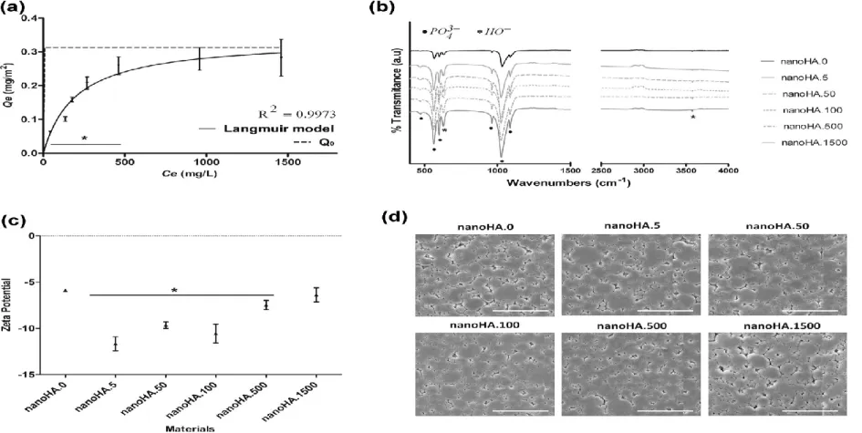
Adsorption isotherm data of CHX adsorption onto HA, was fitted to Langmuir and Freundlich functions, and the obtained regression coefficients (R2) were 0.9973 and 0.9482, respectively. The regression coefficient and the shape of isotherm predicted that Langmuir isotherm model had been suitable to describe the biosorption equilibrium. Therefore, the CHX adsorption equilibrium for Langmuir isotherm is presented in Fig. 1a. Adsorption saturation occurred above 1000 mg/L CHX. The Langmuir equation is described by Qe = KL Ce/(1 +
aL Ce), where Qe is the CHX equilibrium concentration on nanoHA surface
(mg/m2), KL (L/m2) and aL are Langmuir constants (L/mg) and Ce is the final equilibrium concentration of CHX in solution (mg/L) [17]. The isotherms were fitted and the shape is compatible with Langmuir isotherm model. The calculated Langmuir constants, KL and aL, were 2.30 × 10⁻³ L/m2 and 7.34 × 10⁻³ L/mg, respectively. The theoretical monolayer capacity is Q0 and is numerically equal to KL/aL [17]. The obtained Q0 was 0.313 mg/m2, i.e., the maximum amount
of the adsorbed CHX per unit area of nanoHA to form a complete monolayer on the surface. A dimensionless constant separation factor (RL) of 0.98 was determined by equation: RL = 1/(1 + KL × C0), where C0 (mg/L) is the highest initial concentration of adsorbate and KL is the Langmuir constant (L/m2) [34].
3.1.1. Physic-chemical characterization of CHX-loaded nanoHA discs
The FTIR spectra of CHX-loaded nanoHA discs are represented in Fig. 1b. The FTIR spectrum of nanoHA.0 was characterized by the presence of HO− bands at 3570 cm−1 and 629 cm−1, and PO43− bands at 1090, 1037, 962, 602, 564, and
471 cm−1. The FTIR spectra of CHX-loaded nanoHA discs showed the same characteristic bands for phosphate and hydroxyl groups, but the intensity of PO43− bands significantly increased as compared to nanoHA.0 discs.
Fig. 1c shows the net charge of CHX-loaded nanoHA discs. All surfaces were negatively charged. NanoHA.5 discs were the most negatively charged and the nanoHA discs loaded with high CHX concentrations (500 and 1500 mg/L) showed a value of zeta potential (ZP) closer to nanoHA.0 discs. Fig. 1c shows that the ZP increased as the concentration of CHX in the nanoHA discs increased.
Fig. 1d shows the morphology of CHX-loaded nanoHA discs using Scanning Electron Microscopy (SEM). No differences were observed among materials. All surfaces presented a regular structure and the presence of nanoparticles micro-aggregates.
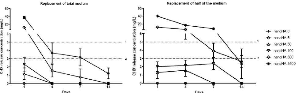
Fig. 2 shows the release profiles of CHX-loaded nanoHA discs for the two protocols of medium replacement mentioned above. For both protocols, nanoHA.5 discs were the only material which did not show measurable release of CHX over time. Fig. 2 shows also the MIC of CHX for E. coli (5 mg/L), S. aureus (3 mg/L) and S. epidermidis (3 mg/L).
3.2.1. Replacement of the total medium at each medium change
The nanoHA discs exhibited a rapid initial release of CHX followed by much slower release kinetics. The amount of CHX released from nanoHA.50 and nanoHA.100 discs occurred over 1 day, and it was lower than the MIC for all bacteria strains. From nanoHA.500 and nanoHA.1500 discs, the CHX was released during 7 days (nanoHA.500) and 14 days (nanoHA.1500). The CHX release from nanoHA.500 discs was higher than the MIC only in first day for all bacteria strains. From nanoHA.1500 discs, the CHX release was higher than the MIC for E. coli in first day and for S. aureus and S. epidermidis up to 7 days.
3.2.2. Replacement of half of the medium at each medium change
The CHX release kinetics was slower than that observed in the previous medium replacement protocol. The CHX release from nanoHA.50 and nanoHA.100 discs
was observed over 4 and 7 days, respectively. The amount of CHX released from these materials was lower than MIC for all bacteria strains. NanoHA.500 and nanoHA.1500 discs released CHX during 14 days. The released CHX concentration from nanoHA.500 discs was higher than MIC for E. coli over 4 days and for S. aureus and S. epidermidis up to 7 days. The amount of CHX released from nanoHA.1500 discs was higher than the MIC for all bacteria strains over 7 days.
3.3. Antimicrobial activity of CHX-loaded nanoHA discs 3.3.1. Quantification of sessile bacteria
The cultivable bacteria data (Fig. 3) show that CHX-loaded nanoHA discs presented a strong and significant reduction in the sessile bacteria, compared to nanoHA.0 discs, except for nanoHA.5 discs. The nanoHA.50 and nanoHA.100 discs showed a higher and significantly anti-adhesive property for E. coli ( 85%) and S. aureus and S. epidermidis ( 90%) compared with nanoHA.0 discs. The nanoHA.500 discs reduced 80% of sessile E. coli and 100% of sessile S. aureus and
S. epidermidis. The nanoHA.1500 discs did not allow the bacteria attachment.
The metabolically active bacteria (RFUs) attached on CHX- loaded nanoHA discs are in accordance with the cultivable bacteria data (Fig. 3). NanoHA.50 to nanoHA.1500 discs showed significant reductions of metabolically active bacteria on their surfaces.
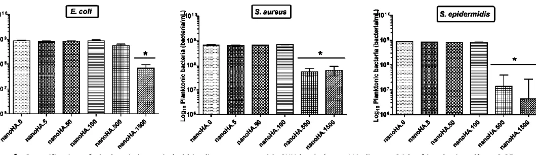
3.3.2. Planktonic bacteria assessment
As shown in Fig. 4, the nanoHA.5 to nanoHA.100 discs did not affect the planktonic bacteria growth. The nanoHA.500 hindered the planktonic S. aureus and S. epidermidis growth but did not affect the growth of planktonic of E. coli. The nanoHA.1500 discs significantly hindered the planktonic growth of all bacteria when compared with nanoHA.0.
3.4. Cytotoxicity of CHX-loaded nanoHA discs
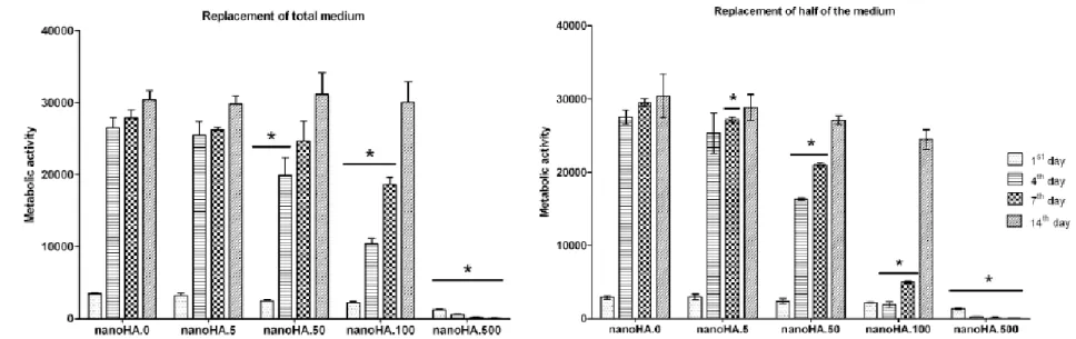
Fig. 5 shows the metabolic activity/proliferation of L929 fibro- blasts on nanoHA discs over a period of 14 days, for the two protocols of medium replacement mentioned above.
On nanoHA.0 discs (control), the metabolic activity increased along the culture time, especially during the first 4 days. NanoHA.5 discs presented similar behavior. However, a dose- and time- dependent inhibition was observed on nanoHA.50 to nanoHA.1500 discs. For nanoHA.50 and nanoHA.100, the inhibitory effect was higher in the first days, i.e., on days 4 and 7, but with the culture medium changes, cells were able to recover and, at day 14, the values were similar to the control ones. Cell proliferation over nanoHA.500 was negligible. NanoHA.1500 was highly cytotoxic, and cell death occurred within minutes (data not shown). This pattern of behavior was similar for the two
protocols of medium withdrawn and replacement. However, the inhibitory effect was lower with the protocol in which the culture medium was totally removed at each medium change and replaced by new medium.
Observation of the nanoHA discs by CLSM provided information that is in line with the results observed in the Alamar Blue assay. Fig. 6 shows representative images of the nanoHA discs cultured with L929 fibroblasts, for the cultures in which the culture medium was totally replaced at each medium change. Cell adhesion was observed in all CHX-loaded nanoHA discs, as evident by the images at day 1, showing randomly distributed cells over the material surface. NanoHA.0 to nanoHA.50 presented similar behavior, exhibiting a progressive increase in the cell numbers with the culture time. At days 1 and 4, nanoHA.0 to nanoHA.50 presented a higher number of attached cells compared to nanoHA.100 and nanoHA.500. On nanoHA.100, cells were able to recover along the culture time and, at days 7 and 14, cultures presented an appearance similar to control. On nanoHA.500, few cells were visible at day 1 and, at later culture times, only some cellular debris were observed.
4.
Discussion
Given the variety of bacteria causative of infections related to biomedical devices and implants, the emergence of bacteria resistant to antimicrobial agents, the resistance to immune defense mechanism and the cytotoxicity of some antimicrobial agents [25], new approaches for the development of antimicrobial agents loaded biomaterials are necessary. These new strategies must have a selective biointeraction pattern, allowing the normal host cellular activities whilst preventing/reducing the microbial colonization and infection [3]. The CHX, a broad spectrum antimicrobial agent, has been described as a good candidate for the development of such materials and devices that do not add to the burden on antibiotics, due to low risk of microbial resistance to CHX [9].
NanoHA biomaterials are widely used in orthopedic and dental applications, given the unique surface properties compared to its bulk-phase counterpart. Thus, the present work reports the interaction of CHX with nanoHA surface, in terms of adsorption and release kinetics profile, antibacterial activity, as well as the cytotoxicity profile, aiming the development of an antibacterial adhesive and cytocompatible material (CHX-loaded nanoHA discs) for further application on indwelling biomedical devices.
Concerning the adsorption of CHX on nanoHA discs, by linear plot of specific adsorption (Ce/Qe) against the equilibrium concentration (Ce), a high regression coefficient (R2 = 0.9973) was observed for Langmuir isotherm model. Therefore, the regression coefficient and the shape of isotherm predicted that Langmuir isotherm had been suitable for to describe the biosorption equilibrium of CHX on nanoHA discs. The Langmuir isotherm assumes mono- layer adsorption onto
4
a surface containing a finite number of adsorption sites of uniform strategies with no transmigration of adsorbate in the plane surface [3,17,34–36]. Once a site is filled, no further sorption can take place at that site [35,36]. Thus, the Langmuir isotherm indicates that the surface of nanoHA reaches a saturation point from 1000 mg/L where the maximum adsorption of the surface was achieved (Q0 = 0.313 mg/m2). The parameter RL expresses the molecule–molecule binding interaction [9,34,35], to predict the affinity between the sorbate and sorbent [35]. Considering that the value of RL indicates the type of Langmuir isotherm to be unfavorable (RL > 1), favorable (0 < RL < 1), irreversible (RL = 0) and linear (RL = 1) [34,35,37], in the present work, the obtained value of RL indicates a favorable adsorption of CHX on nanoHA discs with molecule–molecule interactions. The FTIR spectra of CHX-loaded nanoHA showed similar chemical composition when compared with pure nanoHA. However, CHX-loaded nanoHA spectrum showed a higher intensity of PO43− bands compared to nanoHA.0 discs and this
difference may be due to the binding between chlorhexidine (cationic group) and trivalent phosphate groups in nanoHA, which is in accordance to literature [18,37,39]. Moreover, this bond did not affect the surface morphology of CHX- loaded nanoHA discs, as confirmed by SEM images. The evolution of zeta potential depends on the concentration of CHX in the nanoHA discs that can support the molecule–molecule interactions observed by FTIR and adsorption isotherms. This proportionality of zeta potential values of material surface versus adsorbed bioactive compound may be provide information about electro- static interactions between charged surfaces, according to others authors [17,40,41]. Therefore, as the nanoHA discs are negatively charged and CHX has positive charge [38], the electrostatic interactions between the cationic group of CHX and phosphate groups of nanoHA surface discs may play a major role in the adsorption process.
Taking into account that the release kinetics of CHX from CHX- loaded nanoHA discs may influence its biological performance, two protocols were used to quantify the release profile of CHX, i.e., replacement of the total or half of the medium at specific time- points during the 14-day incubation period. CHX release results from desorption or partial dissolution of the CHX-phosphate binding. As expected, CHX release kinetics was clearly affected by the protocol used, because CHX is prone to leach, especially close to the discs’ surface where rapid diffusion of water molecules was facilitated [13,4,42]. This process depends on the concentration gradient between the bulk solution and the disc surface. Replacement of the total medium originates a high concentration gradient, justifying the rapid initial release, followed by slower release kinetics. On the protocol in which only half of the medium was replaced, concentration gradient was lower, explaining the slower CHX release kinetics over the 14 days incubation. The observed behavior is in agreement with several studies showing that controlled drug release profiles usually reveal an initial burst release of short
duration that is followed by a longer period of continuous but declining release [4,13,18,42–44], and that the release pattern is dependent on the amount of antimicrobial agent adsorbed on biomaterials [13,44].
The interactions between CHX-loaded nanoHA discs and E. coli, S. aureus and S.
epidermidis strains were evaluated in terms of attachment and
anti-planktonic properties.
Concerning the anti-attachment properties, the nanoHA.50 to nanoHA.1500 discs showed a strong capacity to reduce/prevent the sessile bacteria with the reduction of viable bacteria on materials’ surfaces. These materials showed a strong anti-sessile bacteria property, since they were able to inhibit bacterial adhesion and, consequently the biofilm formation. Preventing the adhesion and aggregation of early bacterial colonizers on biomaterial surfaces is relevant because it prevents the attachment of later colonizers that require more demanding growth conditions [45]. Consequently, the reduction of pioneer bacterial attachment affects the successive stages of biofilm formation, which is essential in preventing device-associated infection. This has been suggested in several studies addressing antimicrobial agents loaded biomaterials, e.g., furanone-incorporated polystyrene discs, nanoHA-ZnO composite, Ti/PBA-CHX, ZnO/PVC composite and polyurethane nanocomposites-CHX [3,20,28,46,47].
The anti-planktonic activity of CHX-loaded nanoHA discs was proportional to the CHX concentration used. Only nanoHA.500 and nanoHA.1500 discs released CHX concentrations above MICs for the tested bacteria strains, negatively affecting the planktonic bacteria growth. Other studies reported also the antimicrobial activity of CHX released from several surfaces (e.g., dentin slabs, glass ionomer cements, PLGA microspheres, and hydroxyapatite) [14–16,18,19]. However, results on drug delivery in planktonic cells should not be extrapolated to bacterial biofilms, since the biofilm structure protects bacteria against the action of many antimicrobial agents [3].
Results revealed that the binding of CHX to nanoHA did not affect the antibacterial activity of this compound. The CHX was more effective against Gram-positive bacteria than Gram-negative bacteria. This is in line with several works that also reported that Gram-positive bacteria are more sensitive to CHX [11,14,48]. CHX binds to phospholipids of cell membrane by electrostatic attraction causing its rupture, resulting in fluid leaking, leading to lysis and bacterial death. Probably due to the lack of an outer cell wall membrane, when compared with Gram-negative bacteria, Gram positive-bacteria have low resistance to physical disruption and higher susceptibility to CHX [48].
CHX-loaded nanoHA discs were also evaluated for its cytocompatibility, i.e., for the ability to support the cell growth, using fibroblast cell cultures. Fibroblasts are ubiquitous cells that not only function to sustain various organs and tissues as stromal cells but also act directly to regulate adjacent cell behavior including migration, proliferation, and differentiation. In this way, biological evaluation with
fibroblast cell cultures might be regarded as a general bioassay, providing reliable information concerning acute and long-term cytotoxicity or other biological responses. Results showed a dose- and time-dependent inhibitory effect on the cell proliferation over nanoHA.50—nanoHA.1500 discs. The same pattern was observed by other authors on CHX-loaded to titanium/polybenzyl acrylate and hydroxyapatite [3,19]. The inhibitory effect of CHX was more pronounced in the protocol in which half of culture medium was removed and replaced at specific time-points, due to the cumulative effect of CHX, as confirmed by the release kinetics of CHX. With both experimental protocols, nanoHA.50 discs allowed the adhesion and proliferation of fibro- blast cells in a way similar to control.
Overall, CHX-loaded nanoHA discs, namely nanoHA.50, showed strong anti-sessile bacteria properties, i.e., they significantly reduced bacteria attachment and further accumulation of bacteria, allowing, simultaneously, the proliferation of fibroblast cells. Therefore, the nanoHA.50 materials might be a good alternative to be applied on medical devices to treat devices-related infections.
5.
Conclusions
The adsorption of CHX on nanoHA discs occurred by elec- trostatic interactions between the cationic group of CHX and the phosphate group of nanoHA discs. The release of CHX from CHX-loaded nanoHA discs showed a fast initial rate followed by slower release kinetics, substantiating a system constrained by dilution and diffusion-limiting processes. The CHX-loaded nanoHA discs showed anti-sessile and anti-planktonic properties. The nanoHA.50—nanoHA.1500 showed a strong anti-sessile activity, inhibiting bacterial adhesion and, consequently the biofilm for- mation. The anti-planktonic activity of CHX-loaded nanoHA discs was proportional to the CHX concentration used, and was observed for nanoHA.500 and nanoHA.1500. Binding of CHX to nanoHA did not affect its antimicrobial activity, with CHX-nanoHA being more effective against the tested Gram-positive bacteria than Gram-negative bacteria. The CHX-loaded nanoHA caused dose- and time-dependent inhibitory effects on the adhesion and proliferation of fibroblast cells. The nanoHA.5 and nanoHA.50 reveal similar behavior compared to control. Overall, results suggest that appropriate CHX-loaded nanoHA compositions might be useful as a biomedical material to prevent the occurrence of DRIs.
Acknowledgements
This work was co-financed by FEDER funding through Programa Operacional Factores de Competitividade—COMPETE and by national (Portuguese) funds through FCT—Fundac¸ão para a Ciência e a Tecnologia within project NanoBiofilm (PTDC/SAU- BMA/111233/2009).
References
[1] K. Vasilev, J. Cook, H.J. Griesser, Antibacterial surfaces for biomedical
devices, Expert. Rev. Med. Devices 6 (2009) 553–567.
[2] A. Simchi, E. Tamjid, F. Pishbin, A.R. Boccaccini, Recent progress in
inorganic and composite coatings with bactericidal capability for orthopaedic applications, Nanomedicine 7 (2011) 22–39.
[3] M.C. Cortizo, T.G. Oberti, M.S. Cortizo, A.M. Cortizo, M.A.F.L. de Mele,
Chlorhexidine delivery system from titanium/polybenzyl acrylate coating: evaluation of cytotoxicity and early bacterial adhesion, J. Dent. 40 (2012) 329–337.
[4] M. Baro, E. Sanchez, A. Delgado, A. Perera, C. Evora, In vitro–in vivo
characterization of gentamicin bone implants, J. Control. Release 83 (2002) 353–364.
[5] C. Castro, E. Sanchez, A. Delgado, I. Soriano, P. Nunez, M. Baro, A. Perera,
C. Evora, Ciprofloxacin implants for bone infection. In vitro–in vivo characterization, J. Control. Release 93 (2003) 341–354.
[6] D. Neut, E.P. de Groot, R.S. Kowalski, J.R. van Horn, H.C. van der Mei,
H.J. Busscher, Gentamicin-loaded bone cement with clindamycin or fusidic acid added: biofilm formation and antibiotic release, J. Biomed. Mater. Res. A 73 (2005) 165–170.
[7] H. Alvarez, C. Castro, L. Moujir, A. Perera, A. Delgado, I. Soriano, C.
Evora, E. Sanchez, Efficacy of ciprofloxacin implants in treating experimental osteomyelitis, J. Biomed. Mater. Res. B: Appl. Biomater. 85 (2008) 93–104.
[8] M.P. Ferraz, A.Y. Mateus, J.C. Sousa, F.J. Monteiro, Nanohydroxyapatite
micro- spheres as delivery system for antibiotics: release kinetics, antimicrobial activity, and interaction with osteoblasts, J. Biomed. Mater. Res. A 81A (2007) 994–1004.
[9] M.E. Barbour, D.J. O’Sullivan, D.C. Jagger, Chlorhexidine adsorption to
anatase and rutile titanium dioxide, Colloid Surf. A 307 (2007) 116–120.
[10]C.H.J. Hauman, R.M. Love, Biocompatibility of dental materials used in
contemporary endodontic therapy: a review. Part 1. Intracanal drugs and substances, Int. Endod. J. 36 (2003) 75–85.
[11]A.D. Russel, Mechanisms of resistance to antiseptics, disinfectants and
preservatives. 2, Pharm. Int. 7 (1986) 305–308.
[12]M. Morra, C. Cassinelli, G. Cascardo, A. Carpi, M. Fini, G. Giavaresi, R.
Gia- rdino, Adsorption of cationic antibacterial on collagen-coated titanium implant devices, Biomed. Pharmacother. 58 (2004) 418–422.
[13]N. Hiraishi, C.K.Y. Yiu, N.M. King, F.R. Tay, Chlorhexidine release and
antibacterial properties of chlorhexidine-incorporated polymethyl methacrylate-based resin cement, J. Biomed. Mater. Res. B 94B (2010)
134–140.
[14]C.F. Franco, A.L. Pataro, L.C.R.E. Souza, V.R. Santos, M.E. Cortes, R.D.
Sinisterra, In vitro effects of a chlorhexidine controlled delivery system, Artif. Organs 27 (2003) 486–491.
[15]E.R. Hook, O.J. Owen, C.A. Bellis, J.A. Holder, D.J. O’Sullivan, M.E. Bar-
bour, Development of a novel antimicrobial-releasing glass ionomer cement functionalized with chlorhexidine hexametaphosphate nanoparticles, J. Nanobiotechnol. 12 (2014) 3.
[16]I.C. Yue, J. Poff, M.E. Cortes, R.D. Sinisterra, C.B. Faris, P. Hildgen, R.
Langer, V.P. Shastri, A novel polymeric chlorhexidine delivery device for the treatment of periodontal disease, Biomaterials 25 (2004) 3743– 3750.
[17]E. Gimenez-Martin, M. Lopez-Andrade, A. Ontiveros-Ortega, M.
Espinosa- Jimenez, Adsorption of chlorhexidine onto cellulosic fibers, Cellulose 16 (2009) 467–479.
[18]A.A. Campbell, L. Song, X.S. Li, B.J. Nelson, C. Bottoni, D.E. Brooks, E.S.
DeJong, Development, characterization, and anti-microbial efficacy of hydroxyapatite- chlorhexidine coatings produced by surface-induced mineralization, J. Biomed. Mater. Res. 53 (2000) 400–407.
[19]C.A. de Souza, A.P. Colombo, R.M. Souto, C.M. Silva-Boghossian, J.M.
Granjeiro, G.G. Alves, A.M. Rossi, M.H. Rocha-Leao, Adsorption of chlorhexidine on synthetic hydroxyapatite and in vitro biological activity, Colloids Surf. B: Bioin- terfaces 87 (2011) 310–318.
[20]N. Fong, A. Simmons, L.A. Poole-Warren, Antibacterial polyurethane
nanocom- posites using chlorhexidine diacetate as an organic modifier, Acta Biomater. 6 (2010) 2554–2561.
[21]N. Ribeiro, S.R. Sousa, F.J. Monteiro, Influence of crystallite size of
nanophased hydroxyapatite on fibronectin and osteonectin adsorption and on MC3T3- E1 osteoblast adhesion and morphology, J. Colloid Interface Sci. 351 (2010) 398–406.
[22]M. Alcaide, M.C. Serrano, R. Pagani, S. Sanchez-Salcedo, A. Nieto, M.
Vallet-Regi, M.T. Portoles, L929 fibroblast and Saos-2 osteoblast response to hydroxyapatite-betaTCP/agarose biomaterial, J. Biomed. Mater. Res. A 89 (2009) 539–549.
[23]M.P. Ferraz, F.J. Monteiro, C.M. Manuel, Hydroxyapatite nanoparticles:
a review of preparation methodologies, J. Appl. Biomater. Biomech. 2 (2004) 74–80.
[24]R. Family, M. Solati-Hashjin, S. Namjoy Nik, A. Nemati, Surface
modification for titanium implants by hydroxyapatite nanocomposite, Caspian J. Intern. Med. 3 (2012) 460–465.
[25]K. Fox, P.A. Tran, N. Tran, Recent advances in research applications of
[26]Y.R. Cai, Y.K. Liu, W.Q. Yan, Q.H. Hu, J.H. Tao, M. Zhang, Z.L. Shi, R.K.
Tang, Role of hydroxyapatite nanoparticle size in bone cell proliferation, J. Mater. Chem. 17 (2007) 3780–3787.
[27]X.C. Dou, X.P. Zhu, J. Zhou, H.Q. Cai, J. Tang, Q.L. Li,
Minocycline-released hydroxyapatite-gelatin nanocomposite and its cytocompatibility in vitro, Biomed. Mater. 6 (2011) 025002.
[28]L. Grenho, F.J. Monteiro, M.P. Ferraz, In vitro analysis of the antibacterial
effect of nanohydroxyapatite-ZnO composites, J. Biomed. Mater. Res. A (2013).
[29]K.P. Tank, K.S. Chudasama, V.S. Thaker, M.J. Joshi, Cobalt-doped
nanohydrox- yapatite: synthesis, characterization, antimicrobial and hemolytic studies, J. Nanopart. Res. 15 (2013).
[30]A. Zamanian, F. Moztarzadeh, S. Kordestani, S. Hesaraki, M.R. Tahriri,
Novel calcium hydroxide/nanohydroxyapatite composites for dental applications: in vitro study, Adv. Appl. Ceram. 109 (2010) 440–444.
[31]P. Shi, Y. Zuo, X. Li, Q. Zou, H. Liu, L. Zhang, Y. Li, Y.S. Morsi,
Gentamicin-impregnated chitosan/nanohydroxyapatite/ethyl cellulose micro- spheres granules for chronic osteomyelitis therapy, J. Biomed. Mater. Res. A 93 (2010) 1020–1031.
[32]J. Barros, L. Grenho, C.M. Manuel, C. Ferreira, L. Melo, O.C. Nunes, F.J.
Monteiro, M.P. Ferraz, Influence of nanohydroxyapatite surface properties on Staphylococcus epidermidis biofilm formation, J. Biomater. Appl. 28 (2014) 1325–1335.
[33]A. Borges, C. Ferreira, M.J. Saavedra, M. Simoes, Antibacterial activity
and mode of action of ferulic and gallic acids against pathogenic bacteria, Microb. Drug Resist. 19 (2013) 256–265.
[34]B.H. Hameed, D.K. Mahmoud, A.L. Ahmad, Sorption equilibrium and
kinetics of basic dye from aqueous solution using banana stalk waste, J. Hazard. Mater. 158 (2008) 499–506.
[35]M.B. Desta, Batch sorption experiments: Langmuir and Freundlich
isotherm studies for the adsorption of textile metal ions onto teff straw (Eragrostis tef) agricultural waste, J. Thermodyn. (2013), http://dx.doi.org/10.1155/2013/ 375830
[36]S.J. Allen, G. Mckay, J.F. Porter, Adsorption isotherm models for basic
dye adsorption by peat in single and binary component systems, J. Colloid Interface Sci. 280 (2004) 322–333.
[37]A. Mittal, L. Kurup, J. Mittal Freundlich, Langmuir adsorption isotherms
and kinetics for the removal of Tartrazine from aqueous solutions using hen feath- ers, J. Hazard. Mater. 146 (2007) 243–248.
[38]J. Kim, T. Uchiyama, M. Carrilho, K.A. Agee, A. Mazzoni, L. Breschi, R.M.
Carvalho, L. Tjäderhane, S. Looney, C. Wimmer, A. Tezvergil-Mutluay, F.R. Tay, D.H. Pashley, Chlorhexidine binding to mineralized versus
demineralized dentin powder, Dent. Mater. 26 (2010) 771–778.
[39]R.N.S. Sodhi, H.A. Grad, D.C. Smith, Examination by X-ray
photoelectron spectroscopy of the adsorption of chlorhexidine on hydroxyapatite, J. Dent. Res. 71 (1992) 1493–1497.
[40]Z. Kolská, Z. Makajová, K. Kolárˇová, N.K. Slepicˇková, S. Trostová, A.
Rˇeznícˇková, J. Siegel, V. Sˇvorcˇík, Electrokinetic potential and other surface properties of polymer foils and their modifications, in: Y. Faris (Ed.), Polymer Science, In Tech d.o.o., Rijeka, Croatia, 2013, pp. 203–228 (Chapter 8).
[41]E. Chibowski, M. Espinosa-Jiménez, A. Ontiveros-Ortega, E.
Giménez-Martin., Surface free energy, adsorption and zeta potential in leacril/tannic acid system, Langmuir 14 (1998) 5237–5244.
[42]C. Castro, E. Sanchez, A. Delgado, I. Soriana, P. Nunez, M. Baro, A. Perera,
C. Evora, Ciprofloxacin implants for bone infection. In vitro–in vivo characterization, J. Control. Release 93 (2003) 341–354.
[43]M. Kazemzadeh-Narbat, J. Kindrachuk, K. Duan, H. Jenssen, R.E.W.
Hancock, R.Z. Wang, Antimicrobial peptides on calcium phosphate-coated titanium for the prevention of implant-associated infections, Biomaterials 31 (2010) 9519–9526.
[44]M. Kazemzadeh-Narbat, B.F.L. Lai, C.F. Ding, J.N. Kizhakkedathu,
R.E.W. Han- cock, R.Z. Wang, Multilayered coating on titanium for controlled release of antimicrobial peptides for the prevention of implant-associated infections, Bio- materials 34 (2013) 5969–5977.
[45]M. Katsikogianni, Y.F. Missirlis, Concise review of mechanisms of bac-
terial adhesion to biomaterials and of techniques used in estimating bacteria–material interactions, Eur. Cell Mater. 8 (2004) 37–57.
[46]E.B.H. Hume, J. Baveja, B.W. Muir, T.L. Schubert, N. Kumar, S. Kjelleberg,
H.J. Griesser, H. Thissen, R. Read, L.A. Poole-Warren, K. Schindhelm, M.D.P. Willcox, The control of Staphylococcus epidermidis biofilm formation and in vivo infec- tion rates by covalently bound furanones, Biomaterials 25 (2004) 5023–5030
[47]J.T. Seil, T.J. Webster, Reduced Staphylococcus aureus proliferation and
biofilm formation on zinc oxide nanoparticle PVC composite surfaces, Acta Biomater. 7 (2011) 2579–2584.
[48]H.Y. Cheung, M.M.K. Wong, S.H. Cheung, L.Y. Liang, Y.W. Lam, S.K.
Chiu, Differential actions of chlorhexidine on the cell wall of bacillus subtilis and Escherichia coli, PLoS One 7 (2012).
Fig. 1. Characterization of CHX-loaded nanoHA discs: (a) Adsorption isotherm of CHX into nanoHA discs, plotted as the CHX mass adsorbed per unit area of nanoHA discs (Qe, mg/m2) versus final equilibrium concentration of CHX in solution (Ce, mg/L). The data fit the Langmuir equation. Theoretical monolayer capacity (Q0, mg/m2) (b) FTIR spectra of CHX-loaded nanoHA discs; (c) Zeta potential values of CHX-loaded nanoHA discs (*significant different (p < 0.05) compared to nanoHA.0, according to Turkey HSD). (d) SEM images of CHX-loaded nanoHA discs. Scale bar 20 D m.
Fig. 2. Release profile of CHX from CHX-loaded nanoHA discs, considering the two protocols of culture medium replacement. Broken lines represent MIC values of (1) E. coli and (2) S. aureus and S. epidermidis strains.
Fig. 3. Sessile bacteria populations determined by cultivable method (CFUs) and metabolic bacteria (RFUs) on CHX-loaded nanoHA discs at 24 h of incubation. (*p < 0.05, significant differences compared to nanoHA.0, according to Turkey HSD.)
Fig. 4. Quantification of planktonic bacteria held in direct contact with CHX-loaded nanoHA discs at 24 h of incubation. (*p < 0.05, significant differences compared to nanoHA.0, according to Turkey HSD.)
Fig. 5. Metabolic activity/proliferation of L929 fibroblasts cultured over CHX-loaded nanoHA discs for 14 days, considering the two protocols of culture medium replacement. (*p < 0.05, significant differences compared to nanoHA.0, according to Turkey HSD.)
Fig. 6. CLSM observation of CHX-loaded nanoHA discs cultured with L929 fibroblasts for 1, 4, 7, and 14 days. The culture medium was totally replaced at each medium change. Nuclei were stained with 4′,6-diamidino-2-phenylindole (blue) and actin filaments with phalloidin (red). Scale bar 100 m.