Treatment of diffuse diabetic maculopathy with intravitreal triamcinolone and
laser photocoagulation: randomized clinical trial with morphological and
functional evaluation
Tratamento da maculopatia diabética difusa com triancinolona intravítrea e fotocoagulação
a laser: ensaio clínico randomizado com avaliação morfofuncional
ALBERTO LUIZ GIL1, MIRELA JOBIMDE AZEVEDO2, GIOVANI GENERALI TOMASETTO1, CARLOS HENRIQUE GERVINI MUNIZ1, JACÓ LAVINSKY1
Submitted for publication: November 29, 2010 Accepted for publication: October 7, 2011
Study carried out at the Hospital de Clínicas de Porto Alegre, Porto Alegre (RS), Brazil. 1Physician, Serviço de Oftalmologia do Hospital de Clínicas de Porto Alegre, Porto Alegre (RS),
Brazil.
2Physician, Serviço de Endocrinologia do Hospital de Clínicas de Porto Alegre, Porto Alegre (RS), Brazil.
Funding: This study was partially funded by FIPE - Hospital de Clínicas de Porto Alegre.
Disclosure of potential conflicts of interest: A.L.Gil, None; M.J.de Azevedo, None; G.G.Tomasetto, None; C.H.G.Muniz, None; J.Lavinsky, None.
Correspondence address: Alberto Luiz Gil. Rua Soledade, 569 Conj. 905 A Porto Alegre (RS) -90470-340 - Brazil - E-mail: [email protected]
ABSTRACT
Purpose: Treatment of diffuse macular edema in diabetes mellitus is currently un-satisfactory. The purpose of this double blind randomized clinical trial was to com-pare the treatment of diffuse diabetic macular edema with intravitreal triamcinolone or laser in type 2 diabetes mellitus patients using a morphofunctional assessment.
Methods: Fourteen patients (21 eyes) with clinically significant diffuse macular-edema, previously untreated and with a macular thickness >250 µm at optical coherence
tomography were randomized for treatment with laser or intravitreal injection of triamcinolone acetate. Optical coherence tomography, biomicroscopy, fundoscopy, fluorescein angiography, tonometry, scotometry, visual and contrast acuities were performed at 0, 1, 3 and 6 months.
Results: At pre-treatment stage, Laser (n=9) and Triamcinolone (n=12) groups did not differ regarding retinal thickness, visual and contrast acuities. In Triamcinolone group macular thickness decreased after 1 month (424.1 ± 19.9 µm to 358.4 ± 18.2 µm;
p=0.04) and started to return to the initial values in the 3rd month (p=0.02). No
changes occurred in macular scotometry and visual and contrast acuities. No side effects were observed with both treatments.
Conclusion: During the study macular thickness diminished in the triamcinolone group, especially in the first month of treatment. At 3 and 6 months there was no difference. Macular thickness did not change during the study in the laser group. In the study sample it was not possible to demonstrate differences relates to visual acuity and scotometry between the two groups.
ClinicalTrials.gov Identifier: NCT00668239
Keywords: Macular edema/drug therapy; Triamcinolone/therapeutic use; Photocoa-gulation; Diabetes mellitus; Tomography, Optical coherence
RESUMO
Objetivo: O tratamento do edema macular difuso diabético atualmente é insatisfatório. O objetivo deste ensaio clínico randomizado duplo cego foi comparar, através de avalia-ção morfofuncional, o tratamento do edema macular difuso diabético com triancinolona intravítrea ou laser em grade em pacientes com DM tipo 2.
Métodos: Quatorze pacientes (21 olhos) com edema macular difuso clinicamente significativo, sem tratamento prévio e com espessura macular >250 µm à tomografia de
coerência óptica (OCT) foram randomizados para tratamento com laser ou injeção intravítrea de acetato de triancinolona. Nos tempos 0, 1, 3 e 6 meses foram realizados OCT, biomicroscopia, fundoscopia, angiografia fluoresceínica, tonometria, escotometria, acuidade visual e de contraste.
Resultados: Na fase pré-tratamento, os grupos Laser (n=9) e Triancinolona (n=12) não diferiram na espessura retiniana, escotometria, acuidade visual e de contraste. No grupo Triancinolona houve redução da espessura macular após 1 mês (424,1 ± 19,9 µm vs.
358,4 ± 18,2 µm; P=0,04) com retorno aos valores iniciais a partir do mês 3 (P=0,02). Não houve modificação significativa na escotometria macular, acuidade visual e de contraste. Não ocorreram efeitos colaterais nos tratamentos.
Conclusão: Durante o estudo observou-se diminuição da espessura macular no grupo tratado com triancinolona, principalmente no primeiro mês de tratamento. Aos 3 e 6 meses de tratamento não houve diferença. A espessura macular não modificou durante o estudo no grupo tratado com laser. Nesta amostra estudada não foi possível demons-trar diferenças relacionadas à escotometria e acuidade visual entre os dois grupos.
Descritores: Edema macular/quimioterapia; Triancinolona/uso terapêutico; Fotocoa-gulação; Diabetes mellitus; Tomografia de coerência óptica
INTRODUCTION
Diabetic retinopathy (DR) is the main cause of visual loss in adults. Macular edema affects 29% of the patients with DR and is the main cause of visual loss in this population(1,2).
The Early Treatment Diabetic Retinopathy Study (ETDRS)(2)
sho-wed a major benefit of treatment with laser photocoagulation of clinically significant macular edema. Although this treatment redu-ces moderate visual loss by 50%, about 24% of treated eyes presen-ted a thickened macula and consequently diminished sight after 36 months, suggesting that there is a subset of patients who are resistant to laser photocoagulation treatment.
Prior studies assessing patients with diabetic retinopathy showed that laser treatment of eyes with diffuse macular edema had a worse result than eyes with focal macular edema(3), raising interest
in other therapeutic options including surgery, with pars plana vitrectomy, pharmacological treatment with protein kinase C inhi-bitors(4), and intraocular corticosteroids.
Corticosteroids treatment for ophthalmological diseases are being studied(5) and used intravitreally in different diseases(6). Among the
related adverse effects are transient increased intraocular pressure(5,7),
corticogenic glaucoma(8), retinal detachment, and infectious
Some studies presented good results using intravitreal triam-cinolone acetate to treat diffuse diabetic macular edema in pa-tients who did not have a significant improvement with laser photo-coagulation (conventional treatment)(10-15). Most of these studies did
not describe the clinical characteristics of the patients studied(11,12,14,15).
On the other hand, data about possible improvement using laser after intravitreal triamcinolone are controversial(16,17). A single
non-controlled study with a high dose of intravitreal triamcinolone (25 mg) assessed this injection as a primary treatment for diabetic macular edema(18).
Most studies with triamcinolone and laser photocoagulation con-sider visual acuity as an outcome parameter, thus evaluating es-pecially foveal function. It is likely that a more complete evaluation of macular morphological and functional aspects will supply fur-ther information about the results of these treatments. Few aspects of diffuse diabetic maculopathy treatment have not yet been defini-tively established, such as, the best method of treatment and the correlation between morphological and functional results. It has also not been defined whether the evaluation of functional results should be done by analysis of foveal function or of the whole macula, using other tools such as macular sensitivity and contrast.
The purpose of this study was to compare the treatment of diffuse diabetic macular edema with intravitreal triamcinolone acetate or grid laser in patients with type 2 diabetes mellitus (DM) using a morphofunctional evaluation.
METHODS
P
ATIENTSIn this double blind randomized clinical trial against active treat-ment, patients with type 2 DM and diffuse macular edema secon-dary to DR were evaluated between September 2004 and June 2006. Consecutively included patients were attended in the Cen-ter of Reference for Diabetic Retinopathy at Hospital de Clinicas de Porto Alegre (HCPA) and at the Ophthalmology Outpatient Clinic of HCPA.
The following inclusion criteria were considered: age greater than or equal to 30 years; DR with clinically significant diffuse macu-lar edema, according to criteria established by ETDRS(2); no prior
treatment with laser and/or intravitreal injection of triamcinolone acetate, and presence of central fixation, demonstrated in the Op-tical Coherence Tomography (OCT). Exclusion criteria were: history of glaucoma or ocular hypertension; intraocular surgery in the six preceding months; opacities of the cornea, lens or vitreous which would prevent performing laser or ophthalmological examina-tions; proliferative diabetic retinopathy; history of allergy to fluo-rescein or corticosteroids, and serum creatinine >2.5 mg/dl.
Type 2 DM was diagnosed in patients who had more than 30 years old at the diagnosis of DM with no previous episodes of ketoa-cidosis, and no insulin treatment in the first 5 years after DM diag-nosis. All patients underwent a clinical and laboratory assessment before treatment, which included clinical history, blood pressure measurements, fasting blood glucose (glucose-peroxidase co-lorimetric enzymatic method- Biodiagnostica Kit, Roche Diagnós-tica, São Paulo, Brazil), glycosilated hemoglobin [A1C test; High Performance Liquid Chromatography (HPLC) in a Merck-Hitachi® (Darmstadt, Germany) 9100 apparatus; Reference values 4.8-6.0%], and serum creatinine (Jaffé reaction) measurements.
O
PHTHALMOLOGICALASSESSMENTThe ophthalmological assessment consisted of: OCT, visual acuity, contrast acuity, scotometry, fluorescein angiography, intraocular pres-sure meapres-surement, indirect fundoscopy, and biomicroscopy. After this evaluation patients were randomized to two treatment groups: triam-cinolone or laser. New clinical, laboratory and ophthalmological eva-luations were performed after one, three and six months.
In order to perform OCT, images of the retinal layers were obtai-ned, comprising the 6 mm in the center of the macula in nine con-centric measures. The examination was performed by the same investigator and at the same time (between 10 a.m. and 11 a.m.). The OCT (Stratus 3, Carl Zeiss®
, Oberkochen, Germany) was perfor-med by an examiner who did not know the type of treatment to which the patient was submitted (G.T.). The Macular Thickness pro-gram was used to analyse macular thickness, using in the analysis the highest obtained measurement (peak) in each assessment. The patient’s fixation (central or excentric) was determined. Visual acui-ty was measured with a table standardized by ETDRS, with im-proved correction of refraction(2). Contrast was evaluated in a
man-ner similar to visual acuity, using the Pelly-Robson table. The Hum-phrey AII 745 [Carl Zeiss®
, Oberkochen, Germany, (4) with operatio-nal system 12.6] was used for scotometry (visual field 10-2). Visual acuity and scotometry were evaluated by an investigator who did not know the randomization group (C.M.). The camera used for fluorescein angiography was FF450 PLUS IR (Carl Zeiss®
, Ober-kochen, Germany). Tonometry was performed on both eyes using the Perkins tonometer. Lens opacity was evaluated using the table LOCS II.
T
REATMENTGROUPSLaser treatment: argon laser (Crystal Focus - EMERED®,Jena, Ger-many) was applied to the macular region according to the modified grid technique in inverted C, preserving 500 µ of the foveolus and avascular zone, with 100 µ diameter shots, energy varying from 0.2 to 0.5 joules, with a exposure time between 0.2 and 0.4 seconds. One hundred and fifty to 200 shots were applied according to the retinal area size(2).
Laser for masking (sham procedure): the same stages used for laser treatment were performed, but energy was set to zero, resul-ting in a simulation of the treatment.
Triamcinolone treatment: 0.1 ml (4 mg) of triamcinolone acetate intravitreous was injected through the pars plana in a surgical envi-ronment. The medication used was manipulated by the Ophthal-mos chemist without a preservative agent.
Triamcinolone for masking: the same stages used for triamcino-lone acetate injection were performed in a surgical environment and the simulation of the injection was done by minimum pressure in the patient’s conjunctiva using the capped needle.
S
TATISTICALANALYSISThe sample size was calculated considering the main outcome, retinal thickness, measured by OCT before and after treatment. Assu-ming a mean reduction of the macular edema of 100 µm(19) after laser and a reduction of 250 µm(12) after intravitreal injection, to have a 90% power and 0.05 alpha, an estimative of 8 eyes for each treatment group (difference of 150 µm) had to be included.
Paired and non-paired t tests were used for comparison pur-poses, as indicated. ANOVA for repeat measures followed by a mul-tiple comparison test, LSD (least significant difference) was used to analyse morphofunctional changes of the macula and clinical va-riables during the study. Systolic and diastolic blood pressure chan-ges were used as covariates to analyse the ophthalmological va-riables at the different evaluation times.
Data were expressed as mean ± standard deviation, as a per-centage of patients with the characteristic, or as means (95% confiden-ce interval). Pvalues <0.05 were considered statistically significant. Statistical analysis was performed using SPSS 14.0 (SPSS®
, Chicago, IL, USA).
E
THICSRESULTS
Fourteen patients with type 2 DM and DR fulfilled the inclusion criteria and agreed to participate in the study. Four male and 10 female patients, aged 59.3 ± 6.0 years (52 to 67 years) with DM duration of 15.0 ± 7.3 years (5 to 28 years) were assessed. Two of these patients were being treated with intermediate action insulin, five were using anti-hyperglycemic drugs (metformin, glibencla-mide) and six patients, insulin and anti-hyperglycemic drugs. All patients were hypertensive and used enalapril maleate. Two patients, one in each treatment group, also used hydrochlorothiazide. No me-dication was changed or included during the study.
Seven patients were randomized to treatment with triamcino-lone and seven to laser treatment. Twenty-one eyes were studied: 19 with non-proliferative moderate DR and two with non-prolife-rative severe DR. Seven patients were treated in both eyes: five patients in the Triamcinolone group and two patients in the Laser group.
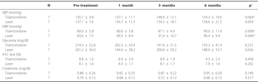
Table 1 describes clinical and laboratory evaluated parameters before treatment and in the 1st, 3rd, and 6th month. These features
were compared between the patients of the Triamcinolone group and the Laser group in the beginning of the study: systolic blood pressure, diastolic blood pressure, fasting blood glucose, AlC test, and serum creatinine and were not different among the two groups. During the study, systolic blood pressure increased in the group treated with triamcinolone. End-of-study diastolic blood pressure values were higher than pre-treatment values in both groups. Fas-ting blood glucose, AlC test, and serum creatinine did not change during the study in the Triamcinolone and Laser groups.
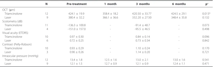
Table 2 shows the ophthalmological parameters before treat-ment and after 1, 3, and, 6 months.
Macular thickness, scotometry, visual and contrast acuity measu-rements, and intraocular pressure were not different in the Triam-cinolone and Laser groups in the pre-treatment period. The analy-sis of OCT measurements during the study was adjusted to systolic and diastolic blood pressure changes (delta) from baseline to the end-of-study. During the study macular thickness diminished in the Triamcinolone group. This reduction was observed in the first month of treatment. At the 3rd and 6th months there was no
diffe-rence in the macular thickness as compared to pre-treatment values. Considering the absolute values of retinal thickness obtai-ned in OCT minus the reference value of retinal thickness (206 µm), the reduction observed in the 1st month of treatment with
triam-cinolone was 30.1 ± 2.0%. The macular thickness returned to the pre-treatment value with recurrence of macular edema that started in the 3rd month. Macular thickness did not change during
the study in the Laser group.
Visual and contrast acuities and intraocular pressure were not evaluated at the end-of-study in all patients due to non-complian-ce with schedules after end-of-study OCT was performed. Those exams did not change during the study in both treatment groups. In the Triamcinolone group the improvement of macular sensiti-vity in the 3rd month, as evaluated by scotometry, did not attain the
statistical adopted significance.
In both treatment groups no patient developed cataract, uveitis, vitreitis, retinal detachment, endophthalmitis, or any possibly treat-ment related side effects.
DISCUSSION
In this study, the eyes of patients with type 2 DM and diffuse macular edema treated with intravitreal triamcinolone had a grea-ter retinal thickness reduction in the 1st month post-treatment as
compared to the eyes treated with laser photocoagulation. This retinal thickness improvement observed with triamcinolone treat-ment remained up to the 3rd month of the study. Subsequently, the
macular edema recurred in all patients.
The morphological evaluation of the macula was performed by OCT, enabling retinal layers visualization. This methodology has a resolution close to that of histology, and allows quantification of the macular thickness. In addition, all measurements were performed at the same time of the day to avoid diurnal variation of retinal thickness. This procedure, associated with the masking of the investigators and patients, ensured accurate evaluation of retinal edema.
In patients with DM and diffuse macular edema, the use of triam-cinolone alone was evaluated in a single study(18). Despite
favoura-ble results in OCT and visual acuity in this non-controlled study, a much higher triamcinolone dose was used as compared to the present study (25 mg vs. 4 mg). Furthermore, a major increase in intraocu-lar pressure that required surgery was observed in 13% of the pa-tients. Only a recently published study compared the results of intravitreal triamcinolone, laser or both treatments(17). In the
com-bined treatment group, the laser was performed 30 days after intravitreal injection of triamcinolone. The results reported were similar to those of the present study, with improved retinal thickness
Table 1. Clinical and laboratory characteristics of the groups treated with triamcinolone and laser during the study
N Pre-treatment 1 month 3 months 6 months pa
SBP (mmHg)
Triamcinolone 7 135.7 ± 9.8 137.1 ± 11.1 148.6 ± 12.1 154.3 ± 19.0 0.004b
Laser 7 137.1 ± 7.6 145.7 ± 11.3 154.3 ± 18.1 158.6 ± 21.2 0.059 DBP (mmHg)
Triamcinolone 7 80.0 ± 5.8 80.0 ± 5.8 87.1 ± 4.9 90.0 ± 11.6 0.008c
Laser 7 83.6 ± 7.5 89.3 ± 8.4 91.4 ± 10.7 96.4 ± 9.4 0.044d
Glycemia (mg/dl)
Triamcinolone 7 214.5 ± 52.8 202.3 ± 54.4 191.8 ± 51.5 193.3 ± 41.4 0.231 Laser 7 201.2 ± 56.0 194.0 ± 38.2 200.8 ± 50.2 188.0 ± 33.7 0.656 A1C test (%)
Triamcinolone 7 8.8 ± 1.6 8.9 ± 2.0 8.9 ± 1.9 9.3 ± 2.3 0.458
Laser 7 8.1 ± 1.6 8.3 ± 1.7 8.1 ± 1.7 7.9 ± 1.6 0.202
Creatinine (mg/dl)
Triamcinolone 7 0.88 ± 0.26 0.92 ± 0.25 0.87 ± 0.22 0.95 ± 0.30 0.140 Laser 7 0.70 ± 0.13 0.68 ± 0.12 0.72 ± 0.12 0.68 ± 0.12 0.317
SBP= systolic blood pressure; DBP= diastolic blood pressure
a= ANOVA for repeated measures; b= multiple comparison test: LSD (Least Significant Difference) p=0.049 for pre-treatment vs. 3 months, p=0.021 for pre-treatment vs. 6 months, p=0.047
only in the patients who received triamcinolone. However, about 40% of the patients had been treated with photocoagulation before they entered the study. Moreover, the authors studied patients with type 1 and type 2 DM and did not supply any informa-tion about glycemic and blood pressure control of the patients. The overall macular function was not evaluated and foveal function was evaluated by visual acuity.
The recurrence of increased retinal thickness in all patients, observed after the 3rd month, was probably related to poor
meta-bolic and blood pressure control of studied patients. Indeed, the mean values of AlC test (>8.8%) were above those usually recom-mended(20) (<7%). Furthermore, during the study the patients
pre-sented a significant increase in blood pressure values unrelated to the change in their anti-hypertensive treatment. It is well known that poor glycemic and pressure control is a risk factor for diabetic ma-culopathy(21). However, this sample of patients is probably
repre-sentative of most diabetic patients seen in outpatient routine. Poor glycemic and blood pressure control is observed both in developing countries like Brazil(22) and in developed countries(23). The influence of
glycemic and pressure control in diabetic maculopathy is reinforced by the observation that in studies that demonstrated a more lasting effect (six months) of intravitreal triamcinolone, the patients had better metabolic and blood pressure control(11,12,14,15).
No improvement was seen in visual acuity as evaluated by the ETDRS Table in the patients of the Triamcinolone group or the Laser group. Other authors also observed improved macular edema without improved visual acuity using triamcinolone(12,13). These
fin-dings can probably be accounted for by the damage caused to the foveal cells as a result of edema and serous detachment (macula-off). In this sense, other overall methods to evaluate the macula (sensitivity to contrast using the Pelly-Robson Table and macular scotometry) should also be used. In the present study, the post-triamcinolone recovery of macular sensitivity measured by scoto-metry was about 34%, but this improvement had borderline statis-tical significance. All the patients whose scotometry improved also presented a reduction in the retinal thickness evaluated by OCT (Figure 1). However, probably because not all patients underwent scotometry, there was no statistical proof of this morphofunctional association with the response to treatment with triamcinolone.
The triamcinolone dose used, 4 mg intravitreally, did not cause increased intraocular pressure in the present study. It has already been shown that probably there was not any advantage in using high triamcinolone doses(24). Furthermore, intraocular pressure
in-crease of 20 to 80% may also occur independent of the
triamcino-lone dose used (5,7,12,13). Possibly in the present study the use of
triam-cinolone manipulated without any addition of preservatives agents may have contributed to the absence of this complication. Also, no progression or onset of cataract, or other complications related to intravitreal injection, such as retinal detachment or endophthalmitis, were observed in the eyes studied.
Among the possible limitations of this study is the number of patients evaluated. Moreover, scotometry could not be performed in all patients. This fact probably accounts for the non-statistical significance in the improvement in macular sensitivity. However, the design used and the adoption of treatment simulation, both for triamcinolone and for laser treatments, support the importance of the present data. This study should be expanded with a greater number of patients and the correlation of macular function with retinal thickness confirmed.
Table 2. Ophthalmological characteristics of the groups treated with triamcinolone and laser during the study
N Pre-treatment 1 month 3 months 6 months pa
OCT (µm)
Triamcinolone 12 424.1 ± 19.9 358.4 ± 18.2 420.50 ± 33.77 424.5 ± 29.1 0.013b
Laser 09 380.4 ± 32.2 366.1 ± 36.6 332.20 ± 27.50 348.4 ± 35.8 0.132 Scotometry (dB)
Triamcinolone 11 -136.3 ± 100.8 - -91.4 ± 48.7 - 0.073
Laser 04 -151.0 ± 157.0 - -95.5 ± 46.5 - 0.498
Visual acuity (ETDRS)
Triamcinolone 10 0.97 ± 0.30 - 0.84 ± 0.14 - 0.096
Laser 06 0.72 ±0.25 - 0.73 ± 0.34 - 0.842
Contrast (Pelly-Robson)
Triamcinolone 10 0.93 ± 0.29 - 1.10 ± 0.24 - 0.170
Laser 03 0.90 ± 0.26 - 1.14 ± 0.20 - 0.721
Intraocular pressure (mmHg)
Triamcinolone 12 13.4 ± 1.8 012.5 ± 1.6 13.0 ± 2.1 13.0 ± 1.6 0.541 Laser 09 12.1 ± 1.5 012.7 ± 0.9 12.1 ± 0.9 12.4 ± 1.1 0.471
OCT= optical coherence tomography
a= ANOVA for repeated measures with systolic and diastolic blood pressure as covariates; b= multiple comparison test: LSD (Least Significant Difference) p=0.040 for pre-treatment vs. 1 month,
p=0.020 for 1 vs. 3 months, p=0.025 for 1 vs. 6 months
Short duration treatment with triamcinolone was effective and safe in this sample of patients with type 2 DM. It is likely that to prolong the beneficial effects of triamcinolone, intensified glycemic and blood pressure control should be part of the treatment of diffuse diabetic maculopathy. The alternative of using laser after intravitreal triamcinolone is still controversial(16,17) and it must be evaluated in
well-designed clinical trials. The possibility of association with other pharmaceuticals used intravitreally, such as the anti-VEGFs (pegapta-nib sodium), may also be a therapeutic alternative to improve the results of the treatment of diabetic maculopathy(25).
During the study macular thickness diminished in the triamci-nolone group, especially in the first month of treatment. At 3 and 6 months there was no difference. Macular thickness did not change during the study in the laser group. In the study sample it was not possible to demonstrate differences related to visual acuity and scotometry between the two groups.
ACKNOWLEDGEMENTS
The triamcinolone used for intravitreal injection was supplied by “Opthalmos - Farmácia de Manipulação”, Porto Alegre, RS, Brazil and OCT exams were performed at “Instituto de Oftalmologia Lavinsky” - Porto Alegre (RS), Brazil.
REFERENCES
1. Klein R, Klein BE, Moss SE, Davis MD, DeMets DL. The Wisconsin epidemiologic study of diabetic retinopathy. IV. Diabetic macular edema. Ophthalmology. 1984;91(12):1464-74. 2. Photocoagulation for diabetic macular edema. Early Treatment Diabetic Retinopathy
Study report number 1. Early Treatment Diabetic Retinopathy Study research group. Arch Ophthalmol. 1985;103(12):1796-806.
3. Bresnick GH. Diabetic macular edema. A review. Ophthalmology. 1986;93(7):989-97. 4. Pendergast SD, Hassan TS, Williams GA, Cox MS, Margherio RR, Ferrone PJ, et al. Vitrectomy
for diffuse diabetic macular edema associated with a taut premacular posterior hyaloid. Am J Ophthalmol. 2000;130(2):178-86.
5. Antcliff RJ, Spalton DJ, Stanford MR, Graham EM, ffytche TJ, Marshall J. Intravitreal triamci-nolone for uveitic cystoid macular edema: an optical coherence tomography study. Ophthalmology. 2001;108(4):765-72.
6. Greenberg PB, Martidis A, Rogers AH, Duker JS, Reichel E. Intravitreal triamcinolone acetonide for macular edema due to central retinal vein occlusion. Br J Ophthalmol. 2002; 86(2):247-8. 7. Jonas JB, Kreissig I, Degenring R. Intraocular pressure after intravitreal injection of
triamcinolone acetonide. Br J Ophthalmol. 2003;87(1):24-7.
8. Kaushik S, Gupta V, Gupta A, Dogra MR, Singh R. Intractable glaucoma following intravitreal triamcinolone in central retinal vein occlusion. Am J Ophthalmol. 2004;137(4):758-60.
9. Moshfeghi DM, Kaiser PK, Scott IU, Sears JE, Benz M, Sinesterra JP, et al. Acute endophthal-mitis following intravitreal triamcinolone acetonide injection. Am J Ophthalmol. 2003; 136(5):791-6. Comment in Am J Ophthalmol. 2004;137(6):1158-9; author reply 1160-1. Am J Ophthalmol. 2004;137(6):1159-60; author reply 1160-1. Am J Ophthalmol. 2004;137(6):1166; author reply 1167. Am J Ophthalmol. 2003;136(5):918-9.
10. Jonas JB, Kreissig I, Söfker A, Degenring RF. Intravitreal injection of triamcinolone for diffuse diabetic macular edema. Arch Ophthalmol. 2003;121(1):57-61.
11. Audren F, Erginay A, Haouchine B, Benosman R, Conrath J, Bergmann JF, et al. Intravitreal triamcinolone acetonide for diffuse diabetic macular edema: 6-month results of a prospecti-ve controlled trial. Acta Ophthalmol Scand. 2006;84(5):624-30.
12. Martidis A, Duker JS, Greenberg PB, Rogers AH, Puliafito CA, Reichel E, Baumal C. Intravitreal triamcinolone for refractory diabetic macular edema. Ophthalmology. 2002;109(5):920-7. 13. Jonas JB, Spandau UH, Kamppeter BA, Vossmerbauemer U, Harder B. Follow-up after
intravitreal triamcinolone acetonide for diabetic macular edema. Eur J Ophthalmol. 2006; 16(4):566-72.
14. Massin P, Audren F, Haouchine B, Erginay A, Bergmann JF, Benosman R, et al. Intravitreal triamcinolone acetonide for diabetic diffuse macular edema: preliminary results of a pros-pective controlled trial. Ophthalmology. 2004;111(2):218-24; discussion 224-5. 15. Gillies MC, Sutter FK, Simpson JM, Larsson J, Ali H, Zhu M. Intravitreal triancinolone for
refractory diabetic macular edema: two-year results of a double-masked, placebo-controlled, randomized clinical trial. Ophthalmology. 2006;113(9):1533-8.
16. Kang SW, Sa HS, Cho HY, Kim JI. Macular grid photocoagulation after intravitreal triam-cinolone acetonide for diffuse diabetic macular edema. Arch Ophthalmol. 2006;124(5): 653-8. 17. Lam DS, Chan CK, Mohamed S, Lai TY, Lee VY, Liu DT, et al. Intravitreal triamcinolone plus sequential grid laser versus triamcinolone or laser alone for treating diabetic macular edema: six-month outcomes. Ophthalmology. 2007;114(12):2162-7.
18. Nicolò M, Nasciuti F, Lai S, Ghiglione D, Borgia L, Calabria G. Intravitreal triamcinolone acetonide as primary treatment for diffuse diabetic macular edema: a prospective noncom-parative interventional case series. Eur J Ophthalmol. 2006;16(1):129-33.
19. Lattanzio R, Brancato R, Pierro L, Bandello F, Iaccher B, Fiore T, Maestranzi G. Macular thickness measured by optical coherence tomography (OCT) in diabetic patients. Eur J Ophthalmol. 2002;12(6):482-7.
20. American Diabetes Association. Standards of medical care in diabetes-2007. Diabetes Care. 2007;30 Suppl 1:S4-S41.
21. Stratton IM, Kohner EM, Aldington SJ, Turner RC, Holman RR, Manley SE, Matthews DR. UKPDS 50: risk factors for incidence and progression of retinopathy in Type II diabetes over 6 years from diagnosis. Diabetologia. 2001;44(2):156-63.
22. Gomes MB, Gianella D, Faria M, Tambascia M, Fonseca RM, Réa R, et al. Prevalence of Type 2 diabetic patients within the targets of care guidelines in daily clinical practice: a multi-center study in Brazil. Rev Diabet Stud. 2006;3(2):82-7.
23. Saydah SH, Fradkin J, Cowie CC. Poor control of risk factors for vascular disease among adults with previously diagnosed diabetes. JAMA. 2004;291(3):335-42.
24. Lam DS, Chan CK, Mohamed S, Lai TY, Li KK, Li PS, et al. A prospective randomised trial of different doses of intravitreal triamcinolone for diabetic macular edema. Br J Ophthalmol. 2007;91(2):199-203.