Arq. Bras. Cardiol. vol.79 número2
Texto
Imagem
Documentos relacionados
For the hypertensive and diabetic patients, the percentage of individuals controlling blood pressure was very similar when values below 140/90 mmHg were considered as the li- mit
Finally, our study showed that, for external electrical cardioversion of atrial fibrillation, a ≥ 150J initial energy level is related to a smaller number of shocks and a
1 - Changes in voltage-duration product (VDP), maximum spatial QRS vector magnitude (QRSmax), and QRS duration (QRSdur) in normotensive WKY rats (stripped columns) and
Results - On electroconvulsive therapy, a significant increase in blood pressure and heart rate was observed and the measurements returned to basal values after 25 minutes.
mocysteine levels in patients with unstable angina, showed the relation between plasma homocysteine levels at 28 days and at 2.5 years after hospital admission as a prognosis
patient had diffuse narrowing of the aorta and its branches, and obstruction of the renal artery with a reduction in renal perfusion, in addition to ventricular septal defect. No
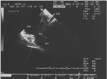
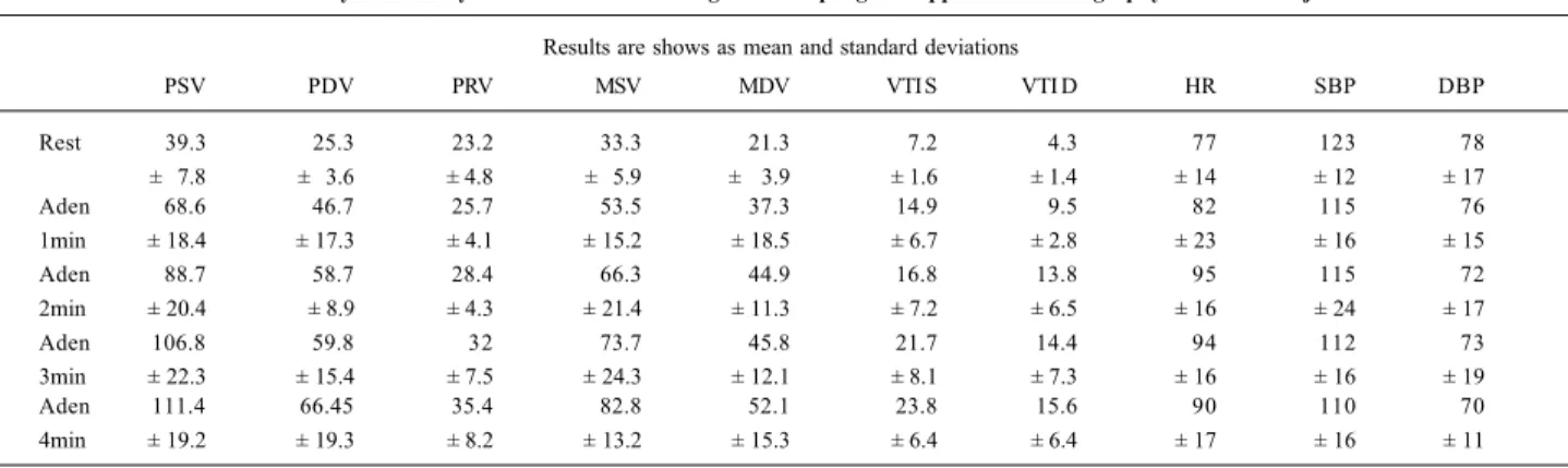
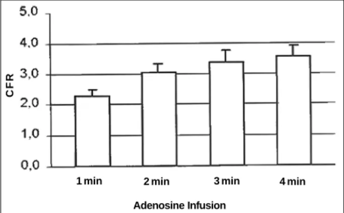
Coronary flow reserve may be assessed by transthoracic Doppler echocardiography, and important functional microcirculation parameters as microcirculation flow velocity, myocardial
A presente investigação, desenvolvida no âmbito do Mestrado em Educação e Formação de Adultos da Faculdade de Psicologia e Ciências de Educação da Universidade do Porto,
