429 https://doi.org/10.1590/0004-282X20170054
ARTICLE
The association of the circle of Willis anomaly
and risk of stroke in patients with carotid
artery disease
A associação de anomalia de Círculo de Willis e o risco de AVC em pacientes
com doença carotídea
Eylem Özaydın Göksu1, Pınar Koç2, Elif Küçükseymen1, Ali Ünal3, Fatma Genç1, Elif Sarıönder Gencer1, Aylin Yaman1
Extracranial atherosclerotic disease accounts for 15–20% of all ischemic strokes1. In patients with carotid
atheroscle-rosis, the risk of stroke is altered by the presence of collat-eral circulation and by the varying demographic properties of the patients2. he incidence of stroke in patients with asymp
-tomatic and symp-tomatic internal carotid artery stenosis is 1, 2–5, 9% and 10% respectively1.
he protective role of collateral circulation depends on
many factors such as anatomic variations, systemic arterial pressure, age and the progression rate of the occlusive
dis-ease. he mainstay of collateral circulation is the circle of
Willis and, in the case of a large artery occlusion, it is readily
available to restore the perfusion. he anatomic properties
of the collateral circulation may determine the subtypes of stroke in patients with occlusive carotid artery disease. In a previous study, up to 50% of anatomical variations in the cere-bral collateral of patients were detected in the circle of Willis2.
In patients with symptomatic carotid artery stenosis,
reduction of blood low due to a stenotic carotid artery is compensated by the increased blood low of the collateral sys -tem3. he circle of Willis is an important collateral pathway,
as it can maintain blood low from the contralateral carotid
and basilar artery to the stenotic carotid artery region. In a study of patients with symptomatic carotid artery disease,
1Antalya Education and Research Hospital, Department of Neurology, Antalya, Turkey;
2Antalya Education and Research Hospital, Department of Radiology, Antalya, Turkey;
3Akdeniz University, Department of Neurology, Antalya, Turkey.
Correspondence: Eylem Özaydın Göksu; Antalya Education and Research Hospital, Department of Neurology, Antalya, Turkey; E-mail: [email protected] Conlict of interest: There is no conlict of interest to declare.
Received 18 July 2016; Received in inal form 20 January 2017; Accepted 14 February 2017.
ABSTRACT
The circle of Willis is an important collateral system that maintains perfusion to the stenotic area from the contralateral carotid and basilar artery to the region of reduced brain perfusion. The aim of the present study was to compare the circle of Willis anomaly in patients with unilateral symptomatic and asymptomatic carotid artery disease. Results: In this retrospective study, we analyzed 175 patients who presented at our outpatient stroke clinic between January, 2013 and June, 2015 with either unilateral symptomatic or asymptomatic carotid artery disease, and who had had CT angiography imaging performed. Demographic properties, carotid artery stenosis and the anomaly of the circle of Willis was recorded. Conclusion: There was no statistically signiicant difference in patients with symptomatic and asymptomatic carotid artery disease in terms of the anomaly of the circle of Willis.
Keywords: circle of Willis; carotid artery diseases; stroke; angiography.
RESUMO
O Círculo de Willis é um importante sistema colateral que mantém a perfusão à área estenótica da carótida contralateral e da artéria basilar para a região de perfusão cerebral reduzida. O objetivo do presente estudo foi comparar a anomalia do Círculo de Willis em pacientes com doença carotídea assintomática e sintomática unilateral. Resultados: Neste estudo retrospectivo, foram analisados 175 pacientes que foram à nossa clínica ambulatorial de AVC, entre janeiro de 2013 e junho de 2015, com doença carotídea assintomática ou sintomática unilateral, e que izeram angiograia por tomograia computadorizada. Propriedades demográicas, estenose da artéria carótida e anomalia do Círculo de Willis foram registradas. Conclusão:Não houve diferença estatisticamente signiicativa em pacientes com doença carotídea sintomática e assintomática em termos de anomalia do Círculo de Willis.
430 Arq Neuropsiquiatr 2017;75(7):429-432
an incomplete circle of Willis was associated with a higher risk of both transient ischemic attack and ischemic stroke4.
However, the results of certain other studies indicated that the prevalence of discontinuity in the circle of Willis in patients with symptomatic carotid disease delivered
con-licting results. Early autopsy studies showed that the preva -lence of absent or hypoplastic segments were associated with increased stroke risk when compared with normal subjects. Conversely, other non-invasive imaging studies showed no
diference in patients experiencing transient ischemic attack
or ischemic stroke due to carotid disease3.
he aim of the present study was to assess the anoma -lies in the circle of Willis with CT angiography in patients with symptomatic and asymptomatic carotid artery disease, to reveal its association with ischemic stroke.
METHODS
A retrospective analysis of patients who presented at our outpatient stroke clinic with carotid artery disease was conducted between January, 2013 and June, 2015.
he study was approved by the ethics committee, and
included 175 patients who had one-sided carotid artery dis-ease detected by multi-detector CT angiography (Philips
Healthcare, 5680 DA Best, he Netherlands). Grading of
the carotid artery stenosis was performed according to
the criteria of the North American Symptomatic Carotid
Endarterectomy Trial5. Patients excluded from the study
were those with > 50% stenosis of the contralateral carotid artery to the symptomatic carotid artery detected by CT angiography, intracranial aneurysm, vascular malforma-tions, and patients with dissection.
he demographic properties of the patients (age, gender),
the risk factors for stroke, the degree of carotid artery steno-sis, whether the patients were symptomatic or
asymptom-atic, and the indings of the circle of Willis were recorded. he symptomatic patients were deined as those patients
who presented with the symptoms of transient ischemic attack or ischemic stroke at the vascular territory of the inter-nal carotid artery and monocular blindness. Asymptomatic
patients were deined as those patients who followed up at
the outpatient stroke clinic without displaying the symptoms of large artery disease.
he circle of Willis of the patients was evaluated as the anterior communicating artery (AcomA), right and left pre
-communicating arteries (ACA A1 segment), posterior com
-municating artery (PcomA) and precom-municating pos
-terior cerebral artery (PCA P1 segment). he hypoplastic
arteries, or the arteries that could not be visualized with CT angiography, were accepted as abnormal.
he presence of A1 segment of asymptomatic ACA,
Acom A, A1 segment of symptomatic ACA, P1 segment of symptomatic PCA, symptomatic PcomA was accepted as a
complete circle of Willis, and thus the absence of any of these arteries was accepted as a circle of Willis anomaly.
CT angiography
A CT angiography examination was performed using the
Philips Brilliance 64 detector CT (Holland) device (Philips Healthcare, 5680 DA Best, he Netherlands). Venous access
was established through the antecubital vein and then an 80 mL non-ionic contrast agent was administered at a rate of 4.5 mL/second, while axial-plane computed tomography images of the carotid and cerebral arteries were obtained using the tracking method. Acquired slices were
trans-ferred to the workstation (Philips IntelliSpace Portal, Philips Healthcare) and multiplane images, maximum intensity pro -jection and volume rendering 3-dimensional images were developed by postprocessing the original slices via the
appro-priate software (AVA). hese images were reviewed with
respect to the vascular plaques and stenosis.
Statistical analysis
he study data were analyzed in SPSS 16.0 for Windows (SPSS Inc., Chicago, Illinois, USA). Demographic and baseline characteristics were summarized as a mean±SD for continu -ous variables and as a percentage of the group for
categori-cal variables. Non-normally-distributed data are presented as medians (inter-quartile range). he normality analysis was performed with the Kolmogorov–Smirnov test. Comparison
of the two groups was performed by the Mann–Whitney
U test for nonparametric comparison and the χ2 test for categorical variables. All the hypotheses were constructed as two-tailed and the α critical value was accepted as 0.05.
RESULTS
During the study period, a total of 175 patients
partici-pated in this study, of whom 121 (69%) were males and 54 (31%) were females. he mean age of the study population
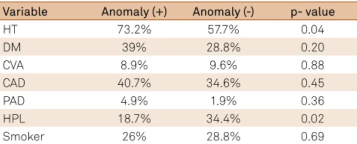
was 66.7 ± 9.2 years. Of the 121 patients, 29.7% did not have an anomaly in the circle of Willis, while 70.3% exhibited an anomaly. Table 1 shows the demographic properties of patients with and without a circle of Willis anomaly.
Table 1. Demographic properties.
Variable Anomaly (+) Anomaly (-) p- value
HT 73.2% 57.7% 0.04
DM 39% 28.8% 0.20
CVA 8.9% 9.6% 0.88
CAD 40.7% 34.6% 0.45
PAD 4.9% 1.9% 0.36
HPL 18.7% 34.4% 0.02
Smoker 26% 28.8% 0.69
431
Göksu EO et al. Circle of Willis and stroke
Hypertension was the most common comorbid condition in groups, followed by coronary artery disease, diabetes mel-litus, smoking, hyperlipidemia, previous stroke and
periph-eral artery disease. he presence of hypertension (p = 0.044) and hyperlipidemia (p = 0.02) was statistically signiicant
between the circle of Willis anomaly positive and negative
patients. he numbers of patients with symptomatic and asymptomatic carotid artery disease were 83 (47.4%) and 92 (52.6%) respectively. An anomaly in the circle of the Willis was detected in 63 (68.7%) patients with asymptomatic and in 60 (72.3%) patients with symptomatic carotid artery dis
-ease (Tables 2 and 3). he overall incidence of anomaly in the circle of Willis was not statistically signiicant between the
patients with symptomatic and asymptomatic carotid artery
disease (p = 0.58). Moreover, there was no statistically signif
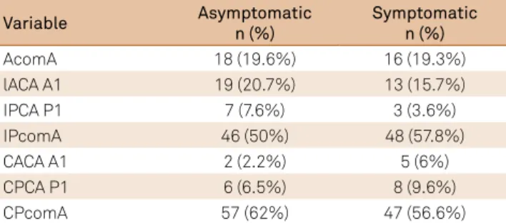
-icant diference in patients with symptomatic and asymp -tomatic carotid artery disease according to the segments of
the circle of Willis (Acoma p = 0.96, IpcomA p = 0.300, IACA A1 p = 0.394, IPCA P1 p = 0.256, KACA A1 p = 0.194, KPCA P1 p = 0.448, KPcomA p = 0.473).
here was a statistically signiicant diference between
symptomatic carotid artery disease and the degree of
steno-sis (p < 0.001) (Table 3). he median carotid artery stenosteno-sis
in symptomatic and asymptomatic carotid artery stenosis patients was 70-90% (25th percentile 70-90% and 75th per-centile preocclusive for the symptomatic and 25th percen-tile 50-69% and 75th percenpercen-tile 70-90% for the asymptomatic
carotid artery stenosis group). he preocclusive and occlusive
carotid artery stenosis comprised 37.4% of the symptomatic and 10.8% of the asymptomatic carotid artery disease patients.
Table 3. Circle of Willis anomaly of individual arteries
between asymptomatic (n = 92) and symptomatic (n = 83) carotid artery disease.
Variable Asymptomatic
n (%)
Symptomatic n (%)
AcomA 18 (19.6%) 16 (19.3%)
lACA A1 19 (20.7%) 13 (15.7%)
IPCA P1 7 (7.6%) 3 (3.6%)
IPcomA 46 (50%) 48 (57.8%)
CACA A1 2 (2.2%) 5 (6%)
CPCA P1 6 (6.5%) 8 (9.6%)
CPcomA 57 (62%) 47 (56.6%)
AcomA: anterior communicating artery; IACA A1: ipsilateral precommunicating arteries A1 segment; CACA A1: contralateral precommunicating arteries A1 segment; IPcomA: ipsilateral posterior communicating artery; CPcomA: contralateral posterior communicating artery; IPCA P1: ipsilateral precommunicating posterior cerebral artery P1 segment; CPCA P1: contralateral posterior communicating artery P1 segment.
DISCUSSION
In this study, no diference could be detected in patients
with symptomatic and asymptomatic carotid artery disease in terms of the circle of Willis anomaly.
he clinical presentation of an occlusive carotid artery
disease is highly variable; while some patients may be diag-nosed incidentally, other patients may present with a dev-astating stroke. In addition, the involvement of intracranial and/or extracranial vessels may accompany this situation.
he presence and efectiveness of the collateral circulation may determine the variability of clinical symptoms. he col
-lateral cerebral circulation may not only provide suicient blood low to the ischemic area, but also perfuse distal to the
occluded artery. In addition, the anatomical features of col-lateral circulation may determine the stroke type in cases of occlusive carotid artery disease. However, it is reported that
up to 50% of signiicant anatomic variations among patients
were seen in the circle of Willis2.
he contribution of the circle of Willis to cerebral hemo -dynamics is not only determined by the stenosis of vertebral and carotid arteries, but also by the presence of the vessels that constitute the circle of Willis.
Previous studies indicated that patients with asymp-tomatic internal carotid artery occlusions have a better pre-served hemodynamic status when compared with symptom-atic patients6,7. However, from this perspective, the role of the
circle of Willis is not clear.
In the study conducted by Waajer et al., instances of the
circle of Willis anomaly were signiicantly higher in patients
with symptomatic carotid disease when compared to the
control patients. In their study, they found signiicantly
more hypoplastic or invisible A1 segments in the symptom-atic carotid artery stenosis group, and the compromised anterior collateral pathway and usually-accompanying posterior pathway occurred more frequently in the symp-tomatic carotid artery stenosis group. In this study, con-trols were retrospectively collected from patients without carotid stenosis. As we had no control group in our study, we could not draw this conclusion3. In an magnetic
reso-nance angiography study by Hartkamp et al., a signiicantly
higher percentage of complete anterior and posterior
circu-lation coniguration was found in the patient group than in
the control subjects8. he prevalence of a circle of the Willis
anomaly was reported in 79.7% of the symptomatic carotid artery disease group9. In our study, the anomaly in the circle
of the Willis was 72.3 % and 68.5% in the symptomatic and asymptomatic carotid artery disease groups respectively. Although we did not recruit a healthy control group for our study, the prevalence we have reported in this study is
sig-niicantly higher than the prevalence of 50% in the normal
population9,10. Although the number of patients with a circle
of the Willis anomaly is more signiicant in the symptom -atic carotid artery disease group, there was no statistically
Table 2. The association between symptomatic and asymptomatic
carotid artery disease and a circle of Willis anomaly.
Variable Anomaly negative n (%)
Anomaly positive
n (%) p-value
Asymptomatic 29 (31.5) 63 (68.5)
432 Arq Neuropsiquiatr 2017;75(7):429-432
signiicant diference among the symptomatic and asymp -tomatic carotid artery disease groups.
In a prior study, it was thought that the hypoplasia of the ACA A1 segment may be a factor that contributes to acute ischemic stroke, due to compromise of the collateral circulation. When compared with healthy individuals, the
anomaly was detected signiicantly more often in acute
ischemic stroke patients11. In a study conducted by Shaban
et al., hypoplastic and absent ACA A1 were detected in 5.9% of acute ischemic stroke patients, although hypoplastic and
absent ACA A1 did not have any efect on vascular distri -bution, side, or the volume of the infarct10. In our study, an
ipsilateral or contralateral presence of the ACA A1 segment
was not signiicantly diferent between the symptomatic and asymptomatic patient population. (IACA A1 p = 0.394, KACA A1 p = 0.194).
In patients with unilateral internal carotid artery
occlu-sion, the presence of collateral low via the posterior com -municating artery in the circle of the Willis was associated with a low prevalence of border zone infarcts12. However,
col-lateral function of the circle of the Willis was not increased in asymptomatic patients with internal carotid artery occlu-sion. In our study, absence of ipsilateral PcomA was not
statistically signiicant diferent between the symptomatic and asymptomatic groups (p = 0.300).
he cerebral collateral circulation maintains cerebral tis -sue perfusion under physiologic and pathologic conditions, such as ischemia. Previous studies revealed the role of col-lateral circulation in patients with carotid artery disease: the absence of collateral circulation in patients with symptom-atic carotid artery disease has a worse prognosis. Evaluation of the cerebral collateral circulation, together with the cere-bral perfusion and the clinical parameters of patients with occlusive carotid artery disease, may give the full spectrum
of the cerebrovascular condition. Nevertheless, the clinical
applications of information regarding collateral circulation in patients with carotid artery disease are still not clear2.
Our study has several limitations, such as its retrospec-tive design, the limited number of patients and the lack of a control group.
In conclusion, we were able to detect a diference between
the symptomatic and asymptomatic internal carotid artery occlusion patient population in terms of a circle of the Willis anomaly. Future prospective studies may, therefore, help to better understand the role of collateral circulation in patients with carotid artery disease.
References
1. Gupta A, Chazen JL, Hartman M, Delgado D, Anumula N, Shao H et al. Cerebrovascular reserve and stroke risk in patients with carotid stenosis or occlusion: a systematic review and meta-analysis. Stroke. 2012;43(11):2884-91. https://doi.org/10.1161/STROKEAHA.112.663716
2. Romero JR, Pikula A, Nguyen TN, Nien YL, Norbash A, Babikian VL. Cerebral collateral circulation in carotid artery disease. Curr Cardiol Rev. 2009;5(4):279-88. https://doi.org/10.2174/157340309789317887
3. Waaijer A, Leeuwen MS, Worp HB, Verhagen HJ, Mali WP, Velthuis BK. Anatomic variations in the circle of Willis in patients with symptomatic carotid artery stenosis assessed with multidetector row CT angiography. Cerebrovasc Dis. 2007;23(4):267-74. https://doi.org/10.1159/000098326
4. Henderson RD, Eliasziw M, Fox AJ, Rothwell PM, Barnett HJ. Angiographically deined collateral circulation and risk of stroke in patients with severe carotid artery stenosis. Stroke. 2000;31(1):128-32. https://doi.org/10.1161/01.STR.31.1.128
5. North American Symptomatic Carotid Endarterectomy Trial (NASCET) investigators. Clinical alert: beneit of carotid endarterectomy for patients with high-grade stenosis of the internal carotid artery. Stroke. 1991;22(6):816-7. https://doi.org/10.1161/01.STR.22.6.816
6. Hoksbergen AW, Legemate DA, Csiba L, Csáti G, Síró P,
Fülesdi B et al. Absent collateral function of the circle of Willis as
risk factor for ischemic stroke. Cerebrovasc Dis. 2003;16(3):191-8. https://doi.org/10.1159/000071115
7. Bozzao A, Floris R, Gaudiello F, Finocchi V, Fantozzi LM, Simonetti G. Hemodynamic modiications in patients with symptomatic unilateral stenosis of the internal carotid artery: evaluation with MR imaging perfusion sequences. AJNR Am J Neuroradiol. 2002;23(8):1342-5.
8. Hartkamp MJ, Der Grond JV, Everdingen KJ, Hillen B, Mali WP. Circle of Willis collateral low investigated by magnetic resonance angiography .Stroke. 1999;30(12)2671-8. https://doi.org/10.1161/01.STR.30.12.2671
9. Kamışlı S, Kamışlı Ö, Teker U, Kablan Y, Saraç K, Özcan C. [Circle of Willis anomalies in stroke patients related with symptomatic carotid artery disease]. Turkish J Cerebrovasc Dis. 2012;18(1):6-9. Turkish https://doi.org/10.5505/tbdhd.2012.36844
10. Shaban A, Albright K, Gouse B, George A, Monlezun D, Boehme A et al. The impact of absent A1 segment on ischemic stroke characteristics and outcomes. J Stroke Cerebrovasc Dis. 2015;24(1):171-5. https://doi.org/10.5505/tbdhd.2012.36844
11. Chuang YM, Liu CY, Pan PJ, Lin CP. Anterior cerebral artery A1 segment hypoplasia may contribute to A1 hypoplasia syndrome. Eur Neurol. 2007;57(4):208-11. https://doi.org/10.1159/000099160