BASIC RESEARCH
1. University of São Paulo - Department of Surgery 2. University of São Paulo - Department of Gastroenterology
3. Southern Cross University - Center of Biological Sciences and the Health 4. University of São Paulo - Department of Biochemistry
Email: [email protected]
Received for publication on January 23, 2007 Accepted for publication on March 21, 2007
OXIDATIVE STRESS IS ENHANCED BY
HYPOTHERMIA IMPOSED ON CERULEIN-INDUCED
PANCREATITIS IN RATS
Andraus, Wellington1, José Jukemura2, Fernando Dutra3, Etelvino José Henriques Bechara4, José E.M. Cunha1, Marcel Cerqueira César Machado1
Andraus W, Jukemura J, Dutra F, Bechara E, Cunha JEM, Leite KRM, César Machado MC. Oxidative stress is enhanced by hypothermia imposed on cerulein-induced pancreatitis in rats. Clinics. 2007;62(4): 483-90.
BACKGROUND: Hypothermia is a frequent event in severe acute pancreatitis (AP) and its real effects on the normal pancreas have not been well demonstrated. Moreover, neither have its effects on the outcome of acute pancreatitis been fully investigated. One hypothesis is that oxidative stress may be implicated in lesions caused or treated by hypothermia.
Aim of the study: To investigate the effect of hypothermia in cerulein-induced acute pancreatitis (CIAP) in rats and the role played by oxidative stress in this process.
METHODS: Male Wistar rats were divided into hypothermic and normothermic groups. Hypothermia was induced with a cold mattress and rectal temperature was kept at 30oC for one hour. Acute pancreatitis was induced with 2 doses of cerulein (20 ìg/kg) administered at a one-hour interval. Serum amylase, pancreas vascular permeability by Evan’s blue method, pancreas wet-to-dry weight ratio and histopathology were analyzed in each group.
RESULTS: When compared with normothermic rats, hypothermic animals, with cerulein-induced acute pancreatitis, showed higher levels of pancreatic vascular permeability (P < 0.05), pancreas wet-to-dry weight ratio (P = 0.03), and histologically verified
edema (P < 0.05), but similar serum amylase levels. The hypothermic group showed a higher oxidized-reduced glutathione ratio
than the normothermic group.
CONCLUSION: Moderate hypothermia produced a greater inflammatory response in established acute pancreatitis induced by cerulein in rats. Moreover, this study suggests that oxidative stress may be one of the mechanisms responsible for the worse outcome in hypothermic rats with cerulein-induced acute pancreatitis.
KEY WORDS: Acute pancreatitis. Cerulein. Hypothermia. Oxidative Stress. Glutathione.
INTRODUCTION
In acute pancreatitis (AP), severity and prognosis are directly related with the intensity of the initial damage.1
In 15% of cases, the pancreatic lesion is severe and the mor-tality varies from 10 to 100%, being greater in those cases where the pancreas is necrotic and infected. Such patients
require intensive care for a longer period as well as multi-ple surgical interventions.2-6 The systemic inflammatory
response and the medical care of these patients may alter their homeostasis by causing modifications in the body tem-perature. These patients may be in shock, with alterations in peripheral vascular resistance, under the effect of seda-tives and curare agents. Frequently, they undergo multiple surgical interventions with prolonged peritoneal exposure and general anesthesia. Therefore, the intravenous infusion of cold fluids, and loss of the protective mechanisms for maintaining their temperature may predispose them to hy-pothermia.7-9
cause deleterious alterations in the pancreas. In the pres-ence of hypothermia, prostacyclin may be inhibited, lead-ing to disruption of pancreatic microcirculation.10
Alterations in the arrangement of acinar cells with enzymatic co-localization and its activation in intracellu-lar space and interstitial tissue have also been demonstrated in an experimental hypothermia study.11 Furthermore, low
body temperature reduces cardiac output, causing hypoten-sion with a further decrease in pancreatic blood flow.12,13
Hypothermia also affects oxygen dissociation from hemoglobin,7 increases blood viscosity, induces alterations
in coagulation, causing thrombosis in pancreatic vessels,14
and may cause ventilatory dysfunction due to lower effi-ciency of the diaphragm.8
In contrast, some authors found a protective effect of mild hypothermia in experimental acute pancreatitis, with less release of amylase, lipase and inflammatory cytokines in the hypothermic groups. They also found less histologi-cal damage with hypothermia. In spite of a decrease in pan-creatic metabolism with low temperatures, the exact mecha-nism of this protective effect is still not clear. 15,16
The ischemia and reperfusion injury has been also treated with hypothermia, and many authors have found an amelio-ration of the lesions in experimental studies.17-21
Several studies have suggested that oxygen radicals play a role in a very early phase of AP,22-28 and that the
eleva-tion of the pancreas oxidative status is related to the se-verity of the disease.26,27 Exacerbated production of free
oxygen radicals is related to the alteration of blood flow and to the ischemia/reperfusion phenomena.29,30
Hypothermia as a modifying factor of oxidative stress in acute pancreatitis has not been fully clarified. Thus, the aims of this study were to investigate the effect of hypo-thermia in cerulein-induced acute pancreatitis (CIAP) in rats and the possible relation of oxidative stress with this condition.
MATERIALS AND METHODS
Animals
Adult male Wistar rats, weighing 220 to 280 g, were in-dividually caged in a temperature controlled room (22-28°C), with a 12 h light-dark cycle. They were fed a standard chow diet (PurinaÒ) and water ad libitum. The experimental
pro-tocol was pre-approved by the Ethics Commission of the Hos-pital das Clínicas, University of São Paulo, Brazil.
Experimental Design
The animals were randomly divided into four groups:
rats without CIAP and normothermia (Control 1; n=10); rats without CIAP and hypothermia (Control 2; n = 10); rats with CIAP and normothermia (Group I; n = 18); and rats with CIAP and hypothermia (Group II; n= 18). In sub-sets of Group I and Group II (n = 8), the ratio of oxidized (GSSG) to reduced (GSH) glutathione was evaluated as in-dicative of oxidative stress.
Induction of Acute Pancreatitis
Acute pancreatitis was induced by an excessive dose of cerulein as described by Lampel et al.31 and modified by
Abdo et al.32 Briefly, two doses of cerulein (20 µg/kg) were administered at a one-hour interval. The first cerulein dose was administered subcutaneously to unanesthetized animals. After 1 h interval, rats were anesthetized by ketamine (Ketalarâ, Parke-Davis; 100 mg/kg, i.p.) and the second dose
of cerulein was injected via the dorsal penis vein. The ani-mals were kept under anesthesia until sacrificed.
Induction of Hypothermia
Hypothermia was induced with the aid of a cold mat-tress during 1 h, immediately after the second dose of cerulein in Group II. Body temperature was kept at 30ºC, as monitored by a rectal thermometer.Immediately follow-ing the hypothermic period of one hour the rats were sub-jected to a median laparotomy and sacrificed by cardiac punction. The pancreas was totally removed and AP was evaluated. The entire pancreas was used to measure GSSG/ GSH ratios.
Evaluation of Pancreatitis
Pancreatitis was evaluated by measuring total water con-tent, vascular permeability (by the Evans’ blue method), and
histopathological examination. Serum amylase levels were
measured as described by Lichtenstein et al.13
The pancreas wet-to-dry weight ratio was calculated as the difference between fresh and dehydrated tissue. This tissue was incubated for 48 hours at 56ºC. Evans’ blue dye was extracted with 3 mL of formamide (4 µg/mg of pan-creatic tissue) for 24 hours. Evans’ blue concentration was determined by spectrophotometry at 620 nm.
Evaluation of GSSG/GSH ratios
Oxidized (GSSG) and reduced (GSH) glutathione were measured by HPLC/electrochemical detection according to Rodriguez-Ariza et al.34 in total pancreas samples obtained
from the subsets of groups I and II.
Preparation of the samples: pancreas samples each weighing 200 mg were homogenized with 1.0 mL of perchloric acid, 1.0 M containing EDTA 2.0 mM; centrifu-gation was performed at 29,000 g for 20 min at 4ºC. The lipid phase was discarded and the supernatant was filtered through a GV Durapore 0.22 mm filter (MillexÒ, Milipore). Samples were kept at -20°C for a maximum period of one hour before being analyzed. The fractions obtained were diluted 10-fold in the mobile phase and 40 mL aliquots were injected into the HPLC apparatus.
Chromatographic method: The separations were achieved in a Shimadzu chromatograph, equipped with a LC-10AD pump, a Nucleosil C-18 (250 x 4.6 mm I.D., 5 mm particle size, 100 Å pore size) column and an ESA Coulochem II electrochemical detector; isocratic elution was performed with 20 mM phosphate buffer pH 2.7. The po-tential settings of the detector were: guard-cell, +0.900 V; detector 1, +0.450 V; detector 2, +0.800 V.
Statistical Analysis
The proximal and distal portions of the pancreas were analyzed separately, except for the glutathione assay, which was performed with the total pancreas. 2-Way ANOVA was used for comparison of the paired groups. The difference was considered statistically significant when P < 0.05.
RESULTS
In the present study the AP was induced by supramaximal stimulation of the endocrine pancreas with cerulein. In this experimental model we evaluated the severity of pancreati-tis by measuring pancreas total water content, vascular per-meability, and histopathological examination.
Our results point out to an enhanced effect of hypother-mia on pancreatic vascular permeability,both in the distal and proximal portions of the pancreas (Figure 1), in CIAP. The control 2 group, submitted to hypothermia, exhibited no significant effect in pancreatic vascular permeability (Figure 1). Serum amylase and the water content were high in groups I and II when compared with controls (p<0.05, Table 1), however, no significant differences were observed in the results of these two groups.
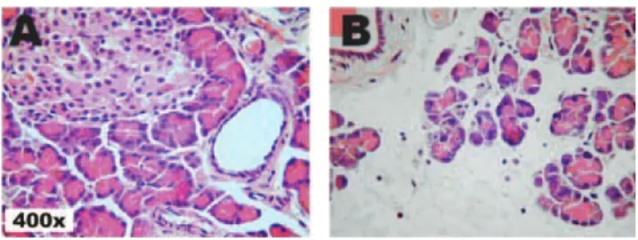
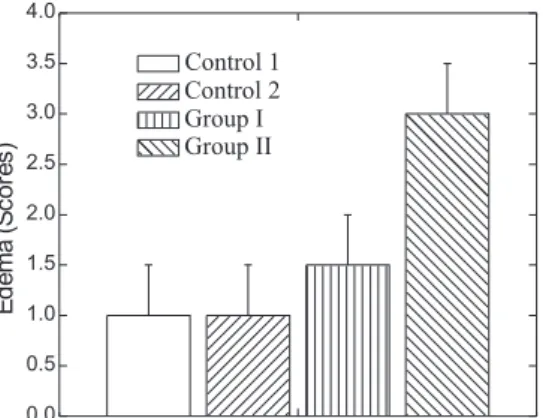
Histological studies have showed minimal edema in group I CIAP rats (figure 2, A) when compared with Group II (Figure 2, B) where a pronounced edema was observed. Accordingly, degrees of tissue damage intensity (scores ac-cording to a standard scale) point to an increased damage in pancreas of CIAP rats submitted to hypothermia ure 3), where differences of acinar cells were observed (Fig-ure 3, compare Group I and Group II).
Figure 1. Comparison of vascular permeability (mg of Evans’ blue stain/g dehydrated tissue) of duodenum proximal and duodenum distal portions of pancreas. (n = 10; P < 0.05)
Table 1: Serum amylase, pancreas water content and GSSG/
GSH ratios in Controls and AP Groups
Control 1 Control 2 Group I Group II Serum amylase 6.2 ± 0.6 5.9 ± 0.9 13.0 ± 2.8 12.7 ± 1.9 (mg/min-1mL-1)a*
Water content 73.8 ± 1.7 72.3 ± 3.3 85.5 ± 5 89.8 ± 1.7 (% total weight)a*
GSSG/GSHb* ———— ———— 195.6 ± 38.3 317.4 ± 20.3 * Data represent the means ± S.D. (n=10 to serum amylase and water content and n=8 to GSSG/GSH)
Diference between groups were tested by 2-way ANOVA with significant levels as indicated:
aP < 0.05 and bP < 0.01
The oxidative stress evaluated through the GSSG/GSH ratio was significantly (P < 0.05) greater in Group II rats in comparison with the Group I and Control 1 rats, show-ing a possible role of reactive species in the observed ef-fects of AP (Table 1).
DISCUSSION
The present study demonstrates that moderate hypother-mia induces an exacerbation of cerulein-induced acute pan-creatitis as shown by enhanced vascular permeability (Fig-ure 1), and changes in the morphology of the AP pancreas (Figure 2 and 3). The worsening of pancreatic lesions by hypothermia may be related to alterations in blood flow and pancreatic ischemia, leading to increased oxidative stress in the organ.
There is no consensus in the literature concerning the real effect of hypothermia in the pancreas or in acute pancreatitis. Recent papers show protection in acute pancreatitis induced by sodium taurocholate and cerulein with moderate hypother-mia.15,16 They reduced the body temperature just after the
in-duction of hypothermia. On the other hand, an experimental animal model in which pancreatic temperature was directly decreased resulted in histological and laboratory pancreatitis11.
This author found co-localization of lysosomal and digestive enzymes, an important step in the initial process in AP.11
Moreover, hypothermia has been reported to be associated with risk for developing AP since the first report by Sano at al.35 in 1940 who found pancreatic perilobular and fat necrosis in pa-tients with accidental hypothermia. Similar findings have been reported by others.36-38
Hypothermia has also been used to add to the preserva-tion of the organs in transplantapreserva-tion, even in the case of pancreas transplantation, on account of decreased organ metabolism.39,40 Several experimental studies showed an
at-tenuation in ischemia and reperfusion injury, in different
organs (liver, lung, neuron), with hypothermia in different degrees.17-21 Hypothermia was also used as a stimulating
factor for the production of heat shock proteins, that showed a prophylactic protection of organ lesions including ischemia and reperfusion injury and acute pancreatitis.41-43
Nevertheless, in clinical practice, some authors have found hyperamylasemia in hypothermic patients.44,45 Moreover, in
an evaluation of 20 patients with accidental hypothermia admitted to ICU for causes other than those related to acute pancreatitis, Lichtenstein et al.13 found hyperamylasemia in 8/9 patients and necrohemorrhagic pancreatitis in 1/2 autopsies.
Heat shock proteins (HSPs) are a group of proteins that repair damaged proteins and are also involved in synthe-sis, degradation, folding, transport, and translocation of pro-teins. Induction of HSPs enhances cells ability to survive a further aggression.41-43 At the start of the inflammation
proc-ess hypothermia showed a protective action in acute pan-creatitis by production of heat shock proteins, in con-trast when acute pancreatitis is already well established, hypothermia may play a deleterious role in pancreatic tis-sue inflammation and worsen its outcome. We used mod-erate hypothermia (30°C), as in others models of experi-mental pancreatitis15,16 such as can be present in clinical
practice, one hour after the first injection of cerulein for induction of acute pancreatitis, and that may explain the worse outcome in hypothermic group.
It’s known that glutathione (table 1) and other sulfhydryl components19 are depleted, while the lipid peroxidation is
higher in the pancreatic tissue during the development of acute pancreatitis,46-48 so the oxidative stress may play an
important role in lesion mechanism. Hypothermia can lead to pancreatic ischemia secondary to alterations in the car-diac output and ventilation, as well as in the pancreatic mi-crocirculation.7,8,12,14 During the ischemic period, xanthine
dehydrogenase is converted to xanthine oxidase, and upon reperfusion xanthine reacts with oxygen yielding reactive oxyradicals.49 It is known that, during the ischemic
proc-ess, levels of antioxidants such as glutathione and superoxide dismutase are low, rendering the tissue prone to free radical lesions.50,51 The results of the present study
demonstrate an increase in the oxidized/reduced glutath-ione ratio in the hypothermic group, suggesting increased oxidative stress determined by hypothermia in the cerulein-induced acute pancreatitis model.
Oxidative stress is one of the main processes during acute pancreatitis28 and the higher concentrations of
reac-tive oxygen species in the hypothermic group suggest that it plays a role in the pathophysiology of the damage proc-ess in induced AP. How hypothermia increases the oxidative stress is still an open question. One might speculate that it
damages the microcirculation, increasing pancreatic ischemia or, alternatively, that it acts directly on the pan-creatic tissue, leading ultimately to the peroxidation of membranes and proteins.
In conclusion, moderate hypothermia aggravates estab-lished cerulein-induced acute pancreatitis inflammation in rats. Perhaps the most important molecular mechanisms
underly-ing this process are those promotunderly-ing a redox imbalance, which can be circumvented by the administration of antioxidant agents. Oxidative stress, like other processes that modify the outcome in acute pancreatitis, should be further investigated for it may be clinically relevant. Based on our results, special attention in controlling the patient’s body temperature should be considered in the management of acute pancreatitis.
RESUMO
Andraus W, Jukemura J, Dutra F, Bechara E, Cunha J, Leite KRM, César Machado MC. Aumento do estresse oxidativo após hipotermia em ratos com pancreatite induzida por ceruleína. Clinics. 2007;62(4):482-90.
BACKGROUND: Hipotermia é um evento freqüente em
episódios de pancreatite aguda, contudo seu efeito real sobre pâncreas normal ainda não esta bem demonstrado. Além do mais, o efeito da hipotermia no decorrer da pancreatite aguda também não está completamente esclarecido. Uma das hipóteses sobre as causas das lesões causadas ou tratadas por hipotermia aventa a implicação de estresse oxidativo.
OBJETIVOS: Investigar o efeito da hipotermia em ratos
com pancreatite aguda induzida por ceruleína e o papel do estresse oxidativo neste processo.
MÉTODOS: Ratos Wistar machos foram divididos em
grupos hipotérmicos e normotérmicos. Hipotermia foi induzida com uma bolsa gelada de forma que a temperatura
retal permanecesse em 30oC por uma hora. Pancreatite
RESULTADOS: Ratos hipotérmicos, com pancreatite aguda induzida por ceruleína, apresentaram maiores níveis de permeabilidade vascular no pâncreas (P < 0.05), razão
peso seco/peso úmido do pâncreas (P = 0.03), e edema
histológico (P < 0.05), mas os níveis de amilase sérica permaneceram iguais aos níveis apresentados pelos ratos normotérmicos. O grupo hipotérmico apresentou maior relação glutationa oxidada/glutationa reduzida em relação ao grupo normotérmico.
CONCLUSÃO: Hipotermia moderada produziu uma maior
resposta inflamatória em ratos com pancreatite aguda estabelecida, induzida por ceruleína, sugerindo que este efeito pode estar ligado a um maior índice de estresse oxidativo em ratos com pancreatite aguda.
UNITERMOS: Pancreatite aguda. Ceruleína. Hipotermia.
Estresse Oxidativo. Glutationa.
REFERENCES
1. Ranson JHC. Stratification of severity for acute pancreatitis. In: Bradley III EL. Acute pancreatitis: diagnosis and therapy. New York: Raven Press; 1994. pp. 13-20.
2. Ranson JHC, Rifkind KM, Roses DF. Prognostic sign and the role of operative management in acute pancreatitis. Surg. Gynecol. Obst. 1974;39:69-81.
3. Machado MCC, Bacchella T, Monteiro da Cunha JE, Jukemura J, Penteado S, Giovanoli ACV, et al. The course of pancreatic necrosis. Influence of infection factors. Rev. Hosp. Clin. Fac. Med. S. Paulo 1985; 40:120-4.
4. Jukemura J, Machado MC, Penteado S, Monteiro da Cunha JE, Pinotti HW. Prognostic value of the sites of pancreatic necrosis determined by computed tomography of the abdomen. Rev. Hosp. Clin. Fac. Med. S. Paulo 1995; 50:147-53.
5. Cunha JEM, Machado MCC, Penteado S, Jukemura J, Bacchella T, Pinotti HW. Pancreatic necrosis in Brazil. In: Bradley III, EL. Acute pancreatitis: diagnosis and therapy. New York: Raven Press; 1994. pp. 121-125.
6. Yokoi H, Naganuma T, Higashiguchi T, Isaji S, Kawarada Y. Prospective study of a protocol for selection of treatment of acute pancreatitis based on scoring of severity. Digestion 1999; 60:14-8.
7. Arndt K. Inadvertent hypothermia in the operating room. AORN J. 1999; 70:204-222.
8. Jones T, Roy RC. Should patients be normothermic in the immediate postoperative period? Ann.Thorac. Surg. 1999; 68:1454-5.
9. Gamal N, Kassabany N, Frank SM, Amar R, Khabar HA, El-Rahmany HK, et al. Age-related thermoregulatory differences in a warm operating room environment. Anesth. Analg. 2000; 90:694-8. 10. Jeremy JY, Mikhailidis DP, Hutton RA, Dandona P. The effect of cooling on in vitro vascular prostacyclin and platelet thromboxane A2 synthesis: relevance to cold-induced pathology Microcirc. Endothelium Lymphatics. 1988; 4:3-20.
12. Eichelter P, Schenk WG. The influence of hypothermia on pancreatic secretion and blood flow. Arch. Surg. 1968; 96:883-6.
13. Lichtenstein A, Onuchic LF, Rocha AS. Accidental hypothermia: glycemic, hematologic and blood amylase changes. Rev. Hosp. Clin. Fac. Med. S. Paulo 1990; 45:173-7.
14. Peng RY, Bongard FS. Hypothermia in trauma patients. J. Am. Coll. Surg. 1999; 188:685-96.
15. Matsuoka K, Ueno T, Morita K, Kawano H, Yamaguchi K, Maekawa T, et al. Effects of moderate hypothermia on proinflammatory cytokine production in a rat model of caerulein-induced pancreatitis. Pancreas. 2003; 26(1):12-7.
16. Wang X, Jiang W, Zhao G, Du D, Zhou M, Hang Y, et al. Mild hypothermia protects against sodium taurocholate (NaTc)-induced acute pancreatitis in rats with adverse effects on serum cytokines. Pancreas. 2005; 30(4):80-6.
17. Gambert S, Bes-Houtmann S, Vandroux D, Tissier C, Vergely-Vandriesse C, Rochette L, et al. Deep hypothermia during ischemia improves functional recovery and reduces free-radical generation in isolated reperfused rat heart. J Heart Lung Transplant. 2004; 23(4):487-91. 18. Shoji T, Omasa M, Nakamura T, Yoshimura T, Yoshida H, Ikeyama K,
et al. Mild hypothermia ameliorates lung ischemia reperfusion injury in an ex vivo rat lung model. Eur Surg Res. 2005; 37(6):348-53. 19. Wang CY, Ni Y, Liu Y, Huang ZH, Zhang MJ, Zhan YQ, et al. Mild
hypothermia protects liver against ischemia and reperfusion injury. World J Gastroenterol. 2005; 11(19):3005-7.
20. Niemann CU, Choi S, Behrends M, Hirose R, Noh J, Coatney JL, et al. Mild hypothermia protects obese rats from fulminant hepatic necrosis induced by ischemia-reperfusion. Surgery. 2006; 140(3):404-12. 21. Kawamura N, Schmeichel AM, Wang Y, Schmelzer JD, Low PA.
Multiple effects of hypothermia on inflammatory response following ischemia-reperfusion injury in experimental ischemic neuropathy. Exp Neurol. 2006; 202(2):487-96.
22. Scott P, Bruce C, Schofield D, Shiel N, Braganza JM, Mccloy RF. Vitamin C status in patients with acute pancreatitis. Br. J. Surg. 1993; 80:750-4. 23. Fu K, Tomita T, Sarras Jr MP, De Lisle RC, Andrews GK. Metallothionein protects against cerulein-induced acute pancreatitis: analysis using transgenic mice. Pancreas 1998; 17:238-46.
24. Abdo EE, Machado MCC, Coelho AMM, Sampietre SN, Leite KRM, Molan NAT, et al. Antioxidative effect of N2-mercaptopropionylglycine (N2 MPG) in experimental acute pancreatitis. Rev. Hosp. Clin. Fac. Med. S. Paulo 1998; 53:169-73.
25. Reinheckel T, Prause J, Nedelev B, Augustin W, Schulz H, Lippert H, et al. Oxidative stress affects pancreatic proteins during the early pathogenesis of rat caerulein pancreatitis. Digestion 1999; 60:56-62. 26. Demols A, Van Laethem JL, Quertinmont E, Legros F, Louis H, Le Moine
O, et al. N-acetylcysteine decreases severity of acute pancreatitis in mice. Pancreas 2000; 20:161-9.
27. Abu-Zidan FM, Bonham MJD, Windsor JA. Severity of acute pancreatitis: a multivariate analysis of oxidative stress markers and modified Glasgow criteria. Br. J. Surg. 2000; 87:1019-23.
28. Curran FJM, Sattar N, Talwar D, Baxter JN, Imrie CW. Relationship of carotenoid and vitamins A and E with the acute inflammatory response in acute pancreatitis. Br. J. Surg. 2000; 87:301-5.
29. Carden DL, Granger DN. Pathophysiology of ischaemia-reperfusion injury. J. Patol. 2000; 190:255-66.
30. Peralta C, Bulbena O, Xaus C, Prats N, Cutrin JC, Poli G, et al. Ischemic preconditioning: a defense mechanism against the reactive oxygen species generated after hepatic ischemia reperfusion. Transplantation 2002; 73:1203-11.
31. Lampel M, Kern HF. Acute interstitial pancreatitis in the rat induced by excessive doses of a pancreatic secretagogue. Virchows Arch A Pathol Anat Histol. 1977; 373:97-117.
32. Abdo EE, Coelho AMM, Montagnini AL, Kubrusly MS, Leite KRM, Sampietre SN, et al. Simplified model of induction of experimental acute pancreatitis with a supra-maximal dose of cerulean. Rev. Hosp. Clin. Fac. Med. S. Paulo 1994; 49:204-7.
33. Schmidt J, Rattner DW, Lewandrowski K, Compton CC, Mandavilli U, Knoefel WT, et al. A better model of acute pancreatitis for evaluation therapy. Ann. Surg. 1992; 215:44-56.
34. Rodriguez-Ariza A, Toribio F, Lopez-Barea J Rapid determination of glutathione status in fish liver using high-performance liquid chromatography and electrochemical detection. J. Chromatogr. B. Biomed. Appl. 1994; 7:311-8.
35. Sano ME, Smith LW. A critical histopathologic study. J. Lab. Clin. Med. 1940; 26:443-56.
36. Read AE, Emslie-Smith D, Gough KR, Holmes R. Pancreatitis and accidental hypothermia. Lancet 1961; 2:1219-21.
37. Duguid H, Simpson RG, Stowers JM. Accidental hypothermia. Lancet 1961; 2:1213-19.
38. Foulis AK. Morphological study of the relation between accidental hypothermia and acute pancreatitis. J. Clin. Pathol. 1982; 35:1244-48. 39. Rivard AL, Gallegos RP, Bianco RW, Liao K. The basic science aspect of donor heart preservation: a review. J Extra Corpor Technol. 2004; 36(3):269-74.
40. Tsujimura T, Kuroda Y, Kin T, Avila JG, Rajotte RV, Korbutt GS, et al. Human islet transplantation from pancreases with prolonged cold ischemia using additional preservation by the two-layer (UW solution/ perfluorochemical)cold-storage method. Transplantation. 2002; 74(12):1687-91.
41. Grise KR, McFadden DW. Peptide YY. Improves local and systemic parameters and prevents death in lethal necrotizing pancreatitis. Pancreas. 2002; 24(1):90-5.
42. Rakonczay Z Jr, Takacs T, Ivanyi B, Mandi Y, Papai G, Boros I, et al. The effects of hypo- and hyperthermic pretreatment on sodium taurocholate-induced acute pancreatitis in rats. Pancreas. 2002; 24(1):83-9.43. akonczay Z Jr, Mandi Y, Kaszaki J, Ivanyi B, Boros I, Lonovics J, et al. Induction of HSP72 by sodium arsenite fails to protect against cholecystokinin-octapeptide-induced acute pancreatitis in rats. Dig Dis Sci. 2002; 47(7):1594-603.
44. Maclean D, Murison J, Griffiths PD. Acute pancreatitis and diabetic ketoacidosis in accidental hypothermic myxoedema. Br. Med. J. 1973; 4:757-61.
Appendix 1: Histopathologic Scoring Criteria. Edema
0 Absent
0.5 Focal expansion of interlobar septae 1 Diffuse expansion of interlobar septae
1.5 Same as 1 + focal expansion of interlobular septae 2 Same as 1 + diffuse expansion of interlobular septae 2.5 Same as 2 + focal expansion of interacinar septae 3 Same as 2 + diffuse expansion of interacinar septae 3.5 Same as 3 + focal expansion of intercellular spaces 4. Same as 3 + diffuse expansion of intercellular spaces Acinar necrosis
0 absent
0.5 Focal occurrence of 1-4 necrotic cells/HPF 1 Diffuse occurrence of 1-4 necrotic cells/HPF
1.5 Same as 1 + focal occurrence of 5-10 necrotic cells/HPF 2 Diffuse occurrence of 5-10 necrotic cells/HPF
2.5 Same as 2 + focal occurrence of 11-16 necrotic cells/HPF 3 Diffuse occurrence of 1 1-16 necrotic cells/HPF
(foci of confluent necrosis)
3.5 Same as 3 + focal occurrence of > 16 necrotic cells/HPF 4 > 16 necrotic cells/HPF (Extensive confluent necrosis) 46. Schoenberg MH, Buchler M, Beger HG. Oxygen radicals in experimental
acute pancreatitis. Hepatogastroenterology. 1994; 41:313-9.
47. Dabrowski A, Gabryelewicz A. Nitric oxide contributes to multiorgan oxidative stress in acute experimental pancreatitis. Scand. J. Gastroenterol. 1994; 29:943-8.
48. Sweiry JH, Mann GE. Role of oxidative stress in the pathogenesis of acute pancreatitis. Scand. J. Gastroenterol. 1996; 31:10-5.
49. Stirpe F, Della Corte E. The regulation of rat liver xanthine oxidase. Conversion in vitro of the enzyme activity from dehydrogenase (type D) to oxidase (type O). J. Biol. Chem. 1969; 244:3855-63.
50. Vreugdenhil PK, Belzer FO, Southard JH. Effect of cold store on tissue and cellular glutathione. Cryobiology 1991; 28:143-9.
51. Franssen C, Defraigne JO, Detry O, Pincemail J, Deby C, Lamy M. Antioxidant defense and free radical production in a rabbit model of kidney ischemia-reperfusion. Transplant. Proc. 1995; 27:2880-3.
Hemorrhage and fat necrotis
0 Absent
0.5 1 focus
1 2 foci
1.5 3 foci
2 4 foci
2.5 5 foci
3 6 foci
3.5 7 foci 4 . 8 foci
Inflammation and perivascular infiltrate