CLINICAL SCIENCE
Comparison between proximal row carpectomy and
four-corner fusion for treating osteoarthrosis
following carpal trauma: a prospective
randomized study
Edgard Novaes Franc¸a Bisneto, Maura Cristina Freitas, Emygdio Jose´ Leomil de Paula, Rames Mattar Jr., Arnaldo Valdir Zumiotti
Department of Orthopedics and Traumatology, Faculdade de Medicina da Universidade de Sa˜o Paulo, Sa˜o Paulo, Brazil.
OBJECTIVE:To compare the functional results of carpectomy and four-corner fusion surgical procedures for treating osteoarthrosis following carpal trauma.
METHODS:In this prospective randomized study, 20 patients underwent proximal row carpectomy or four-corner fusion to treat wrist arthritis and their functional results were compared. The midcarpal joint was free of lesions in all patients.
RESULTS: Both proximal row carpectomy and four-corner fusion reduced the pain. All patients had a decreased range of motion after surgery. The differences between groups were not statistically significant.
CONCLUSIONS: Functional results of the two procedures were similar as both reduced pain in patients with scapholunate advanced collapse/scaphoid non-union advanced collapse (SLAC/SNAC) wrist without degenerative changes in the midcarpal joint.
KEYWORDS: Arthritis; Degenerative; Wrist injuries; Carpal bones; Arthrodesis; Wrist joint.
Franc¸a Bisneto EN, Freitas MC, Emygdio P, Mattar Jr. R, Zumiotti AV. Comparison between proximal row carpectomy and four-corner fusion for treating osteoarthrosis following carpal trauma: a prospective randomized study. Clinics. 2011;66(1):51-55.
Received for publication onSeptember 30, 2010;First review completed onSeptember 30, 2010;Acceptedfor publication onOctober 13, 2010
E-mail: [email protected]
INTRODUCTION
Wrist osteoarthrosis cases are part of the hand surgeons’ daily practice. The etiology of osteoarthrosis may be post-traumatic, degenerative, infectious or rheumatological. Symp-toms are progressive pain and decreased wrist function.
When the etiology is non-traumatic, there is no pattern to the way in which the joints are impaired, since the consequences of this condition have various effects on radial and midcarpal joints. Post-traumatic osteoarthrosis, however, shows a predictable standardized progression, and thus treatment depends on the stage of evolution of this condition. The etiology of post-traumatic wrist osteoarthro-sis is usually secondary to ligament injuries or carpal fractures.1-5In post-traumatic wrist arthrosis cases, 95% are located around the scaphoid, and in 55% of the patients with arthrosis the most common pattern is called scapholunate advanced collapse (SLAC) of the wrist, which results from ligament ruptures. The evolution of this type of arthrosis is
divided into the following three stages:2,6 (1) arthrosis between the styloid process of the radius and the scaphoid; (2) arthrosis in the radial scaphoid fossa, (3) arthrosis between the capitate and the lunate.
The osteoarthrosis can also result from pseudoarthrosis of the scaphoid (scaphoid non-union advanced collapse (SNAC)).7 In this degenerative pattern, pseudoarthrosis of the scaphoid acts biomechanically in the same way as injuries to the scapholunate interosseous ligament; as observed in cases of SLAC wrist, butradial scaphoid fossa was preserved when pseudoarthrosis of the scaphoid occurred.8 The SNAC stages are: (1) arthrosis between the styloid process of the radius and the scaphoid; (2) arthrosis between the scaphoid and the capitate and (3) arthrosis between the capitate and the lunate.
Several surgical approaches for the treatment of post-traumatic osteoarthrosis of the carpal bones have been reported—namely, proximal row carpectomy, four-corner fusion, selective denervation of the wrist, partial styloidect-omy of the styloid process of the radius, fusion of the scaphoid–trapezium–trapezoid joint, fusion of the sca-phoid–capitate joint, ‘‘atlas’’ fusion (lunate–capitate) and the complete fusion of the wrist.4,5,8-22
This study aimed to compare the functional results of proximal row carpectomy and four-corner fusion for the
treatment of post-traumatic wrist osteoarthrosis with no effect on the midcarpal joint.
PATIENTS AND METHOD
Twenty-three patients were selected based on the inclu-sion and non-incluinclu-sion criteria for this study (see below). All patients presented wrist osteoarthrosis, with a diagnosis of SLAC or SNAC, but without involvement of the midcarpal joint. All patients were operated on by the same surgeon at the same hospital, between August 2004 and September 2007. All patients included in this study presented had a range of wrist motion. Three patients did not attend two postoperative assessments and were excluded. Of the 20 patients included, 16 had SNAC and 4 had SLAC.
The evolutional stages of SNAC/SLAC were evaluated by radiography and computed tomography.
The inclusion criteria were the presence of wrist osteoar-throsis grade I or II SLAC/SNAC and acceptance of the statement of free and informed consent. The exclusion criteria were presence of gross deformities in other limbs; rheumatological conditions; infections; involvement of the midcarpal joints, diseased condition in the contralateral wrist and previous fractures of the distal extremity of the radius or the carpal bones. The only other exclusion criterion was patients’ non-attendance at two postoperative functional assessments.
The subjective analysis was based on the disabilities of the arm, shoulder or the hand (DASH) questionnaire and an analog pain scale.23,24
In the objective evaluation, the following were observed: wrist goniometry, grip force (Jamar), grip force of pulp– pulp, lateral (key) and three-finger (tripod) pinches, discrimination between two points on the pulp of the second and fifth fingers, and on the dorsum of the first web, measurements of the hand and wrist volumes and the Jebsen–Taylor functional test.25
Patients underwent preoperative and postoperative assessments and the latter were done 3, 6 and 12 months after the surgical procedure. All assessments were made by the same occupational therapist. Randomization was carried
out by a draw on the day of the surgery. All operations were done by the same surgeon at the same institution. Patients who underwent proximal row carpectomy and those who had four-corner fusion started their rehabilitation with kinesiotherapy and physical means 3 weeks and 2 months after the operation, respectively.
Statistical Analysis
Comparison of the evolutional results within the same surgical group, between the preoperative and postoperative assessment were performed by the Wilcoxon test. Data on the operated and contralateral wrist were also compared using the Wilcoxon test. A non-parametric Mann–Whitney test was used to analyze the results between the two surgical groups. The significance level of 5% (p#0.05) was adopted for this study.
RESULTS
The mean ages of the fusion and the carpectomy groups were 42¡10.6 and 43.4¡10.1 years, respectively. There was no difference in the handedness of the affected limbs between the two groups.
The data were divided into a direct evolutional analysis and comparative analysis within the same group and between the groups.
Direct Evolutional Analysis
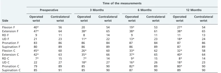
Data on direct evolutional analysis—direct comparison of evolution of the parameters over time—within each group are shown in Table 1. After 12 months despite the radial deviation values, all the wrist parameters decreased in both procedures. Values related to pronation and supination were almost the same as before surgery.
Grip force was evaluated and the fusion group achieved the same values at 12 months as before surgery, this did not occur in the carpectomy group (Table 2).
For pinch force evaluation, all the patients, in both groups, recovered or improved their preoperative values. Discrimination between two point values showed no differences in this study.
Table 1 -Goniometry evaluation.
Time of the measurements
Preoperative 3 Months 6 Months 12 Months
Side
Operated wrist
Contralateral wrist
Operated wrist
Contralateral wrist
Operated wrist
Contralateral wrist
Operated wrist
Contralateral wrist
Flexion F 40* 56 20 54 19* 53 27* 54
Extension F 47* 64 38* 65 38* 61 38* 65
RD F 9 11 8 14 9 11 11 13
UD F 21 23 11* 22 13* 23 18* 24
Pronation F 83 86 86 84 87 84 87 86
Supination F 80 89 86 89 86 89 87 89
Flexion C 45* 60 26* 60 33* 62 32* 58
Extension C 42* 63 35* 66 37* 65 40* 64
RD C 7* 15 7* 14 9* 15 8* 14
UD C 22 27 18* 27 16* 26 18* 23
Pronation C 79 89 85 90 82* 89 80* 90
Supination C 85 91 85 90 87 90 89 90
Values are shown in degrees. *p
#0.05 in the Wilcoxon test.
The operated wrists showed volumetry values that were smaller than those of contralateral wrists by the end of the evaluations, after 12 months of surgery, in both groups.
Both groups showed reduced pain on an analog pain scale. The preoperative values in the fusion and carpectomy groups were 7.6 and 8.2, respectively. After 12 months of surgery the values were 5.1 and 4.8.
DASH values corresponded to the analog pain scale values, which meant that all patients showed an improve-ment in their daily living and work activities. Preoperative DASH values in the fusion and carpectomy groups were 42.7 and 52.4, respectively, and postoperative values were 29.9 and 37.7, respectively.
Jebsen-Taylor test values showed that hand abilities had improved in both groups, from 57 to 40.9 seconds in the fusion group and from 74.1 to 65.2 seconds in the carpectomy group.
Comparative Analysis
Comparative analysis is a proportional comparison of the data between the groups as follows:
Horizontal analysis: evaluation of the data between the operated and the contralateral wrist 12 months after the operation (Table 3).
Vertical analysis: evaluation of the data on the
operated wrist 12 months after the operation in relation to the preoperative ipsilateral data (Table 4).
In the horizontal analysis, the data were compared proportionally and values for the contralateral limb were
used as the baseline. In the vertical analysis, the preopera-tive measurements were used as the baseline values
Complications
In the four-corner fusion group, 1 case of reflex sympathetic dystrophy was seen.
In the proximal row carpectomy group, 3 cases of synovitis with significant wrist edema and 2 cases of reflex sympathetic dystrophy were seen.
All observed complications occurred no later than the second month after the operation and were treated clinically. None of the patients presented any breakage of the synthesis material or deep infection, or any other condition that might have required further surgical inter-vention.
DISCUSSION
The mean age (40–52 years) of patients in this study is simi-lar to that in other studies.8,16-22A period of 5–10 years between the trauma and the start of signs and symptoms of arthrosis
Table 2 -Grip force evaluation.
Time of the measurements
Preoperative 3 Months 6 Months 12 Months
Side
Operated wrist
Contralateral wrist
Operated wrist
Contralateral wrist
Operated wrist
Contralateral wrist
Operated wrist
Contralateral wrist
Fusion 25.7* 35.6 15.4* 35.9 17.7* 30.7 25.7* 35.4
Carpectomy 18.6* 38.3 12.8* 35.9 13.9* 37.6 17.6* 37.6
Values are shown in kgf. *p
#0.05 in the Wilcoxon test.
Table 3 -Horizontal analysis. Proportional evaluation between the operated and the contralateral wrist, 12 months after surgery.
Percentage in relation to the contralateral wrist
Fusion Carpectomy
Flexion 50 55
Extension 58 63
RD 85 57
UD 75 78
Pronation 101 89
Supination 98 99
Key pinch 91 67
Pulp–pulp pinch 69 59
Tripod pinch 73 65
Jamar 73 47
Jebsen–Taylor 98 99
RD = radial deviation; UD = ulnar deviation.
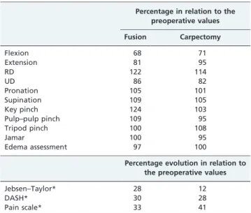
Table 4 -Vertical analysis. Proportional evaluation on the operated wrist between the preoperative assessment and the assessment 12 months after the operation.
Percentage in relation to the preoperative values
Fusion Carpectomy
Flexion 68 71
Extension 81 95
RD 122 114
UD 86 82
Pronation 105 101
Supination 109 105
Key pinch 124 103
Pulp–pulp pinch 109 95
Tripod pinch 100 108
Jamar 100 95
Edema assessment 97 100
Percentage evolution in relation to the preoperative values
Jebsen–Taylor* 28 12
DASH* 30 28
Pain scale* 33 41
*These parameters are represented by inversely proportional values that were obtained by means of the 1002x function, in which x was the directly proportional value).
All the data in this table had a p-value$0.05 according to the Mann– Whitney test.
implies a condition that occurs in patients who are around 35 year old.7
Rigor in applying the non-inclusion criteria was funda-mental for obtaining a homogeneous group of patients. The use of computed tomography to evaluate the joints, particularly the midcarpal joint, contributed greatly towards homogenizing the study groups.
Twelve months after the operation, the overall range of wrist motion was smaller in relation to the preoperative values in both procedures (p#0.05). The arc of flexion– extension was, on average, 25% and 17% less in the cases of four-corner fusion and in proximal row carpectomy, respectively. The radial–ulnar deviation, on average, was, 0.3% and 10% less in fusion patients and carpectomy patients, respectively.
The radial deviation in cases of four-corner fusion was the only goniometry parameter that had improved 12 months after the operation. The postoperative pronation–supination movements were not statistically different in either group. Decreases in the overall range of motion in relation to preoperative values in proximal row carpectomy5,26-28and four-corner fusion5,29 has been reported. In our study,
horizontal analysis within each group showed proportional range-of-motion results favoring proximal row carpectomy, except for radial deviation, for which fusion was favored (p
#0.05). The results presented here are consistent with other published results.8,16-19,22 However, others found that the arc of flexion–extension in cases of four-corner fusion was greater than the arc in cases of proximal row carpectomy.20 Vertical analysis of the four-corner fusion group showed that the grip force recovered to preoperative values (p#0.05). In the carpectomy group, 95% of the preoperative grip force was recovered (p#0.05).
A loss of carpal height in the proximal row carpectomy procedure owing to relative stretching of the flexor and extensor tendons has been reported.2 The improvement in grip force after the operation can be credited to pain relief.27,28 Horizontal data from a series of proximal row carpectomy cases with longer follow-up duration have shown that between 60% and 80% of the grip force in the contralateral wrist is achieved.4,26-28In contrast, only 47% of the grip force was achieved in this study. Patients undergoing proximal row carpectomy may take up to 1 year to achieve complete rehabilitation.28 Horizontal analysis of the grip force in
patients undergoing four-corner fusion provided the best defense of this technique, to the detriment of proximal row carpectomy, because of the preservation of grip force in relation to the non-operated side.2,16
A 10% increase in relative grip force may occur over a 3-year period between postoperative evaluations in cases of four-corner fusion.30Grip force values of between 70% and 87%, in relation to the contralateral wrist have been reported.8,17-20,30 In this study, the grip force was 73% of
the contralateral wrist in patients who underwent four-corner fusion and 47% in patients undergoing proximal row carpectomy compared with the non-operated side (p#0.05). These comparative values between groups were not statistically significant.
Evaluation of the grip force was done by finger pinches (key pinch, pulp–pulp pinch and tripod pinch), together with a test to discriminate between two points. This was done to determine whether surgical procedures might have caused lesions in the peripheral nerves.23 The results
obtained proved that both surgical procedures were safe.
The universal analog pain scale was applied to the study patients. In the four-corner fusion group, there was a 33% reduction in pain, compared with preoperative values (p$0.05). In the proximal row carpectomy group, there was a 41% reduction in pain (p#0.05). The comparative values between groups were not statistically significant.
In the vertical DASH analysis, the patients who under-went four-corner fusion or proximal row carpectomy achieved 30% and 28% evolution, respectively, compared with preoperative values (p$0.05). In the vertical analysis of the Jebsen–Taylor test,25the patients who underwent four-corner fusion achieved, on average, 28% evolution over the task duration, whereas the value was 12% in patients who underwent proximal row carpectomy (p$0.05). In the horizontal analysis, almost all patients recovered their function compared with the contralateral side; 98% in four-corner fusion group and 99% in proximal row carpectomy group. Comparison of the results between groups was not statistically significant.
CONCLUSION
Both proximal row carpectomy and four-corner fusion surgical procedures provided similar functional results for treating degenerative conditions of SLAC/SNAC without the impairment of the midcarpal joint. Indication for the surgical technique should be based on several parameters, such as patient’s age, duration of immobilization, risk of pseudoarthrosis, possibility of breakage of the synthetic material, infection, duration of rehabilitation and the experience of the surgical team.
REFERENCES
1. Watson HK, Ryu J, Akelman E. Limited triscaphoid intercarpal arthrodesis for rotatory subluxation of the scaphoid. J Bone Joint Surg Am. 1986;68:345-9.
2. Watson HK, Ryu J. Evolution of the arthritis of the wrist. Clin Orthop Relat Res. 1986;202:57-67.
3. Taleisnik J, Linscheid RL. Scapholunate instability. In: Cooney WP, Linscheid RL, Dobyns JH, editors. The Wrist: Diagnosis and Operative Treatment. St. Louis: Mosby; 1998. p. 501-26.
4. Wyrick JD. Proximal row carpectomy and intercarpal arthrodesis for the management of wrist arthritis. J Am Acad Orthop Surg. 2003;11:277-81. 5. Weiss AP. Osteoarthritis of the wrist. Instr Course Lect. 2004;53:31-40. 6. Watson HK, Ballet FL. The SLAC wrist: scapholunate advanced collapse
pattern of degenerative arthritis. J Hand Surg Am. 1984;9:358-65. 7. Mack G, Bosse MJ, Gelberman RH, Yu E. The natural history of scaphoid
non-union. J Bone Joint Surg Am. 1984;66:504-9.
8. Krakauer JD, Bishop AT, Cooney WP. Surgical treatment of scapholunate advanced collapse. J Hand Surg. 1994;19:751-9.
9. Garcia-Elias M, Cooney WP, Linscheid RL, Chao EYS. Wrist kinematics after limited intercarpal arthrodesis. J Hand Surg Am. 1989;14:791-9, doi: 10.1016/S0363-5023(89)80077-2.
10. Viegas SF. Limited arthrodesis for scaphoid nonunion. J Hand Surg Am. 1994;19:127-33, doi: 10.1016/0363-5023(94)90236-4.
11. Berger RA. Partial denervation of the wrist: a new approach. Tech Hand Upper Extr Surg. 1998;2:25-35, doi: 10.1097/00130911-199803000-00004. 12. Sauerbier M. Denervation of the wrist. In: Berger RA, Weiss APC,
editors. Hand Surgery. Philadelphia: Lippincott Williams & Williams; 2004. p.1395-403.
13. Hausman M. Conservative surgical treatment of wrist arthritis. Instr Course Lect. 2004;53:23-30.
14. Gohritz A, Gohla T, Stutz N, Moser V, Koch H, Krimmer H, et al. Special aspects of wrist arthritis management for slac and snac wrists using midcarpal arthrodesis results of bilateral operations and conversion to total arthrodesis. Bull Hosp Joint Dis. 2005;63:41-8.
15. Weiss EK, Rodner CM. Osteoarthritis of the wrist. J Hand Surg Am. 2007;32:725–46, doi: 10.1016/j.jhsa.2007.02.003.
17. Tomaino MM, Miller RJ, Cole I, Burton RI. Scapholunate advanced collapse wrist: proximal row carpectomy or limited wrist arthrodesis with scaphoid excision. J Hand Surg Am. 1994;19:134-42, doi: 10.1016/ 0363-5023(94)90237-2.
18. Wyrick JD, Stern PJ, Kiefhaber TR. Motion-preserving procedures in the treatment of scapholunate advanced collapse wrist: proximal row car-pectomy versus four-corner arthrodesis. J Hand Surg Am. 1995;20:965-70, doi: 10.1016/S0363-5023(05)80144-3.
19. Cohen MS, Kozin SH. Degenerative arthritis of the wrist: proximal row carpectomy versus scaphoid excision and four corner arthrodesis. J Hand Surg Am. 2001;26:94-104, doi: 10.1053/jhsu.2001.20160.
20. De Smet L, Degreef I, Robijns F, Truyen J, Deprez P. Salavage procedures for degenerative osteoarthritis of the wrist due to advanced carpal collapse. Acta Orthop Belg. 2006;72:535-40.
21. Severo LA, Costa M, Lopes Junior OV, Piluski P, Lech O. Ana´lise funcional da artrodese dos quatro cantos comparada com a carpectomia proximal. Rev Bras Ortop. 2006;41:14-21.
22. Vanhove W, De Vil J, Van Seymortier P, Boone B, Verdonk R. Proximal row carpectomy versus four-corner arthrodesis as a treatment for SLAC (scapholunate advanced collapse) wrist. J Hand Surg Br. 2008;33E: 118-25.
23. Michlovitz SL. Principles of hand therapy. In: Berger RA, Weiss APC, editors. Hand Surgery. Philadelphia: Lippincott Williams & Williams; 2004. p.105-22.
24. Cheng HM. Disabilities of the arm, shoulder and hand – Dash: ana´lise da estrutura fatorial da versa˜o adaptada para o portugueˆs [dissertac¸a˜o]. Belo Horizonte: Escola de Educac¸a˜o Fı´sica, Fisioterapia e Terapia Ocupacional, Universidade Federal de Minas Gerais; 2006.
25. Jebsen RH, Taylor N, Trieschmann RB, Trotter MJ, Howard LA. An objective and standardised test of hand function. Arch Phys Med Rehabil. 1969;50:311-9.
26. Imbriglia JE, Broudy AS, Hagberg WC, Mckernan D. Proximal row carpectomy: clinical evaluation. J Hand Surg Am. 1990;15:426-30, doi: 10. 1016/0363-5023(90)90054-U.
27. Culp RW, McGuigan FX, Turner MA, Lichtman DM, Osterman AL, McCarroll HR. Proximal row carpectomy: a multicenter study. J Hand Surg Am. 1993;18:19-25, doi: 10.1016/0363-5023(93)90239-Y.
28. Jebson PJ, Hayes EP, Engber WD. Proximal row carpectomy: a minimum 10-year follow-up study. J Hand Surg Am. 2003;28:561-9, doi: 10.1016/ S0363-5023(03)00248-X.
29. Dacho A, Grundel J, Holle G, Germann G, Sauerbier M. Long-term results of midcarpal arthrodesis in the treatment of scaphoid nonunion advanced collapse (SNAC-wrist) and scapholunate advanced collapse (SLAC-wrist). Ann Plast Surg. 2006;56:139–44, doi: 10.1097/01.sap. 0000194245.94684.54.