Cop
yright
© ABE&M t
odos os dir
eit
os r
eser
vados
.
Arq Bras Endocrinol Metab. 2011;55/9 720
1 Endocrinology Division, Hospital
Brigadeiro, São Paulo, SP, Brazil
2 Hospital Universitário João de
Barros Barreto, Belém, PA, Brazil
Correspondence to:
Carlliane Lins P. Martins Endocrinology Division, Universidade Federal do Pará Rua Mundurucus,
66073-000 – Belém, PA, Brazil [email protected]
Received on Dec/13/2010 Accepted on Nov/24/2011
clinical case report
Diabetes mellitus
and spinal
epidural abscess: clinical
or surgical treatment?
Diabetes melito e abscesso epidural espinhal: tratamento clínico ou cirúrgico?
João S. Felício1, Carlliane Lins P. Martins2, Bernardo Liberman1
SUMMARY
Spinal epidural abscess (SEA) is an uncommon condition and its most important predispo-sing factor is diabetes mellitus. Although the treatment of choice is prompt surgical abscess evacuation, followed by antibiotic therapy, successful conservative treatment of SEA has been reported in some cases. We describe a SEA case in a 23-year old white woman with diabetes for 14 years, who was successfully treated only with antibiotics, and achieved full recovery at the fourth month of follow-up. Arq Bras Endocrinol Metab. 2011;55(9):720-2
SUMÁRIO
O abscesso epidural espinhal (AEE) é uma doença incomum e o diabetes melito é o seu fator predisponente mais importante. O tratamento de escolha é a imediata drenagem cirúrgica, seguida de antibioticoterapia, entretanto, casos já foram relatados em que o AEE foi tratado clinicamente com sucesso. Descrevemos um caso de AEE em um paciente diabético tratado sa-tisfatoriamente com uso isolado de antibióticos e que evoluiu com recuperação total no quarto mês de seguimento. Arq Bras Endocrinol Metab. 2011;55(9):720-2
INTRODUCTION
S
pinal epidural abscesses (SEA) have an incidence ranging from 2.5 to 3 per 10,000 hospital admissions (1-4). Back pain, fever and neurological deicit are the three most common symptoms (5-7). Neurological dysfunction occurs when the spinal cord becomes directly compressed or ischemic due to vascular involvement (6,8). Although the treatment of choice is prompt surgical abscess evacuation, followed by antibiotic treatment (9), successful conservative treatment of SEA has been reported in the literature, especially in diabetic patients (8). We described a SEA case in a diabetic patient who was successfully treated only with antibiotics, and who achieved clinical and radiological cure at the fourth month of follow-up.CASE REPORT
Cop
yright
© ABE&M t
odos os dir
eit
os r
eser
vados
.
721 Arq Bras Endocrinol Metab. 2011;55/9
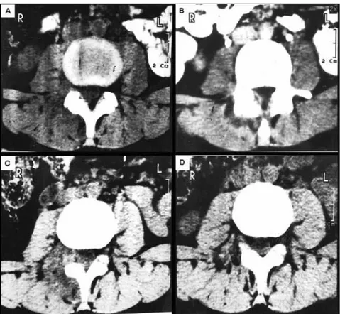
achieved and ESR normalized after 6 weeks of empi-ric intravenous treatment with clindamycin (2.4 g/ day) and amikacin (15 mg/kg/day). The treatment was maintained orally with cephalexin (4,0 g/day) for other 6 weeks. CT showed improvement after one month (Figure 1C) and normalization occurred after 4 months of the initial treatment (Figure 1D).
DISCUSSION
Clinical progression of SEA is graduated into four sta-ges: 1. back pain; 2. nerve-root pain radiating from the involved spinal area; 3. motor weakness, sensory deicit, and bladder and bowel dysfunction; 4. paralysis (1,5,8). The clinical triad of back pain, fever, and neurologi-cal deicit should be a warning of SEA diagnosis, even though this triad is only present in a small number of patients (5). Our patient presented the irst two of the-se SEA classic symptoms, and had established stage 2.
Neuroimaging is essential in identifying the location and extent of a lesion. CT and magnetic resonance ima-ging (MRI) are highly sensitive options in SEA diag-nosis (2,5,6,8). Leys and cols. (10) described ive SEA cases that were diagnosed using CT with an IV contrast
injection, in which the most common signs were loss of physiological epidural fat and unusual ixation of the contrast material at the dural sheath, surrounded by a higher density area situated between the bone and the dural sheath. Our indings were similar to those.
Diabetes mellitus is the mainly predisposing factor
as-sociated with SEA (4). Karikari and cols. conirmed these data in the revision of his 104 patients, and found diabe-tes incidence of 33.6% (11). A factor that could be invol-ved at the SEA pathogenesis in diabetic patients would be the dificulty of polymorphonuclear cells in phagocyting
S. aureus (12), the most common agent involved in the
disorder (3,13,14). As diabetes incidence is on the rise, it may be an important factor in the doubling in the inci-dence of SEA observed in the past two decades (5).
Isolated clinical treatment with broad spectrum an-tibiotics, with activity against S. aureus, anaerobes and
Gram-negative organisms was chosen for this patient, since no pathogen was isolated, neither the source of the infection was identiied, as occurs in about 20.5% of patients (5,9). CT imaging did not conirm bone lesion in our patient, but the treatment was prolonged orally for other 6 weeks, since vertebral osteomyelitis coexists with SEA in up to 80% of the cases (5).
Figure 1. CT at L3-L4 level. A: right dorsal paraspinal and epidural collection before treatment; B: unusual fixation of the IV contrast; C: first month after the treatment; D: CT normalization at the fourth month after the treatment.
Cop
yright
© ABE&M t
odos os dir
eit
os r
eser
vados
.
722 Arq Bras Endocrinol Metab. 2011;55/9
Optimal management of SEA is still being debated in the literature, and there are no clear indications of when medical approach is more appropriate than sur-gery (8,11). Leys and cols. (10) deined four patient groups there are candidates for clinical management: high-risk surgical patients, neurologically intact and stable patients, those with complete paresis for more than 72 hours, and those with extensive diffuse abscess formation (6,8). Rigamonti and cols. (15) also argued that conservative management could be a reasonable option in selected patients, but the clinical outcome of patients who were speciically managed with conserva-tive therapy was not mentioned.
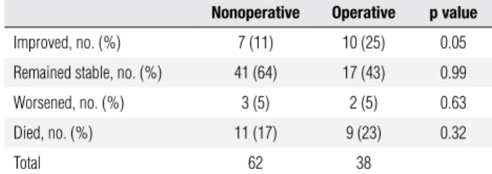
In a recent review, Karikari and cols. (11) did not support the hypothesis that surgical management is su-perior to conservative treatment, either. However, they have not clearly shown if there was radiological cure among the seven (11%) of 62 patients who presented improvement with nonoperative treatment (Table 1). Besides, this kind of treatment was not necessarily re-presented by antibiotic therapy alone, because CT-gui-ded aspiration was also considered.
Few articles reported clear clinical and radiological cure. Our case showed that antibiotic therapy could be indicated as an initial treatment in selected patients, and that more studies are necessary to identify the real safety of the clinical approach in comparison with surgery. The latter is still considered irst-line treatment (5,16), but presents higher mortality (10,11). The option of nono-perative treatment is justiied for patients without marked neurological deicits, according to Siddiq and cols. (17).
In our case report, the high level of suspicion was crucial to SEA diagnosis. The cure was achieved only with antibiotic therapy. Clinical and radiological reco-very occurred at the fourth month of follow-up.
therapy itself is an eficient SEA treatment and should be considered in cases of early diagnosis and absence of advanced neurological deicit.
Acknowledgements: according to Ethics Committee’s recom-mendations, the authors had the consent of the patient and their legal representatives to report this case.
Disclosure: no potential conlict of interest relevant to this article was reported.
REFERENCES
1. Danner RL, Hartman BJ. Update of spinal epidural abscess: 3 ca-ses and review of the literature. Rev Infect Dis. 1987;9:265-74. 2. Kourbeti IS, Tsiodras S, Boumpas DT. Spinal infections: envolving
concepts. Curr Opin Rheumatol. 2008;20(4):471-9.
3. Verner EF, Musher DM. Spinal epidural abscess. Med Clin North Am. 1985;69:375-85.
4. Rea GL, McGregor JM, Miller CA, Miner ME. Surgical treat-ment of the spontaneous spinal epidural abscess. Surg Neurol. 1992;37:274-9.
5. Darouiche RO. Spinal epidural abscess. N Engl J Med. 2006;355(19):2012-20.
6. Alvarez M. Spinal epidural abscess, from onset to rehabilitation: case study. J Neurosci Nurs. 2005;37(2):72-8.
7. Mackenzie AR, Laing RBS, Smith CC, Kaar GF, Smith FW. Spinal epidural abscess: the importance of early diagnosis and treat-ment. Neurol Neurosurg Psychiatry. 1998;65:209-12.
8. Quinines-Hinojosa A, Jun P, Jacobs R, Rosenberg WS, Weinstein PR. General principles in the medical and surgical management of spinal infections: a multidisciplinary approach. Neurosurg Fo-cus. 2004;17(6):E1.
9. Hlavin ML, Kaminski HG, Ross JS, Ganz E. Spinal epidural abs-cess: a ten-year perspective. Neurosurgery. 1990;27(2):177-83. 10. Leys D, Lesoin F, Viscel C, Pasquier F, Rousseaux M, Jomin M, et
al. Decrease morbidity from acute bacterial spinal epidural abs-cess using computer tomography and nonsurgical treatment in selected patients. Ann Neurol. 1985;17:350-5.
11. Karikari IO, Powers CJ, Reynolds RM, Mehta AI, Isaacs RE. Mana-gement of a spontaneous spinal epidural abscess: a single-center 10-years experience. Neurosurgery. 2009;65:919-24.
12. Musclow CE, Farkas-Himsley H, Durbahn G, Spragg L. Fluores-cence assay to monitor phagocytosis by blood – clot derived polymorphonuclear leucocytes. Study of patients with diabetes and phagocytosis of different Staphylococcal species. Cytobios. 1991;65(260):15-24.
13. Heusner AP. Nontuberculous spinal epidural infections. N Engl J Med. 1948;239:845-54.
14. Kaufman DM, Kaplan JG, Litman N. Infections agents in spinal epidural abscess. Neurology. 1980;30:844.
15. Rigamonti D, Liem L, Sampath P, Knoller N, Namaguchi Y, Schreibman DL, et al. Spinal epidural abscess: contemporary trends in etiology, evaluation, and management. Surg Neurol. 1999;52:189-97.
16. Curry WT Jr, Hoh BL, Amin-Hanjani S, Eskandar EN. Spinal epi-dural abscess: clinical presentation, management, and outcome. Surg Neurol. 2005;63:364-71.
17. Siddiq F, Chowfin A, Tight R, Sahmoun AE, Smego RA. Medical vs surgical management of spinal epidural abscess. Arch Intern Med. 2004;164:2409-12.
Diabetes mellitus and spinal epidural abscess
Table 1. Clinical outcome based on treatment plan (Karikari et al. Neurosurgery. 2009;65:919-24)
Nonoperative Operative p value
Improved, no. (%) 7 (11) 10 (25) 0.05 Remained stable, no. (%) 41 (64) 17 (43) 0.99 Worsened, no. (%) 3 (5) 2 (5) 0.63 Died, no. (%) 11 (17) 9 (23) 0.32 Total 62 38
CONCLUSION
Since there is a high incidence of diabetes mellitus, SEA