Boerhaave syndrome – case report
Síndrome de Boerhaave – relato de caso
Biljana Radovanovic Dinic
I, Goran Ilic
II, Snezana Tesic Rajkovic
III, Tatjana Jevtovic Stoimenov
IVMedical School, University of Niš, and Gastroenterology and Hepatology Clinic, Niš Clinical Center, Niš, Serbia
ABSTRACT
CONTEXT: Boerhaave syndrome consists of spontaneous longitudinal transmural rupture of the esopha-gus, usually in its distal part. It generally develops during or after persistent vomiting as a consequence of a sudden increase in intraluminal pressure in the esophagus. It is extremely rare in clinical practice. In 50% of the cases, it is manifested by Mackler’s triad: vomiting, lower thoracic pain and subcutaneous emphysema. Hematemesis is an uncommon yet challenging presentation of Boerhaave’s syndrome. Compared with rup-tures of other parts of the digestive tract, spontaneous rupture is characterized by a higher mortality rate.
CASE REPORT: This paper presents a 64-year-old female patient whose vomit was black four days be-fore examination and became bloody on the day of the examination. Her symptoms included epigastric pain and sufocation. Physical examination showed hypotension, tachycardia, dyspnea and a swollen and painful abdomen. Auscultation showed lateral crackling sounds on inspiration. Ultrasound examination showed a distended stomach illed with luid. Over 1000 ml of fresh blood was extracted by means of na-sogastric suction. Esophagogastroduodenoscopy was discontinued immediately upon entering the proxi-mal esophagus, where a large amount of fresh blood was observed. The patient was sent for emergency abdominal surgery, during which she died. An autopsy established a diagnosis of Boerhaave syndrome and ulceration in the duodenal bulb.
CONCLUSION: Boerhaave syndrome should be considered in all cases with a combination of gastroin-testinal symptoms (especially epigastric pain and vomiting) and pulmonary signs and symptoms (es-pecially sufocation).
RESUMO
CONTEXTO: A síndrome de Boerhaave é uma ruptura longitudinal transmural espontânea do esôfago, normalmente da parte distal. Ela geralmente se desenvolve durante ou após vômitos persistentes como consequência do aumento repentino da pressão intraluminal no esôfago. É extremamente rara na prática clínica. Em 50% dos casos, manifesta-se pela tríade de Mackler: vômitos, dor torácica inferior, enisema subcutâneo. Hematêmese é uma apresentação incomum porém desaiadora da síndrome de Boerhaave. Em comparação com rupturas de outras partes do tubo digestivo, a ruptura espontânea é caracterizada pela taxa de mortalidade mais elevada.
RELATO DO CASO: O artigo apresenta uma paciente do sexo feminino de 64 anos de idade, cujo vômito era preto, quatro dias antes do exame, e continha sangue no dia do exame. Os sintomas incluíam dor epigástrica e sufocação. No exame físico, foi veriicada hipotensão, taquicardia, dispneia e abdômen in-chado e doloroso. Ausculta revelou estertores laterais na inspiração. A ultrassonograia mostrou estômago dilatado, preenchido com conteúdo líquido. Sucção nasogástrica evacuou mais de 1.000 ml de sangue fresco. Esofagogastroduodenoscopia foi abortada imediatamente ao se entrar no esôfago proximal, onde foi observada grande quantidade de sangue fresco. A paciente foi encaminhada com urgência para ci-rurgia abdominal, durante a qual faleceu. Autópsia estabeleceu diagnóstico de síndrome de Boerhaave e úlcera no bulbo-duodenal.
CONCLUSÃO: A síndrome Boerhaave deve ser considerada em todos os casos com uma combinação de sintomas gastrointestinais (especialmente dor epigástrica e vómitos) e sintomas e sinais pulmonares (especialmente sufocação).
IMD. Associate Professor and Attending
Physician, Medical School, University of Niš, and Gastroenterology and Hepatology Clinic, Niš Clinical Center, Niš, Serbia.
IIMD. Associate Professor, Medical School,
University of Niš, and Institute of Forensic Medicine, Niš, Serbia.
IIIMD. Attending Physician, Gastroenterology and
Hepatology Clinic, Niš Clinical Center, Niš, Serbia.
IVMD. Associate Professor, Medical School,
University of Niš, and Institute of Biochemistry, Niš, Serbia.
KEY WORDS:
Esophagus. Rupture, spontaneous. Hematemesis. Pneumothorax. Emphysema.
PALAVRAS-CHAVE:
Esôfago.
INTRODUCTION
Boerhaave syndrome consists of spontaneous longitudinal trans-mural rupture of the esophagus. he syndrome is named ater a German doctor, Herman Boerhaave, who irst described it in 1724.1 In comparison with iatrogenic rupture, which may
develop during diagnostic or therapeutic endoscopic procedures, traumas or various esophageal diseases, spontaneous rupture most commonly develops during or ater persistent vomiting, as a consequence of a sudden increase in intraluminal esophageal pressure. Spontaneous rupture encompasses 15% of all esopha-geal ruptures.2 It is extremely rare in clinical practice. he true
incidence of Boerhaave syndrome in the general population is unknown. However, it is considered to be more common than once thought, since many cases of Boerhaave syndrome are only diagnosed postmortem, thus resulting in underreporting and underestimation with regard to both incidence and mortality.1,3
Boerhaave syndrome is seen most frequently among patients aged 50-70 years.1
he clinical manifestation of spontaneous rupture of the esophagus depends on the rupture location. In 50% of the cases, it is manifested by Mackler’s triad: vomiting, lower thoracic pain and subcutaneous emphysema.3,4
If the diagnosis is not established in time and if appropriate therapeutic measures are not undertaken, serious complications can develop and this may lead to a poor outcome. Compared with ruptures of other parts of the digestive tube, spontaneous rupture of the esophagus has the highest mortality rate.1,5
CASE REPORT
he patient was a 64-year-old female, with a history of long-term arterial hypertension, who was brought to the Gastroenterology and Hepatology Clinic of the Niš Clinical Center by the emer-gency medical services. She was admitted presenting with vom-iting of fresh blood, black stools, epigastric pain, sufocation and exhaustion.
he problems had irst appeared four days before admission in the form of poorly formed black stools and vomiting of small amounts of black substance. She did not see a doctor about these problems. On the day of admission, ater vomiting an excessive amount of black substance, she developed a pain in the epigastric region and then began to vomit fresh blood. It was at this stage that she rang the emergency medical services.
Physical examination showed that the patient was alert, ady-namic, tachycardiac and easily dyspneic, and her skin was pale. Her blood pressure was 60/40 mmHg. Auscultation of the heart was normal. Auscultation of the lungs showed baseline crackles on inspiration on both sides. he abdomen was tense, especially in the epigastric area and let hypochondrium, with tenderness in the epigastric area. he liver and spleen were of normal size.
Appropriate therapy was administered (one ampoule of pranto-pazole, a total of about 3000 ml of continuous infusion of saline solution and lactated Ringer’s solution). he oxygen saturation was 95%. A urinary catheter was placed for monitoring diuresis. An electrocardiogram (ECG) showed sinus tachycardia.
Because of the indings in the abdomen, an ultrasound exam-ination was performed and this showed a distended stomach illed with a large amount of luid. No free luid was found in the abdominal cavity. A nasogastric probe was placed in order to extract the contents and perform esophagogastroduodenoscopy (EGD). Ater inserting the nasogastric probe, about 1,000 ml of fresh blood was extracted. Ater the hemodynamic status had improved, esophagogastroduodenoscopy was attempted. Immediately upon insertion of the endoscope into the proximal esophagus, relux of a large amount of fresh blood was observed; further examination was cancelled. he patient was sent for emergency abdominal surgery. However, she died one hour ater the irst examination.
he laboratory indings and coagulation factors, which were received subsequently, were within normal values. he blood count showed reduced hemoglobin of 70 g/l (reference values: 115-170 g/l) and increased leukocyte count of 12.0 x 10⁹/l
(refer-ence values: 4.0-10.0 x 10⁹/l).
he autopsy showed 650 ml of dark red to black thick luid content in the right hemithorax and 600 ml in the let hemitho-rax (Figure 1). he heart size measurements were 110 x 105 mm. he heart weighed 380 g. he thickness of the cardiac muscle of the let ventricle was 18 mm and of the right ventricle, 6 mm. A rupture along the longitudinal axis was found in the esopha-gus, in the posterior let section of the esophageal wall, 15 mm from the cardia.
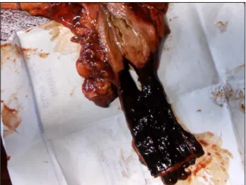
he rupture was 30 x 20 mm in size. he esophageal mucosa was smooth and almost completely covered in bloody-black con-tent (Figure 2). here were no foreign bodies in the abdomi-nal cavity. A small amount of blackish liquid was found in the
Figure 1. Macroscopic indings from the intrathoracic contents
stomach. Numerous small shallow erosions were found in the fundus and body of the stomach.
A mucosal injury of depth 13 mm, covering an area of 20 mm x 15 mm with irm borders and blackish background, consistent with a duodenal bulb ulcer, was observed (Figure 3). he walls were irm and vallum-like and the bottom was partially black. Greenish and black content was present throughout the intestines.
Chemical and toxicological analysis on samples of organ tis-sues, blood and urine did not reveal the presence of any psycho-active substances or pesticides.
he autopsy report declared that the immediate cause of death was hemopneumothorax due to esophageal injury and a chronic duodenal ulcer.
DISCUSSION
Spontaneous rupture of the esophagus is a rare clinical entity with a high mortality rate.5,6 he pathophysiology of Boerhaave
syndrome involves a sudden rise in intraluminal esophageal pressure, thereby forcing the gastric contents against a tight cri-copharyngeus muscle.3,6 It most oten develops during or ater
intense vomiting caused by excessive eating or drinking alcohol.7
However, spontaneous rupture of the esophagus may occur in the absence of predisposing factors. here are cases of spontane-ous esophageal rupture during sleep. In some patients, a muscu-lar layer was missing and this may point to the possibility of ana-tomical predisposition for the development of rupture.1,3
In the literature, there are cases in which the rupture was also associated with gastroesophageal relux disease (GERD), Barrett’s esophagus, peptic stricture of the esophagus, esophageal dysmotility, paraesophageal hernia or bleeding from a duodenal ulcer, which was the case with our patient.5,8,9 In our patient, the
esophageal rupture was a consequence of excessive vomiting due to the bleeding from the duodenal ulcer.
Spontaneous rupture may occur just above the diaphragm in the posterolateral wall of the esophagus. Perforations are usually longi-tudinal (0.6-8.9 cm long), with the let side more commonly afected than the right (90%). his is probably due to an anatomical weakness of the let posterolateral aspect of the esophagus just above the dia-phragm. Spontaneous rupture is rare below the diaphragm or in the thoracic part of the esophagus.3,7 In our case, the rupture was located
in the distal esophagus, 15 mm from the cardia.
he clinical manifestation of Boerhaave syndrome depends on the location of the rupture and the time between its devel-opment and examination. Patients with cervical perforation feel pain in the neck and upper half of the thorax. In cases of perfora-tion in the rest of the esophagus, pain is present in the lower part of the thorax and/or upper abdomen. Considering that sponta-neous rupture most oten happens in the distal esophagus, the majority of patients have Mackler’s triad of symptoms and signs: vomiting, lower thoracic pain and subcutaneous emphysema.3,4
However, this triad is rare, which may delay the diagnosis.10 In a
series of 14 patients with Boerhaave syndrome, only a small per-centage had typical signs and symptoms.3
he symptoms of Boerhaave syndrome can be nonspeciic. Compared with Mallory-Weiss syndrome, Boerhaave syndrome is rarely manifested through hematemesis or other signs of gas-trointestinal bleeding, including melena.1,3,6,10,11 In Boerhaave
syndrome, the rupture is transmural, which leads to esophageal perforation. In our patient, hematemesis was the chief complaint. To begin with, she was vomiting an excessive amount of black substance as a result of bleeding from ulcers. Excessive vomiting led to spontaneous rupture of the esophagus, which manifested as vomiting of fresh blood.
Figure 2. Gross examination of the distal esophagus showing
a longitudinal complete rupture 15 mm from the cardia. Note the darkened esophageal mucosa.
Figure 3. Gross indings from the stomach and duodenum
During physical examination of patients, subcutaneous emphysema is observed in 28%-66% within the irst 24 hours. his inding is signiicant for the initial diagnosis. More typi-cally, subcutaneous emphysema is found later. Besides typical symptoms, atypical symptoms such as hypotension, tachycar-dia, tachypnea, feverishness and cyanosis may also be present.1,7
Atypical symptoms may be prevented through timely diag-nosis. Pneumomediastinum is a signiicant clinical inding.10
Pneumomediastinum is suspected when, during lung ausculta-tion, crunching sounds that are synchronous with the heartbeat are heard (Hamman’s sign). his sign is present in around 20% of the cases.7
Esophageal rupture may be followed by serious complica-tions, of which the most important ones are mediastinitis and multiple organ dysfunction. Sepsis may develop within a few hours. In such cases, the clinical picture is dominated by signs and symptoms of sepsis, which additionally prevents timely diag-nosis and appropriate therapeutic measures.6,7,12
Laboratory indings are not speciic for diagnosing sponta-neous esophageal rupture. Serum albumin is normal but may be low, while the globulin fraction may be normal or slightly elevated.7 Radiography of the heart and lungs is valuable for
the diagnosis. Radiographs usually show signs of pneumome-diastinum or pneumothorax or hydropneumothorax if pleural efusion is concurrent.3,13 In cases of perforation of the
mid-dle third of the esophagus, pleural efusion is present on the right side, while in cases of rupture of the distal esophagus, pleural efusion is present on the let side.5 Diagnostic
thora-centesis shows the presence of food remnants, increased amy-lase and pH below 6. he presence of pneumomediastinum with data including vomiting and chest pain are almost dei-nite signs of Boerhaave syndrome. Overall, 10% of chest radio-graphs are normal.7,14
Esophagography is an important imaging examination for conirming the diagnosis and the location of perforation because it shows extravasation of contrast into the pleural space. he procedure is performed with water-soluble contrast, such
as Gastrograin, since barium may cause severe mediastinitis. Esophagography with Gastrograin is 90% sensitive.7
horacic computed tomography imaging is indicated for making the diagnosis in patients who do not tolerate esopha-gography. During the procedure, localized luid collection is observed, as well as periesophageal air collection.1,15,16 he role
of EGD in the early diagnostic work-up of patients with sus-pected esophageal perforation has been disputed.17 EGD is not
recommended for diagnosing Boerhaave syndrome, since it may increase the rupture and the amount of air in the mediastinum and pleural space.13 In cases with hematemesis, such as in our
patient, the procedure was attempted in order to ascertain the source of bleeding.
he treatment for Boerhaave syndrome is both conservative and surgical. he goals of pharmacotherapy are to reduce mor-bidity and to prevent complications. Surgical management is generally required for both spontaneous rupture and traumatic perforation.14,18 Endoscopic stent insertion ofers a promising
alternative. he mortality rate varies depending on the time that has elapsed since development of the rupture and its recognition and treatment. If treatment is not started within 24 hours from the onset of symptoms, the mortality rate is 25%; ater 24 hours, it is 65%; and ater 48 hours, it is 75%-89%.19
We reviewed the literature in Medline, PubMed, Embase and Lilacs using the English keywords “Esophagus”, “Rupture, sponta-neous”, “Hematemesis” and “Pneumothorax”; and the Portuguese words “Esôfago”, “Ruptura espontânea”, “Hematêmese” and “Pneumotórax” (Table 1).
CONCLUSION
Boerhaave syndrome should be considered in all patients with a combination of gastrointestinal symptoms (epigastric pain and vomiting) and pulmonary symptoms (sufocation), even when all the signs and symptoms (lower thoracic pain and subcutaneous emphysema) of this disease are absent. Early clinical suspicion will lead to timely diagnosis and maximize the survival chances for the patient.
Database Search strategies Papers found Related papers
MEDLINE (via PubMed)
Esophagus AND Rupture, spontaneous AND Hematemesis
AND Pneumothorax AND “case reports” [Publication Type] 9 2
Embase (via Elsevier)
Esophagus AND Rupture, spontaneous AND Hematemesis
AND Pneumothorax AND “case reports” [Publication Type] 0 0
LILACS (via Bireme)
(Esofago [DeCs]) OR (esophagus [MeSH]) AND (Ruptura espontanea [DeCs]) OR (Rupture, spontaneous [MeSH]) AND (Hematemese [DeCs]) OR (Hematemesis [MeSH]) AND (Pneumotorax [DeCs] OR Pneumothorax [MeSH]) AND ” relato de caso”
0 0
REFERENCES
1. Dellon ES, Shaheen NJ. Miscellaneous diseases of the esophagus: foreign bodies, physical injury and systemic and dermatological diseases. In: Yamada T, editor. Textbook of Gastroenterology. 5th ed.
Chichester: Blackwell Publishing: 2009. p. 871-88.
2. Brinster CJ, Singhal S, Lee L, et al. Evolving options in the management of esophageal perforation. Ann Thorac Surg. 2004;77(4):1475-83. 3. Garas G, Zarogoulidis P, Efthymiou A, et al. Spontaneous esophageal
rupture as the underlying cause of pneumothorax: early recognition is crucial. J Thorac Dis. 2014;6(12):1655-8.
4. Venø S, Eckardt J. Boerhaave’s syndrome and tension pneumothorax secondary to Norovirus induced forceful emesis. J Thorac Dis. 2013;5(2):E38-40.
5. Reardon ES, Martin LW.Boerhaave’s syndrome presenting as a mid-esophageal perforation associated with a right-sided pleural efusion. J Surg Case Rep. 2015(11). pii: rjv142.
6. de Schipper JP, Pull ter Gunne AF, Oostvogel HJ, van Laarhoven CJ. Spontaneous rupture of the oesophagus: Boerhaave’s syndrome in 2008. Literature review and treatment algorithm. Dig Surg. 2009;26(1):1-6.
7. Roy PK, Murphy ME, Kalapatapu V, Bashir S, Mujibur R. Boerhaave Syndrome. Medscape. Available from: emedicine.medscape.com/ article/171683. Accessed in 2016 (Sep 8).
8. Tsalis K, Vasiliadis K, Tsachalis T, et al. Management of Boerhaave’s syndrome: report of three cases. J Gastrointestin Liver Dis. 2008;17(1):81-5.
9. Yang ST, Devanand A, Tan KL, Eng PC. Boerhaave’s syndrome presenting as a right-sided pleural efusion. Ann Acad Med Singapore. 2003;32(3):415-7.
10. Fikfav V, Gaur P, Kim MP. Endoscopic management of Boerhaave’s syndrome presenting with hematemesis. J Surg Case Rep. 2014(11). pii:rju110.
11. Søreide JA, Viste A. Esophageal perforation: diagnostic work-up and clinical decision-making in the irst 24 hours. Scand J Trauma Resusc Emerg Med. 2011;19:66.
12. Woo KM, Schneider JI. High-risk chief complaints I: chest pain--the big three. Emerg Med Clin North Am. 2009;27(4):685-712, x.
13. Eckstein M, Sean O. Henderson. Thoracic trauma, esophagus perforation. In: Marx J A, ed. Rosen’s Emergency Medicine. Concepts and Clinical Practice. 8th ed. Philadelphia: Mosby; 2014. p. 455-8.
14. Kollmar O, Lindemann W, Richter S, et al. Boerhaave’s syndrome: primary repair vs. esophageal resection--case reports and meta-analysis of the literature. J Gastrointest Surg. 2003;7(6):726-34. 15. Duman H, Bakırcı EM, Karadağ Z, Uğurlu Y. Esophageal rupture
complicated by acute pericarditis. Turk Kardiyol Dern Ars. 2014;42(7):658-61.
16. Vial CM, Whyte RI. Boerhaave’s syndrome: diagnosis and treatment. Surg Clin North Am. 2005;85(3):515-24, ix.
17. Arantes V, Campolina C, Valerio SH, et al. Flexible esophagoscopy as a diagnostic tool for traumatic esophageal injuries. J Trauma. 2009;66(6):1677-82.
18. Huber-Lang M, Henne-Bruns D, Schmitz B, Wuerl P. Esophageal perforation: principles of diagnosis and surgical management. Surg Today. 2006;36(4):332-40.
19. Schweigert M, Beattie R, Solymosi N, et al. Endoscopic stent insertion versus primary operative management for spontaneous rupture of the esophagus (Boerhaave syndrome): an international study comparing the outcome. Am Surg. 2013;79(6):634-40.
Sources of funding: None
Conlict of interest: None
Date of irst submission: May 4, 2016
Last received: June 17, 2016
Accepted: June 22, 2016
Address for correspondence:
Biljana Radovanovic Dinic
Faculty of Medicine - University of Niš, Serbia Bulevar Zorana Djindjica 48, Niš 18000 Serbia