INTRODUCTION
Article/Artigo
1. Programa de Pós Graduação em Doenças Infecciosas e Parasitárias, Faculdade de Medicina, Universidade Federal de Mato Grosso do Sul, Campo Grande, MS. 2. Sanidade Animal, Empresa Brasileira de Pesquisa Agropecuária Gado de Corte, Campo Grande, MS. 3. Laboratório de Parasitologia Humana, Universidade Federal de Mato Grosso do Sul, Campo Grande, MS.
Address to: Dr. Renato Andreoti. Lab. Biologia Molecular/Sanidade Animal/EMBAPA Gado de Corte. BR 262, km 04, Caixa Postal 15479, 79002-970 Campo Grande, MS, Brasil.
Phone: 55 67 3368-2173
e-mail: [email protected] Received in 09/07/2011 Accepted in 03/08/2011
Epidemiological factors related to the transmission risk of
Trypanosoma cruzi
in a Quilombola community, State of Mato Grosso do Sul, Brazil
Aspectos epidemiológicos relacionados ao risco de transmissão de
Trypanosoma cruzi
em comunidade Quilombola, Estado de Mato Grosso do Sul, Brasil
Marlon Cezar Comineti1, Renato Andreoti2, Elisa Teruya Oshiro3 and Maria Elizabeth Moraes Cavalheiros Dorval3
ABSTACT
Introduction: his work was an epidemiological investigation of the risk of Trypanosoma cruzi transmission in the rural Quilombola community of Furnas do Dionízio, State of Mato Grosso do Sul, Brazil. Methods: Of the 71 animals examined, seven were captured (two opossums, Didelphis albiventris; four rats, Ratus ratus; and one nine-banded armadillo, Dasypus novemcinctus) and 64 were domestic (one canine, Canis familiaris; ive pigs, Sus scrofa; two bovines, Bos taurus; ive caprines, Capra sp.; and 51 ovines, Ovis aries). Parasitological tests were performed to detect parasites in the blood and to identify the morphology of lagellates. hese methods included fresh examinations, bufy coat tests and blood cultures. Molecular analysis of DNA for identiication of trypanosomatids was performed by polymerase chain reaction (PCR) with primers S35 and S36. Results: he parasitological tests showed lagellates in an opossum and two catle. he molecular tests showed DNA from T. cruzi in an opossum and a pig. Triatoma sordida was the only triatomine species found in the community, and it colonized households (four specimens) and the surrounding areas (124 specimens). Twenty-three specimens tested positive for lagellates, which were subsequently identiied as T. cruzi by PCR. Conclusions: Data analysis demonstrated that T. cruzi has a peridomestic life cycle that involves both domestic and wild mammals.
Keywords: Tripanosomatides. Triatoma sordida. Triatomines. Synanthropic animals. PCR.
RESUMO
Introdução: Este trabalho foi uma investigação epidemiológica do risco de transmissão de Trypanosoma cruzi na comunidade rural Quilombola de Furnas do Dionízio, Estado de Mato Grosso do Sul. Métodos: Dos 71 animais examinados, sete foram capturados (dois gambás, Didelphis albiventris; quatro ratos, Ratus ratus; e um tatu, Dasypus novemcinctus) e 64 eram domésticos (um canídeo, Canis familiaris; cinco suínos, Sus scrofa; dois bovinos, Bos taurus; cinco caprinos, Capra sp; e 51 ovinos, Ovis aries). Exames parasitológicos foram realizados para detectar parasitas no sangue e para identiicar a morfologia dos lagelados. Estes métodos incluíram exame a fresco, exame do creme leucocitário e hemocultura. A análise molecular de DNA para identiicação de tripanossomatídeos encontrados foi feita pela reação em cadeia da polimerase (PCR) com os primers S35 e S36. Resultados: Os exames parasitológicos mostraram lagelados em um gambá e nos dois bovinos. Os testes moleculares mostraram a presença do DNA de T. cruzi em um gambá e um suíno. Triatoma sordida foi a única espécie de triatomíneo encontrada na comunidade colonizando domicílio (quatro espécimes) e peridomicílio (124 espécimes). Vinte e três amostras foram positivas para lagelados e identiicados como T. cruzi pela PCR. Conclusões: A análise dos dados aponta para o ciclo peridoméstico do parasita e envolve tanto animais domésticos como selvagens.
Palavras-chaves: Tripanosomatideos. Triatoma sordida. Triatomíneos. Animais sinantrópicos. PCR.
The genus Trypanosoma belongs to the order
Kinetoplastida and comprises parasite species that affect vertebrates of all orders (fish, amphibians, reptiles, birds and mammals) and are transmitted by various blood-sucking invertebrate vectors. One
of these parasites is Trypanosomacruzi, which is
the etiologic agent of Chagas disease. Although initially a woodland enzootic, Chagas has become anthropozoonotic, mainly because the occupation of these woodland areas has reshaped transmission cycles, thereby incorporating humans and domestic animals
into the epidemiological chain of T. cruzi stocks that
are exchanged between sylvatic and domestic cycles1.
Trypanosoma cruzi is usually transmited by a vector, mainly by the hematophagous Reduviidae insects, with the parasite penetrating into the host through skin lesions or mucosal or oral routes, and this last being the main way of parasite transmission to animals. The other transmissions are blood
transfusions and transplacentally way2-4.
The vectors of T. cruzi, the triatomines, are
members of the hemipterous family Reduviidae. Of
the 138 species cataloged in Brazil5, no more than
ive play a direct role in the epidemiology of the
parasite6: Triatoma infestans (Klug, 1834), which is
considered to be the main vector of the disease in Brazil considering it is oten present in housing and
is markedly anthropophilic, T. brasiliensis (Neiva,
1911), Panstrongylus megistus (Burmeister, 1835),
T. pseudomaculata (Corrêa & Espínola, 1964) and T. sordida (Stal, 1859).
The latter species is native to the cerrado, including transitional areas of Maranhão, Piaui, Bahia,
Pantanal and Chaco Oriental7. Despite its noted bird
tropism, this species can use other food sources when its environment is disturbed; as a result, it invades human homes. It is the second most common of the triatomines, with the highest number of positive
METHODS
Entomological surveys veriied the existence of three major
species in MS: T. brasiliensis (Neiva, 1911), P. megistus (Burmeister,
1835) and T. sordida (Stal, 1859) being infestation rates for
domiciliary and peridomestics areas were significant only for T. sordida (9.3% and 86.6%, respectively), while T. brasiliensis and P. megistus showed less than 0.2% infestation for both8.
The hosts of T. cruzi are mammals, and natural infection
by this parasite has been detected in the mammalian orders Didelphimorphia, Xenarthra, Chiroptera, Rodentia, Lagomorpha, Artiodactyla, Carnivora and Primates, which makes them the foci for the maintenance of the parasite in the sylvatic, peridomestic and domestic cycle. In domestic animals, the protozoan mainly infects
dogs and cats, but it has also been found in pigs and goats2,9.
Furnas do Dionízio is a Quilombola community founded by Antonio Vieira, a former slave who migrated from Minas Gerais State in 1890 and setled with his family in an isolated, forested area that has always had limited contact with the surrounding communities.
(The term Quilombola refers to the inhabitants of ethnically
homogeneous, typically isolated rural communities of descendants of former Afro-Brazilian slaves. he term also refers to the culture of these communities). Furnas do Dionízio now numbers 96 families. Historically, basic public services have been deicient or absent in Brazilian Quilombola communities.
he major sources of income for these communities are farming (cassava, sugarcane and their derivatives) and raising livestock.
Study area
Furnas do Dionízio is a Quilombola rural community located in the county of Jaraguari, 45km from Campo Grande, which is the capital City of Mato Grosso do Sul. he community covers approximately 1,031ha, with its center at the coordinates
20°9′1.34″S and 54°34′27.17″W (Figure 1). Modest masonry
houses predominate, usually in close proximity to wooden sties, chicken coops and corrals.
Although it practices subsistence agriculture, the community depends on government subsidies. Cultural traits dating from the earliest members have been nurtured to the present day, typically in the form of community gatherings devoted to prayers, church bazaars to celebrate the patron saints of the month of June, and the practice
of the catira (a group dance).
To beter understand the parasite distribution and identify risk areas in the State of Mato Grosso do Sul, this study investigated the
risk of transmission of T. cruzi in the Quilombola rural community of
Furnas do Dionízio, Mato Grosso do Sul, by verifying the conditions of dwellings and peridomicile structures, determining entomological indices and identifying potential mammalian hosts with molecular and parasitological tests.
Jaraguari
Campo Grande
Goiás Mato Grosso
Bolivia
Paraguay São Paulo
JARAGUARI
Municipal headquarters
Furnas do Dionízio
0 6 KM 12 KM
0 70 KM 140 KM
he capture and parasitological testing of triatomines and the calculation of entomological indices
From May to August 2009, triatomines were collected from one corral, 20 chicken coops, 20 sties and 12 barns associated with 20 households at the core of the community. his area was the most densely populated and had the most peridomicile structures and debris associated with the houses. Any area with debris, wood or tile near to or within the atached structures was investigated and classiied as part of appendix closer.
Each residence and atached structure included in the triatomine research was actively searched. However, the man-hours method was not used because of the peculiarities of each environment, particularly the large volume of debris found in the annexes that required a longer and more thorough search. No dislodged products (Pirisa 5%) were used in the study.
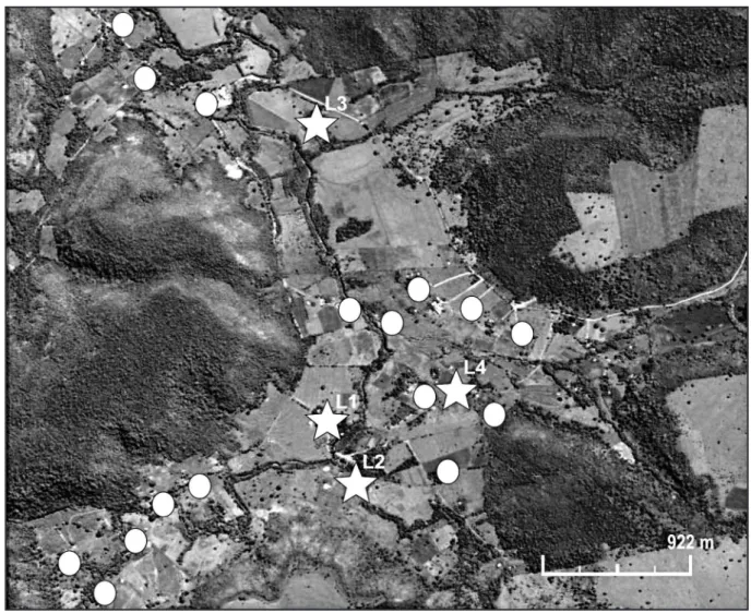
The sites where insects were found were classified as: L1 (sty built of masonry and purchased wooden planks), L2 (corral fenced made with purchased wood), L3 (chicken coop built of masonry and timber extracted from the local forest) and L4 (masonry house).
Insects, when found, were captured using anatomical tweezers, stored in perforated plastic bags and sent to the Laboratory of Human Parasitology at the Universidade Federal de Mato Grosso do Sul (UFMS) to be examined for natural infection by lagellates. Insects were identiied according to their external morphology using
graphical keys for tribes, genera and species10.
To test for parasites, each insect’s abdomen was compressed to collect fecal mater in a saline solution. he material was then smeared
on a slide and examined under a light microscope at 40×. For each
insect that tested positive for lagellates, portions of the stool sample were placed into two tubes containing solid medium NNN (McNeal, Novy & Nicolle) and 1mL of Schneider’s medium. Ater seven days, the samples were tested weekly for a period of four months. In total, 46 samples were obtained. he slides of the positive samples were ixed in methanol and stained with Giemsa. Ater examination, the specimens were individually stored in 2mL Eppendorf tubes containing 70% ethyl alcohol for later molecular examination.
To determine the risk of parasite transmission by insects in the community, the entomological indices of the geographical distribution were calculated. hese indices are deined by the World
Health Organization (WHO)11 and include the rate of natural
infection and the infestation rate:
infected triatomines
captured triatomines
Natural infection = χ 100
households with triatomine
households surveyed
Infestation rate = χ 100
A household was deined as a home and its associated structures.
Mammalian examination, sample collection, and parasitological examination
Conined animals and animals that were captured upon visitation of the ecotopes of the insect were examined in this study. For capture, 10 Tomahawk traps with dimensions of 50 x 22.5 x 20.5cm were used and placed at least 5m apart, depending on size. Near homes, the traps were placed at a distance of 10m from the house as well as
inside and around peridomicile structures. In each residence and atached structure, the same procedure was performed with the remaining traps in places where the insects were found by 10 days of waiting, and the bait (peanut buter and banana) was switched daily.
Blood samples from the captured mammals were collected in 5mL syringes using 25x0.6mm BD needles (Zhejiang Ouijian Medical Apparatus, China) and then transferred to test tubes containing 5mL of EDTA. No more than three hours passed between the time of collection and the laboratory tests. Samples were examined in the laboratory with a bufy coat test (three capillaries) and fresh exam (four slides).
A portion of each blood sample (six drops) was also seeded on solid medium NNN (McNeal, Novy & Nicolle) containing 1mL of Schneider’s medium. Ater seven days, the samples were tested weekly for four months. In total, 142 samples were obtained from the collected mammal blood.
Identiication of protozoa
Ater thin blood smear slides were prepared, ixed and stained
with Giemsa, they were examined under a light microscope at 100×
magniication to detect parasites and characterize their morphology12.
Molecular analysis included DNA extraction from whole animal blood and from a pool obtained from 46 cultures of the intestinal contents of triatomines that tested positive for lagellates. his pool was divided into two aliquots.
A 200µL aliquot from each blood and culture sample was transferred into a 2mL Eppendorf tube, and then 400µL of lysis bufer was added, followed by homogenization for 20s in a shaker. Next, 100µL of 1% SDS was added and the solution was homogenized for 2min, followed by the addition of 40µL of proteinase K (20mg/mL) and a inal 20s homogenization. he resulting solution was incubated
in a water bath at 55°C for 2h.
A 500mL volume of chloroform: isoamyl alcohol solution (24:1) was added to each sample, which was then homogenized for
20s and centrifuged at 15,700g for 15min in an Eppendorf 5415D
microcentrifuge. he supernatant was pipeted into another 1.5mL Eppendorf tube, and twice its volume in isopropyl alcohol cooled to
4°C was added. he resulting volume was homogenized by inverting
the tube 50 times and incubated overnight at 4°C, ater which it
was centrifuged at 15,700g for 10min at 4°C. he supernatant was
discarded and the remaining material was washed. A 500mL volume
of 70% ethanol at 4°C was added, and the sediment was centrifuged
at 13,400g for 5min at 4°C, ater which the supernatant was discarded.
his last step was repeated twice. he resulting pellet was dried in a
dry bath at 60°C and resuspended in 100mL of autoclaved ultrapure
water. he samples were stored at -20°C.
Parasite DNA identification was based on the polymerase chain react ion (PCR), using t wo pr imer s, i .e., S35 (5’-AAATAATGTACGGG(T/G)GAGATGCATGA-3’) and S36 (5’-GGGTCGATGGGGTGGTGT-3’), that amplify a 330-base pair (bp) fragment and anneal to the sequences of the conserved
region of kDNA minicircles in T. cruzi13.
he ampliication consisted of an initial denaturation at 95°C
(10min), followed by 35 denaturation cycles at 94°C (30s each),
annealing at 50°C (1min), extension at 72°C (1min), and a inal
extension (10min) in an XP thermal cycler (Bioer).
electrophoresis at 80V and 400mA for 1h40min. he gels were visualized under UV ater ethidium bromide staining.
Ethical considerations
Permission for capture and examination of wild animals was granted by the Brazilian Institute for the Environment and Renewable Natural Resources (IBAMA) (permit 16611-1) and the UFMS Ethics Commission for the Use of Animals (permit 207/2009, issued 19 March 2009).
RESULTS
Figure 2 shows the households where the insects were found as well as where the mammals were captured and/or contained.
he infestation rate was 20%. he highest number of captures
occurred in the peridomicile structures (Table 1). hese structures
also sheltered livestock such as pigs, sheep and/or chickens.
Figure 2 shows that the points of capture of the insects were very irregular and isolated (except for L4).
he rate of natural infection with lagellates in triatomines was 18% (Table 1).
TABLE 1 - Triatomine capture sites and lagellate infection rates forwet smears. Furnas do Dionízio, Jaraguari, State of Mato Grosso do Sul, Brazil, 2009.
Positive
Site Number* n %
L1 60 16 26.7
L2 42 1 2.4
L3 22 6 27.3
L4 4
-Total 128 23 18.0
L1: sty built of masonry and new wooden planks, L2: corral made with new wooden fencing, L3: chicken coop built of masonry and timber extracted from the local forest, L4: masonry house. *number of captured triatomines, n: number.
In the potential vertebrate hosts (Table 2), flagellates were
observed in the bufy coat tests and blood cultures of two catle and an opossum.
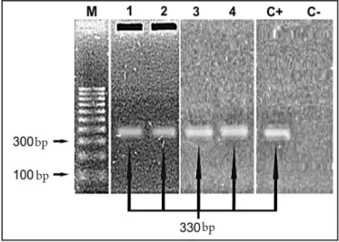
Figure 3 shows the results of T. cruzi DNA ampliication with S35 and S36 primers.
Two cultures from the pooled triatomine intestinal contents
were positive for T. cruzi. Of 71 total blood samples from domestic,
synanthropic and wild animals, two [from one opossum (3) and one pig (4)] were positive.
FIGURE 3 - Ampliication products obtained using S35 and S36 primers for
Trypanosoma cruzi.
M: marker, 1 and 2: pooled triatomine stool cultures, 3: opossum, 4: pig, C+: positive control, C-: negative control, bp: base pair.
DISCUSSION
he vectors of T. cruzi are oten associated with mud and wood
houses because these locations simulate the conditions found in their natural ecotopes. he homes in the study region were typically of this type, and some were improved in the last ive years. However, with rare exceptions, no signiicant improvements were made in associated structures, such as pens, corrals and chicken coops. hese structures are built or refurbished mostly from the timber extracted from the forest that surrounds the community, and the same wood can serve as a vehicle for triatomines.
Piles of wood and tiles were also observed near or even within the associated structures. his type of situation creates a favorable environment for insect colonization and reproduction. Although insects were not found to colonize the homes (except for L4), they were found no more than 30m away from the homes, and triatomines
usually suck the blood of the closest animal14. It is noteworthy that
almost all of the insects infested peridomicile areas, suggesting litle possibility that the houses will be colonized.
In the studied households, the only vector species found was T. sordida. he ability of T. sordida to colonize both natural and artiicial ecotopes, along with its high tolerance to environmental
change15, makes it an important vector in the parasite cycle and
diicult to control. Despite T. sordida being considered a species
that prefers to infect birds, the high rates of peridomestic infestation (20%) and parasitic infection (18%) suggest that surveillance is necessary because the mere coexistence with a vector increases the
possibility of human infection16 besides presenting a higher rate of
infection when compared with other surveys8,17,18.
Other studies showed that T. sordida is the most frequent in
captures17 - also peridomestic and domiciliary infestation reaching
86.6%8 and 47.7%17, respectively - and that infection rates range from
0.2%8 to 2.3%18.
Furthermore, the insects may move and setle closer to the houses if entomological control measures are not taken. It is believed that
this has not yet occurred for two reasons. First, the places where the insects were found were more isolated than the rest of the community, and L1 and L2 (which had the highest number of captures) were very close and had vegetable aisles that latched, which was not observed for other homes. However, as the rate of infestation was high, this region is at risk of becoming a new focus of vector insect colonization. Second, triatomines were found only where there were conined animals that could serve as a food source.
Because these new housing units can be built at any time, new infection foci could be found if preventive measures are not taken.
Another observation for this area is that most of the sugar cane and cassava plantations, which are the main sources of income for the community, are usually on the hillsides, which alters the vegetation cover and may facilitate the movement of triatomines closer to artiicial ecotopes19. Moreover, having established a human habitation
in the deforested area, it is common practice to breed livestock such as chickens, pigs, sheep and catle to provide support for the residents and pets such as dogs and cats for protection and company. Domestic animals represent a source of food for insects and are another factor favoring the establishment of the parasite transmission cycle, as observed by Souza20, who found that 10.7% of dogs in this same area
tested positive for T. cruzi.
In this work, the domestic animal that tested positive for T. cruzi
was the pig (Sus scrofa). Other studies have shown that this animal hosts the parasite21-23. he results indicate a low parasitemia because
it was not possible to verify the infection by parasitological testing; only the molecular testing by PCR detected the parasite DNA. his result suggests a low level of parasitemia and that the pig may be a possible source for protozoan maintenance in the Furnas do Dionízio.
Synanthropic animals were captured when the associated structures were visited, which is relevant because them are a normal part of the transmission cycle of T. cruzi9.
Only the opossum (Didelphis albiventris) tested positive for
T. cruzi(Table 2), which suggests that, along with pigs, this animal may transmit the parasite in the community because it was captured where there were colonies of triatomines.
TABLE 2 - Wild, synanthropic and domestic mammals infected with
Trypanosoma sp., by site and parasitological exam. Furnas do Dionízio, Jaraguari, State of Mato Grosso do Sul, Brazil, 2009.
Trypanosoma sp. Number positive cases
Species of samples Site BCE WS BC
Didelphis albiventris 2 L1 - - 1
Ratus ratus 4 L2 - -
-Dasypus novemcinctus 1 L1 - -
-Bos taurus 2 L1 2 - 2
Canis familiaris 1 L1 - -
-Capra sp. 5 L2 - -
-Sus scrofa 5 L1 - -
-Ovis aries 51 L2 - -
-Total 71 2 - 3
L1: sty built of masonry and new wooden planks, L2: corral made with new wooden fencing.
BCE: bufy coat examination, WS: wet smears, BC: blood culture.
bp
bp
he concern with the opossum is that because of its ecological and behavioral characteristics, it easily acclimates to environments modiied by humans when it finds suitable conditions for survival. With the construction of houses and peridomestic structures, the anthropic environment provides the opossum a niche by ofering food from the accumulation of organic waste, laying hens in open spaces and poorly stored animal feed. hese factors lead the marsupials to invade peridomicile areas and were prevalent in the Furnas do Dionízio community.
The finding of hemoflogellates in two cattle is still under study. Although a molecular conirmation was not performed, the protozoans that were found were morphologically very similar to Trypanosoma theileri, which is a cosmopolitan parasite common in mammals of the order Artiodactyla that has been found in catle from
Mato Grosso do Sul24.
Although T. theileri is not considered to be pathogenic, it can
induce chronic infections when associated with concomitant
diseases25,26. New surveys, cultures and molecular biology studies
of the parasite are in progress.
hese results suggest that in the study region, the parasite is active in the peridomestic environment because almost all of the triatomines, as well as the infected pig and the opossum that tested
positive for T. cruzi, were captured near the peridomicile structures.
hese results may aid eforts to prevent parasite transmission in the community and help raise awareness in the population about the need for improvements to peridomicile structures as well as the risks involved in establishing colonies of vectors and shelters for their protozoan hosts.
ACKNOWLEDGMENTS
he authors declare that there is no conlict of interest.
CONFLICT OF INTEREST
FINANCIAL SUPPORT
REFERENCES
he authors wish to thank the Núcleo Estadual de Entomologia and Centro de Controle de Vetores do Estado de Mato Grosso do Sul, with special thanks to the civil servants Guilmara M.A. Gonçalves, João Nascimento and to Ezequiel P. Ramos for their kind atention.
Financial support was provided by Empresa Brasileira de Pesquisa
Agropecuária Gado de Corte (EMBAPA) and Fundação de Apoio
ao Desenvolvimento do Ensino, Ciência e Tecnologia do Estado de
Mato Grosso do Sul (FUNDECT). A master’s program grant was
provided by Coordenação de Aperfeiçoamento de Pessoal de Nível
Superior (CAPES).
1. Tartaroti E, Azeredo-Oliveira MTV, Ceron CR. Problemática vetorial da Doença de Chagas. Arq Cienc Saude 2004; 11:44-47.
2. Rey L. Parasitologia: parasitos e doenças parasitárias do homem nos trópicos, 4nd ed. Rio de Janeiro: Guanabara Koogan; 2008.
3. Valente SAS, Valente VC, Fraiha Neto H. Considerations on the epidemiology and transmission of Chagas disease in the Brazilian Amazon. Mem Inst Oswaldo Cruz 1999; 94:395-398.
4. Yeo M, Acosta N, Llewellyn M, Sánchez H, Adamson S, Miles GAJ, et al. Origins of Chagas disease: Didelphis species are natural hosts of T. cruzi I and armadillos hosts of T. cruzi II, including hybrids. Int J Parasitol 2005; 35: 225-233.
5. Galvão C, Carvalho R, Rocha DS, Jurberg J. A checklist of the current valid species of the subfamily Triatominae Jeannel, 1919 (Hemiptera, Reduviidae) and their geographical distribution, with nomenclatural and taxonomic notes. Zootaxa 2003; 202:1-36.
6. Vinhaes MC, Dias JCP. Doença de Chagas no Brasil. Cad Saude Publica 2000, 16:7-12.
7. Foratini OP. Biogeograia, origem e distribuição da domicilização de triatomíneos no Brasil. Rev Saude Publica 2006; 40:964-998.
8. Almeida PS, Cereti Júnior W, Obara MT, Santos HR, Barata JM. Survey of Triatominae (Hemiptera: Reduviidae) fauna in domestic environments and natural infection by Trypanosomatidae in the State of Mato Grosso do Sul. Rev Soc Bras Med Trop 2008; 41:374-380.
9. Coura JR, Dias JCP. Epidemiology, control and surveillance of Chagas disease - 100 years ater its Discovery. Mem Inst Oswaldo Cruz 2009; 104:31-40. 10. Carcavallo RU, Rodrigues MEF, Galvão C, Rocha DS, Girón IG, Arocha MAO,
et al. Habitats e fauna relacionada. In: Carcavallo RU, Girón IG, Jurberg J, Lent H, editors. Atlas dos vetores da doença de Chagas nas Américas. Rio de Janeiro: Fundação Oswaldo Cruz; 1997. p. 561-600.
11. World Health Organization. Control of Chagas disease. Technical Report Series. 2005; 905:40-55.
12. Souza MA. Morphobiological characterization of Trypanosoma cruzi Chagas, 1909 and its distinction from other trypanosomes. Mem Inst Oswaldo Cruz 1999; 94:205-210.
13. Sturm NR, Degrave W, Morel C, Simpson L. Sensitive detection and schizodeme classiication of Trypanosoma cruzi cells by ampliication of kinetoplast minicircle DNA sequences: use in diagnosis of Chagas’ disease. Mol Biochem Parasitol 1989; 33:205-214.
14. Wisnivesky-Colli C. Feeding paterns of triatominae in relation to transmission of american trypanosomiasis. In: Brenner RR, Stoka AM, editors. Chagas disease vectors. Boca Raton: CRC Press; 1989. p. 99-117.
15. Diotaiuti L, Loiola CF, Falcão PL, Dias JCP. he ecology of Triatoma sordida in natural environments in two diferent regions of the state of Minas Gerais, Brazil. Rev Inst Med Trop Sao Paulo 1993; 35:237-245.
16. Toledo MJO, Kühl JB, Silva SV, Gasperi V, Araújo SM. Estudo sobre triatomíneos e reservatórios silvestres de Trypanosoma cruzi no Estado do Paraná, sul do Brasil. Resultados preliminares. Rev Soc Bras Med Trop 1997; 30:197-203. 17. De Paula MBC, Costa IN, Freitas PA, Limongi JE, Pajuaba Neto AA, Pinto
RMC, et al. Occurrence of positivity for Trypanosoma cruzi in triatomine from municipalities in Southeastern Brazil, from 2002 to 2004. Rev Soc Bras Med Trop 2010; 43:9-14.
18. Silveira AC, Feitosa VR, Borges R. Distribuição de triatomíneos capturados no ambiente domiciliar, no período de 1975-83, Brasil. Rev Bras Malariol Doenças Trop 1984; 36:15-312.
19. Siqueira-Batista R, Gomes AP, Rôças G, Cota RMM, Rubião ECN, Pissinati A. Chagas’s disease and deep ecology: the anti-vectorial fight in question. Cien Saude Colet 2011; 16:677-687.
20. Souza AI. Estudo clínico da infecção natural por T. cruzi em cães residentes em uma área rural de Mato Grosso Do Sul, Brasil [Mestrado]. [ Jaboticabal]: Universidade Estadual Paulista; 2007. 95 p.
21. Salazar-Schetino PM, Bucio MI, Cabrera M, Bautista J. First Case of Natural Infection in Pigs. Review of Trypanosoma cruzi Reservoirs in Mexico. Mem Inst Oswaldo Cruz 1997; 92:499-502.
22. Valente VC. Potencial de domiciliação de Panstrongylus geniculatus (Latreille, 1811) (Hemiptera, Reduviidae, Triatominae) no município de Muaná, Ilha de Marajó, nordeste do Estado do Pará, Brasil. Rev Soc Bras Med Trop 1999; 32:595-597.
23. Herrera HM, Abreu AUGP, Keuroghlian A, Freitas TP, Jansen AM. he role played by sympatric collared peccary (Tayassu tajacu), white-lipped peccary (Tayassu pecari), and feral pig (Sus scrofa) as maintenance hosts for Trypanosoma evansi and Trypanosoma cruzi in a sylvatic area of Brazil. Parasitol Res 2008; 103:619-624. 24. Martins JR, Leite RC, Doyle RL. Tripanosomatides like Trypanosoma theileri in
the catle tick Boophilus microplus. Rev Bras Parasitol Vet 2008; 17:113-114. 25. Doherty ML, Windle H, Voorheis HP, Larkin H, Casey M, Clery D, et al. Clinical
disease associated with Trypanosoma theileri infections in a calf in Ireland. Vet Rec 1993; 132:653-656.