80
SUMMARY
BACKGROUND AND OBJECTIVES: Reduced pres-sure pain thresholds (PPT) and presence of muscular trig-ger points are often observed in patients with migraine. Physical therapy is frequently helpful in these patients. The
objective of this study was to demonstrate the beneits of
static ultrasound in the treatment of patients with migraine. CASE REPORT: Female patient, 25 years-old with mi-graine since the age of 15 years. She was referred by a headache specialist due to refractoriness to pharmaco-logical treatment. She had around 8 disabling attacks per months, lasting from 2 to 3 days. We examined the cranio-cervical muscles, measured the PPT and cranio-cervical range of motion. She participated in 20 sessions twice a week, last-ing from 40 to 50 minutes, of global stretchlast-ing, stretchlast-ing and cervical traction, as well as myofascial release and
1. PhD’s Student Department of Biomechanics Medicine and Rehabilitation of the Locomotor Apparatus. School of Medicine at Ribeirao Preto, University of Sao Paulo, Brazil.
2. Physiotherapist Graduated from the Faculty School of Medici-ne at Ribeirao Preto, University of Sao Paulo, Brazil.
3. Professor Department of Neuroscience and Behavioral Scien-ces, School of Medicine at Ribeirao Preto, University of Sao Pau-lo, Brazil.
4. M.D, PhD University of São Paulo, Department of Neuros-cience and SNeuros-ciences of Behavior, School of Medicine at Ribeirao Preto, University of Sao Paulo. Sao Paulo, Brazil.
5. Professor Department of Biomechanics Medicine and Reha-bilitation of the Locomotor Apparatus. School of Medicine at Ribeirao Preto, University of Sao Paulo, Brazil.
Corresponding Author: Debora Bevilaqua Grossi, M.D.
Departamento de Biomecânica Medicina e Reabilitação do Apa-relho Locomotor
Av. Bandeirantes, 3900 14049-900 Ribeirão Preto, SP. Phone/Fax: (16) 3602-4413 E-mail: [email protected]
Static ultrasound and manual therapy in refractory migraine. Case
report *
Ultrassom estático e terapia manual para tratamento da enxaqueca refratária. Relato de caso
Maria Claudia Gonçalves
1, Elaine Regina Teixeira da Silva
2, Thaís Cristina Chaves
3, Fabíola Dach
4,
José Geraldo Speciali
3, Rinaldo Roberto de Jesus Guirro
5, Débora Bevilaqua-Grossi
5.
* Received from (Department of Biomechanics Medicine and Rehabilitation of the Locomotor Apparatus. School of Medicine at Ribeirao Preto, University of Sao Paulo, Brazil).
deactivation of muscular trigger points. From the 6th
sec-tion after, static ultrasound was added to the protocol. CONCLUSION: There has been signiicant reduction in the frequency and duration of migraine attacks, as well as increased PPT. Physical therapy using Static Ultrasound may be of value for patients with refractory migraine. Keywords: Physical Therapy Modalities, Migraine with-out Aura, Myofascial Pain Syndromes, Ultrasound therapy.
RESUMO
JUSTIFICATIVA E OBJETIVOS: Limiares reduzi-dos de dor a pressão (LDP) e presença de pontos de ga-tilho musculares costumam ser observadas em
pacien-tes com enxaqueca. A isioterapia costuma ser útil para
esses pacientes. O objetivo deste estudo foi demonstrar os benefícios do ultrassom estático no tratamento de pacientes com enxaqueca.
RELATO DE CASO:Paciente do sexo feminino, 25 anos, com enxaqueca desde os 15 anos de idade. Foi enviada por especialista em cefaleia devido à refratariedade ao tratamento farmacológico. Tinha aproximadamente 8 ataques incapaci-tantes por mês que duravam 2 a 3 dias. Foram examinados
os músculos craniocervicais, medido o LDP e a amplitude
de movimento cervical. Participou de 20 sessões, duas vezes por semana com duração de 40 a 50 minutos, de alonga-mento global e tração cervical, além de vibração miofascial e desativação dos pontos de gatilho musculares. Após a 6a
sessão introduziu-se o ultrassom estático ao protocolo. CONCLUSÃO: Houve redução signiicativa na fre -quência e duração dos ataques de enxaqueca, além de
aumento do LDP. A isioterapia com ultrassom estático pode ser útil para pacientes com enxaqueca refratária.
INTRODUCTION
Migraine is a prevalent and debilitating condition in the general population1. Its central mechanisms are well
described. Activation of the trigeminal vascular system plays a central role in the pathophysiology of migraine2
.
Pericranial tenderness, allodynia, and referred pain are frequently seen during and between attacks of migraine headache3. Accordingly, peripheral nociceptive input,
including input from the muscles, may be of import-ance in the pathophysiology of migraine4,5. Of interest is
that palpation of trigger points often initiatesor worsens headaches6. Patients with migraine often present lower
craniocervical muscles pressure pain thresholds (PPT) as well as forward head position6. Additionally, they
often have several active myofascial trigger points in the craniocervical region4.
Static Ultrasound is a non-invasive method, sometimes used to deactivate trigger points, since it yields increased muscular temperature which, in turn, accelerates muscu-lar metabolic rates, reducing spasms, pain and chronic
inlammation, while increasing local blood low7,8. Static
Ultrasound may also increase PPT, therefore reducing local peripheral tenderness9,10. To the best of our
know-ledge, this technique has never been used before to de-activate myofascial trigger points.
Similarly, manual therapy techniques, such as massage, stretching, and progressive compression may yield
bene-its for individuals with migraine11-12. These techniques
change blood low by mobilizing supericial tissues rela -tive to deeper structures relieving muscular tension13.
In this study, we report the case of a patient with refrac-tory migraine with incomplete relieve after manual ther-apy techniques, but with important improvement in the
frequency and duration of migraine after Static Ultra-sound was associated.
CASE REPORT
Female patient, 25 years old, nurse had migraine without aura since the age of 15 years. Pain was unilateral, mainly on the left side. Frequency of attacks was around 8 per month, lasting from 2 to 3 days. Pain was severe (from 8 to 10 in a numerical 10 point scale). Analgesics provided only modest relief. Migraine caused disability was im-portant and clinical antecedents were unremarkable. Before being referred to our physical therapy service, she had been followed by a headache specialist and treated with several standard medication protocols, as well as with nerve blocks (greater and lesser occipital nerves), the patient related transient improvement.
Posture was assessed and it was veriied forward head position, rectiication of the chest and lumbar hypolor -dosis. Muscular palpation by digital pressure (up to 4 kg) was conducted bilaterally in the following craniocer-vical muscles: suboccipitalis, upper portion of trapezius (insertion and body), temporalis (anterior, medium and posterior portions), masseter (origin, body, insertion), and sternocleidomastoid. A total of 16 trigger points were
identiied and reproduced the headache when pressed.
PPTs were assessed using a pressure algometer (In-strutherm DD-200) with 1 cm2 surface, and a speed of 1
kg/s14. Measurements were taken 3 times with the
excep-tion of assessments on the thenar region of the right hand (assessed only once and used as a control). Measure-ments were taken at 3 different times: before treatment, at the 11th week of treatment (interim) and at the 22nd and
last session (Table 1).
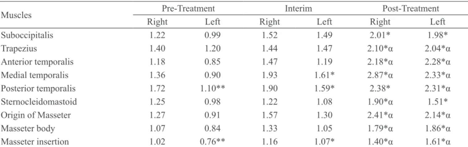
Table 1 – Pressure pain threshold before treatment, at the 11th week of treatment (interim) and at the 22th session (last session).
Muscles Pre-Treatment Interim Post-Treatment
Right Left Right Left Right Left
Suboccipitalis 1.22 0.99 1.52 1.49 2.01* 1.98*
Trapezius 1.40 1.20 1.44 1.47 2.10*α 2.04*α
Anterior temporalis 1.18 0.85 1.47 1.19 2.18*α 2.28*α Medial temporalis 1.36 0.90 1.93 1.61* 2.87*α 2.33*α Posterior temporalis 1.72 1.10** 1.90 1.59* 2.38* 2.31*α Sternocleidomastoid 1.25 0.98 1.22 1.08 1.90*α 1.51* Origin of Masseter 1.27 0.91 1.57 1.30 2.41*α 2.14*α
Masseter body 1.07 0.84 1.33 1.05 1.79*α 1.86*α
Masseter insertion 1.02 0.76** 1.16 1.07* 1.40*α 1.61*α
& Difference between both sides; * Difference in relation to pre-treatment; α Difference in relation to the interring assessments; ANOVA test
Cervical range of motion (CROM) was evaluataed with
a speciic CROM device15, measuring lexion, extension,
lateral inclination, and rotation of the head. All assess-ments were conducted by the same professional. Treatment sessions happened twice a week, lasting from 40 to 50 minutes each, over 18 weeks, corresponding to
20 sessions plus baseline and inal assessments. Sessions
consisted of manual therapy techniques as well as orien-tation for daily stretching of the cervical region and of the chest. The patient was also instructed to walk at least 3 times per week for at least 30 minutes, and received recommendations about proper posture.
The following procedures were conducted in all ses-sions: global stretching, practicing for diaphragmatic breathing, cervical stretching, cervical traction, myofa-scial liberation, and deactivation of trigger points. Global stretching inthe positions frog on the ground or frog in the air was alternated at different sessions, with duration of 20 to 30 minutes. During the exer-cises, the patient was instructed to use diaphragmatic breathing (Figure 1).
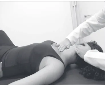
Stretching, traction, and myofascial release: cervical stretching was maintained for 30 seconds for each
position (lexion, extensions, rotations and inclina -tion)16; tractions were maintained for 2 minutes. After
that, myofascial release was conducted during 20 minutes (Figure 2).
Deactivation of trigger points: progressive compres-sion of trigger points was used. When the patient re-ported pain, compression was suspended until total
pain relief. After that, digital pressure was again in-creased until new onset of pain. Procedures were re-peated until total relief of pain, or maximum duration of 60 seconds per trigger point.
After 6 sessions, considerable changes were not ob-served. She had important muscular tenderness, and had lost motivation to continue on therapy. From the 6ª ses-sion on we added static Static Ultrasound (IBRAMED´s Sonopulse compact 1MHz) at the dose of 1.5 W/cm2, per
1.5 minutes on the TPs that could trigger the headache. This resource was always used toward the end of the ses-sions and on no more than 2 trigger points.
PPT at the three assessment times was compared with
two-way ANOVA with post test (Bonferroni) (p < 0.05).
CROM values were compared with the chi-square test . After 20 sessions the frequency of headaches was
sig-niicantly reduced, from 8 to 2 attacks per month. Pain
intensity ranged from 5 to 10 at baseline and from 5 to 8 at treatment end. Duration decreased from over 1 day to a mean of 7 hours.
In the pre-treatment assessment, the left side had signii -cantly lower PPT as contrasted to the right side. In the in-terim assessment, PPT increased overall bilaterally and
reached signiicant values for the medium and posterior
temporal muscles on the left side and for the insertion of the masseter on the left side. At treatment end, PPT
val-ues were signiicantly increased relative to pre-treatment
and interim assessments. For the occipital muscles (both sides), right posterior temporal muscle, and left stern-ocleidomastoid, differences happened only relative to
baseline. For all others, differences were also signiicant
as contrasted to the interim assessments (Table 1). In the initial assessment she had 16 trigger points that Figure 1 – Global stretching
could trigger the headache, which decreased to 14 in the interim assessment, and to just 5 at the end of the program.
As for the cervical range of motion, pre-treatment as-sessment demonstrated small reduction in movement
amplitude for lexion, lateral and left inclination and cer -vical extension. Cer-vical range of motion increased for
all parameters, although without signiicant difference.
DISCUSSION
The patient responded poorly to manual therapy tech-niques and traditional physical therapy. This may have
relected a possible baseline state of central sensitiza -tion. Because her pain was reproduced during myofa-scial release, as well as following deactivation of trigger points, peripheral manipulation may have contributed to the central sensitization. In the initial assessment she had 16 trigger points that could trigger the headache; they were 14 in the interim assessment, and only 5 at the end of the program. After onset of Static Ultrasound therapy, pain improved considerably, and was no longer triggered by manual therapy techniques.
Although Static Ultrasound did not cause relevant tem-perature increase, since parameters involved low heat-ing rate and short application time, trigger points were probably stimulated by Static Ultrasound non-thermal effects which knowingly yield physiological effects and segmental analgesia8. Reduction in muscular hyperalgia
was probably followed by decreased nociceptive input to the central nervous system and, as a consequence, central and peripheral sensitization were decreased. We found important reduction in the PPT as a function of therapy. Furthermore, there was a reduction in the PPT between both sides of the head, suggesting once more that peripheral and central sensitization were reduced. Headache severity may be result of nociceptive mechan-isms from the periphery, as well as from intra and extra-cranial tissues. The input converges into the second or-der neurons, located in the trigeminal nucleus caudalis. Because brain hypersensitivity is a dynamic condition
inluenced by nociceptive inputs17, nociceptive
lateral-ization may facilitate unilateral hypoalgesia in patients with headache18. This may have explained the increased
severity of pain on the head, as well as the lower PPT values pre-treatment.
Although the cervical range of motion did not signii -cantly vary as a function of treatment, it may be that minor changes were enough to yield better muscular balance and cervical mobility. Finally, the outcome
may have also relected the effects of adjuvant therapy,
such as Global stretching, diaphragmatic breathing and others, since they improve corporal image, increasing self-perception and muscular tonus rebalance and tend to decrease muscular tension, therefore helping to relieve the trigger points. Altogether, the intervention improved migraine headaches that had been previously resistant to pharmacological therapies.
The association of the two types of therapies, Static Ultrasound and manual therapy techniques, has de-creased duration and frequency of migraine attacks and
the number of active trigger points, as well as signii -cantly increasing PPT. Physical therapy provides several non-invasive methods that focus on triggering and per-petuating headache factors20.
Limited studies demonstrated the beneits of Static
Ultrasound treatment for the release of trigger points in migraine patients. This case report demonstrated that the combination of manual therapy techniques and Static Ultrasound offers an interesting non-pharmaco-logical alternative for migraine patients with myofa-scial involvement. Future studies, as randomized
clin-ical studies, must be conducted to conirm our results
in a large scale.
REFERENCES
1. Stovner Lj, Hagen K, Jensen R, et al. The global burden of headache: a documentation of headache prevalence and disability worldwide. Cephalalgia 2007;27(3):193-210. 2. Goadsby PJ, Lipton RB, Ferrari MD. Migraine--current understanding and treatment. N Engl J Med 2002;346(4):257-70.
3. Bevilaqua-Grossi D, Chaves TC, Gonçalves MC;
Moreira VC, Canonica AC, Florencio LL, Bordini CA,
Speciali JG, Bigal ME. Pressure Pain Thereshold in the Craniocervical muscles of women with episodic and chronic migraine: A Controlled Study. Arquivos de neu-ropsiquiatria 2011.
4. Fernández-de-Las-Peñas C, Cuadrado ML, Pareja JA. Myofascial trigger points, neck mobility and forward head posture in unilateral migraine. Cephalalgia. 2006 Sep; 26(9):1061-70.
5. Giamberardino MA, Tafuri E, Savini A, et al. Contri-bution of myofascial trigger points to migraine symp-toms. J Pain. 2007 Nov;8(11):869-78.
6. Bevilaqua-Grossi D, Pegoretti KS, Goncalves MC et al. Cervical Mobility in Women With Migraine. Head-ache 2009;49:726-731.
stretch-ing window, part two: rate of thermal decay in deep muscle following 1-MHz ultrasound. J Athl Train 1996;31(2):139-43.
8. Draper DO, Castel JC, Castel D. Rate of temper-ature increase in human muscle during 1 MHz and 3 MHz continuous ultrasound. J Orthop Sports Phys Ther 1995;22(4):142-50.
9. Baker RJ, Bell GW. The effect of therapeutic
modal-ities on blood low in the human calf. J Orthop Sports
Phys Ther 1991;13(1):23-7.
10. Srbely JZ, Dickey JP, Lowerison M, et al. Stimu-lation of myofascial trigger points with ultrasound in-duces segmental antinociceptive effects: a randomized controlled study. Pain 2008;139(2):260-6.
11. Majlesi J, Unalan H. High-power pain threshold ultra-sound technique in the treatment of active myofascial trigger points: a randomized, double-blind, case-control study. Arch Phys Med Rehabil 2004;85(5):833-6.
12. Fryer G, Hodgson L. The effect of manual pressure release on myofascial trigger points in the upper trapez-ius muscle. J Bodywork Mov Ther 2005;9(4):248-55. 13. Fernandez de las Penas C, Alonso-Blanco C, Fer-nandez-Carnero J, et al. The immediate effect of is-chemic compression technique and transverse friction massage on tenderness of active and latentmyofascial trigger points: a pilot study. J Bodywork Mov Ther 2006;10(1):3-9.
14. Gerwin RD. A review of myofascial pain and ibro -myalgia factors that promote their persistence. Acupunct
Med 2005;23(3):121-34.
15. Hou CR, Tsai LC, Cheng KF, et al. Immediate effects of various physical therapeutic modalities on cervical myofascial pain and trigger-point sensitivity. Arch Phys Med Rehabil 2002;83(10):1406-14.
16. Chaves TC, Nagamine HM, de Sousa LM, et al. Intra and Inter agreement of pressure pain threshold for masti-catory structures in children reporting orofacial pain re-lated to temporomandibular disorders and symptom-free children. J Orofac Pain 2007;21(2):133-42.
17. Youdas JW, Carey JR, Garrett TR. Reliability of meas-urements of cervical spine range of motion--comparison of three methods. Phys Ther. 1991 Feb;71(2):98-104 18. Cunha AC, Burke TN, França FJ, et al. Effect of global posture reeducation and of static stretching on pain, range of motion, and quality of life in women with chronic neck pain: a randomized clinical trial. Clinic 2008;63(6):763-70.
19. Herren-Gerber R, Weiss S, Arendt-Nielsen L, et al. Modulation of central hyper-sensitivity by nociceptive input in chronic pain after whiplash injury. Pain Med 2004;5(4):366-76.
20. Lawler SP, Cameron LD. A randomized, controlled trial of massage therapy as a treatment for migraine. Ann Behav Med. 2006;32(1):50-9.
Presented in September, 09, 2011. Accepted in January, 24, 2012.