136e
Rev Bras M ed Esporte _ Vol. 12, Nº 3 – M ai/Jun, 2006 1. Fisioterapeuta Graduado pela Universidade Estadual do Oeste doPara-ná (UNIOESTE) – Campus de Cascavel.
2. M édico Radiologista, Docente da Disciplina de Radiologia e Diagnósti-co por Imagem da UNIOESTE.
3. Docente da Disciplina de Histologia da UNIOESTE. 4. Acadêmico de Fisioterapia da UNIOESTE.
5. Docente da Disciplina de Eletrotermofototerapia e Supervisor de Está-gio em Fisioterapia Ortopédica e Traumatológica da UNIOESTE. Received in 20/6/05. Final version received in 27/9/05. Approved in 4/1/06.
Correspondence to: M ono.
Endereço para correspondência: Gladson Ricardo Flor Bertolini, Rua Luiz Vilw ock, 125, B, Nova Cidade – 85803-177 – Cascavel, PR. E-mail: [email protected]; [email protected]
Effect of continuous therapeutic ultrasound
in rabbit grow th plates
Andersom Ricardo Fréz1, Déborah Ariza1, José Roberto Leonel Ferreira2, Éder Paulo Belato Alves3,
Gláucia Ritter Breda4, Lígia Aline Centenaro4, Tiago Kijoshi Ueda4 and Gladson Ricardo Flor Bertolini5
O
RIGINALA
RTICLEKeyw ords: Ultrasound therapy. Grow th plate.
ENGLISH VERSION
ABSTRACT
The therapeutic efficiency of ultrasound has become an indis-pensable tool of physical therapy treatment in cases of alteration by lesions and in many kinds of sickness. How ever, in pediatric cases the use of ultrasound is controversial due to possible distur-bance and damage to the grow th plate. The aim of this study is to find out if the continuous ultrasound presents alteration effects on the grow th plate of female rabbits. Eight New Zealand female rab-bits w ith tw o months of age w ere tested. They w ere treated by
continuous therapeutic ultrasound w ith doses of 1 W/cm2 in the
lateral region of the right knee joint for five minutes, during 10 days, w ith an interval of tw o days after five applications. The left knee joint w as used as a control. The histological analysis show ed an alteration in the thickness of the grow th plate on the treated side 24.40% bigger than in the left knee joint of the control (p < 0.0001). On other hand, the radiological analysis did not show any difference betw een the limbs. The conclusion w as that the thera-peutic ultrasound produced significant histological alterations in the cartilage thickness on the treated side according to the manner it w as used in the experiment. Such fact suggests an acceleration in the grow th plate metabolism.
INTRODUCTION
The ultrasound is know n as a therapeutic treatment w hich pre-sents clinical evidence for the use in lesions of the soft tissues
w ith the aim to speed healing(1-3), decrease articulatory stiffness,
reduce pain and muscular spasm(4), increase the colagen
synthe-sis(5), and regeneration of the peripherical innervation(6).
How ever, many reports on its application in the
muscular-skel-etical system have been focusing in the fractures consolidation(4),
presenting good results in the formation(7-8) and consolidation of
the bone corn, in animal models and humans as w ell(7,9-10). M
ore-over, the ultrasound can increase the osteoblasts density(11),
stim-ulate the cellular matrix secretion(12-13) and of grow th factors
en-volved in fractures consolidation(14).
All these effects make therapeutic ultrasound to be chosen for the treatment of alterations caused by lesions and many illnesses. How ever, there is some controversy about its use in pediatric
pa-tients, since some studies show possible disturbs and damage to
the grow th plate(4,15), w hich may lead them to a premature fusion,
w ith consequent discrepancy in the limbs length or an uneven
grow th in one of the sides of the bone(16).
Being an efficient resource in many cases in the clinical prac-tice, it is believed that further studies are need to evaluate w heth-er its use in regions of endochondral ossification should be ap-plied, once the literature is not conclusive in relation to the possible deleterial effects(15,17-18).
The aim of this study is to evaluate the effects of the treatment w ith continuous therapeutic ultrasound on the grow th plate of New Zealand female rabbits’ tibia during the development phase through histological analysis. M oreover, after the initial grow th phase, w e verify w hether there are alterations in the tibia through radiological exam.
M ETHODS
The present w ork is characterized as experimental w ith qualita-tive and quantitaqualita-tive analysis 08 New Zealand female rabbits w ith 2 months of age w ere used. The animals w ere kept in cages and standardized food and drinkable w ater w ere given ad libitum. All of them received the therapeutic ultrasound application in the lateral region of grow th plate of the right tibia and the left back paw w as used as control.
The ultrasound equipment used w as the Avatar II model by KLD,
w ith 1 M Hz head and Effective Radiation Area (ERA) of 4 cm2. It
w as previously calibrated. The used parameters w ere: dose of 1,0
W/cm2 (Spacial and Temporal Averages – SATA), during 5 min, in a
subaquatic manner (immersion in w ater), in continuous emission, using a plastic dish w ithout filling as a container for application to absorb the ultrasound, and treated w ater as conduction means. The animals had their right back limbs tricotomized in the first and sixth days of the therapy.
The procedures occurred in 10 sessions, daily conducted, w ith an interval of 2 days after the 5th session, simulating the proce-dure in the physical therapy in humans. The animals w ere manual-ly held during the session.
10 days after the last session, 4 animals w ere sacrificed. The back limbs w ere desarticulated in the hips and ankles, and the fe-mur and tibia cleaned from the soft parts, only the junction by the ligaments and the knee articulatory capsule being kept. They w ere all kept in 10% formol.
Rev Bras M ed Esporte _ Vol. 12, Nº 3 – M ai/Jun, 2006
137e
checked, that is, their thickness, and 9 measurement in the lateral, intermedium and medium regions w ere done. The program used for the measurements w as the Image-Pro-Plus 3.0.1. The paired t test of the Prism 2 01 program w as used for the statistical analy-sis.
The 4 remaining animals w ere evaluated through radiological exams at 4 months old, trying to observe any epiphysary alteration and/or significative discrepancy betw een the length of the treated limb and the control. The equipment used for the radiological exam w as the Tridoros 812E model by Siemens The used parameters w ere: 150 mA, t = 0,02 s and kV 49, Emic Limex, and anterior-posterior incidence. The analysis w as conducted by tw o radiology physicians, members of the Brazilian College of Radiology, both w ith 20 years of experience in the field. Initially, a comparative analysis of the right and left low er limbs w as done. The radiolo-gists did not have previous know ledge of w hich limb w as the ob-ject of the study. The physicians w ere then notified about the meth-odology and the aim of the experiment and a new analysis w as conducted.
RESULTS
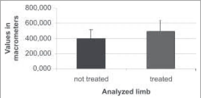
The obtained values in the histological analysis show ed that in the control side, the size of the grow th plates w as around 393,359
± 117,934 µm, and, the treated side presented thickness of 489,354
± 147,386 µm, having a significant variation of 24,40% more on
the treated side (p < 0,0001) (graphic 1).
proteoglycan cellular matrix. Reher et al.(5) credit the stimulus of
bone formation to the colagen synthesis, Li et al.(11) credit the
ac-celeration to the. E2 prostaglandin synthesis, though..
Zhang et al.(13) applied pulsed ultrasound of low intensity for 20
min, during 7 days, in sternum bone culture of chicken embrios and concluded that the acceleration in the bone formation occurs by the increase in the condrocytes hypertophy directly in the ter-minal differentiation, w ithout causing hypertrophy in the hyaline
cartilage. How ever, Nolte et al.(17) using the same parameters, but
in rat fetuses cells culture, observed increase in the endochondral calcification after 7th day of application, and the histological analy-sis presented healthy conditions, similarly to the control. They also credited the ossification to the direct effect in the osteoblasts and cartilage calcification.
Spadaro and Albanese(21) studied the effect of the low intensity
ultrasound in grow ing rats’ long bones during four w eeks, w ith daily 20 min applications. The results show ed that the ultrasound did not interfere in the femur and tibia’s grow th nor allow ed in-crease of the bone mineral density.
Wiltink et al.(15) using metatarsus fragments of mice fetuses,
treated w ith 1 M Hz ultrasound, pulsed 100 Hz (2 ms: 8 ms), w ith
0,77 W/cm2 (Spacial Average and Temporal Peak – SATP) observed
increase in the length of the proliferative zone of the grow th carti-lage and lack of alterations in the hypertrofic carticarti-lage region, con-cluding that there w as proliferation w ithout stimulation of the cel-lular differentiation.
Rabbits w ere chosen for this w ork due to their suitable size and easy positioning of the ultrasound head. The tibia proximal region w as selected due to its skin localization, making the positioning of the transductor easy, and, the ultrasound application w ay w as sub-aquatic due to greater easiness for handling of the head in the animal’s back limb region. M oreover, there are few acoustic prob-lems betw een w ater and the soft tissue, leading to little reflex-ion(22).
Deleterious action of the ultrasound may be observed in this present study, once its action over the grow th plate w as seen, increasing its thickness and leading to evidence of increase in the metabolism in the plate w ith probable cellular proliferation.
These data agree w ith the histological findings by Lyon et al.(4)
w ho using continuous ultrasound, 1 M Hz, 2,2 W/cm2, for 20 min,
in New Zealand female rabbits, obtained a significant increase in the cartilage thickness. Nevertheless, alterations in the metaphys-is contour, undmetaphys-istinctive epiphysmetaphys-is line and tibia plateau in curved shape causing varo angulation, occurred in these authors’ radio-logical analysis, being the last one not pointed in the actual study.
The radiologists did not find suggestive or characteristic of pos-sible vipos-sible bone alterations radiological signs through the meth-od. During the reevaluation the physicians w ere uncertain about the epiphyses spaces, w hich w as credited to the slight asymme-try of positioning.
Correlating to the clinical practice, the results point that the ther-apeutic ultrasound applied w ith the present parameters, should not be used for treatment in sites w ith open grow th plate, since the histological exam show ed significant alterations, presenting epiphyses stimilation. Although the reports presented in the radio-logical evaluation did not point any visible alterations, the continu-ation of these exams w ith the adult animal could present deformi-ties, a fact neglected in the present study.
In the present study, the small number of animals used for the histological and radiological analysis w as seen as a restriction. Besides that, other w ays of the ultrasound presentation w ere not evaluated, namely variations in the density of exit pow er, in the base frequency (1 or 3 M hz) and in the w ay of giving (continuous or pulsed).
Graphic 1 – Comparison betw een the obtained values in the histological analysis of the thickness of the plates on the not-treated side and treated side
In the radiological analysis the medical report discriminated: soft tissues w ith usual attenuation of the X-rays; bone trabeculae w ith homogeneous distribution and density, w ithout areas of osteope-nia visible through the method; straight and regular surfaces w ith preserved spaces; symmetrical grow th plates w ith usual shape and density. No radiological evidence of bone demineralization ar-eas w as found. After the explanation on the w ork methods, there w as a new evaluation from the radiologists, w ho w ere in doubt about the plates spaces, how ever, they attributed such doubts to a slight positioning asymmetry.
DISCUSSION
The objective of this study w as to determine w hether the ther-apeutic ultrasound may cause alterations in the grow th plate and clinically detectable alterations to the radiological exam. According to Yang et al.(19) and Gebauer et al.(20) the ultrasound stimulus to the
formation of the bone corn is due to the synthesis increase of the proteins of the extracellular matrix in the cartilage, possibly alter-ing the chondrocytes maturation and the formation of the
endoch-ondral bone. Parvizi et al.(12) credit the acceleration in the
138e
Rev Bras M ed Esporte _ Vol. 12, Nº 3 – M ai/Jun, 2006CONCLUSIONS
The therapeutic ultrasound of 1 M Hz, w ith dosage in 1 W/cm2
SATA, w henever applied in the continuous w ay does not produce alterations tangible through the radiological exam in female rabbits at 4 months old. On the other hand, w hen a histological analysis is done, significant alterations in the cartilage thickness on the treat-ed side are observtreat-ed, suggesting an acceleration in the plate me-tabolism. New studies are necessary for further know ledge on the morphological and histological responses on the grow th plate. Fur-thermore, considering correlation betw een studies in animal mod-els and human modmod-els, a safe dosage for pediatric patients, guar-anting the lack of possible deleterial effects, should be tried.
ACKNOWLEDGM ENTS
To Presmam Enterprises – Dealing and M aintenance Ltda., for the ther-apeutic ultrasound equipment calibration used in the present study.
All the authors declared there is not any potential conflict of inter-ests regarding this article.
REFERENCES
1. Karnes JL, Burton HW. Continuous therapeutic ultrasound accelerates repair of contraction-induced skeletal muscle damage in rats. Arch Phys M ed Rehabil 2002; 83:1-4.
2. Shomoto K, Takatori K, M orishita S, Nagino K, Yamamoto W, Shimohira T, et al. Ef fects of ultrasound therapy on calcificated tendonitis of the shoulder. J Jpn Phys Ther Assoc 2002;5:7-11.
3. Zeqiri B, Hodnett M . New method for measuring the effective radiating area of physiotherapy treatment heads. Ultrasound M ed Biol 1998;24:761-70. 4. Lyon R, Liu XC, M eier J. The effects of therapeutic vs. high-intensity ultrasound
on the rabbit grow th plate. J Orthop Res 2003;21:865-71.
5. Reher P, Elbeshir ELI, Harvey W, M eghji S, Harris M . The stimulation of bone formation in vitro by therapeutic ultrasound. Ultrasound M ed Biol 1997;23:1251-8.
6. Crisci AR, Ferreira AL. Low -intensity pulsed ultrasound accelerates the regener-ation of the sciatic nerve after neurotomy in rats. Ultrasound M ed Biol 2002;28: 1335-41.
7. Busse JW, Bhandari M , Kulkarni AV, Tunks E. The effect of low -intensity pulsed ultrasound therapy on time to fracture healing: a meta-analysis. CM AJ 2002;166: 437-41.
8. Warden SJ, Favaloro JM , Bennell KL, M cmeeken JM , Ng KW, Zajac JD, et al. Low -intensity pulsed ultrasound stimulates a bone forming response in UM R-106 cells. Biochem Biophys Res Commun 2001;3:443-50.
9. Haar G. Therapeutic ultrasound. Eur J Ultrasound 1999;9:3-9.
10. Rodriguez O, M onreal R. Low -intensity pulsed ultrasound therapy for the accel-eration of bone fracture repair. Anais do Congresso Brasileiro de Engenharia Biomédica 2000;650-655. Florianópolis, 2000.
11. Li JGR, Chang WHS, Lin JCA, Sun JS. Optimum intensities of ultrasound for PGE2 secretion and grow th of osteoblasts. Ultrasound M ed Biol 2002;26:683-90.
12. Parvizi J, Parpura V, Greenleaf JF, Bolander M E. Calcium signaling is required for ultrasound-stimulated aggrecan synthesis by rat chondrocytes. J Orthop Res 2002; 20:51-7.
13. Zhang ZJ, Huckle J, Francomano CA, Spencer RGS. The influence of pulsed low -intensity ultrasound on matrix production of chondrocytes at different stag-es of differentiation: an explant study. Ultrasound M ed Biol 2002;28:1547-53.
14. Ito M , Azuma Y, Ohta T, Komoriya K. Effects of ultrasound and 1,25-dihydroxyvi-tamin d3 on grow th factor secretion in co-cultures of osteoblasts and endothelial cells. Ultrasound M ed Biol 2000;26:161-6.
15. Wiltink A, Nijw eide PJ, Oosterbaan WA, Hekkenberg RT, Helder PJM . Effects of therapeutic ultrasound on the endochondral ossification. Ultrasound M ed Biol 1995;21:121-7.
16. Carty H. Children’s sports injuries. Eur J Radiol 1998;26:163-76.
17. Nolte PA, Klein-Nulend J, Albers GHR, M arti RK, Semeins CM , Goei SW, et al. Low intensity ultrasound stimulates endochondral ossification in vitro. J Orthop Res 2001;19:301-7.
18. Pessina AL, Volpon JB. Aplicação do ultra-som terapêutico na cartilagem de cres-cimento de coelhos. Rev Bras Ortop 1999;34:347-54.
19. Yang KH, Parvizi J, Wang SJ, Lew allen DG, Kinnick RR, Greenleaf JF, et al. Expo-sure to low -intensity ultrasound increases aggrecan gene expression in a rat femur fracture model. J Orthop Res 1996;5:802-9.
20. Gebauer GO, Lin SS, Beam HA, Vieira PJ, Parsons R. Low -intensity pulsed ultra-sound increases the fracture callus strength in diabetic BB w istar rats but does not affect cellular proliferation. Clin Orthop Res 2002;20:587-92.
21. Spadaro JA, Albanese SA. Application of low -intensity ultrasound to grow ing bone in rats. Ultrasound M ed Biol 1998;24:567-73.