Cancer survival among children and
adolescents at a state referral hospital in
southeastern Brazil
Sobrevida do câncer em crianças e
adolescentes em hospital de referência
estadual na região sudeste do Brasil
Gláucia Perini Zouain-Figueiredo 1
Eliana Zandonade 2
Maria Helena Costa Amorim 3
1Hospital Infantil Nossa Senhora da Glória. Rua Mary Ubirajara, 205. Santa Lúcia. Vitória, ES, Brasil. CEP: 29.027-080.
E-mail: [email protected]
2 Departamento de Estatística. Centro de Ciências Exatas. Universidade Federal do Espírito Santo. Vitória, ES, Brasil. 3Departamento de Enfermagem. Centro Biomédico. Universidade Federal do Espírito Santo. Vitória, ES, Brasil.
Abstract
Objectives: to analyze the patient characteristics and evaluate overall survival, survival according to demographic variables, the most common tumor groups and subgroups, the stages of disease, and risk factors after at least 5 years among children and adolescents with cancer who were admitted to a state referral hospital between 2000 and 2005.
Methods: the Kaplan-Meier method was employed to estimate survival. The survival curves were compared using the log-rank test. The Cox regression model was used to estimate the effect of independent variables.
Results: a total of 571 new cases were registered. The most frequent cancer groups were leukemia (34%), lymphoma (18%), and central nervous system (CNS) tumors (15%).The overall survival rate was 59%. The risk factors associated with lower survival were an age of more than 4 years or less than 1 year, the presence of CNS tumors, and non-localized disease.
Conclusion: although this was not a population-based study, it provides important epidemiological information about a state where population data on childhood and adolescent cancer are scarce and where hospital-based data do not exist. The survival rate found here should serve as a framework for future improvements, helping to guide policymakers focused on pediatric oncology in the state.
Key words Cancer, Children, Adolescents, Survival analysis
Resumo
Objetivos: analisar as características dos pacientes, avaliar sobrevida global e sobrevida de acordo com variáveis demográficas, grupos e subgrupos dos tumores mais comuns, extensão da doença e fatores de risco após pelo menos cinco anos em crianças e adolescentes com câncer, admitidos em hospital de referência estadual entre 2000 e 2005.
Métodos: método de Kaplan-Meier foi utilizado para estimar função de sobrevida. Curvas de sobre-vida foram comparadas utilizando teste de log-rank. Modelo de Regressão de Cox foi utilizado para estimar o efeito das variáveis independentes.
Resultados: um total de 571 novos casos foi ana-lisado. Os grupos mais frequentes foram leucemias (34%), linfomas (18%) e tumores de sistema nervoso central (SNC) (15%). Taxa de sobrevida global foi 59%. Fatores de risco associados à menor sobrevida: idade superior a quatro anos ou menor de um ano, presença de tumores do SNC e doença não localizada. Conclusões: embora não seja um estudo popula-cional, este trabalho fornece importantes informações epidemiológicas de um estado onde estudos de base populacional do câncer na infância e adolescência são escassos e hospitalares não existem. A taxa de sobrevida encontrada deve servir como um marco para melhorias futuras ajudando os formuladores de políticas com foco na área de oncologia pediátrica no Estado.
Introduction
In Brazil, it is estimated that nearly 3% of all new cases of cancer occur in individuals who are less than 19 years old.1The proportion of childhood cancers is related to the population structure. Low-income countries have younger median ages and higher proportions of children in their populations than do high-income countries. In Africa as a whole, 4.8% of cancers occur in children younger than 15 years, compared with only 0.4% in Europe.2 Overall, an estimated 11,530 cases among Brazilians under 19 years old were reported in 2012, which corre-sponds to a median incidence of 160 per million (154.3 per million children).3Approximately 230 of these cases are expected to occur in the State of Espírito Santo (ES) in southeastern Brazil.1
Advances in childhood (ages 0-14 years) and adolescent (ages 15-19 years) cancer treatment and in diagnostic techniques have resulted in an increase in survival in developed countries over the last four decades. The five-year survival has increased from less than 30% in the 1960s to approximately 80% in the 2000s for all childhood cancers and occasionally surpasses 90% in certain subgroups.4-6
Multiple interrelated factors are responsible for the outlook for childhood cancer. Factors inherent to the disease itself affect survival rates (e.g., the type of tumor and its location). However, improvements in the survival of children and adolescents with cancer have been observed over the past few decades, including the integration of high-quality referral centers, clinical research, and education, and an approach to primary care emphasizing the early detection and improvement of pediatric cancer units by well-trained healthcare professionals. Indeed, the strong link between the quality of care and research activities depends on the alignment of systematic investment and national healthcare priorities to support the improvement of care for children with cancer.7-10
In 2011, cancer was the second leading cause of death (7.9% of the total) in Brazil for people aged between 1 and 19 years, surpassed only by acci-dents.11
The aim of the present study was to analyze the characteristics and overall survival after at least 5 years of children and adolescents aged under 19 years who were admitted to the Hospital Infantil Nossa Senhora da Glória (HINSG), the exclusive state referral center, for cancer treatment between 2000 and 2005. The study also aimed to analyze survival according to different demographic va-riables, groups of diseases, stages of disease, and the
most common disease subgroups and risk factors.
Methods
A survival study was conducted using hospital-based, retrospective secondary data.
The HINSG is a general pediatric hospital located in Vitória, Espírito Santo, a state in south-eastern Brazil, and it is the exclusive state referral center of the Sistema Único de Saúde - SUS (Unified Health System) for the treatment of cancer in indi-viduals under 19 years of age. Every year, nearly 100 children and adolescents from all areas of ES and neighboring states are referred to this center for the treatment of various malignancies. Bone marrow transplants (BMTs) and local treatment for retinoblastoma (Rb) are not provided in the state of ES through the SUS.
The study population consisted of children (aged 0-14 years) and adolescents (aged 15-18 years and 364 days) who were admitted to the HINSG between January 2000 and December 2005 as new cases of primary malignant neoplasms. Patients with a history of treatment with chemotherapy or radio-therapy and patients who had a second neoplasm or a benign neoplasm, except for neoplasms of the central nervous system (CNS), were excluded from this study.
Groups and subgroups were created in accor-dance with the third edition of the International Classification of Childhood Cancer, 3rd version (ICCC-3). Cases of nonmalignant tumors of the brain and CNS were thus included in the sample.
All of the neoplasms were confirmed cytomor-phologically or histopathologically, except for certain CNS tumors for which a biopsy could not be performed because of technical and surgical limita-tions. In these cases, diagnoses were made using imaging studies.
The data were obtained from the information included in the medical records of the HINSG Oncology Unit. This study examined the number of cases by sex, age group, state of origin, most frequent groups and subgroups of disease, and the extent of disease. These variables were considered as predictors in the survival model, whose exposure variable was the survival time in months.
following clinical staging systems, which have been adopted worldwide by collaborative groups focusing on childhood and adolescent cancer treatment, were adopted to control this variable: Ann Arbor staging for Hodgkin’s lymphoma (HL), Murphy staging for non-Hodgkin’s lymphoma (NHL), the National Wilms’ Tumor Study Group staging for nephroblas-toma, Evans staging for neuroblastoma (NB), the tumor-node-metastasis (TNM) cancer staging system for bone tumors, and the TNM staging system for soft tissue tumors in cases of osteosarcoma (OTS) or rhabdomyosarcoma (RMS).
Special attention was paid to leukemia, lymphoma, and CNS tumors, which are the most frequent forms of cancer in these age groups.
The outcome variable was the time (in months) from the date of diagnosis until death from cancer (the event of interest). Other related events (discharge from the health service, abandonment of treatment, and loss to follow-up) were censored. The deadline to determine the patients’ outcomes was December 31st, 2010.
A follow-up analysis was conducted by exa-mining the medical records for at least 60 months after a diagnostic conclusion. The Health Mortality Information System, made available by the State of Espírito Santo Department of Health, provided confirmation and the dates of deaths that did not occur in the HINSG.
A survival analysis was used to analyze the data. The Kaplan-Meier method was used to determine the overall survival time and the survival time according to the study variables. The survival curves were
compared among the different categories using the log-rank method. A multivariate Cox proportional hazard model (hazard ratio, or HR) was used to verify the independent effects of the study variables that had a statistical significance of up to 10%. Tests were performed to verify the proportionality of the risks (Schoenfeld residuals, test of proportional hazard in the Cox model, and graphical tests) and to analyze the model residual (martingale residual graphics and deviance from the fitted model).12The final significance level adopted in the model was 5%.
The statistical analysis of the data was performed using Statistical Package for the Social Sciences (SPSS) software (version 18.0) and the R language.
The present study was conducted in accordance with Resolution 196/96 of the National Health Council and was approved by the HINSG’s Human Research Ethics Committee (number 88/2010) on November 23rd, 2010.
Results
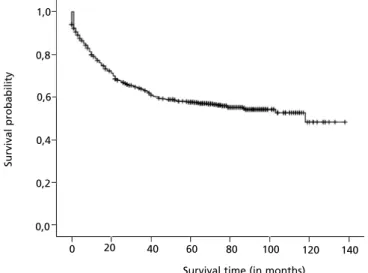
The sample population consisted of 571 patients with new cases of cancer who were admitted during the 6-year study period. Among the subjects, there was a survival rate of 59% after 5 years, and 247 patients died. The mean survival time was 82.7 (95% CI, 77.3-88.1) months (maximum observation time of 120 months), with a standard error of 2.7 months. Figure 1 shows the survival function calculated from the Kaplan-Meier curves for the outcome of death from cancer.
Figure 1
Survival curve using the Kaplan-Meier method for 571 children and adolescents under 19 years of age admitted to the HINSG with new cases of cancer. City of Vitória, ES, Brazil. 2000-2005.
1,0
0,8
0,6
0,4
0,2
0,0
20
0 40 60 80 100 120 140
Survival time (in months)
Su
rvi
va
l
p
ro
b
a
b
il
it
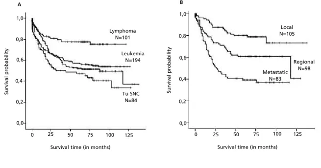
Table 1 shows a description of the survival time according to the independent variables of interest. Figures 2A and 2B show the Kaplan-Meier curves
that reflect the results in the table for the most frequent groups and the extent of the disease, respec-tively.
Table 1
Absolute and relative frequencies of the categories of the variables, number of deaths and mean survival time, with confidence intervals obtained using the Kaplan-Meier method and corresponding p-values of the log-rank tests for children and adolescents under 19 years of age admitted to the HINSG with new cases of cancer. City of Vitória, ES, Brazil, 2000-2005.
Survival time (in months)
Clinical-epidemiological variables N % p-value
Deaths Mean 95% LL 95% UL
ICCC-3= International Classification of Childhood Cancer, 3rdversion; * Proportion in relation to the reference group; †In total, 278 cases
of leukemia and CNS tumors were excluded. No information was available for seven patients. Sex
Female Male
Age group (in years) 0 |-- 1
1 |-- 5 5 |-- 10 10 |-- 15 15 |-- 19 Region of origin Espírito Santo North Central Metropolitan South Other States
Most common ICCC-3 groups I Leukemia
II Lymphoma III CNS tumors IV-XII Other I-XII Total
Most common ICCC-3 subgroups (n=415) I (a) Acute lymphocytic leukemia I (b) Acute myeloid leukemia II (a) Hodgkin’s lymphoma
II (b) Non-Hodgkin’s and non-Burkitt’s lymphoma II (c) Burkitt’s lymphoma
III (b) Astrocytoma IV (a) Neuroblastoma VI (a) Nephroblastoma VIII (a) Osteosarcoma IX (a) Rhabdomyosarcoma Extent of disease †(n=286)
278 cases of leukemia and CNS tumors were excluded from the analysis of the extent of the disease. We analyzed the disease variables for 10 groups comprising a total of 286 cases. Data could not be obtained from the medical records for seven patients (2.4%). Advanced diseases (regional and metastatic) were the most common, representing 63% of cases. As this variable was correlated with the survival time, the Cox regression analysis was performed with an N of 286 patients.
The patients’ ages varied from 22 days to 18 years and 11 months. The majority of patients were in the 1- to 4-year age group (34%). Most subjects were male (52%), with a male to-female ratio of 1.1/1.
508 patients (89%) lived in the state of ES, and an absolute majority lived in the metropolitan area of Vitória. The neighboring state of Bahia was home to 9% of the patients.
In the 12 groups, leukemia was the most frequent malignant neoplasm (34%), and acute lymphocytic leukemia was the most frequent subgroup (73%). The second most frequent group was lymphoma (18%), which was predominantly represented by HL and Burkitt’s lymphoma (BL). HL and BL accounted for 31% and 30% of the lymphoma subgroups,
respectively. CNS tumors were the third most common neoplasm (15%), and astrocytoma (AST) was the most common subgroup (36%). Combined, these three groups comprised most of the cases (67%). The remaining 33% of the cases were tumors of the sympathetic nervous system (5%), Rb (< 1%, two cases), renal tumors (9%), liver tumors (< 1%), bone tumors (6%), soft tissue sarcomas (7%), germ cell tumors (3%), carcinomas (2%), and other unspecified malignant tumors (< 1%, one case).
Table 1 shows the main functions of unadjusted survival for the study variables. The probability of overall survival for girls was higher than for boys, although this difference was not statistically signifi-cant (p=0.078). There were differences in the proba-bility of survival according to age group (p=0.011) and the three most frequent groups of cancer (p=0.001) (Figure 2A). Comparison of the 10 most common subgroups revealed differences in terms of the probability of overall survival (p=0.001). Advanced disease was associated with a significant reduction in the probability of survival (Figure 2B). Figures 2A and 2B show excessive mortality after the 60-month survival period for CNS tumors and regional diseases.
Figure 2
Survival curves for 571 children and adolescents under 19 years of age admitted to the HINSG with new cases of cancer according to the most frequent groups. City of Vitória, ES, Brazil. 2000-2005.
(A) The extent of disease*, except leukemia and CNS tumors; (B) using the Kaplan-Meier method. * The extent of disease data for seven patients could not be obtained from the medical records.
1,0
0,8
0,6
0,4
0,2
0,0
25
0 50 75 100 125
1,0
0,8
0,6
0,4
0,2
0,0
25
0 50 75 100 125
Lymphoma N=101
Leukemia N=194
Tu SNC N=84
Local N=105
Regional N=98 Metastatic
N=83
A B
Su
rvi
va
l
p
ro
b
a
b
il
it
y
Su
rvi
va
l
p
ro
b
a
b
il
it
y
There were no differences in survival associated with the patients’ region of origin.
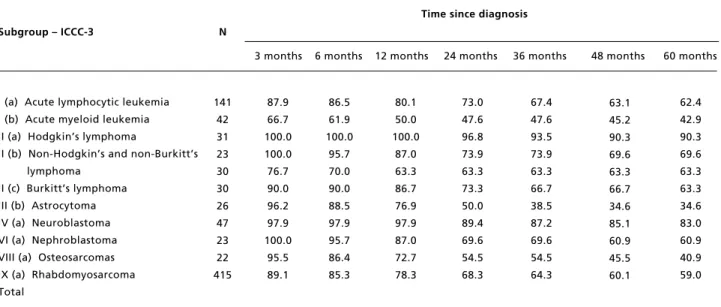
The probability of survival was calculated for the 10 most frequent subgroups, totaling 415 cases (Table 2). Higher survival was found for individuals with HL (90.3%) and nephroblastoma (83%) at 60 months. In addition, all deaths from BL occurred within the first 12 months after the date of diagnosis. Patients with acute myeloid leukemia (AML) had one of the lowest mean survival rates, both at 3 (66.7%) and 60 (42.9%) months. As cases of NB and RMS advanced, the survival time steadily decreased. The lowest survival rates occurred at the end of the 5-year period.
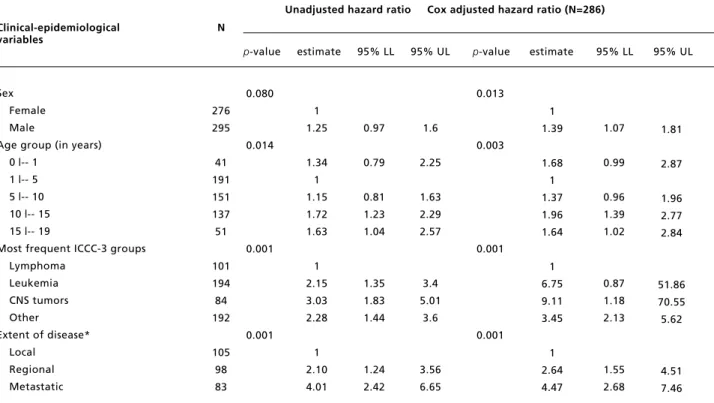
After adjusting for variables in the model, the multivariate analysis showed that males had a 1.39 (1.07-1.81) times greater risk of death (Table 3). Similarly, infants (< 1 year old), older children (the 5- to 9- and 10- to 14-year age groups), and adoles-cents had 1.68, 1.37, 1.96, and 1.64 times greater risk of death, respectively, compared with the risk in the 1- to 4-year age group. Patients with leukemia or
CNS tumors showed 6.75 and 9.11 times greater risk of death than patients with lymphoma. Regional and metastatic diseases were associated with a signifi-cantly higher risk of death. These patients had 2.64 and 4.47 times the risk associated with local diseases, respectively.
In the Cox analysis, we tested the assumptions of the proportional hazard model by graphing the Schoenfeld residuals. Using hypothesis testing, the violation of the assumption was observed for sex and the most frequent ICCC-3 groups, which after a new graphic analysis,12 did not present evidence of serious violation of the assumption of proportional risks for these variables. In the residual analysis, the martingale residuals suggested the presence of two atypical points (less than -2). However, when the deviance residuals, which are less asymmetric that the martingale residuals, were analyzed, the observed outliers were deemed to be acceptable. We conclude that there is evidence that the model adequately fits the data.
Table 2
Probability of survival for the 10 most frequent subgroups of cancer in children and adolescents under 19 years of age admitted to the HINSG with new cases of cancer. Vitória, ES, Brazil, 2000-2005.
Time since diagnosis
Subgroup – ICCC-3 N
3 months 6 months 12 months 24 months 36 months 48 months 60 months
ICCC-3= International Classification of Childhood Cancer, 3rdversion.
I (a) Acute lymphocytic leukemia I (b) Acute myeloid leukemia II (a) Hodgkin’s lymphoma
II (b) Non-Hodgkin’s and non-Burkitt’s lymphoma
II (c) Burkitt’s lymphoma III (b) Astrocytoma IV (a) Neuroblastoma VI (a) Nephroblastoma VIII (a) Osteosarcomas IX (a) Rhabdomyosarcoma Total
73.0 47.6 96.8 73.9 63.3 73.3 50.0 89.4 69.6 54.5 68.3
62.4 42.9 90.3 69.6 63.3 63.3 34.6 83.0 60.9 40.9 59.0 63.1
45.2 90.3 69.6 63.3 66.7 34.6 85.1 60.9 45.5 60.1 67.4
47.6 93.5 73.9 63.3 66.7 38.5 87.2 69.6 54.5 64.3 87.9
66.7 100.0 100.0 76.7 90.0 96.2 97.9 100.0
95.5 89.1
80.1 50.0 100.0
87.0 63.3 86.7 76.9 97.9 87.0 72.7 78.3 86.5
61.9 100.0
95.7 70.0 90.0 88.5 97.9 95.7 86.4 85.3 141
Table 3
Hazard ratios associated with the study variables and a multivariate model of 571 new cases of cancer among children and adolescents under 19 years of age admitted to the HINSG. City of Vitória, ES, Brazil, 2000-2005.
Unadjusted hazard ratio Cox adjusted hazard ratio (N=286)
Clinical-epidemiological N
variables
p-value estimate 95% LL 95% UL p-value estimate 95% LL 95% UL
ICCC-3: International Classification of Childhood Cancer, 3rdversion; * In total, 278 cases of leukemia and CNS tumors were excluded. The
extent of disease data for seven patients could not be obtained from the medical records. Sex
Female Male
Age group (in years) 0 |-- 1
1 |-- 5 5 |-- 10 10 |-- 15 15 |-- 19
Most frequent ICCC-3 groups Lymphoma
Leukemia CNS tumors Other
Extent of disease* Local
Regional Metastatic
1.6
2.25
1.63 2.29 2.57
3.4 5.01
3.6
3.56 6.65
1.07
0.99
0.96 1.39 1.02
0.87 1.18 2.13
1.55 2.68 1
1.39
1.68 1 1.37 1.96 1.64
1 6.75 9.11 3.45
1 2.64 4.47 0.013
0.003
0.001
0.001 0.080
0.014
0.001
0.001
0.97
0.79
0.81 1.23 1.04
1.35 1.83 1.44
1.24 2.42 1
1.25
1.34 1 1.15 1.72 1.63
1 2.15 3.03 2.28
1 2.10 4.01 276
295
41 191 151 137 51
101 194 84 192
105 98 83
1.81
2.87
1.96 2.77 2.84
51.86 70.55 5.62
4.51 7.46
Discussion
This is the first descriptive analysis of childhood and adolescent cancer in the state of ES, where data on childhood and adolescent cancer are limited to the information found in the population-based cancer registries (PBCRs) of the Metropolitan Area of Vitória (the capital and five neighboring cities) for the year 1997 alone.3,13,14
The proportional distribution of the three most common cancers found in the HINSG study according to the ICCC-3 is similar to that reported by 14 Brazilian PBCRs (Vitória was not included because the data covered less than 3 years), which have shown that the main groups of cancers are leukemia (18-41%), lymphoma (13-24%), and CNS tumors (7-17%).3
In other areas, leukemia has been reported to represent 25-35% of all cases. Leukemia was also the most common cancer (34%) in our study, occur-ring predominantly in acute forms. The second commonest childhood cancer in the HINSG series was lymphoma (18%), followed by CNS tumors (15%). The order of frequency of these first three
groups differs from the observations of PBCRs in developed countries, where CNS tumors are more common than lymphomas. However, the order is compatible with results found in the medical litera-ture for countries displaying the characteristics of developing countries, including Brazil.3,13,15-18
The order of frequency of subgroups in our series was similar to that found in other population studies. The percentage of the acute lymphoid leukemias (73%) in the present study is in accor-dance with the literature, in which this subgroup totaled 75-80% of all leukemias.15,17Likewise, the astrocytoma subgroup (36%) predominated among the CNS tumors, showing a similar trend to that of other populations in all countries. This finding was also consistent with the results of a Brazilian study conducted at a single center, where astrocytomas represented 37% of CNS tumors in patients less than 21 years old.15,19
of cancers in these subgroups (31% and 30%, respectively) in the present study. There is a high prevalence of anti-EBV antibodies in the population of Vitória.22This may explain the high proportion of lymphomas in our study, but it is also possible that CNS tumors were underdiagnosed, which would explain the lower number of cases compared with lymphoma. These observations need to be explored in further population-based registry studies.
According to data from a European PBCR, for all types of cancer, boys of any age have a higher risk of cancer than girls (overall sex ratio of 1.2:1). However, this ratio varies between different groups and subgroups of neoplasia.15 Between 1983 and 2005, the hospital-based cancer registry of the Instituto Nacional de Câncer observed that, of all cancer cases in children and adolescents, 54.5% were male, and 45.5% were female.13 Cancer in males was slightly more prevalent than in females, with a ratio of 1.1:1 in the HINSG study.
The pattern of diagnosis varies greatly between age groups, but on the whole, children at 1-4 years of age have the highest rates of diagnosis in most PBCRs.3,15In the present study, the most common age range was 1-4 years old (34%). Therefore, the results obtained in this study concerning the sex ratio and the most prevalent age group are similar to those found in other populations.
The analysis of survival time enabled compar-isons to be made between different categories of each study variable. The following were significant risk factors for lower survival rates in the HINSG sample: a higher age (compared with the 1- to 4-year age group), an age younger than 1 year, the presence of CNS tumors, and advanced-stage disease. There were no significant differences in survival rates for gender. In general, these results are comparable with those reported in a population-based setting.23-25
The survival rates varied between different groups and subgroups. Of the 10 most frequent subgroups in this analysis, the highest 5-year survival rates were found for HL and nephroblas-toma, whereas the worst outcomes were recorded for neuroblastoma, AML, and rhabdomyosarcoma. The low survival rate of patients with neuroblastoma or rhabdomyosarcoma in the HINSG sample can be partially attributed to the large number of patients with advanced stages of the disease. According to the literature, survival is inversely proportional to the advanced stage of disease, which underlines the benefits of early therapeutic intervention.2,25
Overall, the 5-year survival rate for all cancers combined for children and adolescents diagnosed at the HINSG between 2000 and 2005 was 59%. The
survival rates for childhood cancers vary substan-tially from country to country around the world.2,9,15,17,26The 5-year survival rates reported by the EUROCARE-4 study for all cancers combined were 81% in children and 87% in adoles-cents/young adults diagnosed with cancer between 1995 and 2002.24In Shanghai, China, from 2002-2005, the rate was 55.7%.23In sub-Saharan Africa, the overall 5-year survival varies from 5% in Côte d’Ivoire (before 2001) to 70% in South Africa.27
It is important to note that 5-year survival is not equivalent to cure. In a large population-based study in Nordic European countries, the 5-year survivors who died during subsequent follow-up experienced recurrence or progression of the primary tumor or had a second malignancy. Some patients also died from causes unrelated to cancer and its treatment.28 The decline in the curves corresponding to survival rates beyond 5 years observed in the present study indicates a need for future follow-up studies focusing on the causes of death and the risk factors. These studies will allow therapies to address the causes of death. It would also be interesting to inves-tigate the morbidity and the quality of life of the survivors.
The establishment of effective population-based childhood cancer registries is one of the key policies for improving the care of children with cancer around the world.10
The analytical limitations of this study primarily concern the scarcity of PBCR data in the state of ES. It was thus difficult to make comparisons with other referral services in Brazil or with the population data for cancer in children and adolescents from the state of ES. For these reasons, it was not possible to corre-late the cases admitted to the HINSG from the state of ES (508 cases in 6 years) with the incidence of cases in the state as a whole.
treated by private practices in the city of Vitória or go to another treatment center in another state. There is a substantial need for future population-based studies to establish the reliability of the relationship between the groups and subgroups of neoplasms among children and adolescents in the state of ES.
The present study summarized the occurrence of childhood cancer and survival rates at the single public state treatment center for oncologic diseases in children and adolescents. Although this was not a population-based study, it supplies important
epidemiological data from a state where population-based studies of childhood and adolescent cancer do not exist.
The clinical outcomes of the children and adoles-cents included in this sample reflect the health care that they received in both primary and tertiary care. The survival rate found in this analysis still lies below that found in developed countries’ population reports23,24 and should serve as a framework for future improvements, helping to guide policymakers to focus on pediatric oncology in the state of ES.
References
1. Instituto Nacional de Câncer. Estimativa 2012: incidência de câncer no Brasil. Rio de Janeiro: INCA; 2011. 2. Magrath I, Steliarova-Foucher E, Epelman S, Ribeiro RC,
Harif M, Li CK, Kebudi R, Macfarlane SD, Howard SC. Paediatric cancer in low-income and middle-income coun-tries. Lancet Oncol. 2013; 14: e104-16.
3. De Camargo B, de Oliveira Santos M, Rebelo MS, Reis RS, Ferman S, Noronha CP, Pombo-de-Oliveira MS. Cancer incidence among children and adolescents in Brazil: First report of 14 population-based cancer registries. Int J Cancer. 2010; 126: 715-20.
4. Gatta G, Capocaccia R, Stiller C, Kaatsch P, Berrino F, Terenziani M. Childhood cancer survival trends in Europe: a EUROCARE Working Group study. J ClinOncol. 2005; 23: 3742-51.
5. Smith MA, Seibel NL, Altekruse SF, Ries LAG, Melbert DL, O'Leary M, Smith FO, Reaman GH. Outcomes for children and adolescents with cancer: challenges for the twenty-first century. J Clin Oncol. 2010; 28 (15): 2625-34. 6. Stiller CA, Kroll ME, Pritchard-Jones K. Population
survival from childhood cancer in Britain during 1978-2005 by eras of entry to clinical trials. Ann Oncol. 2012; 23: 2464-9.
7. Arora B, Banavali SD. Pediatric oncology in India: past, present and future. Indian J Med Paediatric Oncol. 2009; 30: 121-3.
8. Pritchard-Jones K, Sullivan R. Children with cancer: driving the global agenda. Lancet Oncol. 2013; 14: 189-91. 9. Howard SC, Metzger ML, Wilimas JA, Quintana Y, Pui CH, Robison LL, Ribeiro RC. Childhood cancer epidemi-ology in low-income countries. Cancer. 2008; 112: 461-72. 10. Pritchard-Jones K, Pieters R, Reaman GH, Hjorth L,
Downie P, Calaminus G, Naafs-Wilstra MC, Steliarova-Foucher E. Sustaining innovation and improvement in the treatment of childhood cancer: lessons from high-income countries. Lancet Oncol. 2013; 14 (3): e95-104.
11. Brasil. Ministério da Saúde. Secretaria Executiva. Datasus Informações de Saúde. Estatísticas vitais. Mortalidade 2011 [access Jul 07, 2013]. Available at: http://www.datasus.gov.br
12. Colosimo EA, Giolo SR. Análise de sobrevivência aplicada. ABE, Projeto Fisher. São Paulo: Editora Edgard Blücher; 2006.
13. Ministry of Health. National Cancer Institute (INCA). Childhood and adolescent cancer in Brazil: data from mortality and population-based registries. Rio de Janeiro: National Cancer Institute; 2009. [access Jun 20, 2013].
Available at:
http://www2.inca.gov.br/wps/wcm/connect/inca/portal/hom e
14. Reis RS, Santos MO, Thuler LCS. Incidência de tumores pediátricos no Brasil. Rev Bras Cancer. 2007; 53 (1): 5-15. 15. Kaatsch P. Epidemiology of childhood cancer. Cancer Treat
Rev. 2010; 36: 277-85.
16. Arora RS, Eden T, Kapoor G. Epidemiology of childhood cancer in India. Indian J Cancer. 2009; 46: 264-73. 17. Braga PE, Latorre MRDO, Curado MP. Câncer na infância:
análise comparativa da incidência, mortalidade e sobrevida em Goiânia (Brasil) e outros países. Cad Saúde Pública. 2002; 18 (1): 33-44.
18. Mirra AP, Latorre MRDO, Veneziano DB, Editores. Incidência, mortalidade e sobrevida do câncer da infância no município de São Paulo. São Paulo: Tomgraf Editora; 2004. Disponível em: http://bvsms.saude.gov.br/bvs/publi-cacoes/inca/registro_de_cancer_2004.pdf
19. Pinho RS, Andreoni S, Silva NS, Cappellano AM, Masruha MR, Cavalheiro S, Vilanova LCP. Pediatric central nervous system tumors: a single-center experience from 1989 to 2009. J Pediatr Hematol Oncol. 2011; 33: 605-9.
20. Pedrosa MF, Pedrosa F, Lins MN, Pontes NT, Hanois Falbo G. NonHodgkin’s lymphoma in childhood: clinical and epidemiological characteristics and survival analysis at a single center in Northeast Brazil. J Pediatr (Rio J.). 2007; 83: 547-54.
21. Magrath I. Epidemiology: clues to the pathogenesis of Burkitt lymphoma. Br J Haematol. 2012; 156: 744-56. 22. Figueira-Silva CM, Pereira FEL. Prevalence of
23. Bao PP, Zheng Y, Wu CX, Peng P, Gong YM, Huang ZZ, Fan W. Population-based survival for childhood cancer patients diagnosed during 2002-2005 in Shanghai, China. Pediatr Blood Cancer. 2012; 59: 657-61.
24. Gatta G, Zigon G, Capocaccia R, Coebergh JW, Desandes E, Kaatsch P, Pastore G, Peris-Bonet R, Stiller CA; EURO-CARE Working Group. Survival of European children and young adults with cancer diagnosed 1995-2002. Eur J Cancer. 2009; 45 (6): 992-1005.
25. Baade PD, Youlden DR, Valery PC, Hassall T, Ward L, Green AC, Aitken JF. Population-based survival estimates for childhood cancer in Australia during the period 1997-2006. Br J Cancer. 2010; 103 (11): 1663-70.
26. Sullivan R, Kowalczyk JR, Agarwal B, Ladenstein R, Fitzgerald E, Barr R, Steliarova-Foucher E, Magrath I, Howard SC, Kruger M, Valsecchi MG, Biondi A, Grundy P, Smith MA, Adamson P, Vassal G, Pritchard-Jones K. New policies to address the global burden of childhood cancers. Lancet Oncol. 2013; 14 (3): e125-35.
27. Hadley LG, Rouma BS, Saad-Eldin Y. Challenge of pedi-atric oncology in Africa. Semin Pediatr Surg. 2012; 21 (2): 136-41.
28. Moller TR, Garwicz S, Barlow L, Winther JF, Glattre E, Olafsdottir G, Olsen JH, Perfekt R, Ritvanen A, Sankila R, Tulinius H. Decreasing late mortality among five-year survivors of cancer in childhood and adolescence: A popu-lation-based study in the Nordic countries. J Clin Oncol. 2001; 19: 3173-81.