1 . Full Professor. Scientific Coordinator of Fundação Cardiovascular São Francisco de Assis, Belo Horizonte, MG, Brazil.
2 . MSc. Fellow Postgraduation Course of Cardiovascular Surgery Francisco de Assis Truth is Jesus Cardiovascular Foundation; Cardiologist Physician, Belo Horizonte – MG, Brazil.
3 . MSc Fellow Postgraduation Course of Cardiovascular Surgery Francisco de Assis Truth is Jesus Cardiovascular Foundation; Resident Fellow Cardiovascular Surgery Madre Tereza Hospital, Belo Horizonte-MG, Brazil.
4 . Full Professor Cardiovascular Surgery, School of Medicine Vale do Sapucaí University, Pouso Alegre-MG, Brazil; Cardiovascular Surgery – BSCVS.
Otoni Moreira Gomes
1, Mônica de Mônico Magalhães
2,
Rafael Diniz Abrantes
3,
Elias Kallás
4Pantoprazol oferece proteção miocárdica semelhante ao pré-condicionamento isquêmico: estudo em
corações isolados de ratos
Pantoprazole provides myocardial protection
similar to ischemic preconditioning: experimental
study of isolated hearts of rats
Research performed at the Experimental Laboratory São Francisco de Assis Truth is Jesus Cardiovascular Foundation, Belo Horizonte, MG, Brazil.
Correspondence
Otoni Moreira Gomes. Rua José do Patrocínio, 522 – Santa Mônica – Belo Horizonte, MG, Brazil – CEP 31525-160
E-mail: [email protected]
Article received on April 8th, 2011 Article accepted on June 23rd, 2011
Abstract
Objective: To evaluate pantoprazole effect in the functional
recovery of isolated hearts of rats, submitted to ischemia and reperfusion with and without ischemic preconditioning.
Methods: In four groups of eight Wistar breed rats, the
hearts were removed after anesthesia and perfused with Krebs-Henseleit solution (95% O2, 5% CO2, 37°C). GI, GII, GIII and GIV hearts were submitted to ischemia (20 min) and reperfusion (30 min). In GII and GIV, preconditioning was performed with 5 min of ischemia and 5 min of reperfusion before 20 min of the ischemia period induction. In GIII and GIV pantoprazole 100 mg was done before a 20 min-period of ischemia induction. Heart Rate (HR), Coronary Flow (CoF), Systolic Pressure (SP), +dP/dt and -dP/dt were registered before (t0) and after reperfusion (t30). Kruskal-Wallis (P<0.05) test was used.
Results: There were no differences (P>0.05) between
groups among HR and CoF values. Differences occurred between groups, I and II, III and IV at t30 with SP reduced for 32% mean value in GI, 65% GII, 65% GIII, and 73% GIV; The t30 + dP/dtmax were 34% in GI, 61% GII, 63% GIII and 72% GIV. The t30 -dP/dtmax were GI 28%, GII 63%, GIII 75
% and GIV 75%; (P<0.05). There were no significant differences in the SP, +dP/dtmax, and -dP/dtmax between Groups II, III and IV results.
Conclusion: The administration of pantoprazole before
induction of ischemia significantly protected the myocardial functional recovery with the results of SP, + dP / dtmax and -dP/dtmax similar to the ischemic preconditioning against ischemia-reperfusion.
Descriptors: Ischemic preconditioning. Myocardium.
Myocardial ischemia. Myocardial infarction.
Resumo
Objetivo: Avaliar o efeito do pantoprazol na recuperação
funcional de corações isolados de ratos submetidos à isquemia e reperfusão com e sem pré-condicionamento isquêmico.
Métodos: Em quatro grupos de oito ratos Wistar, após
anestesia os corações foram removidos e perfundidos com Krebs-Henseleit (95% O2, 5% CO2, 37°C). Os corações de GI, GII, GIII e GIV foram submetidos a 20' de isquemia e 30’de reperfusão. Em GII e GIV realizou-se
condicionamento com 5' de isquemia e 5' de reperfusão antes dos 20' de isquemia. Em GIII e GIV, pantoprazol 100mcg foram injetados imediatamente antes dos 20' de isquemia. Frequência cardíaca (FC), Fluxo Coronariano (FCo), Pressão Sistólica (PS), + dP/dt e -dP/dt foram registrados em (T0) e (t30). Estatística: Kruskal-Wallis (P <0,05).
Resultados: Não houve diferenças (P> 0,05) entre grupos
nos valores de FC e de CFo. Diferenças (P <0,05) ocorreram entre GI e GII, GIII e GIV, com PS t30 reduzida para 32% GI, 65% GII, 65% GIII e 73% GIV. Em t30 + dP/dtmax 34% GI, 61% GII, 63% GIII e 72% GIV. A -dP/dtmax t30 GI 28%, GII
63%, GIII 75% e GIV 75%. Não houve diferença estatística (P< 0,05) nos valores de PS, +dP/dtmax e -dP/dtmax entre os GII, GIII e GIV.
Conclusão: A administração do pantoprazol antes da
indução da isquemia protegeu significativamente a recuperação funcional miocárdica com resultados de SP, +dP/ dtmax e -dP/dtmax semelhantes aos do pré-condicionamento isquêmico contra lesão de isquemia-reperfusão.
Descritores: Precondicionamento isquêmico. Miocárdio.
Isquemia miocárdica. Infarto do miocárdio.
INTRODUCTION
The reduction or interruption of blood flow to the myocardium implies the limitation or cessation of oxygen and metabolic substrates to cardiomyocyte, with consequent functional, structural and metabolic diseases. The diversion of cellular aerobic metabolism to anaerobiosis leads to accumulation of intracellular ions and metabolites. The Ischemia promotes rapid depletion of cellular reserves of adenosine triphosphate (ATP) and creatine phosphate, leading to depression of contractile function and ATP-dependent ion pumps [1]. The accumulation of Ca++ and Na + in the cytosol, with concomitant loss of K+ to the extracellular environment, affects the membrane potential and transmembrane ion gradient.
These changes lead to accumulation of metabolites, cellular acidosis, increased osmotic load, the formation of reactive oxygen species and eventually to activation of enzymes sensitive to Ca++. In response, morphological changes may begin to occur. The activated proteases, especially by the sudden increase in the concentration of Ca++ and free radicals, degrade the myofibrillar proteins of the cytoskeleton, while the lipases disrupted membranes, causing their rupture and cell death [2].
The return of oxygen and nutrients causes the cytosolic Ca++, already elevated during ischemia, to increase further with reperfusion, due to the additional entry of these ion channels by voltage-gated Ca++. Besides these channels, Ca++ also increases the cytosol, entering through the sarcolemma by the Na+/ Ca++ and also by the release of Ca++ by sarcoplasmatic reticulum [3,4].
Therefore, during ischemia/reperfusion, cardiomyocytes are exposed to a harmful sequence of events with two main components, namely: ischemia and reperfusion injury and data from the experimental study suggest that the damage caused by reperfusion is proportional to that caused by
ischemia [5]. It is also known that involvement of the myocardium is not homogeneous throughout its length, with areas of greater or lesser involvement, depending on the duration and intensity of ischemia, or both. Thus, cardiomyocytes can be found in different states of reversible and irreversible damages [6].
The reversible lesion is characterized functionally by complete restoration of myocardial function after normalization of blood flow. This myocardial dysfunction is reversible, and it may last hours or days, despite the restoration of blood flow. This type of damage may not result in changes of cell ultrastructure [7].
This is why the change of heart function is one of the earliest and most frequent clinical signs and hemodynamically perceived in most cases, avoiding the urgent need for histopathological studies on trans-and post-operative and clinical care of patients with myocardium ischemia.
Among the mechanisms and resources to protect against the degeneration of myocardial ischemia reperfusion has highlighted the special technique of ischemic preconditioning by Murry et al. [8] discovery in 1988, awarded the Nobel Prize for Medicine, demonstrating a significant reduction in the area of myocardial necrosis by a succession of short periods of myocardial ischemia and reperfusion prior to the continuous maintenance of coronary reperfusion.
Although capable of use in the prevention of induced lesions in surgical procedures involving percutaneous or direct surgery and periods of coronary occlusion, the surgical ischemic preconditioning may not be used in the vast majority of patients susceptible to coronary occlusions in environments outside hospitals or inside hospitals without cardiovascular surgical routine.
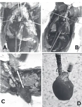
A perfusion cannula was introduced and secured in the ascending aorta, preventing injury to the aortic cups to ensure adequate coronary perfusion; then the left atrium was opened and a multiperforated cannula was inserted and externalized through the left ventricle apex. Finally, the pulmonary artery was opened at its origin to facilitate the drainage of the right ventricle. Following, the hearts were removed at a maximum of a 3-minute time after the procedure, and after 10-minute reperfusion, the multiperforated cannula was substituted by the catheter with balloon for the ventricular function control (Figure 1).
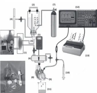
All hearts were reperfused with Krebs-Henseleit solution (aered/gassed with 95% O2 and 5% CO2, at 37°C, using a modified, disposable Langendorff system, model São Francisco de Assis Truth is Jesus Cardiovascular Foundation disposable Langendorff modified system. ServCor, Comex Ind. & Com Ltda) (Figure 2) with circulation system and water heating and telethermometer models (Braile Biomédica), biomonitor BESE®, and a matricial printer Epson®. After ten minutes of coronary reperfusion, the transventricular catheter was removed, and a pre-calibrated balloon catheter was introduced into the left ventricle. [9], in 1986, demonstrating the inhibitory effect of the H+/
K+ pump in the parietal cells of the stomach to reduce the acidity of gastric juice and treatment of peptic ulcer. Later, other H+/ K+ pump inhibitors had been developed, including the lansoprazole, pantoprazole, rabeprazole and tenatoprazole [10] being the most frequent the prescription of omeprazole and pantoprazole.
Although they have been developed with the specific goal of therapy for treating gastric hyperacidity in recent years the proton pump inhibitors were associated with benefits in the treatment of angina pectoris and myocardial ischemia, including the fact that his administration has reduced the frequency of anginal attacks and electrocardiographic signs of myocardial ischemia during an exercise treadmill test in studies of coronary heart disease [11-13]. It was recently proposed and demonstrated the protective effect of omeprazole on induced myocardial ischemia in isolated rat hearts [14], suggesting the effect of this drug by induced preconditioning.
Another particular aspect inherent to the clinical use of H+/ K+ pump inhibitor is the fact that pantoprazole has interactive reactions with less significant drug antagonism as proved with antiplatelet treatment suggesting their preferred choice in these circumstances.
However, there are still no specific experimental demonstrations showing the protective effect of pantoprazole in coronary ischemia-reperfusion nor his action and interference with the phenomenon of ischemic preconditioning of the myocardium.
The aim of the present study was to analyze the effects of pantoprazole in the protection of the functional recovery of isolated rat hearts subjected to ischemia-reperfusion with and without ischemic preconditioning.
METHODS
The study was approved by the local Institutional Ethics Committee and was conducted according to the guidelines for animal experimentation of the Brazilian College on Animal Experimentation (COBEA). It was studied the isolated hearts of 32 Wistar breed rats with 280 g mean body weight. After analgesia induced by sulphuric ether P.A spontaneous inhalation and anaesthesia by intraperitoneal injection of ketamine 10 mg and xylazine 2 mg. The animals underwent a wide-range median thoracotomy and 500 IU of heparin were regularly injected into the right atrium. For safety and quickness in the procedure preparation, the ascending aorta was isolated with the 3-wire technique [14,15]. The first wire pulls and lifts the aortic root to protect the cusps and to prevent ventricular cannulation; the second wire pulls and lifts the cranial (superior) end of the ascending aorta; and the third wire is passed between the first two to tie off the aorta on the cannula after it was inserted.
The eight hearts of Group I (GI) and Group II (GII) were submitted to 20-min ischemia and 30-min reperfusion. In GII heart’s intracoronary injection of pantoprazole100 mg was done immediately before the 20 min ischemia period of induction. The eight hearts of Group III and Group IV were submitted to 5-min ischemia and 5-min reperfusion preconditioning before the 20-min ischemia induction In Group IV heart’s intracoronary injection of pantoprazole 100 mcg was done immediately before the 20-min ischemia period of induction. In summary, Group I consisted of hearts without preconditioning and without pantoprazole; Group II without preconditioning and with pantoprazole; Group III with preconditioning and without pantoprazole; and Group IV with preconditioning and with pantoprazole.
The following parameters were registered after the 15-min stabilization period (t0), and after a 30-minute period past coronary reperfusion was initiated (t30): Heart Rate (HR/bpm), Coronary Flow (CoF ml/min), Systolic Pressure (SP mmHg), +dP/dtmax and -dP/dtmax. Kruskal-Wallis nonparametric statistical analysis (P<0.05) was used. All P values < 0.05 were considered statistically significant.
RESULTS
No differences (P>0.05) were found between groups regarding HR and CoF results (Table 1). The SP, +dP/dtmax, and -dP/dtmdifferences from t0/t30 was significant (P<0.05) in all groups but regarding SP, +dP/dtmax and –dP/dtmax after the reperfusion period (t30) differences (P<0.05) occurred between the results of Group I and Groups II, III and IV, with SP averages ranging from 139 (t0) to 44 mmHg, 32% (t30) in GI, from 134 to 87 mmHg, 65% in GII, from 132 to 86 mmHg, 65% in GIII and 132 to 96 mmHg, 73% in GIV the The +dP/dtmax declined from 3365 mmHg.min-1(t
0) to 1144 mmHg.min-1, 34% (t
30) in GI, from 3382 mmHg.min -1(t
0) to 2036 mmHg.min-1, 60% (t
30) in GII, from 3300 mmHg.min -1(t
0) to 2175 mmHg.min-1, 63% (t
30) in GIII and from 3226 mmHg.min-1(t
0) to 2334 mmHg.min
-1, 72% (t
30) in GIV. The – dP/dtmax declined from 1569 mmHg.min-1(t
0) to 468 mmHg.min-1, 28% (t
30) in GI, from 1366 mmHg.min -1(t
0) to 768 mmHg.min-1, 56% (t
30) in GII, from 1042 mmHg.min -1(t
0) to 725 mmHg.min-1, 67% (t
30) in GIII and from 940 mmHg.min
-1(t
0) to 706 mmHg.min
-1, 75% (t
30) in GIV (Table 2, Figure 3). Differences in the SP, +dP/dtmax, and –dP/dtmax results between Groups II, III and IV were no significant.
Fig. 2 – Isolated Heart System Diagram - 1. Termal exchanger, 2. Perfusate reservoir. 3. Microfilter (20 ì), 4. Gauge/Manometer, 5. Telethermometer, 6. Chamber, 7. Carbogen (95% 02 +5% C02), 8. Heart, 9. Intraventricular balloon, 10. Drug injector, 11. Flow Collector, 12. ECG and Ventricular Pressure monitor, 13. Printer, 14. Disposable set
Table 1. Heart rate and Coronary flow average results Groups I II III IV t0 292 ± 10.2 271 ± 23.0 269 ± 38.7 277 ± 19.0
t30 255 ± 20.6 257 ± 51.2 242 ± 31.4 240 ± 44.0
% 87 94 90 87 t0 20 ± 1.6 20 ± 0.5 18 ± 2.1 19 ± 1.4
t30 12 ± 1.5 13 ± 3.0 13 ± 0.2 12 ± 2.2
% 60 65 67 63
FC-bpm FCo – ml/min
Groups: I - Ischemia, II – Ischemia + preconditioning
III – Ischemia + Pantoprazole, IV – Preconditioning + pantoprazole
Table 2. Systolic pressure, +dp/dt and -dp/dt average results Groups I II III IV t0 139±14.1 134± 11.0 132±10.5 132±10.0 t30 39±13.6 87±11.5 86±19.3 96±10.0 % 32 65 65 73 t0 3123±57.9 3605± 471.3 3300±32.7 3226±306.0 t30 930±7536 2011±446.5 2091±63.4 2334±316.0 % 34 61 63 72 +dp/dt mmHg.min-1
Groups: I-Ischemia, II-Ischemia + preconditioning, III-Ischemia + Pantoprazole, IV-Preconditioning + pantoprazole
PS mmHg t0 1277±19.3 1366±265.3 1254±16.1 940±146.0 t30 338±68.3 869±291.7 946±161.1 706±75.0 % 28 63 75 75
DISCUSSION
In 1986, Parks & Granger [16] and Murry et al. [8] demonstrated by the first time that reperfusion can be more harmful than ischemia separately and the phenomenon of myocardial protection by the ischemic preconditioning with reduction of the myocardium ischemia–reperfusion injury in dogs. Yellon et al. [17] demonstrated that the ischemic preconditioning protection also occurs in the human myocardium which raised a great interest extending this study also to other organs. However, the surgical ischemic preconditioning may not be used in the vast majority of patients susceptible to coronary occlusions in environments outside hospitals or inside hospitals without cardiovascular surgical routine.
New horizons for myocardial protection have arisen with research on proton pump H+/K+ performed by Lindberg et al. [9], in 1986, and by Nagashima et al. [18] demonstrating, in 1994, the existence of the proton pump H+/K+ ATPase in the myocardium as well. They studied their electrophysiological interference in the hearts of Guinea pigs [19], motivating further research, such as the multicenter trials GUARDIA and ESCAMI. These trials have carefully analyzed inhibitors such as eniporide and cariporide - the proton pump Na+/H+ blockers, emphasizing the protection against myocardial ischemia-reperfusion injuries, but with contrasting results and doubts about the prophylactic effect and indication of its therapeutic use [20]. More recently, Budzynski et al. [13], in 2008, described the beneficial effects of omeprazole in the protection of anginal attacks and ischemic electrocardiographic changes during the exercise test in patients with coronary heart diseases. Moffatand & Karmazyn [21], in 1993, demonstrated the possibility of pharmacological protection against degeneration of ischemia-reperfusion by inhibiting the Na+/ H+ ATPase proton pump. This was also confirmed
in subsequent research with Na+/ H+ ATPase inhibitors and amiloride cariporida [22,24]. In a recent study [14], it was found a significant protection of omeprazole against ischemia-reperfusion in isolated rat hearts.
These results associate the benefits of omeprazole with the results reported by studies addressing myocardial protection with ischemic preconditioning [8,25-28]. The pharmacokinetics basis may be related to changes in transmembrane H+/K+ gradients, the first markers of myocardial ischemia, responsible for the repolarization expressed in T-wave morphology of the electrocardiogram [29]. In 1998, the studies by Hotta et al. [30] in isolated hearts of Guinea pigs, using an analytic method for detecting fluorescent compounds, the fluorometry and magnetic resonance imaging, indeed confirmed the acute ionic intracellular changes of the myocardial ischemia-reperfusion with changes in pH and concentrations of cytosolic Na+, H+, and Ca++, thus confirming the blockade and protection by omeprazole as well.
Harmful effects of proton pump inhibitors, Na+/ H+ATPase, favoring respiratory failure [31] and stroke [32] provide important constraints for clinical use. It is also shown that among K+/H+ATPaseblocker proton pump inhibitors omeprazole has an effect more important than pantoprazole in the antiplatelet action of aspirin and clopidogrel drugs of wide clinical use in patients with cardiovascular diseases [33,34]. However, there are still no specific experimental demonstrations showing the protective effect of pantoprazole in coronary ischemia-reperfusion.
In the current investigation no differences (P>0.05) were found between groups regarding HR and CoF results. The SP, +dP/dtmax, and -dP/dtm differences from t0/t30 was significant (P<0.05) in all groups but regarding SP, +dP/ dtmax and –dP/dtmax after the reperfusion period differences occurred between the results of Group I and Groups II, III, and IV with SP averages reduced to 32% in the 30 minutes of reperfusion (t30) following the ischemia period in the hearts of Group I. It reduced to 65% in GII; 65% in GIII and 73% at (t30) in GIV. The +dP/dtmax declined to 34% (t30) in GI; 60% (t30) in GII; 63% (t30) in GIII and 72% (t30) in GIV. The –dP/dtmax declined 28% (t30) in GI; 56% (t30) in GII; 67% (t30) in GIII, and 75% (t30) in GIV, without no significant differences (P<0.05) in the SP, +dP/ dtmax, and –dP/dtmax results between Groups II, III and IV. Although with limited result’s interpretation considering only one period of induced myocardium ischemia and only one sequence of the ischemia and reperfusion periods for the preconditioning induction, the isolated organ study with exclusion of the in situ body physiological responses; the possible different physiopathology responses of different animal species and mainly regarding the particularities of the clinical application in human beings, it
REFERENCES
1. Jennings RB, Reimer KA. The cell biology of acute myocardial ischemia. Annu Rev Med. 1991;42:225-46.
2. Sommerschild HT, Kirkeboen KA. Preconditioning: endogenous defense mechanisms of the heart. Acta Anaesthesiol Scand. 2002;46(2):123-37.
3. du Toit EF, Opie LH. Modulation of severity of reperfusion stunning in the isolated rat heart by agents altering calcium flux at onset of reperfusion. Circ Res. 1992;70(5):960-7.
4. Kusuoka H, Camilion de Hurtado MC, Marban E. Role of sodium/calcium exchange in the mechanism of myocardial stunning: protective effect of reperfusion with high sodium solution. J Am Coll Cardiol. 1993;21(1):240-8.
5. Du XJ, Anderson KE, Jacobsen A, Woodcock EA, Dart AM. Suppression of ventricular arrhythmias during ischemia-reperfusion by agents inhibiting Ins(1,4,5)P3 release. Circulation. 1995;91(11):2712-6.
6. Zaugg M, Schaub MC. Signaling and cellular mechanisms in cardiac protection by ischemic and pharmacological preconditioning. J Muscle Res Cell Motil. 2003;24(2-3):219-49.
7. Solaro RJ. Integration of myofilament response to Ca2+ with cardiac pump regulation and pump dynamics. Am J Physiol. 1999;277(6 Pt 2):S155-63.
8. Murry CE, Jennings RB, Reimer KA. Preconditioning with ischemia: a delay of lethal cell injury in ischemic myocardium. Circulation. 1986;74(5):1124-36.
9. Lindberg P, Nordberg P, Alminger T, Brandström A, Wallmark B. The mechanism of action of the gastric acid secretion inhibitor omeprazole. J Med Chem. 1986;29(8):1327-9.
was demonstrated in the present investigation that the pantoprazole administration before ischemia induction significantly protected the myocardium function recovery with similar results of the SP, +dP/dtmax and -dP/dtmax obtained with the ischemic preconditioning technique against the ischemia-reperfusion injury.
CONCLUSION
The pantoprazole administration before ischemia induction significantly protected the myocardium function recovery with similar results of the SP, +dP/dtmax and -dP/ dtmax obtained with the ischemic preconditioning technique against the ischemia-reperfusion injury.
10. Sachs G, Shin JM, Howden CW. Review article: the clinical pharmacology of proton pump inhibitors. Aliment Pharmacol Ther. 2006;23(Suppl 2):2-8.
11. Libby P, Bonow RO, Mann DL, Zipes D. Braunwald’s heart disease. Philadelphia:Saunders;2008.
12. Dobrzycki S, Baniukiewicz A, Korecki J, Bachórzewska-Gajewska H, Prokopczuk P, Musial WJ, et al. Does gastro-esophageal reflux provoke the myocardial ischemia in patients with CAD? Int J Cardiol. 2005;104(1):67-72.
13. Budzynski J, Klopocka M, Pulkowski G, Suppan K, Fabisiak J, Majer M, et al. The effect of double dose of omeprazole on the course of angina pectoris and treadmill stress test in patients with coronary artery disease: a randomised, double-blind, placebo controlled, crossover trial. Int J Cardiol. 2008;127(2):233-9.
14. Gomes OM, Magalhães MM, Abrantes RD. Proteção da recuperação functional do miocárdio pelo omeprazol após isquemia-reperfusão em corações isolados de ratos. Rev Bras Cir Cardiovasc. 2010;25(3):388-98.
15. Gomes OM, Gomes ES, Carvalho JI, Faraj M. Adaptações técnicas na preparação de Langendorff para estudo de corações isolados de pequenos animais. Coração. 1999;9:36-8.
16. Parks DA, Granger DN. Contributions of ischemia and reperfusion to mucosal lesion formation. Am J Physiol. 1986;250(6 Pt 1):G749-53.
17. Yellon DM, Alkhulaifi AM, Pugsley WB. Preconditioning the human myocardium. Lancet. 1993;342(8866):276-7.
18. Nagashima R, Odashiro K, Morita S. Evidence for the existence of myocardial H+-K+ATP and its electrophysiological effects. Jpn Heart J. 1994;35(suppl):473-4.
19. Nagashima R, Tsuda Y, Maruyama T, Kanaya S, Fujino T, Niho Y. Possible evidence for transmembrane K(+)-H+ exchange system in guinea pig myocardium. Jpn Heart J. 1999;40(3):351-64.
20. Avkiran M, Marber MS. Na(+)/H(+) exchange inhibitors for cardioprotective therapy: progress, problems and prospects. J Am Coll Cardiol. 2002;39(5):747-53.
21. Moffat MP, Karmazyn M. Protective effects of the potent Na/H exchange inhibitor methylisobutyl amiloride against post-ischemic contractile dysfunction in rat and guinea-pig hearts. J
Mol Cell Cardiol. 1993;25(8):959-71.
22. Hurtado C, Pierce GN. Inhibition of Na(+)/H(+) exchange at the beginning of reperfusion is cardioprotective in isolated, beating adult cardiomyocytes. J Mol Cell Cardiol. 2000;32(10):1897-907.
and nonfatal myocardial infarction in patients undergoing coronary artery bypass graft surgery: The EXPEDITION study. Circulation. 2003;108:3M.
24. Zhao ZQ, Corvera JS, Halkos ME, Kerendi F, Wang NP, Guyton RA, et al. Inhibition of myocardial injury by ischemic postconditioning during reperfusion: comparison with ischemic preconditioning. Am J Physiol Heart Circ Physiol. 2003;285(2):H579-88.
25. Saini HK, Dhalla NS. Modification of intracellular calcium concentration in cardiomyocytes by inhibition of sarcolemmal Na+/H+ exchanger. Am J Physiol Heart Circ Physiol. 2006;291(6): H2790-800.
26. Gomes OM, Valladares UF, Santos CH, Abrantes RD. Preconditioning abolishion by midazolam in isolated hearts of rats. Acta Cir Bras. 2009;24(3):173-6.
27. Pinheiro BB, Fiorelli AI, Gomes OM. Efeitos do pós-condicionamento isquêmico na função ventricular esquerda de corações isolados de ratos. Rev Bras Cir Cardiovasc. 2009;24(1):31-7.
28. Oliveira DM, Gomes ES, Mussivand T, Fiorelli AI, Gomes OM. Efeitos da N-acetilcisteína no precondicionamento isquêmico: estudo em corações isolados de ratos. Rev Bras Cir Cardiovasc. 2009;24(1):23-30.
29. Gomes OM, Gomes ES, Faraj M. Doença da discinesia miocárdica de estresse. Rev Bras Cir Cardiovasc. 2004;19(4):378-85.
30. Hotta Y, Fujita M, Nakagawa J, Ando H, Takeya K, Ishikawa N, et al. Contribution of cytosolic ionic and energetic milieu change to ischemia- and reperfusion-induced injury in guinea pig heart: fluorometry and nuclear magnetic resonance studies. J Cardiovasc Pharmacol. 1998;31(1):146-56.
31. Eichler W, Bechtel MJ, Klaus S, Heringlake M, Hernandez M, Toerber K, et al. Na /H+ exchange inhibitor cariporide: effects on respiratory dysfunction after cardiopulmonary bypass. Perfusion. 2004;19(1):33-40.
32. Gruberg L.EXPEDITION: Sodium-proton exchange inhibition to prevent coronary events in acute cardiac conditions tri. http://www.medscape.com/viewarticle/464672
33. Gilard M, Arnaud B, Cornily JC, Le Gal G, Lacut K, Le Calvez G, et al. Influence of omeprazole on the antiplatelet action of clopidogrel associated with aspirin: the randomized, double-blind OCLA (Omeprazole CLopidogrel Aspirin) study. J Am Coll Cardiol. 2008;51(3):256-60.