Abstract
Although many regenerative cell therapies are being developed to replace or regenerate ischaemic muscle, the lack of vasculature and poor persistence of the therapeutic cells represent major limiting factors to successful tissue restoration. In response to ischaemia, stromal cell-derived factor-1 (SDF-1) is up-regulated by the affected tissue to stimulate stem cell-mediated regenerative responses. Therefore, we encapsulated SDF-1 into alginate microspheres and further incorporated these into an injectable collagen-based matrix in order to improve local delivery. Microsphere-matrix impregnation reduced the time for matrix thermogelation, and also increased the viscosity reached. This double-incorporation prolonged the release of SDF-1, which maintained adhesive and migratory bioactivity, attributed to chemotaxis in response to SDF-1. In vivo, treatment of ischaemic hindlimb muscle with microsphere-matrix led to increased mobilisation of bone marrow-derived progenitor cells, and also improved recruitment of angiogenic cells expressing the SDF-1 receptor (CXCR4) from bone marrow and local tissues. Both matrix and SDF-1-releasing matrix were successful at restoring perfusion, but SDF-1 treatment appeared to play an earlier role, as evidenced by arterioles that are phenotypically older and by increased angiogenic cytokine production, stimulating the generation of a qualitative microenvironment for a rapid and therefore more effi cient regeneration. These results support the release of implanted SDF-1 as a promising method for enhancing progenitor cell responses and restoring perfusion to ischaemic tissues via neovascularisation.
K e y w o r d s: C y t o k i n e s , h y d r o g e l s , i n j e c t a b l e s , neovascularisation, regenerative medicine, vascular biology
*Address for correspondence: E.J. Suuronen
Division of Cardiac Surgery, University of Ottawa Heart Institute,
40 Ruskin Street, Ottawa K1Y4W7, Canada
Telephone Number: 1-613-798-5555 x19087 FAX Number: 1-613-671-5367; E-mail address: [email protected]
Introduction
Myopathies, such as ischaemic heart disease and peripheral arterial disease (PAD) are signifi cant killers in developed nations. In particular, PAD affl icts an estimated 27 million people in Europe and North America (Belch et al., 2003). This disease manifests when the fl ow of blood to extremities is acutely or chronically reduced (Selvin and Erlinger, 2004; Shamoun et al., 2008). Attributed to the lack of blood fl ow, symptoms typically include: pain, cramping, weakness and a poor ability to heal. To restore perfusion, current treatments include stent or vascular transplants with surgical intervention, or amputation; however, these options are invasive, carry risks, and amputation signifi cantly reduces quality of life. Therefore, non-invasive options for restoring perfusion would be invaluable for PAD patients. Recent years have seen the generation of many novel approaches for developing tissue substitutes to replace or regenerate ischaemic muscle; however, the supply of suffi cient and appropriate vasculature represents a major limiting parameter for a successful therapeutic approach (Ko et al., 2007; Kaully et al., 2009).
To augment reparative processes, regenerative therapies using bone marrow-, blood-, or tissue-derived progenitor cells have emerged; however, the results of these treatments are controversial and there is growing evidence to suggest that non-embryonic transplanted cells do not successfully integrate with host tissue (Murry et al., 2006; Suuronen et al., 2007). It is now believed that functional improvement results from neovascularisation and the restoration of blood fl ow to ischaemic muscle, and that this phenomenon is initiated, maintained, and enhanced by paracrine factors and secondary recruitment of progenitor cells (Cho et al., 2007; Formigli et al., 2007).
After ischaemic injury, an endogenous response to recruit the endogenous progenitor cells is initiated (Hofmann et al., 2005). Regarded as critical to this process is the up-regulation and release of the cytokine stromal cell-derived factor-1 (SDF-1) from ischaemic muscle (Askari et al., 2003) and the subsequent recruitment of circulating progenitor cells (CPCs), mobilised from distal tissues, such as bone marrow, into ischaemic tissue (De Falco et al., 2004). However, the stem cell recruitment response is short-lived and tissue accumulation is low (Wojakowski et al., 2004; Fazel et al., 2006). Recruited CPCs are observed to be pro-angiogenic (Park et al.,
A STROMAL CELL-DERIVED FACTOR-1 RELEASING MATRIX ENHANCES THE
PROGENITOR CELL RESPONSE AND BLOOD VESSEL GROWTH IN ISCHAEMIC
SKELETAL MUSCLE
D. Kuraitis1,2,3, P. Zhang1, Y. Zhang1,2, D.T. Padavan1, K. McEwan1,4, T. Sofrenovic1,2, D. McKee1, J. Zhang5, M. Griffi th2,6, X. Cao4, A .Musarò3, M. Ruel1,2 and E.J. Suuronen1,2,*
1Division of Cardiac Surgery, University of Ottawa Heart Institute, Ottawa, Canada 2Department of Cellular and Molecular Medicine, University of Ottawa, Ottawa, Canada
3DAHFMO-Unit of Histology and Medical Embryology, IIM, University of Rome La Sapienza, Rome, Italy 4Faculty of Engineering, University of Ottawa, Ottawa, Canada
D Kuraitis et al. Controlled SDF-1 release for treating ischaemia
2004; Suuronen et al., 2009) and are thought to augment functional recovery by promoting neovascularisation.
This study aims to use the SDF-1 signalling mechanism in an effort to amplify the endogenous response to ischaemia and the recruitment of vasculogenic progenitor cells. Herein, we report on the encapsulation of SDF-1 into microspheres, which are added to an injectable collagen-based matrix that thermogels at physiological temperature. We provide evidence that treatment with an SDF-1 releasing collagen matrix improves the vasculogenic response of ischaemic muscle, mediated by the recruitment of progenitor cells.
Methods
All reagents were obtained from Sigma-Aldrich (Oakville, Canada), unless otherwise indicated.
Generation of microspheres
Through a physical cross-linking reaction, blank (no peptide added) alginate microspheres were created with 1.25 % sodium alginate (w/v). The solution was added to a syringe and forced through a J1 Encapsulation Device (Nisco, Zürich, Switzerland) at 1 mL/min, using a syringe pump, and 9.8 L/min N2 gas. Microspheres were allowed to fall into a 2 % CaCl2(w/v) cross-linking bath, and kept stirring for 20 min before washing with phosphate-buffered saline (PBS), and snap-freezing in liquid nitrogen. SDF-1 loaded microspheres were created by adding 200 ng SDF-1 per g of alginate solution during sodium alginate solubilisation. Microspheres with both human (Cedarlane Laboratories, Hornby, Canada) and murine SDF-1 (Biovision, San Francisco, CA, USA) were generated, and stored at -80 ºC.
Microsphere morphology
Microspheres were thawed and suspended in PBS. Images were taken using an inverted phase contrast microscope (Olympus 1x81F), and microsphere size was assessed using Image-Pro Plus. Microsphere ultrastructure was observed using a Philips/FEI XL-30 scanning electron microscope (Hillsboro, OR, USA); WD = 7.0 mm and keV = 1.2. Microspheres were fi xed in 3 % glutaraldehyde for 2 h, washed and subsequently dehydrated in various ethanol dilutions (30 %, 50 %, 70 %, 80 %, 90 %, 95 %, 99 %) for 5 min each before being critically point-dried. Specimens were mounted on stubs and coated with Pd/Au using a Hummer Sputter Coater (Ladd Research, Williston, VT, USA).
Matrix preparation
As described previously (Suuronen et al., 2009), a collagen matrix was created on ice, using a cross-linking mixture containing a 1:1 molar ratio of N-(3-dimethylaminopropyl)-N’-ethylcarbodiimide hydrochloride and N-hydroxysuccinimide (EDC/NHS; 13 mM) in 2(-N-morpholino) ethanesulfonic acid (MES) buffer, a solution of 1 % porcine type I atelocollagen (w/v; Nippon Ham, Tskuba, Japan) and 40 % chondroitin
sulphate-C (CSC) (w/v; Wako Chemicals, Osaka, Japan). The cross-linked collagen solution was diluted with PBS before adjusting the pH to 7.2 ±0.2 using 1 N NaOH or HCl. The fi nal concentration of collagen and CSC were 0.59 % (w/v) and 2.4 % (w/v), respectively. The proportion of microspheres in the matrix was 21 % (w/v), and when included, the SDF-1 concentration was 40 ng/ mL. SDF-1 microsphere-matrix was prepared using the same procedure, except 400 mg of microspheres was added in the PBS dilution step. For in vitro use, matrices were thermogelled for 20 min at 37 °C.
Reagent sterilisation
All liquid reagents (PBS, MES, CSC, alginate) were sterile
fi ltered (0.45 μm). Collagen (1 %) was prepared with sterile water and the resulting solution was sterilised by exposure to ultraviolet (UV) light for a period of 15 min.
Rheology
The rheological properties of the collagen matrix were measured using a Brookfield R/S Plus Rheometer as previously described (Deng et al., 2010). Samples of collagen matrix or microsphere-collagen matrix (1.5 mL) were subjected to a constant shear rate of 5 s-1 for 20-30 min using a C50-2 spindle (spindle gap of 4 μm, according to the spindle specifi cations), and the temperature was maintained at 37 °C. Rheo3000 v1.2 software was used to monitor viscosity (Pa·s) and time to gelation (s). The time at which maximum viscosity was reached was considered to be the material’s time to gelation. In addition to time viscosity profiles, elastic storage (G’) and loss (G”) modulus as a function of temperature at a frequency of 1 Hz was also measured.
SDF-1 release
Microspheres containing human SDF-1 were used to assess release kinetics. Microspheres alone or embedded in a collagen matrix were added to a 20 mL fl ask with 5 mL of PBS. At various time points, samples were taken and immediately frozen at -80 ºC and fresh PBS was added to replace the removed aliquot volume. SDF-1 content in the supernatant was assessed using an ELISA kit (R&D Systems, Minneapolis, MN, USA), according to the manufacturer’s protocol. Data is reported as a ratio of concentration at time t, relative to the maximal release.
Release kinetics
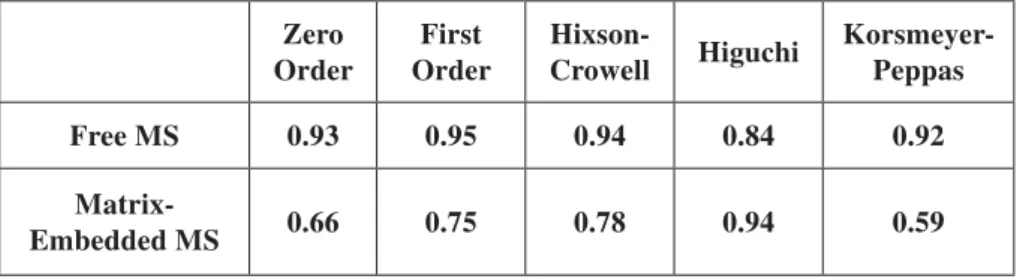
To analyse SDF-1 release from microspheres (+/- matrix embedding), various release kinetic models were used to describe the observed release kinetics. Correlation coeffi cients were determined for data fi t to zero-order release (Hadjiioannou et al., 1993), fi rst-order release (Bourne et al., 2002), Higuchi (Higuchi, 1963), Hixson-Crowell (Hixson and Hixson-Crowell, 1931) and Korsmeyer-Peppas (Korsmeyer et al., 1983) models.
Cell culture
After obtaining informed consent, total peripheral blood mononuclear cells (PBMCs) were isolated from the blood of healthy human volunteers by Histopaque 1077 density-gradient centrifugation, as previously described (Ruel et al., 2005). Cells were cultured on fi bronectin-coated plates in endothelial basal medium (EBM-2; Clonetics, Guelph, Canada) containing 5 % FBS (v/v), VEGF, R3-IGF-1, and hEGF supplements. After 4 d in culture, supernatant and non-adherent cells were removed, and adherent populations were considered to be circulating progenitor cells (CPCs).
Adhesion assay
CPCs (2×10⁴) were resuspended in 1 mL of medium and seeded in 12-well dishes with fi bronectin-coated coverslips, containing medium with 50 mg of blank microspheres or medium with 50 mg of human SDF-1 loaded microspheres. After 1 h at 37 ºC, medium was aspirated and adherent cells were fixed with 4 % paraformaldehyde (PFA). Coverslips were washed with PBS and mounted on slides with 4’,6-diamidino-2-phenylindol-(DAPI)-containing mounting medium (Vector Laboratories, Burlington, Canada). Six random fi elds-of-view were imaged using a Zeiss Z1 fl uorescent microscope and DAPI+ cells were counted.
Migration assay
CPCs (2×10⁴) in VEGF-free medium were added to the top chamber of a Transwell tissue culture well (Corning, New York, NY, USA). The lower chamber contained VEGF-free medium with 50 mg of blank microspheres or VEGF-free medium with 50 mg of human SDF-1 loaded microspheres. The bottom of each well contained a fi bronectin-coated coverslip. After 24 h of incubation, cells were fi xed, DAPI-stained, visualised, and counted.
Chemokinesis versus chemotaxis assay
To investigate the mode of action through which SDF-1 induces CPC migration, assays, based on a previously described protocol for the evaluation of chemokinesis versus chemotaxis (Misiak-Tloczek and Brzezinska-Blaszczyk, 2009), were carried out. Three treatment conditions (represented as the presence of SDF-1 in the upper well/lower well) were tested: SDF-1/0 (SDF-1 only in the upper well); SDF-1/SDF-1 (SDF-1 in both upper and lower wells); 0/SDF-1 (SDF-1 in the lower well only). SDF-1 was used at a concentration of 9 ng/mL, as this is the approximate amount released from microspheres after 24 h. Quantifi cation was performed in the same manner as described for the migration assay above.
Animal model
All procedures were performed with the approval of the University of Ottawa Animal Care Committee, in compliance with the National Institute of Health’s Guide for the Care and Use of Laboratory Animals. Bone marrow transplantation was performed as previously described (Whitman et al., 2004). Briefl y, female C57BL/6J mice (8-9 weeks old, Jackson Laboratories, Bar Harbor, ME, USA) were irradiated with a total of 900 rads from a caesium source, delivered in 2 equal doses, 3 h apart. Donor
bone marrow cells (7×10⁶) from green
fl uorescent protein (GFP) transgenic mice (C57BL/6-Tg(CAG-EGFP)10sb/J, Jackson Laboratories) were injected into the tail vein of irradiated recipient mice. Six weeks after transplantation, proximal femoral arteries in left hindlimbs were ligated as described (Limbourg et al., 2009), using 4.0 silk thread, under 2 % isofl urane. Limbs subsequently received 80 μL
injection of: 1) PBS (n = 9); 2) collagen matrix (n = 8); or 3) collagen matrix containing murine SDF-1 microspheres (n = 8). Treatments were delivered by 3 equivolumetric injections into the adductor muscle downstream of the ligation site, using a 27-gauge needle.
Blood perfusion of both hindlimbs was measured before and after ligation, and at days 4, 7 and 14 post-operatively using a multifi bre needle probe (8 separate collecting fi bres), and a laser Doppler blood fl ow monitor (Moor Instruments, Axminster, UK).
Blood samples (~100 μL) were procured from the right saphenous veins on days 0 (pre-operative baseline), 1, 4, 7 and 14 post-operatively. PBMCs were isolated using density-gradient centrifugation and immediately characterised using fl ow cytometry as described below.
Flow cytometry
Cells were labelled with antibodies against the following antigens: c-kit (Southern Biotech, Birmingham, AL, USA), CXCR4 (BD Biosciences, Mississauga, Canada), and fl k-1 (eBioscience, San Diego, CA, USA), and analysed with a FACSAria fl ow cytometer (BD Biosciences). Isotype-matched immunoglobulin antibodies were used as controls.
Immunohistochemistry
Animals were sacrifi ced at day 14. Hindlimb muscle tissue was collected and fi xed overnight in PFA before paraffinisation. All samples were analysed in cross-section. Samples were de-paraffi nised and hydrated with sequential washes in toluene and decreasing concentrations of ethanol. Antigen retrieval was performed using boiling citrate buffer. All staining was performed in PBS containing 10 % normal horse serum (Vector Laboratories). The following antibodies were used: anti-α-smooth muscle actin (SMA; pre-diluted, Abcam, Cambridge, MA, USA), anti-GFP (1:100; Abcam), and anti-CXCR4 (1:50; Abcam). For all tissue sections, mounting medium with DAPI (Vector Laboratories) was used to visualise nuclei. All measurements and cell counts were determined from 6 random microscopic fi elds-of-view and averaged from 2 blinded observers.
Cytokine Antibody Arrays
Relative cytokine levels in hindlimb lysates and serum from sacrifi ced animals (n = 5 per treatment group) were analysed using RayBio Mouse Cytokine Antibody Array Kits (Raybiotech, Norcross, GA, USA), according to the manufacturer’s protocol.
Statistical Analysis
D Kuraitis et al. Controlled SDF-1 release for treating ischaemia
continuous data between groups were performed with a one-way analysis of variance and comparisons between individual groups were performed with a two-tailed Student’s t-test. For in vitro CPC analysis, results were paired by donor, and subjected to a paired t-test. Probability values of p < 0.05 were considered statistically signifi cant.
Results
Generation of SDF-1 microspheres
Microspheres had a mean diameter of 38.5 μm (±14.0 μm (SD); Fig. 1A). When hydrated, microspheres had a smooth, spherical morphology (Fig. 1B), and a rougher surface upon dehydration as visualised by scanning electron microscopy (SEM) (Fig. 1C).
Microsphere-matrix impregnation
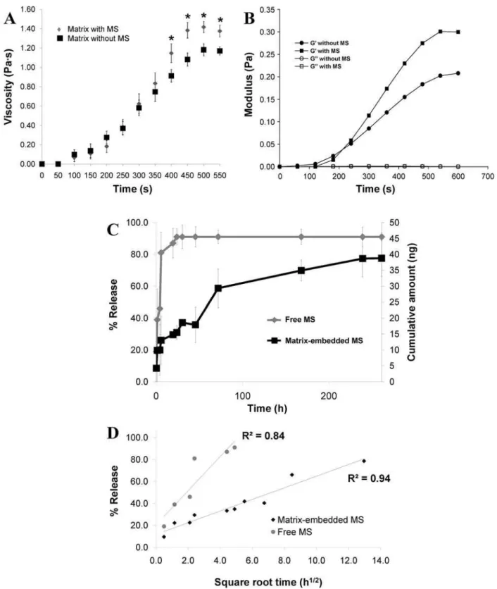
The addition of microspheres to the collagen matrix reduced the time to gelation and caused the matrix to solidify at a greater viscosity, as early as 400 s after
application (p = 0.05; Fig. 2A, 2B), which was maintained over time. Maximum viscosity reached was greater for the matrix containing microspheres (1.42 Pa·s), compared to matrix alone (1.18 Pa·s; p = 0.003). Microspheres in solution released their SDF-1 content within 1 d, but matrix impregnation prolonged the maximal release up to approximately 10 d (Fig. 2C). Analysis of SDF-1 release kinetics shows that release from microspheres best fi ts a fi rst order model, indicated by the greatest correlation coeffi cient, but after impregnation in a matrix, release follows the Higuchi model (Table 1, Fig. 2D).
Bioactivity of SDF-1 microspheres
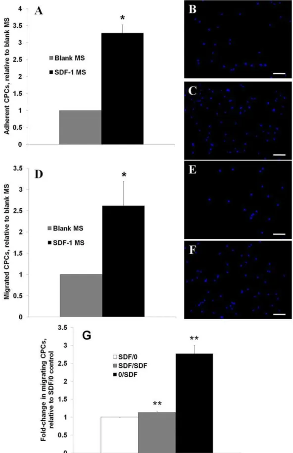
Blank microspheres did not confer any difference in adhesion potential with cultured CPCs, but the addition of SDF-1 loaded microspheres supported an increase in adhesive CPCs by 2.3-fold after 1 h of exposure (p = 0.04; Fig. 3A-C). When CPCs were given a chemotactic stimulus of blank or SDF-1 loaded microspheres, 3.2-fold more cells migrated towards the SDF-1-releasing microspheres than the blank ones after 24 h (p = 0.004; Fig. 3D-F).
Fig. 1. Characterisation of SDF-1-containing alginate microspheres. Average size of microspheres was 38.5 μm (A). Microspheres displayed a rounded morphology in saline solution (B; 400x), and a lattice structure after drying and imaging using scanning electron microscopy (C; 379x). Scale bars = 50 μm.
Zero Order
First Order
Hixson-Crowell Higuchi
Korsmeyer-Peppas
Free MS 0.93 0.95 0.94 0.84 0.92
D Kuraitis et al. Controlled SDF-1 release for treating ischaemia
SDF-1 mediated-migration of CPCs is mainly chemotactic
Compared to the lack of a chemotactic stimulus (absence of SDF-1 in the lower well), chemokinesis, stimulated by equivalent amounts of SDF-1 in the upper and lower chambers, increased CPC movement to the lower chamber by 13 % (p = 0.04; Fig. 3G). The chemotactic stimulus with SDF-1 only in the lower chamber induced the greatest migration of CPCs, by 177 % (p = 0.006; Fig. 3G).
In vivo mobilisation of CPCs by SDF-1 treatment
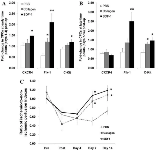
Based on the in vitro chemotactic properties of SDF1 microsphere matrix, we defi ned whether the construct is able to enhance the recruitment of CPC in an in vivo system. GFP+ (bone marrow-derived) CPCs in the peripheral blood were analysed over time, and compared to baseline. Interestingly, the SDF-1 microsphere matrix treatment signifi cantly increased circulating CXCR4+ cells (by 45 %; p = 0.02), fl k-1+ cells (by 105 %; p = 0.001), and c-kit+
cells (by 18 %; p = 0.04) at early time point post injury (Fig. 4A) and was able to maintain elevated numbers of circulating fl k-1+ cells (by 149 %; p = 0.002) and c-kit+ cells (by 48 %; p = 0.01) for an extended period (7-14 d) post operation (Fig. 4B). Notably, the collagen matrix alone increased circulating fl k+ cells (by 20 %; p = 0.009) at the earlier evaluation and c-kit+ cells at both early (by 48 %; p = 0.01) and late time point (by 29 %; p = 0.02; Fig. 4A, B).
Restoration of perfusion by matrix treatments
Indeed, both matrix treatments (with or without SDF-1 microspheres) were able to restore perfusion to baseline levels by one week post-treatment, compared to PBS controls (Fig. 4C; p < 0.04), which was maintained at two weeks post-treatment (p < 0.05).
Arteriole density of ischaemic hindlimbs
Arteriole density in ischaemic hindlimbs was not different between collagen and SDF-1 collagen matrices (p = 0.9);
D Kuraitis et al. Controlled SDF-1 release for treating ischaemia
however, both matrix and SDF-1 microsphere matrix treatments increased arteriole density compared to PBS by 67 % (p = 0.01) & 69 % (p = 0.02), respectively (Fig. 5A-E). Mean arteriole size was greater in the SDF-1 microsphere matrix treatment group compared to PBS-treated mice (p < 0.05; Fig. 5B-E). There was also a trend for greater arteriole cross-sectional area with SDF-1 microsphere matrix treatment compared to collagen matrix (p = 0.09), and for collagen matrix treatment compared to PBS (p = 0.08; Fig. 5B).
Recruitment of bone marrow-derived cells to treated hindlimbs
SDF-1 microsphere matrix and collagen matrix treatments recruited 17.7- and 4.2-fold more GFP+ cells to treated hindlimbs, compared to PBS (p = 0.0007 and 0.02), respectively (Fig. 6A-D). There was a trend for SDF-1 microsphere matrix treatment to recruit more GFP+ cells than collagen matrix alone (p = 0.06; Fig. 6A).
Engraftment of CXCR4+ cells in ischaemic hindlimbs
Overall, SDF-1 microsphere matrix treatment recruited 4.3- and 1.8-fold more CXCR4+ cells to treated hindlimbs compared to PBS and collagen (p = 0.0004 and 0.05; Fig. 7A-D). Separating the analysis of CXCR4+ cells into those recruited from the marrow versus those recruited locally, SDF-1 microsphere matrix treatment recruited 4.9-fold more CXCR4+GFP+ (bone marrow-derived) and 4.5-fold more CXCR4+GFP- (local) cells, compared to PBS and collagen (p = 0.008 and 0.02; Fig. 7A). Collagen matrix alone also recruited 2.8-fold more CXCR4+GFP+ cells compared to PBS (p = 0.03; Fig. 7A).
Cytokine profi les
In the hindlimb (Fig. 8A), interleukin-1α (IL-1α; p = 0.04) and monocyte inhibitory protein-3α (MIP-3α; p = 0.04) were reduced, and vascular cell adhesion molecule-1 (VCAM-1; p = 0.03) and insulin-like growth factor-1 (IGF-1; p = 0.03) were increased with SDF-1 microsphere
matrix treatment. In the serum (Fig. 8B): L-selectin was increased with SDF-1 microsphere matrix treatment (p = 0.01) and reduced with collagen alone (p = 0.01); basic fi broblast growth factor (bFGF) was increased with collagen alone (p < 0.001) and with SDF-1 microsphere matrix treatment (p = 0.02); and VCAM-1 was reduced with SDF-1 microsphere matrix treatment compared to collagen matrix (p = 0.007) and PBS (p < 0.001). For both serum and hindlimb, no signifi cant differences between any treatments were observed for the following infl ammatory cytokines: GM-CSF, TNF-α, IL-1β and IL-6 (all p > 0.4).
Discussion
In this study, we demonstrated that the release of SDF-1 from alginate microspheres can be effectively prolonged by incorporation of the microspheres into a thermogelling collagen matrix. Released SDF-1 was bioactive, supported
rapid adhesion and migration of CPCs, and also stimulated the mobilisation of CPCs from bone marrow when applied to ischaemic muscle. Both matrix alone and SDF-1-releasing matrix restored perfusion and improved arteriole density by 2 weeks, but the SDF-1 treatment best supported the growth of arterioles. SDF-1 microsphere matrix treatment also recruited more bone marrow-derived cells and local CXCR4+ cells to the ischaemic muscle, and altered levels of local angiogenic factors.
Incorporation of alginate microspheres into the collagen matrix increased the hydrogel’s viscosity and reduced the time to gelation. This is thought to be mediated by the cross-linking reaction, whereby functional groups on collagen (–NH2) and alginate (–COOH) are covalently bound by EDC/NHS (Mitra et al., 2011), increasing the total number of cross-links within the material and enhancing its strength, as has been previously reported (Liu et al., 2008). An earlier onset of gelation is advantageous
Fig. 6. Recruited bone marrow-derived cells. More GFP+ cells were engrafted in ischaemic hindlimbs treated with collagen or SDF-1 microsphere-matrix (SDF-1), with a trend for more GFP+ cells in the SDF-1 microsphere-matrix treatment, compared to matrix alone (A). Representative images of GFP+ cells recruited to PBS- (B), collagen matrix- (C), or SDF-1 microsphere-matrix- (D) treated hindlimbs. Scale bars = 100 μm; n = 6-8 per group. *p ≤
D Kuraitis et al. Controlled SDF-1 release for treating ischaemia
in a clinical setting, allowing for a reduction in the time required for stable material integration within host tissue. Additionally, a positive correlation between hydrogel viscosity and resistance to degradation has been noted (Yang et al., 2010), suggesting a better persistence in vivo of microsphere-containing hydrogels.
During the period of release, SDF-1 kinetics from matrix-embedded microspheres fi t the Higuchi model of release whereby the initial drug concentration >> drug solubility in the matrix, swelling is negligible, perfect sink conditions are maintained and edge effects are negligible, suggesting that diffusion is the primary mechanism of SDF-1 release (Higuchi, 1963). In contrast, microspheres alone initially demonstrated a burst release followed by a
fi rst order release of SDF-1. Although a burst-release effect was observed with microspheres alone, the embedding of microspheres in matrix was able to prolong the SDF-1 release by 10-fold. This observation can be explained by the properties of the microspheres and the collagen-hydrogel system. Hydrogels have an innate ability to retain small peptides (Cadee et al., 2002; Ruvinov et al., 2010), and incorporation of peptide-containing microspheres into hydrogels has been shown previously to extend the release profi le of the peptide (Kempen et al., 2008).
SDF-1 bioactivity was maintained during the microsphere generation and cross-linking procedure; upon its release, SDF-1 augmented rapid adhesion of CPCs, as well as inducing chemotaxis of CPCs, rather than chemokinesis, indicating its ability to induce CPC homing towards a chemotactic gradient rather than the induction of random mobilisation.
In the ischaemic hindlimb study, marrow-mobilised cells expressing the SDF-1 receptor CXCR4 were increased in circulation of SDF-1 microsphere matrix-treated animals at early time points, but this effect was later lost. This can be explained by previous studies showing the eventual down-regulation of CXCR4 following cytokine-induced mobilisation from the bone marrow (Kim et al., 2006). More specifi cally, CXCR4 expression is dose-dependent on SDF-1 levels; doses of ≤ 1 μg in a mouse model have shown CXCR4 levels equivalent to controls that did not receive SDF-1 (Kimura and Tabata, 2010). Interestingly, higher doses of SDF-1 have the same effect, suggesting a potential negative feedback loop for the SDF-1/CXCR4 axis. Notably, SDF-1 microsphere matrix treatment also stimulated an early mobilisation of fl k-1+ and c-kit+ cells from the bone marrow into circulation, and maintained this effect up to 2 weeks post-treatment.
CXCR4 is SDF-1’s exclusive receptor and CXCR4+ CPCs are reduced over time, mechanisms other than SDF-1 release are needed to explain the increase in mobilised CPCs expressing fl k-1 and c-kit. We have previously shown that systemic transplantation of CPCs can induce a potent response from the host’s CPCs (Suuronen et al., 2009). Additionally, this effect has been documented in humans, and CPC persistence in the circulation has been observed up to 1 year after cell transplantation (Turan et al., 2010). Therefore, it is likely that the initiation of the SDF-1/ CXCR4 mobilisation and recruitment response by SDF-1 microsphere matrix treatment activates other endogenous progenitor cell mechanisms.
Ischaemia was induced in all mice (average of 55 % of normal perfusion). By one week post-treatment, animals that received matrix treatment (with or without SDF-1 microspheres) had hindlimb perfusion restored to baseline levels, which is similar to previous reports of matrix treatment for hindlimb ischaemia (Suuronen et al., 2009). It has been previously demonstrated that an increase in perfusion is attributable to increased local vasculature (Kim et al., 2010; Suuronen et al., 2010), an observation
that was seen in the current study; matrix treatments increased arteriole density in ischaemic hindlimbs. It was hypothesised that SDF-1 microsphere matrix treatment would confer superior restoration of perfusion, but instead, the matrix-only treatment was equally effective. Kimura and Tabata (2010) also attempted to enhance neovascularisation using an SDF-1-releasing hydrogel, and observed increased capillary density with SDF-1 treatment at 4 d post-treatment, which was abrogated by 10 d. Another SDF-1 release study did not show a difference in vascular density between SDF-1 and controls at 9 d post-treatment (Rabbany et al., 2010). It may be that the addition of SDF-1 accelerates the regenerative response to collagen matrix treatment. This is supported by the observation that SDF-1 treatment increased the cross-sectional size of arterioles. Cross-sectional area of new vasculature has been shown to increase over a period of 4 weeks (Ruvinov et al., 2011), demonstrating that vessel area is indicative of vessel maturity. Therefore, it is plausible that SDF-1 treatment had an earlier effect on neovascularisation, allowing for more rapid growth/maturation of arterioles, compared to both matrix and PBS treatments.
D Kuraitis et al. Controlled SDF-1 release for treating ischaemia
The SDF-1 microsphere matrix treatment recruited the most GFP+ marrow-mobilised cells in our animal model. Other SDF-1-release studies have also shown recruitment of stem cells, positive for c-kit (Zhang et al. 2007; Thevenot et al., 2010) expression. In particular, it was expected that SDF-1 treatment would increase the recruitment and engraftment of CXCR4+ cells. Both the matrix and SDF-1 microsphere matrix treatments increased homing of bone marrow-derived CXCR4+ cells to the treated hindlimbs (SDF-1 microsphere matrix treatment had the most pronounced effect), but the SDF-1 microsphere matrix treatment also demonstrated an improved recruitment of CXCR4+ cells of non-bone marrow origin. Recently, it was shown that local pools of progenitor cells co-expressing CXCR4 and endothelial markers reside in tissues (Sandstedt et al., 2010), suggesting a potential role in neovascularisation. In our experiment, SDF-1 treatment may be recruiting a similar population, as evidenced by CXCR4+GFP- staining. Microvascular endothelial cells have also been documented to express CXCR4 (Takagi et al., 2009), suggesting another CXCR4-expressing population that is possibly activated in treated hindlimbs. Regardless, CXCR4+ fractions have been observed to be superior to whole CXCR4- fractions with respect to potential for invasion, neovascularisation, and restoration of perfusion (Seeger et al., 2009).
Compared to controls, infl ammatory cytokines (IL-1α
and MIP-3α) were reduced in hindlimbs treated with SDF-1 microsphere matrix, a result similar to a study by Thevenot et al. (2010). The latter examined cytokine responses to SDF-1 delivery in a synthetic scaffold; however, our results also suggest that the SDF-1 microsphere matrix supports a favourable environment for angiogenic activity through local factors, as evidenced by increased IGF-1 and VCAM-1 in hindlimb lysates. IGF-VCAM-1 is a stimulator of angiogenesis (Su et al., 2003) and is cytoprotective (Li et al., 1997), and elevated IGF-1 is ideal for recovery. Pelosi et al. (2007) have shown that persistent IGF-1 expression accelerates the regenerative response and restores architecture and structure soon after skeletal muscle injury. VCAM-1 is a cell adhesion molecule expressed by endothelial cells. When VCAM-1 is solubilised and detected in serum, it is used as an indicator of dysfunctional endothelium (Balciunas et al., 2009). Our SDF-1 microsphere matrix treatment had signifi cantly less circulating VCAM-1, further suggesting a vasculo-protective role of this therapy. Furthermore, both matrix treatments increased systemic bFGF, which is a potent angiogenic cytokine (Hosseinkhani et al., 2006).
Conclusion
In this study, we demonstrated that SDF-1 can be successfully encapsulated in an alginate microsphere system and further incorporated into an injectable matrix for non-invasive delivery. The treatment of ischaemic mouse hindlimbs with SDF-1-releasing matrix enhanced progenitor cell mobilisation and recruitment. Compared to collagen-only treatment, the addition of SDF-1 appears to confer an earlier effect on neovascularisation, as suggested by greater arteriole maturity. These fi ndings suggest that
application of an SDF-1-releasing matrix constitutes a suitable therapy for prevalent myopathies with reduced perfusion, with the potential to augment progenitor cell mobilisation and homing, as well as its ability to rapidly support neovascularisation.
Acknowledgments
DK was supported by a Canadian Institutes of Health Research Canadian Graduate Scholarship; PZ is a recipient of the Lawrence Soloway Research Fellowship Award; YZ was supported by a Heart & Stroke Foundation of Ontario Doctoral Research Award; DTP was supported by a University of Ottawa Cardiology Research Endowment Fellowship; KM was supported by an Ontario Graduate Scholarship; DK and TS were supported by Heart & Stroke Foundation of Ontario Master’s Studentships; JZ was supported by a Natural Sciences and Engineering Research Council of Canada postdoctoral fellowship. The authors would like to thank Suzanne Crowe for her fl ow cytometry expertise and Ann Fook Yang for assistance with SEM.
This work was supported by grant-in-aid T6793 from the Heart and Stroke Foundation of Ontario (to EJS), by grant MOP-77536 from the Canadian Institutes of Health Research (to MR and EJS), by a Natural Sciences and Engineering Research Council of Canada Research Tool and Instruments Grant (to XC), by a Canadian Stem Cell Network grant (to MG), and by Fondazione Roma and 7FP-Myoage (to AM). Funding sources did not have a role in experimental design, data management, or manuscript preparation.
References
Askari AT, Unzek S, Popovic ZB, Goldman CK, Forudi F, Kiedrowski M, Rovner A, Ellis SG, Thomas JD, DiCorleto PE, Topol EJ, Penn MS (2003) Effect of stromal-cell-derived factor 1 on stem-cell homing and tissue regeneration in ischaemic cardiomyopathy. Lancet
362: 697-703.
Balciunas M, Bagdonaite L, Samalavicius R, Baublys A (2009) Markers of endothelial dysfunction after cardiac surgery: soluble forms of vascular-1 and intercellular-1 adhesion molecules. Medicina (Kaunas) 45: 434-439.
Belch JJ, Topol EJ, Agnelli G, Bertrand M, Califf RM, Clement DL, Creager MA, Easton JD, Gavin JR 3rd, Greenland P, Hankey G, Hanrath P, Hirsch AT, Meyer J, Smith SC, Sullivan F, Weber MA; Prevention of Atherothrombotic Disease Network (2003) Critical issues in peripheral arterial disease detection and management: a call to action. Arch Intern Med 163: 884-892.
Bourne D (2002) Pharmacokinetics. In: Modern Pharmaceuticals, 4th ed. Marcel Dekker Inc., New York, pp 67-92.
Cadee JA, de Groot CJ, Jiskoot W, den Otter W, Hennink WE (2002) Release of recombinant human interleukin-2 from dextran-based hydrogels. J Control Release 78: 1-13.
cell transplantation into the ischaemic heart. J Exp Med
204: 3257-3269.
De Falco E, Porcelli D, Torella AR, Straino S, Iachininoto MG, Orlandi A, Truffa S, Biglioli P, Napolitano M, Capogrossi MC, Pesce M (2004) SDF-1 involvement in endothelial phenotype and ischemia-induced recruitment of bone marrow progenitor cells. Blood 104: 3472-3482.
Deng C, Zhang P, Vulesevic B, Kuraitis D, Li F, Yang AF, Griffi th M, Ruel M, Suuronen EJ (2010) A collagen-chitosan hydrogel for endothelial differentiation and angiogenesis. Tissue Eng Part A 16: 3099-3109.
Fazel S, Cimini M, Chen L, Li S, Angoulvant D, Fedak P, Verma S, Weisel RD, Keating A, Li RK (2006) Cardioprotective c-kit+ cells are from the bone marrow and regulate the myocardial balance of angiogenic cytokines. J Clin Invest 116: 1865-1877.
Formigli L, Perna AM, Meacci E, Cinci L, Margheri M, Nistri S, Tani A, Silvertown J, Orlandini G, Porciani C, Zecchi-Orlandini S, Medin J, Bani D (2007) Paracrine effects of transplanted myoblasts and relaxin on post-infarction heart remodelling. J Cell Mol Med 11: 1087-1100.
Hadjiioannou TP, Christian GD, Koupparis MA, Macheras PE (1993) Quantitative Calculations in Pharmaceutical Practice and Research. VCH Publishers Inc., New York, pp 345-348.
Higuchi T (1963) Mechanism of Sustained-Action Medication. Theoretical Analysis of Rate of Release of Solid Drugs Dispersed in Solid Matrices. J Pharm Sci 52: 1145-1149.
Hixson AW, Crowell JH (1931) Dependence of reaction velocity upon surface and agitation (I) theoretical consideration. Ind Eng Chem 23: 923-931.
Hofmann M, Wollert KC, Meyer GP, Menke A, Arseniev L, Hertenstein B, Ganser A, Knapp WH, Drexler H (2005) Monitoring of bone marrow cell homing into the infarcted human myocardium. Circulation 111: 2198-2202.
Hosseinkhani H, Hosseinkhani M, Khademhosseini A, Kobayashi H, Tabata Y (2006) Enhanced angiogenesis through controlled release of basic fibroblast growth factor from peptide amphiphile for tissue regeneration. Biomaterials 27: 5836-5844.
Kaully T, Kaufman-Francis K, Lesman A, Levenberg S (2009) Vascularisation – the conduit to viable engineered tissues. Tissue Eng Part B Rev 15: 159-169.
Kempen DH, Lu L, Hefferan TE, Creemers LB, Maran A, Classic KL, Dhert WJ, Yaszemski MJ (2008) Retention of in vitro and in vivo BMP-2 bioactivities in sustained delivery vehicles for bone tissue engineering. Biomaterials
29: 3245-3252.
Kim HK, De La Luz Sierra M, Williams CK, Gulino AV, Tosato G (2006) G-CSF down-regulation of CXCR4 expression identifi ed as a mechanism for mobilization of myeloid cells. Blood 108: 812-820.
Kim SW, Kim H, Cho HJ, Lee JU, Levit R, Yoon YS (2010) Human peripheral blood-derived CD31+ cells have robust angiogenic and vasculogenic properties and are effective for treating ischemic vascular disease. J Am Coll Cardiol 56: 593-607.
Kimura Y, Tabata Y (2010) Controlled release of stromal-cell-derived factor-1 from gelatin hydrogels
enhances angiogenesis. J Biomater Sci Polym Ed 21: 37-51.
Ko HC, Milthorpe BK, McFarland CD (2007) Engineering thick tissues – the vascularisation problem. Eur Cells Mater 14: 1-18.
Korsmeyer RW, Gurny R, Doelker E, Buri P, Peppas NA (1983) Mechanisms of solute release from porous hydrophilic polymers. Int J Pharm 15: 25-35.
Li Q, Li B, Wang X, Leri A, Jana KP, Liu Y, Kajstura J, Baserga R, Anversa P (1997) Overexpression of insulin-like growth factor-1 in mice protects from myocyte death after infarction, attenuating ventricular dilation, wall stress, and cardiac hypertrophy. J Clin Invest 100: 1991-1999.
Limbourg A, Korff T, Napp LC, Schaper W, Drexler H, Limbourg FP (2009) Evaluation of postnatal arteriogenesis and angiogenesis in a mouse model of hind-limb ischemia. Nat Protoc 4: 1737-1746.
Liu W, Griffi th M, Li F (2008) Alginate microsphere-collagen composite hydrogel for ocular drug delivery and implantation. J Mater Sci 19: 3365-3371.
Misiak-Tloczek A, Brzezinska-Blaszczyk E (2009) IL-6, but not IL-4, stimulates chemokinesis and TNF stimulates chemotaxis of tissue mast cells: involvement of both mitogen-activated protein kinases and phosphatidylinositol 3-kinase signalling pathways. APMIS 117: 558-567.
Mitra T, Sailakshmi G, Gnanamani A, Raja ST, Thiruselvi T, Gowri VM, Selvaraj NV, Ramesh G, Mandal AB (2011) Preparation and characterization of a thermostable and biodegradable biopolymers using natural cross-linker. Intl J Biol Macromol 48: 276-285.
Murry CE, Reinecke H, Pabon LM (2006) Regeneration gaps: observations on stem cells and cardiac repair. J Am Coll Cardiol 47: 1777-1785.
Park S, Tepper OM, Galiano RD, Capla JM, Baharestani S, Kleinman ME, Pelo CR, Levine JP, Gurtner GC (2004) Selective recruitment of endothelial progenitor cells to ischemic tissues with increased neovascularization. Plast Reconstr Surg 113: 284-293.
Pelosi L, Giacinti C, Nardis C, Borsellino G, Rizzuto E, Nicoletti C, Wannenes F, Battistini L, Rosenthal N, Molinaro M, Musarò A (2007) Local expression of IGF-1 accelerates muscle regeneration by rapidly modulating infl ammatory cytokines and chemokines. FASEB J 21: 1393-1402.
Rabbany SY, Pastore J, Yamamoto M, Miller T, Rafi i S, Aras R, Penn M (2010) Continuous delivery of stromal cell-derived factor-1 from alginate scaffolds accelerates wound healing. Cell Transplant 19: 399-408.
Ruel M, Suuronen EJ, Song J, Kapila V, Gunning D, Waghray G, Rubens FD, Mesana TG (2005) Effects of off-pump versus on-off-pump coronary artery bypass grafting on function and viability of circulating endothelial progenitor cells. J Thorac Cardiovasc Surg 130: 633-639.
Ruvinov E, Leor J, Cohen S (2010) The effects of controlled HGF delivery from an affi nity-binding alginate biomaterial on angiogenesis and blood perfusion in a hindlimb ischemia model. Biomaterials 31: 4573-4582.
D Kuraitis et al. Controlled SDF-1 release for treating ischaemia
Sandstedt J, Jonsson M, Lindahl A, Jeppsson A, Asp J (2010) C-kit+ CD45- cells found in the adult human heart represent a population of endothelial progenitor cells. Basic Res Cardiol 105: 545-556.
Seeger FH, Rasper T, Koyanagi M, Fox H, Zeiher AM, Dimmeler S (2009) CXCR4 expression determines functional activity of bone marrow-derived mononuclear cells for therapeutic neovascularization in acute ischemia. Arterioscl Thromb Vasc Biol 29: 1802-1809.
Selvin E, Erlinger TP (2004) Prevalence of and risk factors for peripheral arterial disease in the United States: results from the National Health and Nutrition Examination Survey, 1999-2000. Circulation 110: 738-743.
Shamoun F, Sural N, Abela G (2008) Peripheral artery disease: therapeutic advances. Expert Rev Cardiovasc Ther
6: 539-553.
Su EJ, Cioffi CL, Stefansson S, Mittereder N, Garay M, Hreniuk D, Liau G (2003) Gene therapy vector-mediated expression of insulin-like growth factors protects cardiomyocytes from apoptosis and enhances neovascularization. Am J Physiol Heart Circ Physiol 284: H1429-1440.
Suuronen EJ, Price J, Veinot JP, Ascah K, Kapila V, Guo XW, Wong S, Mesana TG, Ruel M (2007) Comparative effects of mesenchymal progenitor cells, endothelial progenitor cells, or their combination on myocardial infarct regeneration and cardiac function. J Thorac Cardiovasc Surg 134: 1249-1258.
Suuronen EJ, Zhang P, Kuraitis D, Cao X, Melhuish A, McKee D, Li F, Mesana TG, Veinot JP, Ruel M (2009) An acellular matrix-bound ligand enhances the mobilization, recruitment and therapeutic effects of circulating progenitor cells in a hindlimb ischemia model. FASEB J
23: 1447-1458.
Suuronen EJ, Hazra S, Zhang P, Vincent R, Kumarathasan P, Zhang Y, Price J, Chan V, Sellke FW, Mesana TG, Veinot JP, Ruel M (2010) Impairment of human cell-based vasculogenesis in rats by hypercholesterolemia-induced endothelial dysfunction and rescue with L-arginine supplementation. J Thorac Cardiovasc Surg 139: 209-216. e2.
Takagi Y, Hashimoto N, Phan SH, Imaizumi K, Matsuo M, Nakashima H, Hashimoto I, Hayashi Y, Kawabe T, Shimokata K, Hasegawa Y (2009) Erythromycin-induced CXCR4 expression on microvascular endothelial cells. Am J Physiol 297: L420-431.
Thevenot PT, Nair AM, Shen J, Lotfi P, Ko CY, Tang L (2010) The effect of incorporation of SDF-1alpha into PLGA scaffolds on stem cell recruitment and the infl ammatory response. Biomaterials 31: 3997-4008.
Turan RG, Bozdag-Turan I, Ortak J, Akin I, Kische S, Schneider H, Turan CH, Rehders TC, Rauchhaus M, Kleinfeldt T, Adolph E, Brehm M, Yokus S, Steiner S, Sahin K, Nienaber CA, Ince H (2010) Improved mobilization of the CD34(+) and CD133(+) bone marrow-derived circulating progenitor cells by freshly isolated intracoronary bone marrow cell transplantation in patients with ischemic heart disease. Stem Cells Dev, in press.
Whitman SC, Rateri DL, Szilvassy SJ, Yokoyama W, Daugherty A (2004) Depletion of natural killer cell function decreases atherosclerosis in low-density lipoprotein
receptor null mice. Arterioscl Thromb Vasc Biol 24: 1049-1054.
Wojakowski W, Tendera M, Michalowska A, Majka M, Kucia M, Maslankiewicz K, Wyderka R, Ochała A, Ratajczak MZ (2004) Mobilization of CD34/CXCR4+, CD34/CD117+, c-met+ stem cells, and mononuclear cells expressing early cardiac, muscle, and endothelial markers into peripheral blood in patients with acute myocardial infarction. Circulation 110: 3213-3220.
Yang J, Wang F, Tianwei T (2010) Degradation behavior of hydrogel based on crosslinked poly(aspartic acid). J Appl Polym Sci 117: 178-185.
Zhang G, Nakamura Y, Wang X, Hu Q, Suggs LJ, Zhang J (2007) Controlled release of stromal cell-derived factor-1 alpha in situ increases c-kit+ cell homing to the infarcted heart. Tissue Eng 13: 2063-2071.
Discussion with Reviewers
Reviewer II: Could the authors foresee applications for their injectable hydrogel loaded with SDF1 microspheres in damaged bone tissue with compromised vascularity?
Authors: Vascularisation is believed to play an important role in bone wound healing (Giannoudis et al., 2008, additional reference), yet defective neovascularisation in some patients leads to an avascular fracture site. A recent study reported that inhibiting angiogenesis in a rat tibia osteotomy model resulted in non-union of the fracture over the early healing stages, demonstrating the importance of the vasculature for proper bone wound repair (Fassbender et al., 2011, additional reference). Notably, the local application of SDF-1 was able to increase neo-blood vessel maturation leading to accelerated bone regeneration, characterised by increased callus formation in a distraction osteogenesis model (Fujio et al., 2011, additional reference). Therefore, we can envision that a strategy such as the system reported here may be applicable in bone trauma, whereby blood vessel growth is stimulated at the early stages after bone injury, possibly allowing for more effective wound healing to occur, and without delay.
Reviewer II: In the discussion, the authors suggest that the matrix is important to support the reperfusion and that SDF-1 supports early neovascularisation. Would it be possible that a burst release of SDF-1 from a hydrogel matrix would be equivalent or even better than the material solution proposed with a relatively long release profi le?
reference). However, in the present study, we observed an increase in vessel size, but no difference in vascular density between the two matrix treatments (+/- SDF-1 release), suggesting that the SDF-1 effect was early and accelerated the regeneration response conferred by the injected collagen matrix. Therefore, it is conceivable that a burst release of SDF-1 from microspheres to augment the body’s natural SDF-1 release response may be as good as, or better than, prolonged release for treatment strategies involving therapeutic matrix injections. However, we also cannot exclude the possibility that the prolonged presence of SDF-1 may have a role in other effects, such as the vessel maturation process, or the prevention of apoptosis, which have been reported previously by others (Reddy et al., 2008, additional reference; Ho et al., 2010, additional reference), but were not examined in the present study.
Additional References
Fassbender M, Strobel C, Rauhe JS, Bergmann C, Schmidmaier G, Wildemann B (2011) Local inhibition of angiogenesis results in an atrophic non-union in a rat osteotomy model. Eur Cells Mater 22: 1-11.
Fujio M, Yamamoto A, Ando Y, Shohara R, Kinoshita K, Kaneko T, Hibi H, Ueda M (2011) Stromal cell-derived factor-1 enhances distraction osteogenesis-mediated skeletal tissue regeneration through the recruitment of endothelial precursors. Bone, in press.
Giannoudis PV, Einhorn TA, Schmidmaier G, Marsh D (2008) The diamond concept – open questions. Injury
39: 5-8.
Ho TK, Tsui J, Xu S, Leoni P, Abraham DJ, Baker DM (2010) Angiogenic effects of stromal cell-derived factor-1 (SDF-1/CXCL12) variants in vitro and the in vivo expressions of CXCL12 variants and CXCR4 in human critical leg ischemia. J Vasc Surg 51: 689-699.
Reddy K, Zhou Z, Jia SF, Lee TH, Morales-Arias J, Cao Y, Kleinerman ES (2008) Stromal cell-derived factor-1 stimulates vasculogenesis and enhances Ewing’s sarcoma tumor growth in the absence of vascular endothelial growth factor. Int J Cancer 15: 831-837.
Segers VF, Tokunou T, Higgins LJ, MacGillivray C, Gannon J, Lee RT (2007) Local delivery of protease-resistant stromal cell derived factor-1 for stem cell recruitment after myocardial infarction. Circulation 116: 1683-1692.
Sundararaman S, Miller TJ, Pastore JM, Kiedrowski M, Aras R, Penn MS (2011) Plasmid-based transient human stromal cell-derived factor-1 gene transfer improves cardiac function in chronic heart failure. Gene Ther, in press.