Article
Functional interactome of Aquaporin 1 sub-family reveals new physiological
functions in Arabidopsis Thaliana
Mohamed Ragab Abdel Gawwad1, Jasmin Šutković1, Lavinija Mataković2, Mohamed Musrati1, Lizhi Zhang3
1
Genetics and Bioengineering department, International University of Sarajevo, Ilidza, 71220 Bosnia and Herzegovina 2
University of Josip Jurja Strossmayer, Biology Department, Croatia 3
Department of Molecular Genetics, The Ohio State University, 484 West 12th Avenue, Columbus, OH 43210, USA E-mail: mragab@ius.edu.ba
Received 12 July 2013; Accepted 25 July 2013; Published online 1 September 2013
Abstract
Aquaporins are channel proteins found in plasma membranes and intercellular membranes of different cellular compartments, facilitate the water flux, solutes and gases across the cellular plasma membranes. The present study highlights the sub-family plasma membrane intrinsic protein (PIP) predicting the 3-D structure and analyzing the functional interactome of it homologs. PIP1 homologs integrate with many proteins with different plant physiological roles in Arabidopsis thaliana including; PIP1A and PIP1B: facilitate the transport of water, diffusion of amino acids and/or peptides from the vacuolar compartment to the cytoplasm, play a role in the control of cell turgor and cell expansion and involved in root water uptake respectively. In addition we found that PIP1B plays a defensive role against Pseudomonas syringae infection through the interaction with the plasma membrane Rps2 protein. Another substantial function of PIP1C via the interaction with PIP2E is the response to nematode infection. Generally, PIP1 sub-family interactome controlling many physiological processes in plant cell like; osmoregulation in plants under high osmotic stress such as under a high salt, response to nematode, facilitate the transport of water across cell membrane and regulation of floral initiation in Arabidopsis thaliana.
Keywords Aquaporins; Arabidopsis thaliana; interactome; 3-D structure.
1 Introduction
Transport of materials across biological membranes is a fundamental process in all living cells. Charged and polar molecules, however, require special pathways to cross the cellular membrane, as the hydrophobic tails of lipid molecules create a considerable energetic barrier against their diffusion. Membrane channels provide such pathways for selective exchange of water-soluble materials (water, ions, and other nutrients) across the
Network Biology ISSN 22208879
URL: http://www.iaees.org/publications/journals/nb/onlineversion.asp RSS: http://www.iaees.org/publications/journals/nb/rss.xml
Email: networkbiology@iaees.org EditorinChief: WenJun Zhang
membrane. Three major functional characteristics of membrane channels, which are furnished by specific arrangements of amino acids in their structure, are permeation, selectivity, and gating. Aquaporins (AQP) are a family of membrane channels primarily responsible for conducting water across cellular membranes. These channels are widely distributed in all kingdoms of life, including bacteria, plants, and mammals. They form tetramers in the cell membrane, and facilitate the transport of water and, in some cases, other small solutes across the membrane (Wang and Tajkhorshid, 2007; Li et al., 2011). These proteins are members of the larger family of major intrinsic proteins (MIPs) with 26–34 kDa, and all protein members have six transmembrane helices, with both N- and C-termini on the cytosolic side of the membrane (Fraysse et al., 2005).
Water permeation through aquaporins is a passive process that follows the direction of osmotic pressure across the membrane. Although many aquaporins function as always-open channels, a subgroup of aquaporins, particularly in plants have evolved a sophisticated molecular mechanism through which the channel can be closed in response to harsh conditions of the environment, under which exchange of water can be harmful for the organism (Törnroth-Horsefield et al., 2005). Plants respond to drought or flood conditions by shutting down almost all of their AQP .In humans, 13 different AQP (AQP0–AQP12) have been characterized in various organs (kidneys, eyes, and the brain). The solved structures of several AQP at high resolution are indicative of a conserved protein architecture in the whole family (Wang and Tajkhorshid, 2007).
Tetramerization is a common structural feature of AQPs (Yu et al., 2006). Four functionally independent pores provide highly selective pathways for water permeation across the low dielectric barrier of lipid bilayers. Another pore, known as the central pore, is formed between the 4 monomers (Muller et al., 2002).
Plant aquaporins are categorized as either tonoplast intrinsic proteins (TIPs) or plasma membrane intrinsic proteins, PIPs (Chaumont et al., 2000). Among the members of the plant MIP family, the PIPs form the most highly conserved subfamily (Fraysse et al., 2005). The PIP subfamily can be further subdivided into two groups PIP1 and PIP2, of which the PIP1 isoforms are most tightly conserved, sharing >90% amino acid sequence identity (Fraysse et al., 2005).
The members of these groups differ in N- and C-termini lengths, the N-terminus being longer in PIP1 aquaporins. In plant cells, the transport of PIP proteins to the plasma membranes or the integration in the membrane appears to be dependent on PIP2 expression (Zelazny et al., 2007). The genome of Arabidopsis thaliana encodes 35 full-length aquaporin homologues. Thirteen of them belong to the PIP subfamily (Santoni et al., 2003).
The importance of aquaporins in environmental stress responses has been demonstrated through gene expression analyses of various plants species and the characterization of transgenic plants expressing these aquaporins.
Aquaporin-1 (AQP1), a membrane channel protein, is the first characterized member of the aquaporin (AQP) family (Hohmann et al., 2000). The protein is abundantly present in multiple human tissues, such as the kidneys. AQP1 forms homotetramers in cell membranes, each monomer forming a functionally independent water pore, which does not conduct protons, ions, or other charged solutes.
Members of the AQP1 family include: PIP 1-1, PIP1-2, PIP1-3, PIP1-4 and PIP1-5. The PIP1 subfamily of aquaporins constitutes about 1% of the plasma membrane (PM) proteins from Arabidopsis thaliana leaves (Robinson et al., 1996).
Biologically PIP1 subfamily proteins respond to salt stress. It was shown that these proteins are also involved in other processes, such as acetyl-CoA metabolic process, brassinosteroid biosynthetic process, sterol
biosynthetic process and in cellular response to iron ion starvation and iron ion transport. Furthermore, they have important role in carbon dioxide transport and regulation of protein localization (Hohmann et al., 2000; Li et al., 2010).
The subfamily of PIP1 proteins is located in chloroplast envelope, integral to membrane, membrane, mitochondria, plasma membrane, plasmodesma and vacuole. Analyses of mRNAs show that the two spinach
(Spinacia oleracea), PIP1 isoforms, SoPIP1; 1 and SoPIP1; 2 according to the nomenclature proposed by Johanson, are differentially expressed (Johanson et al., 2001). Recent study reported that PIP1; 2, the most abundant PIP in spinach leaves, is localized in phloem sieve elements in source and sink tissues (Fraysse et al.,
2005).
In this study, the 3-D structure of PIP1 homologs had been predicted in order to understand the functional
interactome of its homologs. Moreover, new physiological functions had been assigned to Aquaporins generally and specifically PIP1protein sub-family.
2 Materials and Methods
2.1 Retrieval of PIP1 homologs protein sequences
PIP1 protein sequences were obtained from the sequence database of National center of Biotechnology information (NCBI) (Sayers et al., 2009) and the Arabidopsis Information Resource (TAIR) (Lamesch et al., 2012), as shown in Table 1.
2.2 Multiple sequence alignment and phylogenetic tree
Amino acid multiple sequence alignment was made in using ClustalW2 program (Larkin et al., 2007), the
same programwas used forphylogenetic tree construction. 2.3 3-D structure prediction and conformation
The aquaporin protein sequences of PIP1 and PIP2 subfamilies from Arabidopsis thaliana was obtained from NCBI. The FASTA sequence of Aquaporin proteins from Arabidopsis thaliana was obtained from NCBI and 3-D structure prediction was made for each PIP protein by Swiss Model server (Schwede et al., 2003) and
then the models were visualized by PyMOL molecular visualization program (The PyMOL Molecular Graphics System).
Table 1 PIP1s subfamily members and their gene ID
codes from NCBI and TAIR.
2.4 Protein-protein interaction prediction
Protein interaction networks were determined by using STRING - Known and Predicted Protein-Protein Interactions (Franceschini and Szklarczyk, 2013). The functional interactome of PIP1 homologs was calculated
at 0.7 confeident factor.
PIP1 proteins NCBI Gene IDs TAIR IDs
PIP1-1 332646681 AT3G61430
PIP1-2 330255530 AT2G45960
PIP1-3 332189192 AT1G01620
PIP1-4 30023776 AT4G00430
2.5 Subcellular localization
Subcellular localization for each protein was predicted in Protein Localization Prediction Software (WOLF
PSORT) (Letunic et al., 2012).
3 Results
3.1 Multiple sequence alignment and phylogenetic tree
The protein sequences of PIP1 aquaporin sub-family of Arabidopsis thaliana was obtained from the TAIR. Multiple alignment of the primary structure of the target proteins highlights the degree of sequence conservation and high sequence similarity. Moreover, conserved Asn-Pro-Ala (NPA) motifs were found in all PIP1 homologs (Fig. 1).
Fig. 1 Phylogenetic tree constructed by ClustalW2.
3.2 3-D structure prediction and conformation
3.3 Subcellular localization
The Subcellular localizations of PIP1s are analyzed with using WoLF PSORT program. Results showed good
and significant reliabilities that all of the subfamily; PIP1-1, PIP1-2, PIP1-3, PIP1-4, PIP1-5 are mainly located in the plasma membrane, alongside other sites including; cytoplsm, plastid, mitochondrion, golgi and E.R Table 2.
3.4 Interactome analysis
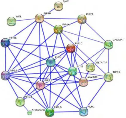
According to the data obtained from Simple molecular architecture tool, we found many interactions belonging to the five PIP1 protein sub-family in various cellular localizations, but mainly the partners were localized in
the plasma membrane, the interactome network of PIP1 subfamily showed significant interaction with almost all members in PIP2 subfamily and PIP3 subfamily. Additionally strong interaction of PIP1A, PIP1C and PIP1;
5 is shown with RD28, DELTA-TIP, and ANAC098 whereas PIP1B weakly interacts with WOL and Rps2 and Aquaporin 3 family. PIP1; 4 showed significant interaction with RD28, PIP3 family and PIP2; 8 protein (Fig.
4).
Fig. 4 Interactome of PIP1s subunits as obtained from string interaction. Interactome is shown in a confidence view where
stronger associations are represented by thicker lines.
Protein Subcellular localization
PIP1-1 Plasma membrane
PIP1-2 Plasma membrane, plastid
PIP1-3 Plasma membrane, plastid
PIP1-4 plasma membrane,mitochondrion
PIP1-5 Plasma membrane, gologi, E.R.
Table 2 Subcellular sites of PIP1s subfamily obtained
4 Discussion
Plant aquaporins is large family with at least 38 homologs, divided into four major subfamilies: plasma
membrane intrinsic protein (PIP), tonoplast intrinsic protein (TIP), nodulin 26-like intrinsic proteins (NIP), and small and basic intrinsic proteins (SIP) 1. The fourth subfamily of small and basic intrinsic proteins is not well characterized so far. Plasma membrane intrinsic protein (PIP) is divided into two groups: PIP1 and PIP2. In
Arabidopsis thaliana, PIP1 has five homologs; PIP1a, PIP1b, PIP1c, PIP1; 4 and PIP1; 5 which are comprising six membrane-spanning domains tilted along the plane of the membrane and linked by five loops (A to E)
located on the intra- (B, D) or extra-cytoplasmic (A, C, E ) side of the membrane. The N- and C-terminal extremities are both exposed to the cytosol Fig. 3. A central aqueous pore is delineated by the transmembrane domains and loops B and E, which both carries a conserved Asn-Pro-Ala (NPA) motif and dip from either side
of the membrane into the center of the molecule (Horton et al., 2007).
The functional interactome of PIP1A showed 7 interactions with PIP1B, PIP2; 5, PIP3, RD28 (PIP2C),
TIP2; 2, GAMMA-TIP and PIP2A. The interactors: PIP2; 5, PIP3, RD28 have been confirmed experimentally by yeast-two hybrid system, assigning a new functions to PIP1A protein like osmo-regulation in plants under high osmotic stress, earlier examined under a high salt condition (Maurel et al., 2008). Other predicted
interactor of PIP1A such as PIP1B is sharing water transport while TIP2; 2, GAMMA-TIP and PIP2A have the following functions; ammonia transporter/ methyl-ammonium transmembrane transporter/ water channel,
facilitate the transport of water, diffusion of amino acids and/or peptides from the vacuolar compartment to the cytoplasm, play a role in the control of cell turgor and cell expansion and involved in root water uptake respectively (Zardoya and Villalba, 2001; Ishikawa et al., 2010; Braun et al., 2011).
PIP1B is essential for the water permeability of the plasma membrane and for the morphology of the root system. Our data showed that PIP1B is sharing 96% homology similarities of PIP1A and 3 interactors as well;
PIP2A, PIP2; 5 and PIP3. These proteins had been experimentally confirmed by yeast-two hybrid as interactors of PIP1B (Maurel et al., 2008). The coordination between PIP1A and PIP1B in plant membranes
regulates the osmotic pressure under salinity stress and control the cell turgor. Among the functional interactome of PIP1B, there are two interesting interactors; WOL (histidine kinase 4) is cytokinin-binding receptor that transducers cytokinin signals across the plasma membrane and Rps2 which is plasma membrane
protein with leucine-rich repeat, leucine zipper, and P loop domains that confers resistance to Pseudomonas syringae infection by interacting with the virulence gene avrRpt2. RPS2 protein interacts directly with plasma membrane (Dortay et al., 2008; Kuwagata et al., 2012). Two other interactors of PIP1Bare PIP2E and PIP2; 8 having the common aquaporins function of water transport through plant cell membranes.
PIP1C and PIP1D are sharing 99.7% homology similarities, 3-D structure and functional interactome. The
profile of their interactome comprises PIP2; 5, PIP3, RD28 and PIP2; 8 proteins which regulate the osmotic pressure under abiotic stress. Moreover, PIP1C interacts with anac089 protein (Arabidopsis NAC domain
containing protein 89) which is negatively regulating floral initiation in Arabidopsis thaliana (Qi and Katagiri, 2009). PIP1C also interacts with DELTA-TIP; ammonia transporter/methylammonium transmembrane transporter which is the main channel of ammonia. It expresses especially in flowers, shoot and stem. PIP1; 3,
PIP1; 5 and DELTA-TIP interacts with anac089 protein, involved in transcription factor activity (Li et al., 2010; Li et al., 2011). In addition PIP2E interacts with PIP1C assigning the function of response to nematode plus active water channel. PIP2E induce signals to plant immune system as response to nematode infection.
PIP1; 5 protein, as all PIP1 members, regulates the water channel activity and response to salt stress. The interactome profile of PIP1; 5 comprises PIP2; 5, PIP3, RD28, PIP2; 4, anac089 and PIP2; 8 which are the
osmotic stress such as under a high salt, response to nematode, facilitate the transport of water across cell membrane and regulation of floral initiation in Arabidopsis thaliana (Kaldenhoff et al., 2007). Additionally, PIP1; 5 has a strong interaction with NLM1 (arsenite transmembrane transporter), assigning the PIP1; 5 member to share the coordination of arsenite transport and tolerance. NLM1 also acts as water channel regulator, probably required to promote glycerol permeability and water transport across cell membranes
(Kamiya et al., 2009).
AQPs are a protein network in plant cell integrating in all physiological processes. Uncovering this
network will enable us to infiltrate to core of cellular and molecular levels of the cell complexity. The functional interactome of PIP1protein sub-family is the first step to comprehend this complexity. The subcellular localization of PIP1 homologs is the key to unlock the complexity of their functional interactome,
giving more details about AQPs network global function and physiological processes which they are interfering and where?
Abbreviations
AQP: Aquaporin
MIP: major intrinsic proteins TIP: Tonoplast intrinsic proteins
PIP: Plasma membrane intrinsic proteins
SoPIP: Spinaciaoleracea plasma membrane intrinsic proteins ClustalW2: multiple sequence alignment program, version 4
MSA: Multiple Sequence Alignment
Pymol: program to obtain 3D structure of the target proteins
WoLF PSORT: Subcellular localization prediction for each protein PDB: Protein data bank
Acknowledgment
Authors thank the board of trustees, International University of Sarajevo for financial and moral support to
realize this work.
References
Braun P, Carvunis AR, Charloteaux B, et al. 2011. Evidence for network evolution in an Arabidopsis
interactome map. Science, 333(6042): 601-607
Dortay H, Gruhn N, Pfeifer A, et al. 2008. Toward an interaction map of the two-component signalling pathway of Arabidopsis thaliana. Journal of Proteome Research, 7(9): 3649-3660
Chaumont F, Moshelion M, Mark J. 2000. Daniels. Regulation of plant aquaporin activity. Franois Bilogy Cells, 97(10): 749-764
Hohmann I, Bill RM, Kayingo I, et al. 2000. Microbial MIP channels. Trends in Microbiology, 8: 33-38 Horton P, Park KJ, Obayashi T, et al. 2007. WoLF PSORT: Protein Localization Predictor. Nucleic Acids
Research, 35: W585-W587
Franceschini A, Szklarczyk D, et al. 2013. STRING v9.1: protein-protein interaction networks, with increased coverage and integration. Nucleic Acids Research, 41(Database issue): D808-D815
Ishikawa H, Uenishi Y, Takase T, et al. 2010. Involvement of phytochrome A in regulation of water dynamics and aquaporin expression in Arabidopsis roots. 21st International Conference on Arabidopsis Research
Johanson U, Karlsson M, Johansson I, et al. 2001. The complete set of genes encoding major intrinsic proteins in Arabidopsis provides a framework for a new nomenclature for major intrinsic proteins in plants. Plant Physiology, 126: 1358-1369
Kaldenhoff R, Bertl A, Otto B, et al. 2007. Characterization of plant aquaporins. Methods in Enzymology, 428: 505-531
Kamiya T, Tanaka M, et al. 2009. NIP1;1, an aquaporin homolog, determines the arsenite sensitivity of Arabidopsis thaliana. Journal of Biological Chemistry, 284(4): 2114-2120
Kuwagata T, Ishikawa-Sakurai J, Hayashi H, et al. 2012. Influence of low air humidity and low root
temperature on water uptake, growth and aquaporin expression in rice plants. Plant and Cell Physiology, 53(8): 1418-1431
Larkin MA, Blackshields G, Brown NP, et al. 2007. ClustalW and Clustal X version 2.0. Bioinformatics, 23(21): 2947-2948
Li J, Zhang J, Wang X, et al. 2010 A membrane-tethered transcription factor ANAC089 negatively regulates
floral initiation in Arabidopsis thaliana. Science China Life Sciences, 1299-1306
Lamesch P, Berardini T.Z and Li D.2012.The Arabidopsis Information Resource (TAIR): improved gene
annotation and new tools. Nucleic Acids Research, 40: 1202-1210
Letunic I, Doerks T, Bork P. 2012. SMART 7: recent updates to the protein domain annotation resource.
Nucleic Acids Research, 40(1): 302-305
Maurel C, Verdoucq L, Luu DT, et al. 2008. Plant aquaporins: Membrane channels with multiple integrated functions. Annual Review of Plant Biology, 59: 595-624
Muller DJ, Janovjak H, Lehto T, et al. 2002. Observing structure, function and assembly of single proteins by AFM. Progress in Biophysics & Molecular Biology, 79: 1-43
Ping Li, Wind J, Shi XL, et al. 2011. Fructose sensitivity is suppressed in Arabidopsis by the transcription factor ANAC089 lacking the membrane-bound domain. Proceedings of the National Academy of Sciences of USA, 108: 3436–3441
Qi Y, Katagiri F. 2009. Purification of low-abundance Arabidopsis plasma-membrane protein complexes and identification of candidate components. Plant Journal, 57(5): 932-944
Robinson BH, Brooks RR, Kirkman JH, et al. 1996. Plant-available elements in soils and their influence on the vegetation over ultramar (serpentine) rocks in New Zealand. Journal of the Royal Society of New Zealand, 26: 457-468
Santoni V, Javot H, Lauvergeat V, et al. 2003. Role of a single aquaporin isoform in root water uptake. Plant Cell, 15: 509-522
Sayers E W, Barrett T, Benson S H. 2009. Database resources of the National Center for Biotechnology Information. Nucleic Acids Res, 37: 5-15.
Schwede T, Bordoli L, Kopp J, et al. 2006. The SWISS-MODEL Workspace: A web-based environment for
protein structure homology modelling. Bioinformatics, 22: 195-201 The PyMOL Molecular Graphics System, Version 1.5.0.4. Schrödinger, LLC
Törnroth-Horsefield S, Wang Y, Hedfalk K, et al. 2005. Structural mechanism of plant aquaporin gating. Nature, 439(7077): 688-694
Wang Y, Tajkhorshid E. 2007. Molecular mechanisms of conduction and selectivity in aquaporin water
Yu J, Yool AJ, Schulten K, et al. 2006. Mechanism of gating and ion conductivity of a possible tetrameric pore in Aquaporin-1. Structure, 14: 1411-1423
Zardoya R. and Villalba S. 2001. A phylogenetic framework for the aquaporin family in eukaryotes. Journal of Molecular Evolution, 52(5): 391-404
Zelazny E, Borst JW, Muylaert M, et al. 2007. FRET imaging in living maize cells reveals that plasma