Advanced Pharmaceutical Bulletin, 2013, 3(1), 51-55 doi: http://dx.doi.org/10.5681/apb.2013.009
http://apb.tbzmed.ac.ir/
*Corresponding author: Behzad Baradaran, Assistant Professor of immunology, Immunology Research Center, Faculty of Medicine, Tabriz University of Medical Sciences, Tabriz, Iran. Tel: (+98) 411 3364665, Fax: (+98) 411 3364665, Email: behzad_im@yahoo.com
Copyright © 2013 by Tabriz University of Medical Sciences
Inhibitory and Cytotoxic Activities of
Salvia Officinalis L.
Extract
on
Human Lymphoma and Leukemia Cells by
Induction of Apoptosis
Fatemeh Zare Shahneh1, Samira Valiyari1, Behzad Baradaran1*, Jalal Abdolalizadeh1, Ali Bandehagh2, Abbas Azadmehr3, Reza Hajiaghaee4
1
Drug Applied Research Center, Tabriz University of Medical Sciences, Tabriz, Iran.
2Plant Breeding and Biotechnology Department, Faculty of Agriculture, University of Tabriz, Iran. 3
Immunology Department, Qazvin University of Medical Sciences, Qazvin, Iran.
4
Pharmacognosy & Pharmaceutics Department of Medicinal Plants Research Center, Institute of Medicinal Plants, ACECR, Karaj, Iran.
Introduction
The conventional modality for cancer therapy includes surgery, radiation and drugs, separately or in combination. An effective anticancer drug should kill cancer cells without affecting abnormal to normal cells. Therefore, the identification of new cytotoxic drug with low side effects on immune system has developed as
important area in new studies of
immunopharmacology.1 This ideal circumstance is feasible by inducing apoptosis in cancer cells. Apoptosis (Programmed cell death) an active physiological suicide that occur normally during development and aging and as a homeostatic mechanism to maintain cell populations in tissues. Apoptosis is characterized by distinct morphological and biochemical changes, including cell shrinkage, membrane blebbing, chromatin condensation, and formation of apoptotic bodies. Maintenance of organelle integrity, condensation and DNA
fragmentation, followed by removal of dying cells by macrophage-mediated phagocytosis.2 Oncogenic mutations can disrupt apoptosis and lead to tumor initiation, progression and metastasis. Dysfunction of the apoptotic program can promote tumor initiation, progression and treatment resistance. Defects in apoptosis can result in cancer, autoimmune diseases and spreading of viral infections.3 Traditional medicines have been used for maintaining heath, boosting immune system function, prevention, therapy and remission of cancer. Natural product can serve as chemoperventive and chemotherapeutic agent.4
Recently, extensive studies have been dedicated to the apoptosis and the role of this process in intervening the lethal properties of anti-neoplastic agents in cancer cells. Anticancer agents induce apoptosis, so that disruption of apoptotic cell death reduces treatment sensitivity. Extensive varieties of natural compounds A R T I C L E I N F O A B S T R A C T
Article Type: Research Article
Article History: Received: 6 October 2012 Accepted: 7 October 2012 ePublished: 7 February 2013
Keywords:
Salvia officinalis L Cytotoxic Anti-tumor Apoptosis Cancer
Zare Shahneh et al.
possess significant cytotoxic as well as chemopreventive activity, which act via apoptosis.5 Extracts of plants used in traditional medicine also have a similar property. Fifty-eight species of the genus
Salvia (Lamiaceae) are found in Iran, which 17 are endemic. The genus Salvia shows diverse biological activities manifested by the different components that allow for the many medicinal and pharmaceutical applications of the plant materials and/or extracts. In fact, many diterpenes, isolated from plants of several species of the genus Salvia, have been demonstrated interesting pharmacological properties, such as antioxidant, anti-microbial,6 anti-inflammatory, analgesic,7 antipyretic, hemostatic,8 hypoglycemic9 and antitumor.10
The objective of this study was to examine the in vitro cytotoxic activities of methanol standardized extract, on a non-Hodgkin’s B-cell lymphoma (Raji) and human leukemic monocyte lymphoma (U937), Human acute myelocytic leukemia (KG-1A) and Human Umbilical Vein Endothelial (HUVEC) cell lines, using a MTT cytotoxicity assay. The study also tested whether the mechanism of action involves induction of apoptosis. Cell death ELISA was employed to quantify the nucleosome production resulting from nuclear DNA fragmentation during apoptosis.
Materials and Methods Preparation of plant extract
Salvia officinalis L. plants were collected from the central part of Iran (Karaj) in April 2011. Mr. Ajani from the Department of Botany, Institute of Medicinal Plants (IMP) of Karaj, Iran identified the plant. A voucher specimen was deposited in the herbarium of the above mentioned (herbarium No.1471). The aerial parts of the plant were separated, shade dried and grinded into powder with mortar and pestle. The prepared powder was kept in tight containers protected completely from light. Extraction of methanolic extract was carried out by macerating 100 g of powdered dry plant in 500 ml of 70% methanol for 48 h at room temperature. Then, the macerated plant material was extracted with 70% methanol solvent by percolator apparatus (2-liter volume) at room temperature. The plant extract was removed from percolator, filtered through Whatman filter paper (NO.4) and dried under reduced pressure at 37 ºC with rotator evaporator before being added to ethanol as the solvent. The methanol extract was filtered and concentrated using a rotary evaporator and then evaporated to dryness. Briefly, the concentrated plant extracts were dissolved in dimethyl sulphoxide (DMSO) (SIGMA, USA) to get a stock solution of 10 mg/ml. The sub-stock solution of
0.2 mg/ml was prepared by diluting 20 μL of the stock solution into 980 μL serum-free culture medium, RPMI 1640 (the percentage of DMSO in the experiment should not exceed 0.5). The stock and sub-stock solutions were both stored at 4 ºC.
Cell cultures
Burkitt’s lymphoma B-cell line (Raji), human leukemic monocyte lymphoma cell line (U937), Human acute myelocytic leukemia cell line (KG-1A) and Human Umbilical Vein Endothelial Cells (HUVEC) were purchased from Pasteur Institute of Iran (Bank cell). The cells was grown and maintained in a humidified incubator at 37 ºC and in 5% CO2 atmosphere. RPMI-1640 medium (SIGMA) was supplemented with 0.01 mg/ml heat inactivated Fetal Calf Serum (FCS), 100 units/ml penicillin, and 100 μg/ml streptomycin ( ALL FROM INVITROGEN GIBCO) was used for cell cultures. Upon reaching confluency, the cells were passaged. After being harvested from sterile T75 culture flasks (NUNC, DENMARK), the cells were counted using a hemocytometer and cell viability was determined by trypan blue exclusion. Ten thousand
cells from log phase cultures were seeded in 100 μl of
RPMI medium supplemented with 10% fetal bovine serum per well of 96-well flat-bottom culture plates (NUNC, DENMARK).11Cells were incubated with the
Salvia officinalis L. extract for a defined time (12, 24 and 48 hours). Proliferative response and cell death of the Salvia officinalis L. extract-treated cells were determined using MTT assay and cell death ELISA Cell Viability Assay, respectively.
MTT Cell viability assay
Salvia officinalis Inhibits Lymphoma Proliferation
calculated by EXCEL software and the mean optical density (OD) ± SD for each group of the replicates calculated. Percent growth inhibition of cells exposed to treatments was calculated as follows: % Inhibition = 100 - (Test OD/Non-treated OD) × 100).
Cell death detection
Cell Death Detection ELISA PLUS (ROCHE APPLIED SCIENCE) was used to quantify histone-complexed DNA fragments (nucleosomes) in cytoplasm of the apoptotic cells after induction of apoptosis. Briefly, after incubation with the methanolic extract (at concentrations determined by MTT assay) for 24 h cells were pelleted and lysed. Mouse monoclonal antibodies against single-strand DNA and histones (H1, H2a, H2b, H3 and H4) specifically detected and bound mononuclesomes and oligonucleosomes derived from cells undergoing apoptosis. Biotinylated anti-histone antibodies then fixed the antibody-nucleosome complexes to the streptavidin-coated microtiter plate. The anti-DNA antibodies were conjugated with a peroxidase that reacted with the substrate ABTS [2,2V-azino-di (3- ethylbenzthiazolin-sulfonate)] to form a colored product. The remaining steps were carried out according to the instructions supplied by the manufacturer. The resulting color development, which was proportional to the amount of nucleosomes captured in the antibody sandwich, was measured at 405 nm wavelength using a Benchmark microtiter plate reader (BIO-RAD).13 Results were expressed as the apoptotic index (AI), calculated from the ratio of absorbance of treated (apoptotic) sample to that of the untreated (control) sample.
Dye exclusion assay
Cellular cytotoxicity induced by the Salvia officinalis L. extract treatment was measured with trypan blue exclusion assay. Briefly, 1×10⁴ cells were seeded into 96-well plates and treated with or without (as control) crude extract at specified doses for 12, 24 and 48 h. After the incubation period, the cultures were harvested and washed twice with PBS. The cell pellet was then
resuspended with 0.5 ml PBS. Then, 20 μL of cell was
mixed with equal volume of 0.4% trypan blue (SIGMA, USA Merck) and was count by Neubauer haemocytometer (WEBER, ENGLAND) by clear field microscopy (NIKON, JAPAN). Only viable cells were counted. Each extract and control was assayed two times in triplicate.
Statistical Analysis
The data are expressed as mean ± standard deviation (SD) for at least three independent determinations in triplicate for each experimental point. The data were analyzed using IBM SPSS Statistics 20 software. For all the measurements, Tow-way ANOVA followed by
Duncan’s New Multiple Range Test (P≤0.05) was used
to assess the statistically significance of difference between control and FA treated.
Results
Effects of methanolic extract of Salvia officinalis L. on proliferation of leukemia and lymphoma cells
Salvia officinalis L.extract at 50 to 800 μg/ml exhibited significant dose-dependent inhibitory effects on the proliferation of Raji (Figure 1A), U937 (Figure 1B), and KG-1A cells (Figure 1C), with more than 80% suppression. However, the extract induced no significant suppression on the proliferation of normal HUVEC cells (Figure 1D).
Figure 1. Effect of different concentrations of Salvia officinalis L. (A) Raji , (B) U937, (C) KG-1A and (D) HUVEC cell lines in 12, 24 and
Zare Shahneh et al.
Table 1 shows the concentrations producing 50% growth inhibition (IC50) of theSalvia officinalis L. extract on the three cell lines in three times,which Raji proliferation was most potently suppressed with the lowest IC50 value. Similar inhibitory effects were found in KG-1A, U937 cells after incubation with the
Salvia officinalis L extract. Proliferation of Raji cells was most significantly reduced by Salvia officinalis L.
extract at 100 to 300 µg /ml, result in a lower IC50 value;while KG-1A and U937 cell growth was most significantly reduced from 200 to 400 µg /ml with higherIC50 values. Table 2 illustrates that Raji, KG-1A and U937 cells were similarly susceptible to the cytotoxicity of Salvia officinalis L. extract with more than 80% growth suppression; however, HUVECcells were much less susceptible to the cytotoxic effect of
Salvia officinalis L. extract. Viability rate was decreased in descending form 50.29%, 47.07% and 44.37% in U937, KG-1A and Raji, respectively (Table 3). In Table 4, Raji, KG-1A and U937 cells was compared to elucidate the cytotoxicity of both Salvia officinalis L. extract and Toxol with more than 80% and 90% growth suppression.
Table 1. Concentration producing 50% growth inhibitions (IC50) of Salvia officinalis L. extract on the three cell lines in
12h, 24h and 48h.
Cell lines
IC50 (µg/ml)
12 hr 24 hr 48 hr
Raji 239.692 163.187 130.597
U937 304.332 229.312 161.825
KG-1A 345.623 214.377 152.182
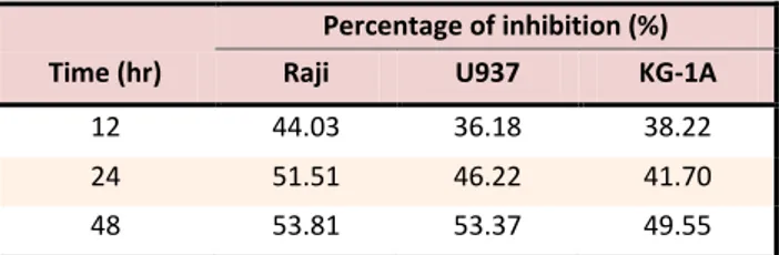
Table 2. Percentage of inhibition in IC50 concentration induced
by Salvia officinalis L. extract on Raji, U937and KG-1A cell
lines respectively.
Time (hr)
Percentage of inhibition (%)
Raji U937 KG-1A
12 44.03 36.18 38.22
24 51.51 46.22 41.70
48 53.81 53.37 49.55
Table 3. Percentage of Viable cells induced by Salvia
officinalis L. extract in IC50 concentration on the three cell
lines. Results are expressed as the mean percentage of dead cells F SD in triplicate experiments with three wells each. Percentage of dead cells was calculated from the ratio of dead cells to total number of cells using trypan blue exclusion test.
Time (hr)
Viable cells (%)
Raji U937 KG-1A
12 53.92 59.81 61.22
24 48.40 51.81 54.88
48 44.37 47.07 50.29
Table 4. Comparison of cytotoxic activity of Salvia officinalis L.
extract (800 μg/ml) and Toxol (50 μg/ml) on the 3 cell lines.
Cell lines
Growth inhibition %
Crude extract Toxol
Raji 87.00 89.1
U937 75.77 99.4
KG-1A 81.00 89.9
Effects of Salvia officinalis L extract on cell death of leukemia and lymphoma
As determined by MTT assay, Salvia officinalis L.
extract at 100, 200 and 300 µg /ml in 24 h were chosen for each cell line in cell death detection ELISA. Before determination of Salvia officinalis L. extract-induced cell death, cell viability of the fourcell lines was evaluated. As shown in Table 3, the proportion of viable Raji cells was increased sharply. However, the percentage of death of U937 and KG-1A cells peaked at 12, 24 and 48 hr, respectively, when incubated with the Salvia officinalis L. extract of the same concentration. These results indicate that the apoptotic response of these three cell lines should be evaluated at different time points.
As shown in Figure 2, the Salvia officinalis L. extract at
300 μg/ml could strongly induce apoptosis of Rajicells after 24 h in a dose-dependent manner. Effects of
Salvia officinalis L. extract on proliferation of (A) Raji, (B) KG-1A, (C) U937, and (D) HUVEC. Cells were incubated withincreasing concentrations (50–800 µg/ml) of Salvia officinalis L. extract in culture medium for 24 h (Raji, U937, KG-1A and HUVEC) and the proliferative response was then assessed with MTT assay. The possible mechanism was via induction of apoptosis, as evidenced by the significant increase in nucleosome production at 200 and 300 µg /ml of Salvia officinalis L. extract after incubation for 24h but the ratio of apoptosis was constant approximately 50±3%.
Figure 2. Effects of Salvia officinalis L. extract on cell death of
Raji, U937, and KG-1A. Cells were incubated with the Salvia
officinalis L extract in culture medium at concentrations derived
Salvia officinalis Inhibits Lymphoma Proliferation
Discussion
The current study has demonstrated that methanolic extract of a commonly used in Persian medicinal herb,
Salvia officinalis L. in its natural form, could significantly suppress the proliferation of Burkitt’s lymphoma B-cell line (Raji), human leukemic monocyte lymphoma cell line (U937) and Human acute myelocytic leukemia cell line (KG-1A) in vitro by means of the MTT assay. Such antiproliferative activity of Salvia officinalis L. extract was characterized by the time and dose-dependent and tumor-selective manner, as reflected by the comparatively low IC50 values and the absence of significant effects on normal HUVEC cells respectively. On the contrary, Toxol at optimal in vitro concentration non-selectively induced at least 90% growth suppression on all the studied cell lines (Table 4). These results suggested that when compared with the commonly used chemotherapeutic antitumor drug (e.g. Toxol), the Salvia officinalis L. extract, albeit at higher concentration, could induce comparable antitumor activity with much less cytotoxic effects on normal cells.
In Raji, U937 and KG-1A cell lines viability were decreased (from 79% to 13%, from 81% to 23% and from 85% to 19%) with increasing treatment concentration and (from 53% to 44%, from 61% to 50% and from 63% to 47%) treatment time respectively. In order to determine the antiproliferative activity of Salvia officinalis L. extract is shown by induction of apoptosis, cell death detection ELISA was used to quantify the nucleosome production during nuclear DNA denaturation of apoptotic cells. Before evaluating the Salvia officinalis L. extract-induced apoptosis, the percentages of death of the 3 cell lines at 24h were determined since the incubation time necessary to induce apoptosis can show broad variation. For Raji cells, the sharp increase in percentage of dead cells paralleled the significant nucleosome production up regulated by the Salvia officinalis L. extract from 12 to 48 h, suggesting the dose- and time-dependent induction of apoptosis. Similar results were seen in KG-1A and U937 cells. These results suggested that the extract exerts its antitumor effect on these leukemia and lymphoma cells possibly via an apoptosis-dependent pathway. Therefore, further studies are required to evaluate whether cell cycle arrest or apoptosis induction contribute more to the Salvia officinalis L. extract-induced cytotoxic effect on panel cells. In order to elucidate the cytotoxic activities of Salvia officinalis L.
extract on the growth of leukemia and lymphoma cells of other sub-types, acute T-cell leukemia cell line
(Jurkat) and Hodgkin’s lymphoma may become the
target cells used in our future studies. In addition, mechanistic studies on cell cycle arrest and early apoptotic events may be conducted to delineate other possible antitumor mechanisms of the Salvia officinalis L. extract. Besides, future in vivo antitumor studies will
be performed in order to confirm these in vitro results. In conclusion, this study provides the evidence that in vitro cytotoxic activity of a methanolic standardized extract from wild Salvia officinalis L. was found dose-dependently inhibit the proliferation of lymphoma and leukemic cells possibly via an apoptosis-dependent pathway.
Acknowledgements
The authors would like to thank the financial support of Drug Applied Research Center of Tabriz University of medical sciences and kind assistance of who contribute for this research.
Conflict of interest
The authors report no conflicts of interest.
References
1. Mehta RG, Murillo G, Naithani R, Peng X. Cancer chemoprevention by natural products: How far have we come? Pharm Res 2010;27(6):950-61.
2. Elmore S. Apoptosis: A review of programmed cell death. Toxicol Pathol 2007;35(4):495-516.
3. Fischer U, Schulze-Osthoff K. Apoptosis-based therapies and drug targets. Cell Death Differ 2005;12 Suppl 1:942-61.
4. Taraphdar AK, Roy M, Bhattacharya RK. Natural products as inducers of apoptosis: Implication for cancer therapy and prevention. Current science 2001; 80: 1387-98. 5. Reed JC, Pellecchia M. Apoptosis-based therapies for
hematologic malignancies. Blood 2005;106(2):408-18. 6. González A, Abad T, Jiménez I, Ravelo A. A first study of
antibacterial activity of diterpenes isolated from some Salvia species. Biochem Syst Ecol 1987; 17: 293-6. 7. Hosseinzadeh H, Haddadkhodaparast MH, Arash AR.
Antinociceptive, antiinflammatory and acute toxicity effects of salvia leriifolia benth seed extract in mice and rats. Phytother Res 2003;17(4):422-5.
8. Hernandez-Perez M, Rabanal RM, de la Torre MC, Rodriguez B. Analgesic, anti-inflammatory, antipyretic and haematological effects of aethiopinone, an o-naphthoquinone diterpenoid from salvia aethiopis roots and two hemisynthetic derivatives. Planta Med 1995;61(6):505-9.
9. Lima CF, Andrade PB, Seabra RM, Fernandes-Ferreira M, Pereira-Wilson C. The drinking of a salvia officinalis infusion improves liver antioxidant status in mice and rats. J Ethnopharmacol 2005;97(2):383-9.
10. Liu J, Shen HM, Ong CN. Salvia miltiorrhiza inhibits cell growth and induces apoptosis in human hepatoma hepg(2) cells. Cancer Lett 2000;153(1-2):85-93.
11. Phelan MC. Basic Techniques for Mammalian to Cell Tissue Culture. Current protocols in cell biology 1998; 7: 1-10.
12. Mosmann T. Rapid colorimetric assay for cellular growth and survival: Application to proliferation and cytotoxicity assays. J Immunol Methods 1983;65(1-2):55-63.