Instituto do Coração (InCor) do Hospital das Clínicas da Faculdade de Medicina da Universidade de São Paulo, São Paulo, SP, Brazil.
Correspondence to: Maria de Lourdes Higuchi, M.D., Laboratório de Anatomia Patológica, Instituto do Coração/InCor, Faculdade de Medicina da Universidade de São Paulo, Av. Dr. Enéas C. Aguiar 44, 05403-000 São Paulo, SP, Brasil. Tel: +55 11 3069- 5252, FAX: +55 11 3069-5251. e-mail: [email protected]
INVITED REVIEW
INFECTIOUS AGENTS IN CORONARY ATHEROMAS: A POSSIBLE ROLE IN THE PATHOGENESIS OF
PLAQUE RUPTURE AND ACUTE MYOCARDIAL INFARCTION
Maria de Lourdes HIGUCHI & Jose A. F. RAMIRES
SUMMARY
In this review we report our recent findings of histopathological features of plaque instability and the association with Mycoplasma pneumoniae (MP) and Chlamydia pneumoniae (CP) infection, studying thrombosed coronary artery segments (CAS) of patients who died due to acute myocardial infarction. Vulnerable plaques are known to be associated with fat atheromas and inflammation of the plaque. Here we demonstrated that vulnerability is also related with focal positive vessel remodeling that maintains relatively well preserved lumen even in the presence of large atheromatous plaques. This phenomena may explain why the cinecoronariography may not detect large and dangerous vulnerable plaques. Greater amount of these bacteria in vulnerable plaques is associated with adventitial inflammation and positive vessel remodeling: the mean numbers of lymphocytes were significantly higher in adventitia than in the plaque, good direct correlation was obtained between numbers of CD20 B cells and numbers of CP infected cells in adventitia, and between % area of MP-DNA in the plaque and cross sectional area of the vessel, suggesting a cause-effect relationship. Mycoplasma is a bacterium that needs cholesterol for proliferation and may increase virulence of other infectious agents. In conclusion, co-infection by Mycoplasma pneumoniae and Chlamydia pneumoniae may represent an important co-factor for plaque instability, leading to coronary plaque thrombosis and acute myocardial infarction, since larger amount of these bacteria strongly correlated with histological signs of more vulnerability of the plaque. The search of CMV and Helicobacter pilori in these tissues resulted negative.
KEYWORDS:Chlamydia pneumoniae; Mycoplasma pneumoniae; Infection; Atherosclerosis; Vulnerable plaque, Myocardial infarction.
INTRODUCTION
Many works have tried to associate infectious agents in the development of atherosclerosis and acute myocardial infarction30,31 mainly the Chlamydia pneumoniae (CP)8,12,23,25. Nevertheless, the results are controversial5,29 and the serological data can not discriminate patients who will develop acute myocardial infarction (AMI)7. Inflammation in the atherosclerotic plaque is accepted to be linked to plaque instability3,10,17. High level of cholesterol in the blood seems to be the most frequent associated risk factor, but it is difficult to explain why cholesterol causes inflammation. On the other hand, the accelerated progression of systemic atherosclerosis in some autoimmune rheumatic diseases, such as systemic lupus erythematosus, antiphospholipid syndrome, rheumatoid arthritis and vasculitis, have led the authors to implicate autoimmune mechanisms in development of atherosclerosis24. We performed a series of works analyzing deeply the histopathological characteristics of ruptured plaque and compared with
stable plaques of the same and different patients, regarding the constituents and size of the plaque, remodeling of the wall and plaque and adventitial inflammation. We also search for some infectious agents, using different techniques (immunohistochemistry, in situ hibridization and electron microscopy) in the same material in order to diminish the false positive or negative results. The results of these works are divided in 4 sections.
1- Fatal thrombosed atheromas are associated with larger plaques and positive remodeling2:Several lines of clinical and angiographic
&
Fig. 1 - Histological views of unstable and stable coronary atheromas. Fig.1A - Ruptured thrombosed segment showing expanded vessel containing a large fat plaque and a thinned medial layer (RC - right coronary - Movat stain). On the right top, a detailed view of the adventitial inflammatory infiltrate at the confocal laser microscopy, the external elastic membrane (EEM) in green fluorescence exhibiting holes (arrow) and the lymphocytes stained in yellow. On the right bottom, histological detail of the adventitial and medial inflammatory infiltrate associated with EEM fragmentation and medial layer attenuation. Fig. 1B is a schematic representation of a thrombosed coronary artery segment presenting inflammation in all layers of the vessel, positive remodeling, large fat plaque, and a wide lumen occluded by a thrombus. Fig. 1C is an equi-stenotic stable segment from the same patient of Fig. 1A, exhibiting a smaller fibrotic plaque that causes more obstructive injury due to the vessel negative remodeling (DA - descending anterior artery - Movat stain). Fig. 1D - Schematic representation of a stable coronary artery segment presenting a small fibrotic plaque causing severe obstruction of the lumen due to the negative remodeling of the vessel; there was no inflammation on the wall.
Table 1
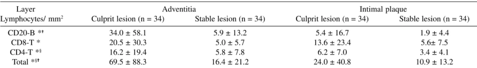
Mean cross-sectional areas (mm2) of the coronary artery segments with stable and unstable (culprit) atherosclerotic lesions from the same individuals
Layer Adventitia Intimal plaque
Lymphocytes/ mm2 Culprit lesion (n = 34) Stable lesion (n = 34) Culprit lesion (n = 34) Stable lesion (n = 34)
CD20-B *= 34.0 ± 58.1 5.9 ± 13.2 5.4 ± 16.7 1.9 ± 4.4
CD8-T * 20.5 ± 30.3 5.0 ± 5.7 13.6 ± 23.4 5.6± 7.5
CD4-T *§ 16.2 ± 19.4 5.8 ± 7.8 6.2 ± 7.0 3.4 ± 4.1
Total *§= 69.5 ± 88.3 16.4 ± 21.2 24.0 ± 40.8 10.9 ± 13.2
' plaques and negative remodeling with stable ones. In the present
post-mortem study we tested the hypothesis that the size of coronary atheroma and the type of remodeling distinguish culprit lesion responsible for fatal AMI from equi-stenotic lesion in the same coronary tree.
The main coronary branches from 36 consecutive patients with fatal AMI were histologically studied. The culprit lesion (Group A), responsible for vascular occlusion and fatal AMI was compared with equi-stenotic (histologically determined) stable plaque (Group B), obtained in another coronary branch from the same patient. Morphometric measurements included areas of the lumen, plaque and vessel and vascular remodeling determined by the relative cross-sectional vessel area (lesion vessel area/reference vessel area) X 100%. Positive remodeling was defined as relative vessel area > 105% and negative remodeling when relative vessel area was < 95%. Plaque composition measurement included the % areas occupied by fibrosis, lipid core, macrophages (CD 68) and smooth muscle cells (HHF 35). Compared to Group B, Group A had larger mean (SD) areas of plaque 9.6 (1.5) vs 4.7 (2.3) mm2, vessel 12.7 (4.9) vs 7.4 (3) mm2 and lumen 1.7 (1.5) vs 1.2 (0.86) mm2; (p < 0.01). Positive remodeling was more frequent in Group A than Group B: 21/30 (70%) vs 8/26 (31%). Plaque area correlated positively with % area of lipid core, r = 0.68 (p < 0.01) and with % area of macrophages, r = 0.41 (p < 0.01). Plaque area related negatively with % area fibrosis, r = -0.64 (p < 0.01) and with % area of smooth muscle cells, r = -0.48 (p < 0.01). Negative remodeling was present in 19% of group B and in only 3% of group A. Fig. 1A exemplifies a thrombosed ruptured plaque of a patient died due to acute myocardial infarction, presenting positive remodeling associated with medial thinning and less adventitial fibrosis. Fig. 1B is a schematic demonstration of the characteristics of a vulnerable segment. Fig. 1C is the histological representation of the stable plaque exhibiting preserved medial layer, less positive remodeling and similar degree of obstruction.
In conclusion, atherosclerotic plaques that cause thrombosis and fatal acute myocardial infarction usually present vessel positive remodeling and tend to be larger than equi-stenotic stable plaques.
2- Adventitial inflammation and attenuation of medial and adventitial layers are associated with positive remodeling of the vessel and plaque rupture11: Analyzing 34 of the CAS studied in above
described work, we searched if adventitial inflammation is associated with positive remodeling and plaque vulnerability. The culprit lesion segments (Group A) and non-culprit segments (Group B) were compared regarding the mean numbers of lymphocytes in the adventitia and in the plaque. Quantity of adventitial microvessels, adventitial fibrosis and, using confocal laser microscopy, external elastic membrane were also compared10,11. In the adventitia, the mean numbers of lymphocytes and microvessels/mm2 were respectively 69.5 ± 88.3 and 60.9 ± 32.1 in the culprit lesions; and 16.4 ± 21.1 and 44.3 ± 16.1 in stable lesions (lymphocytes - p < 0.01; microvessels - p = 0.04). The most numerous lymphocyte in adventitia was the CD20 B cell. Within the plaques, the mean number of total lymphocytes was 24.0 ± 40.8 in culprit lesions and 10.9 ± 13.2.06 in stable ones (p = 0.17), and a significant difference was seen in CD4 T cells 6.2 ± 7.0 vs 3.4 ± 4.1 (p < 0.05); the most numerous lymphocyte in the plaque was CD8 T cell. The mean percent area of adventitial fibrosis/cross sectional area of the vessel was significantly lower in unstable plaques (16.24 ± 5.07% vs 28.95 ± 9.76% respectively; p < 0.001). Fig. 1A, closer views demonstrate inflammatory
infiltrate on the base of the plaque and in the medial and adventitial layers, in a rupture plaque segment, that is not present in stable segments. The inflammatory infiltrate releasing enzymes may have a role in the pathogenesis of plaque rupture injuring the medial layer and adventitial collagen. The confocal laser microscopy demonstrates in 3D view that the external elastic membrane presents several holes associated with lymphocytes (Fig. 1A, right top). Figures 1B and 1D show schematic representation respectively of positive remodeling and inflammation associated with vulnerable thrombosed plaque and negative remodeling and absence of inflammation associated with stable occlusive plaques. In conclusion, unstable plaques frequently exhibit chronic pan-arteritis, accompanied by vessel dilatation, medial thinning, and diminished adventitial collagen. Adventitial inflammation and not only the plaque inflammation seems to be directly related with atheroma instability.
3 - Chlamydia pneumoniae in ruptured plaques: comparison with stable atherosclerotic plaque and nonatherosclerotic coronary artery segments12: In spite of a crescent number of papers trying to clarify if
CP is participating in the pathogenesis of plaque rupture in acute myocardial infarction, this remains a non-clarified matter. Most of the already published papers did not focus the ruptured thrombosed plaques9,14,15,16,32. As above discussed, vulnerable plaques are associated with adventitial inflammation and vessel positive remodeling. Here we searched if CP was present in the adventitia of fatal ruptured thrombosed segments.
Four groups of coronary artery segments from patients died at the Instituto do Coração (InCor) do Hospital das Clínicas da Faculdade de Medicina da Universidade de São Paulo were analyzed. Search of CP+ cells was performed using immunohistochemistry, in situ hybridization, electron microscopy, and confocal laser microscopy.
Group A - fatal ruptured plaque from 23 AMI patients; Group B - 23 nonruptured equistenotic plaque from Group A patients; Group C stable atheromatous plaque from 11 nonAMI patients; Group D -nonatherosclerotic coronary artery segments from 11 patients without evidences of coronary artery disease.
The mean number of CP+ cells/ 400x field was obtained analyzing immunostained slides. As positive controls were utilized a segment of ruptured plaque presenting large number of CP+ cells at electron microscopy and one paraffin block of rabbit lung severely infected with C. pneumoniae.
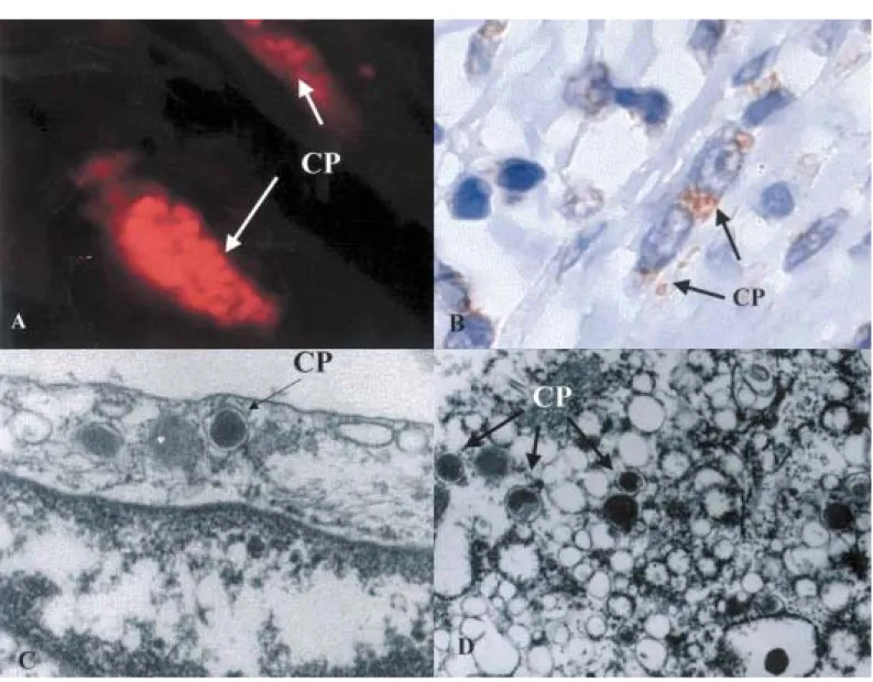
Fig. 2 - Demonstration of Chlamydia pneumoniae (CP) in adventitia cells of vulnerable coronary atheroma segment through many different techniques. Fig. 2A - Two fibroblasts presenting many CP bodies detected by immunofluorescence. Fig. 2B - In situ hybridization technique revealing CP-DNA in fibroblasts and inflammatory cells of adventitia and at the extracellular matrix. Figs. 3C and 3D - electron micrographies respectively showing endothelial cell and macrophage with elementary bodies of CP in the cytoplasm characterized by electron dense core and “pear” shape of the external membrane.
Table 2
Frequency and quantity of C. pneumoniae+ cells in unstable, stable and nonatherosclerotic groups of coronary artery segments
Group A unstable lesions Group B stable plaques Group C stable plaques Group D no atheroma
%C. pneumoniae + coronary artery segments
Adventitia 100% 91% 82% 82%
Plaque 96% 83% 82% 100%
C. pneumoniae + cells/mm2 (median number)
Adventitia 6.19 0.67 1.11 0.17
Plaque 2.33 0.27
expansion of external wall and the typical pear shape form. No difference
in the amount of CP+ cells was found among groups B, C, and D. The mean numbers of CD+20B cells in adventitia correlated positively with the mean numbers of CP+ cells positive by immunohistochemistry (Fig. 4B ), suggesting a cause-effect relationship.
In this item we concluded that CP bacteria are frequently present in cells of adventitia layer, in both atherosclerotic and nonatherosclerotic coronary artery segments. However, significant higher numbers of them are present in vulnerable plaque segments than the others. Increased numbers of CD20 B cells correlated with increased numbers of CP in adventitia, suggesting that CP contributes to the development of vessel inflammation and plaque instability.
4 - Association between Mycoplasma pneumoniae and Chlamydia pneumoniae in fatal thrombosed atherosclerotic coronary plaques13:
If CP is present in the adventitia of almost all coronary artery segments, the next point is to clarify what is favoring CP proliferation and if it is associated with severe inflammation and positive remodeling. Re-analysis of electron microscopy specimens demonstrated another bacterium adjacent to the CP bodies. This bacterium was recognized as mycoplasma because it did not have the external wall and using the in situ hibridization technique, as Mycoplasma pneumoniae (MP).
This finding was reported in a brief communication: the association between MP and CP in thrombosed CAS13. It is known that mycoplasmas are the only bacteria that require cholesterol in their membrane and for proliferation21. Mycoplasmas have been considered as a typical parasite of epithelium (respiratory and genitourinary tracts). It is believed that mycoplasmas are not capable of invading the human body. However here we demonstrated that intima of the arteries are usual habitat for mycoplasmas. Here we tested the hypothesis that MP is in higher amounts in vulnerable segments than in stable ones, associated with higher amounts of fat, CP, inflammation and positive vessel remodeling.
Serial sections of the same 68 CAS described in the above CP study were used.
The electron microscopy specimens were revised.
A double staining for simultaneous detection of CP (by immuno-histochemistry technique using the alkaline phosphatase as chromogen), and of MP by ISH technique was performed in some cases.
The mean % areas of intima MP-DNA in the cases of both groups A and B were compared with the mean numbers of CP + cells, of CD4+ T, CD8+ T and CD20+B cells, and % fat area in the plaques.
We found at the in situ hibridization slides MP bacteria as small brownish granules in the lipidic areas of the plaques, both at the extracellular matrix and in foam cells (Fig. 3A and 3B), mainly in the unstable plaque group (groups A). The stable plaques that usually are more fibrotic than lipidic showed much less MP into the plaque. MP were also detected in the media and in the adventitia, usually in larger forms.
In stable segments (groups B and C), a lower amount of MP was
detected in the plaque and almost absent in the media and absent or scarce in the adventitia.
In group D (non-atherosclerotic group), MP were either absent or present very scarcely in the subendothelial space.
The % area of MP and the mean number of CP+ cells, the % area of fat and the mean number of lymphocytes/mm2 are shown in Table 3.
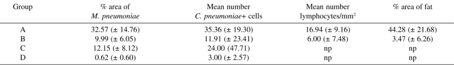
The mean % areas of MP-DNA were significantly higher in group A (ruptured plaque) than in group B (stable plaques of the same patient) (p < 0.01). There was no difference between groups B and C and they were greater than group D.
Analyzing the stable and ruptured plaques from groups A and B altogether, a significant correlation was obtained (Pearson test) between the % area occupied by MP in the plaque and the % area of fat in the plaque (r = 0.69) and with vessel cross-sectional area (r = 0.65). No important correlation was seen with the mean number of lymphocytes CD4+ T (r = 0.37), CD8+ T (r = 0.37), and CD20+ B (r = 0.018) lymphocytes in the plaque.
At the electron microscopy, mycoplasmas could be identified by their morphological unique characteristics: small irregular, tended to round shape, measuring from 0.1 to 0.4 µm, lacking the external wall and containing a granulated chromatin-like material. They were frequently seen adhered to the endothelial cells of “vasa vasorum” and, inside the endothelial cells, in the interstitium, and in the cytoplasm of macrophages usually nearby C. pneumoniae bodies. Necrotic lipidic core of vulnerable plaques contained many mycoplasmas in a rounded or elyptical shape and a lot of membranous structures were compatible with degenerated bacteria (Fig. 3D); in the medial layer and in the adventitia they were usually found in larger, cylindrical shape, presenting more electron dense granules, and absent cytoplasmic organelles (Fig. 3C).
In the stable plaques, a lower amount of MP was found.
Mycoplasmas are known as the smallest self-replicating organisms, requiring cholesterol for surviving because their external membrane is constituted by cholesterol, a unique property among prokaryotes21. They have been considered as a parasite of the epithelial surfaces.
In the present study, we found that MP are present in almost all fat atheroma’s areas, therefore mainly in vulnerable plaques. It believes that mycoplasmas induce a mild immune response constituted basically of T cells, and also immunedepression20. Higher amounts of MP was associated with CD8+ T cells and low number of CD4+ T cells. Mycoplasmas are capable of oxidizing the host cell membrane inducing its apoptosis1, favoring the rupture of the plaque. The mycoplasmas could explain the increased level of cytokines usually observed in vulnerable plaques22 as it is known they increase release of cytokines by inflammatory cells6.
Table 3
Quantity of Mycoplasma pneumoniae,Chlamydia pneumoniae, lymphocytes and fat in the plaques of the different groups
Group % area of Mean number Mean number % area of fat
M. pneumoniae C. pneumoniae+ cells lymphocytes/mm2
A 32.57 (± 14.76) 35.36 (± 19.30) 16.94 (± 9.16) 44.28 (± 21.68)
B 9.99 (± 6.05) 11.91 (± 23.41) 6.00 (± 7.48) 3.47 (± 6.26)
C 12.15 (± 8.12) 24.00 (47.71) np np
D 0.62 (± 0.60) 3.00 (± 2.57) np np
np = non-performed
! between MP and intracellular CP bacteria would favor the proliferation
of CP, increase of the inflammatory infiltrate. The inflammatory infiltrate and many CP would cause enzymatic degradation of the extracellular matrix, vessel positive remodeling and plaque rupture.
CONCLUSION
In conclusion, in this review we report a series of works of our Lab that strongly favor the concept that atherosclerosis and plaque rupture are complications associated with infectious agents. M. pneumoniae is present in almost all coronary atheromas. The close association between M. pneumoniae with C. pneumoniae may favor proliferation of both bacteria that induces to more inflammation and plaque rupture. The great amount of C. pneumoniae in adventitial fibroblasts and smooth muscle cells favors the hypothesis that this bacterium participates on the positive vessel remodeling.
RESUMO
Agentes infecciosos em ateromas coronarianos: um possível papel na patogênese da ruptura da placa e infarto agudo do miocárdio
Nesta revisão relatamos recentes achados nossos sobre aspectos histológicos de instabilidade da placa e a associação com Mycoplasma pneumoniae (MP) e Chlamydia pneumoniae (CP), estudando segmentos de artéria coronária trombosados de pacientes que faleceram por infarto agudo do miocárdio. Placas vulneráveis são conhecidas como sendo placas gordurosas e com inflamação. Aqui demonstramos que a vulnerabilidade está também relacionada com remodelamento positivo do vaso o qual pode preservar a luz do vaso mesmo na presença de uma placa de ateroma grande. Grande quantidade dessas bactérias em placas vulneráveis está associada a inflamação da adventícia e remodelamento
positivo do vaso: o número médio de linfócitos foi significativamente maior na adventícia do que na placa, e boas correlações diretas foram obtidas entre os números médios de células B CD20 e os números de células infectadas por CP na adventícia, e entre as % de áreas positivas para MP na placa e as áreas em secção transversal dos respectivos vasos, sugerindo uma relação de causa - efeito entre esses agentes infecciosos e vulnerabilidade da placa. Micoplasma é uma bactéria que necessita colesterol para a proliferação e pode aumentar a virulência de outros agentes infecciosos. Em conclusão, co-infecção por Mycoplasma pneumoniae e Chlamydia pneumoniae pode representar um importante co-fator de instabilidade da placa, levando a trombose da placa coronariana e infarto agudo do miocárdio, pois a maior quantidade dessas bactérias mostrou forte correlação com sinais histológicos de maior vulnerabilidade da placa. A pesquisa nesses tecidos de CMV e Helicobacter pilori foi negativa.
ACKNOWLEDGEMENT
This work was funded by grants from the Fundação E. J. Zerbini and FAPESP.
REFERENCES
1. ALMAGOR, M.; KAHANE, I. & YATZIV, S. - Role of superoxide anion in host cell injury induced by M. pneumoniae infection. A study in normal and trisomy 21 cells. J. clin. Invest., 73: 842-847, 1984.
2. BEZERRA, H.G.; HIGUCHI, M.L.; GUTIERREZ, P.S. et al. - Atheromas that cause fatal thrombosis are usually large and frequently accompanied by vessel enlargment. Cardiovasc. Path., 10: 189-196, 2001.
3. BOYLE, J.J. - Association of coronary plaque rupture and atherosclerotic inflammation. J. Path., 181: 93-99, 1997.
"
4. CLARKSON, T.B.; PRICHARD, R.W.; MORGAN, T.M.; PETRICK, G.S. & KLEIN, K.P. - Remodeling of coronary arteries in human and nonhuman primates. J. Amer. med. Ass., 271: 289-294, 1994.
5. ERICSON, K.; SALDEEN, T.G.P.; LINDQUIST, O.; PAHLSON, C. & MEHTA, J.L. -Relationship of Chlamydia pneumoniae infection to severity of human coronary atherosclerosis.Circulation, 101: 2568-2571, 2000.
6. GALLILY, R.; AVRON, A.; JAHNS-STREUBEL, G. & MUHLRADT, P.F. - Activation of macrophages and monocytes by mycoplasmas. In: RAZIN, S. & TULLY, J.G., ed.Molecular and diagnostic procedures in mycoplasmology. vol. I. Molecular characterization. San Diego, Academic Press, 1995. p. 421-438.
7. GLADER, C.A.; BOMAN, J.; SAIKKU, P. et al. - The proatherogenic properties of lipoprotein(a) may be enhanced through the formation of circulating immune complexes containing Chlamydia pneumoniae-specific IgG antibodies. Europ. Heart J.,21: 639-646, 2000.
8. GLAGOV, S.; WEISENBERG, E.; ZARINS, C.K.; STANKUNAVICIUS, R. & KOLETTIS, G.J. - Compensatory enlargement of human atherosclerotic coronary arteries.New Engl. J. Med., 316: 1371- 1375, 1987.
9. GUPTA, S. - Chronic infection in the etiology of atherosclerosis - focus on Chlamydia pneumoniae.Atherosclerosis, 143: 1-6, 1999.
10. HIGUCHI, M.L.; BEZERRA, H.G.; PALOMINO, S. et al. - Adventitial fibrosis and inflammation surrounding atheroma: implications for different arterial remodeling in stable and unstable plaques. J. Amer. Coll. Cardiol., 35: 368A, 2000.
11. HIGUCHI, M.L.; GUTIERREZ, P.S.; BEZERRA, H.G. et al. - Comparison between adventitial and intimal inflammation of ruptured and nonruptured atherosclerotic plaques in human coronary arteries.Arq. bras. Cardiol., 79: 20-24, 2002. 12. HIGUCHI, M.L.; CASTELLI, J.B.; AIELLO, V.D. et al. - Great amount of C. pneumoniae
in ruptured plaque vessel segments at autopsy. A comparative study of stable plaques. Arq. bras. Cardiol., 74: 149-151, 2000.
13. HIGUCHI, M.L.; SAMBIASE, N.; PALOMINO, S. et al. - Detection of Mycoplasma pneumoniae and Chlamydia pneumoniae in ruptured atherosclerotic plaques. Braz. J. med. biol. Res., 33: 1023-1026, 2000.
14. HOFFMEISTER, A.; ROTHENBACHER, D.; WANNER, P. et al. - Seropositivity to chlamydial lipopolysaccharide and Chlamydia pneumoniae, systemic inflammation and stable coronary artery disease: negative results of a case - control study. J. Amer. Coll. Cardiol., 35: 112-118, 2000.
15. KUO, C. & CAMPBELL, L.A. - Detection of Chlamydia pneumoniae in arterial tissues. J. infect. Dis., 181(suppl. 3): S432-S436, 2000.
16. LEINONEN, M. & SAIKKU, P. - Interaction of Chlamydia pneumoniae infection with other risk factors of atherosclerosis. Amer. Heart J., 138: S504-S506, 1999. 17. LIBBY, P.; GENG, Y.J.; AIKAWA, M. et al. - Macrophages and atherosclerotic plaque
stability. Curr. Opin. Lipidol., 7: 330-335, 1996.
18. MAASS, M.; BARTELS, C.; KRÜGER, S. et al. - Endovascular presence of C. pneumoniae DNA is a generalized phenomenon in atherosclerotic vascular disease. Atherosclerosis, 140(suppl. 1): S25-S30, 1998.
19. NISHIOKA, T.; LUO, H.; EIGLER, N.L. et al. - Contribution of inadequate compensatory enlargement to development of human coronary artery stenosis: an in vivo intravascular ultrasound study. J. Amer. Coll. Cardiol., 27: 1571-1576, 1996.
20. OPITZ, O.; PIETSCH, K.; EHLERS, S. & JACOBS, E. - Cytokine gene expression in immune mice reinfected with Mycoplasma pneumoniae: the role of T cell subsets in aggravating the inflammatory response. Immunobiology, 196: 575-587, 1997. 21. RAZIN, S.; YOGEV, D. & NAOT, Y. - Molecular biology and pathogenicity of
mycoplasmas.Microbiol. molec. Biol. Rev. , 62: 1094-1156, 1998.
22. ROSS, R. - Atherosclerosis - an inflammatory disease. New Engl. J. Med., 340: 115-126, 1999.
23. SAIKKU, P.; LEINONEN. M.; MATTILA, K. et al. - Serologic evidence of an association of a novel Chlamydia TWAR with chronic coronary heart disease and acute myocardial infarction. Lancet,2: 983-986, 1988.
24. SHOENFELD, Y.; SHERER, Y. & HARATS, D. - Atherosclerosis. An infectious, inflammatory and autoimmune disease. Trends in Immunology. (http://news.bmn.com/ hmsbeagle/102/notes/feature12)
25. SHOR, A. & PHILLIPS, J.I. - Chlamydia pneumoniae and atherosclerosis. J. Amer. med. Ass., 282: 2071-2073, 1999.
26. SMITS, P.C.; BOS, L.; QUARLES VAN UFFORD, M.A. et al. - Shrinkage of human coronary arteries is an important determinant of de novo atherosclerotic luminal stenosis: an in vivo intravascular ultrasound study. Heart, 79: 143-147, 1998.
27. SMITS, P.C.; PASTERKAMP, G.; JAEGERE, P.P.T.; FEYTER, P.J. & BORST, C. -Angioscopic complex lesions are predominatly compensatory enlarged: an angioscopy and intracoronary ultrasound study. Cardiovasc. Res., 41: 458-464, 1999.
28. STACEY, A. & BRADLOW, A. - Arcanobacterium haemolyticum and Mycoplasma pneumoniae co-infection. J. Infect., 38: 41-42, 1999.
29. THOMAS, M.; WONG, Y.; THOMAS, D. et al. - Relation between direct detection of
Chlamydia pneumoniae DNA in human coronary arteries at postmortem examination and histological severity (Stary grading) of associated atherosclerotic plaque. Circulation, 99: 2733-2736, 1999.
30. VAN DER WAL, A.C.; PIEK, J.J.; DE BOER, O.J. et al. - Recent activation of the plaque immune response in coronary lesions underlying acute coronary syndromes. Heart,80: 14-18, 1998.
31. WANISHSAWAD, C.; ZHOU, Y.F. & EPSTEIN, S.E. - Chlamydia pneumoniae-induced transactivation of the major immediate early promoter of cytomegalovirus: potential synergy of infectious agents in the pathogenesis of atherosclerosis. J. infect. Dis., 181: 787-790, 2000.
32. WONG, Y.; THOMAS, M.; TSANG, V.; GALLAGHER, P.J. & WARD, M.E. - The prevalence of Chlamydia pneumoniae in atherosclerotic and nonatherosclerotic blood vessels of patients attending for redo and first time coronary artery bypass graft surgery. J. Amer. Coll. Cardiol., 33: 152-156, 1999.