Universidade Nova de Lisboa
Instituto de Higiene e Medicina Tropical
Hemozoin as an immune stimulant of the mosquito
Anopheles gambiae
response against the malaria parasite
Maria Luísa Simões
DISSERTAÇÃO PARA A OBTENÇÃO DO GRAU DE DOUTOR EM
CIÊNCIAS BIOMÉDICAS, ESPECIALIDADE EM
BIOLOGIA CELULAR E MOLECULAR
Universidade Nova de Lisboa
Instituto de Higiene e Medicina Tropical
Hemozoin as an immune stimulant of the mosquito
Anopheles gambiae
response against the malaria parasite
Maria Luísa Simões
Orientador: Professor Doutor Henrique Silveira
Dissertação apresentada para cumprimento dos requisitos necessários à obtenção do grau de Doutor no Ramo de Ciências Biomédicas, Especialidade em Biologia Celular e Molecular,
segundo o Regulamento n.o 474/2012 de 19 de Novembro. Apoio financeiro da Fundação para
Publications
The work presented in this thesis resulted in the following publications:
Simões, ML & Chandrasekar, R. (2014) Immune Pathways in Anopheles gambiae. In:
Chandrasekar, R, Tyagi, BK, Gui, ZZ & Reeck, G (eds). Short views on Insect
Biochemistry and Molecular Biology. Kansas State University, KS, Manhattan, USA.
Resumo
Hemozoína como estimulador da resposta imunitária do
mosquito
Anopheles gambiae
contra o parasita da malária
Maria Luísa Simões
Palavras-chave:
Anopheles, Plasmodium, Imunidade, Hemozoína, Malária.Hemozoína, um metabolito produzido por Plasmodium spp., tem surgido como um
potente estimulador, activando o sistema imunitário do hospedeiro e levando à produção de citocinas e quimiocinas em tecidos de mamíferos. Neste estudo, desvendamos o papel deste subproduto do parasita como estimulador da imunidade de Anopheles gambiae em
resposta à infecção por Plasmodium berghei. A malária é uma doença infecciosa de
distribuição mundial, causada por parasitas do género Plasmodium e transmitida pelas
fêmeas de mosquitos do género Anopheles. A resposta imunitária do mosquito vector da
malária contra o parasita envolve várias vias metabólicas que não se encontram ainda bem caracterizadas. Resultados laboratoriais revelaram que a hemozoína activa a expressão de vários genes da imunidade, incluindo péptidos microbianos e factores
anti-Plasmodium. Destaca-se a indução, após estimulação com hemozoína, da forma larga
(REL2-F) do factor de transcrição REL2, da via Immune deficiency (Imd). Estes
resultados foram confirmados pela estimulação de tecidos e células de Anopheles gambiae
com hemozoína sintética e silenciamento do gene que codifica REL2-F e do gene que codifica o seu regulador negativo Caspar. Neste trabalho, mostrou-se pela primeira vez o impacto do tratamento com hemozoína na infecção por Plasmodium:a hemozoína reduz
eficientemente tanto a taxa como a intensidade da infecção no mosquito. Propomos, assim, que a hemozoína estimula a imunidade inata de Anopheles, activando a expressão de genes
efectores que tornam o mosquito mais resistente ao Plasmodium, e que esta activação é
mediada por REL2.
Após identificação de um conjunto de genes associados à imunidade induzidos pela hemozoína, e de acordo com as propriedades da via Imd/REL2 sugeridas pelos resultados obtidos, construímos uma linha de mosquitos Anopheles gambiae geneticamente
modificados, através da sobreexpressão do gene anti-plasmódico FBN9 (fibrinogen immunolectin 9), sob regulação de Vitellogenin 1, um promotor específico do corpo
Abstract
Hemozoin as an immune stimulant of the mosquito
Anopheles gambiae
response against the malaria parasite
Maria Luísa Simões
Keywords:
Anopheles, Plasmodium, Immunity, Hemozoin, Malaria.The Plasmodium metabolite hemozoin has emerged as a potent immunostimulator,
targeting the host immune system and activating the production of cytokines and chemokines in mammalian tissues. In this study, we disclose the role of this parasite’s byproduct as stimulator of Anopheles gambiae immunity to Plasmodium berghei. Malaria
is a worldwide infectious disease caused by Plasmodium parasites and transmitted by
female Anopheles mosquitoes. The malaria vector mosquito Anopheles immune response
to the parasite involves several pathways which are not yet well characterized. High throughput analyses revealed that hemozoin activates the expression of several immunity genes, including antimicrobial peptides (AMPs) and anti-Plasmodium factors.
Importantly, we found that the Immune deficiency (Imd) pathway transcription factor REL2, in its full-length form REL2-F, was induced upon hemozoin treatment. These findings were confirmed by stimulation of Anopheles gambiae tissues and cells with
synthetic hemozoin and silencing of REL2-F and its negative regulator Caspar. Notably, we have for the first time shown the impact of hemozoin treatment on Plasmodium
infection, effectively reducing both rate and intensity of the infection. We propose that hemozoin boosts the innate immunity in Anopheles, activating key effector genes that turn
the mosquito more resistant to Plasmodium, and this activation is REL2-mediated.
Following identification of a set of key immunity genes induced by hemozoin and encouraged by the properties of the Imd/REL2 pathway suggested by the obtained results, we have successfully engineered a genetically modified Anopheles gambiae line, by
overexpression of FBN9 (fibrinogen immunolectin 9) antiparasitic gene under regulation
Table of Contents
1. Introduction ... 1
1.1 Malaria: Global Situation ... 1
1.2 Malaria: Infection Cycle Overview ... 1
1.3 Plasmodium Parasite ... 3
1.3.1 Parasite Life Cycle ... 3
1.3.2 Parasite Control Strategies ... 4
1.4 Anopheles Vector ... 6
1.4.1 Vector Life Cycle ... 7
1.4.2 Classical Vector Control Strategies ... 9
1.4.3 Novel Vector Control Strategies ... 11
1.5 Anopheles gambiae Immune System ... 13
1.5.1 Plasmodium-Anopheles Interactions ... 13
1.5.2 Pattern Recognition Receptors (PRRs) ... 14
1.5.3 Serine Proteases ... 16
1.5.4 Antimicrobial Peptides (AMPs) ... 17
1.5.5 Immune Signalling Pathways ... 17
1.6 Hemozoin as an Immunity Activator ... 23
1.7 Aim ... 25
2. Material and Methods ... 27
2.1 Ethics Statement ... 27
2.2 Synthetic Hemozoin (sHz) Preparation ... 27
2.3 Mosquito Treatment and Infection ... 27
2.4 RNA Isolation ... 28
2.5 Microarray Hybridization and Analysis ... 28
2.6 Stimulation of A. gambiae Cells ... 29
2.7 Tissue Collection Before and After Blood Ingestion ... 29
2.8 Real-time qRT-PCR Analysis ... 31
2.9 RNAi Gene Silencing ... 31
2.10 Statistical Analysis ... 32
2.11 DNA Isolation and Amplification ... 32
2.13 Cloning ... 34
2.14 Bacterial Transformation ... 34
2.15 Colony Screening ... 34
2.16 Isolation of Plasmid DNA (Miniprep) ... 35
2.17 Sequencing ... 35
2.18 Isolation of Plasmid DNA (Midiprep) ... 35
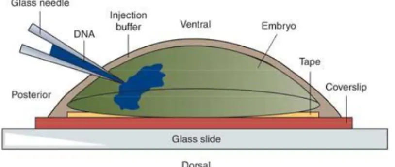
2.19 Embryo Microinjection ... 36
2.20 Larvae Screening ... 37
3. Results ... 38
3.1 Hemozoin Impairs P. berghei Infection in A. gambiae ... 38
3.2 Hemozoin Stimulates the Innate Immune System and Influences anti-Plasmodium Effectors ... 40
3.3 REL2-F, rather than REL2-S or REL1, is Induced by Hemozoin in A. gambiae Cells……….43
3.4 Hemozoin Further up-regulates REL2-F Upon caspar Silencing... 45
3.5 Hemozoin up-regulates REL2-F and Other Immune Related Genes in vivo Before and After P. berghei Infection ... 46
3.6 REL2 Mediates the Hemozoin Effect ... 49
3.7 A. gambiae FBN9, under Regulation of the Vitellogenin 1 Promoter, is Cloned into a Plasmid Vector ... 51
3.8 Transformation and Colony Screening ... 53
3.9 Sequencing Confirmed Successful Cloning ... 54
3.10 Transgenic A. gambiae X1 line overexpressing FBN9 under Regulation of the Vitellogenin 1 Promoter ... 54
4. Discussion and Conclusions ... 57
5. References ... 62
6. Appendices ... 82
6.1 Appendix 1 ... 82
6.2 Appendix 2 ... 89
List of Abbreviations
A.- Anopheles
ACT- artemisinin-based combination therapy
A. gambiae s.s.- Anopheles gambiae sensu stricto
AMA1- apical membrane antigen 1 AMP- antimicrobial peptide
APL1- AnophelesPlasmodium-responsive leucine-rich repeat 1
APL2- Anopheles Plasmodium-responsive leucine-rich repeat 2
β2M- beta-2 microglobulin BC- before Christ
bp- base pair
BSA- bovine serum albumin ºC- degree Celsius
CDC- Centers for Disease Control and Prevention cDNA- complementary DNA
CEC- cecropin
CEC1, 3- cecropin 1, 3
CLIPA2, A7, A8, B14, B15, B17- CLIP-domain serine protease A2, A7, A8, B14, B15, B17
CpG ODN- cytosinetriphosphate deoxynucleotide, phosphodiester, guaninetriphosphate deoxynucleotide oligodeoxynucleotide
cRNA- complementary RNA CSP- circumsporozoite protein CTL- C-type lectin
CTL4- C-type lectin 4
CTLMA2- CTL mannose binding 2 DDT- dichlorodiphenyltrichloroethane DEF- defensin
DEF1- defensin 1
dH2O- distilled water
DNA- deoxyribonucleic acid
DsRed- Discosoma sp. red fluorescent protein
dsRNA- double-stranded RNA
EBA-175- erythrocyte binding antigen 175 EEC- European Economic Community e.g.- exempli gratia (for example)
Fb- fat body
FBN9- fibrinogen immunolectin 9 FeIII, 3+-ironIII, 3+
FREP- fibrinogen-related protein g- acceleration of gravity
GAM1- gambicin 1 gDNA- genomic DNA
GFP- green fluorescent protein GLURP- glutamate-rich protein GPI- glycosylphosphatidylinositol
Gram +, Gram –- Gram-positive, Gram-negative hr, h- hour
i.e.- id est (in other words)
IGC- Instituto Gulbenkian de Ciência Imd- immune deficiency
I-PpoI- intron-encoded endonuclease from Physarum polycephalum
IRS- indoor residual spraying ITN- insecticide-treated bednets IVF- in vitro transcription
Jak/Stat- Janus kinase-signal transducer and activator of transcription L1-L4- larval stages 1 to 4
LB- Luria-Bertani
LLIN- long-lasting insecticide treated net LPS- lipopolysaccharide
LRR- leucine-rich repeat protein
LRRD7- leucine-rich repeat-containing protein M- molar
MeOH- methanol µg- microgram mg- milligram Mg- midgut µl- microliter ml- milliliter µm- micrometre
MSP-1- merozoite surface protein 1 N- total number
NaHCO3- sodium bicarbonate NaOH- sodium hydroxide NF-κB- nuclear factor kappa B nl- nanoliter
NLRP3- NLR family, pyrin domain containing 3 NO- nitric oxide
NOS- nitric oxide synthase ns- not significant
P.- Plasmodium
PAMP- pathogen associated molecular pattern
Pb- Plasmodium berghei
pbm- post blood meal
PBS- phosphate buffered saline PCR- polymerase chain reaction PGN- peptidoglycan
PGRP- peptidoglycan recognition protein PPO- prophenoloxidase
PRR- pattern recognition receptor
REL2-S- REL2 short-length form RHD- Rel-homology domain
RIDL- release of insects carrying a dominant lethal (gene) RNA- ribonucleic acid
RNAi- RNA interference ROS- reactive oxygen species S7- ribosomal protein S7 Sbjct- subject
SD- standard deviation sHz- synthetic hemozoin SIT- sterile insect technique
SOC- super optimal broth with catabolite repression SRPN2- serpin 2
STAT-A, -B- signal transducer and activator of transcription A, B TAK1- transforming growth factor-beta activated kinase 1
TEP- thioester-containing protein
TEP1, 3, 4- thioester-containing protein 1, 3, 4 TIR- Toll-interleukin-1-receptor
TLR- Toll-like receptor TLR9- Toll-like receptor 9 TNF- tumor-necrosis factor USA- United States of America v/v- volume/volume
w/v- weight/volume
1. Introduction
1.1 Malaria: Global Situation
Malaria is one of the world’s most significant insect-borne human diseases, caused by
Plasmodium parasites and transmitted by female Anopheles mosquitoes. It is widespread
in tropical and subtropical regions, and is presently considered endemic in 104 countries (WHO, 2013). It has been estimated that 207 million cases of malaria and 627 000 deaths due to this infectious disease occurred globally in 2012. Most cases/deaths occurred in Africa and most deaths were in children under five years of age (WHO, 2013). Although in the last decade malaria mortality has dropped by 42% due to increased control interventions (WHO, 2013), the challenge of producing an effective vaccine is still to be met, the host defences are unable to control parasite replication, the parasite has been mounting resistance to antimalarial drugs and the mosquito is growing resistance to insecticides. This observation stresses the importance of intensifying the current malaria control strategies and creating new alternative and effective interventions to fight this devastating parasite infection.
1.2 Malaria: Infection Cycle Overview
Malaria is one of the oldest known diseases, which had already been described in detail in ancient Greece by the 5th century BC. The malarial parasites were first discovered by
Charles Laveran (Nobel Prize in Physiology or Medicine, 1907) in 1880, when he observed parasites inside the red blood cells of infected patients. Later, in 1897, Ronald Ross (Nobel Prize in Physiology or Medicine, 1902), observed parasites maturing inside the mosquito, thus discovering how malaria is transmitted and tracing its life cycle. The success of malaria as a worldwide disease is due in part to the fact that it is transmitted by a mosquito vector. The complex parasite life cycle is shared between two hosts: in the vertebrate host, Plasmodium completes the asexual stages of its life cycle (the
1.3
Plasmodium
Parasite
There are five species of protozoan parasites of the genus Plasmodium considered
responsible for human malaria: Plasmodium falciparum, Plasmodium vivax, Plasmodium
ovale, Plasmodium malariae and Plasmodium knowlesi. Of these, Plasmodium
falciparum (P. falciparum) and P. vivax are the most prevalent forms. P. falciparum
malaria is the most deadly of both and predominates in Africa (Guerra et al., 2008), while
P. vivax has a wider distribution (Battle et al., 2014), partly due to its higher adaptability
and also because it is able to undergo a dormant liver stage (the hypnozoite), enabling its longer survival.
1.3.1 Parasite Life Cycle
Plasmodium parasites suffer several morphological changes along their life cycle:
Pre-erythrocytic stage (or exo-erythrocytic schizogony): When an infected Anopheles
female mosquito bites the vertebrate host, sporozoites are injected along with saliva into the host skin, until they reach the liver. There they invade cells (hepatocytes) and transform into hepatic schizonts. Schizonts grow and parasites differentiate into invasive motile forms called merozoites, which are released into the blood stream (reviewed by Prudêncio et al., 2006) (Figure 1A).
Erythrocytic stage (or erythrocytic schizogony): Merozoites infect red blood cells (erythrocytes) and develop into immature trophozoites (or ring stages), which then progress to mature trophozoites. Mature trophozoites develop into schizonts, which release merozoites. Merozoites can either invade another erythrocyte and continue this stage, or differentiate into gametocytes (Figure 1A).
Sporogonic cycle: The sexual phase of the Plasmodium life cycle starts when the female
Anopheles mosquito ingests infected blood containing male and female gametocytes.
migrate through the hemocoel and invade the salivary glands (Figure 1B), until they are transmitted to the vertebrate host upon another blood meal, thus starting the asexual part of the Plasmodium life cycle (reviewed by Cirimotich et al., 2010; reviewed by Yassine
and Osta, 2010).
1.3.2 Parasite Control Strategies
1.3.2.1 Antimalarial Drugs
There are different chemotherapeutic agents for the treatment of malaria. Their value depends on the emergence of parasite resistance. Chloroquine was the predominant antimalarial drug in the past; its action interferes with the parasite ability to metabolise hemoglobin, inhibiting its digestion (Foley and Tilley, 1997). However, due to historical overuse, Plasmodium parasites resistance to this drug is seriously menacing effective
malaria control. Other common antimalarial medications, such as sulfadoxine-pyrimethamine (antifolates which inhibit critical enzymes of the folate pathway, depriving the parasite from essential folate cofactors (Hyde, 2005)) and mefloquine (another aminoquinoline, as chloroquine), are facing the same Plasmodium-resistance
problem.
Current strategies for malaria treatment (WHO, 2013) recommend the use of artemisinin-based combination therapies (ACTs) for uncomplicated P. falciparum malaria. In the case
of P. vivax infection, in areas where resistance to chloroquine is not yet present, this drug
should be used; otherwise, an appropriate ACT must be chosen. In severe malaria cases, treatment should be based on injected artesunate (an artemisinin derivative), followed by a complete course of an effective ACT.
Although ACTs have been adopted as a first-line treatment in malaria-endemic countries, information reporting resistance to artemisinin has recently been disclosed (Dondorp et al., 2009). Parasite resistance to artemisinin has now been detected in four countries of the greater Mekong subregion, in Southeast Asia (WHO, 2013). Nevertheless, many pharmaceutical companies are still marketing artemisinin-based monotherapies in malaria endemic regions, against all advices. The problem is so major, that in April 2013 WHO (World Health Organization) released the Emergency response to artemisinin
resistance in the Greater Mekong subregion: Regional framework for action 2013–2015,
contain artemisinin resistance. Besides, significant research efforts are now focused as well on the new issue of artemisinin resistance monitoring (Ariey et al., 2014).
1.3.2.2 Vaccines
Vaccines represent one of the most cost-effective tools in the control of malaria. Despite years of intensive research, no licensed vaccine is available yet for malaria; therefore, international efforts from research laboratories, pharmaceutical companies and funding agencies are committed towards its development. However, there exist several candidates being evaluated at present.
Vaccine candidate RTS,S/AS01 is in phase 3 clinical trials. It has been engineered based on a protein expressed by Plasmodium sporozoites (circumsporozoite protein, CSP)
together with a hepatitis B surface antigen, for targeting of the pre-erythrocytic stage of the disease. First results with African children have shown a 50% reduction in malaria (Agnandji et al., 2011) and more promising results have followed (Otiene, 2013). Other attempts to target the pre-erythrocytic stage of infection for vaccine production consisted in immunizations with irradiated sporozoites (Hoffman et al., 2010) and development of genetically attenuated parasites incapable of progressing beyond the liver stage of infection (Vaughan et al., 2010).
An antimalarial vaccine will undoubtedly be an achievement, as repeated exposure to
Plasmodium parasites results in some degree of naturally acquired immunity. It is thought
that vaccination with parasite antigens can accelerate immunity, hence some Plasmodium
blood stage antigens (including apical membrane antigen 1 (AMA1), erythrocyte binding antigen 175 (EBA-175), glutamate-rich protein (GLURP) and merozoite surface protein 1 (MSP-1)) have been targeted in vaccine development (Sagara et al., 2009; El Sahly et al., 2010; Hermsen et al., 2007; Hill, 2011).
Another strategy consists in the development of transmission-blocking vaccines that fight the disease at the vector level, thus interrupting its transmission to the human host.Pfs25, Pfs48/45, Pfs230 and Pfs28 are examples of P. falciparum surface proteins expressed
during the sexual stage development inside the mosquito, that are candidate antigens currently being used as anti-Plasmodium transmission blocking targets (Wu et al., 2008;
1.4
Anopheles
Vector
Malaria is spread from one person to another through the bite of the female Anopheles
mosquito. The Anopheles genus comprises more than 400 different species, from which
approximately 70 species can transmit human malaria parasites and about 40 species are dominant vectors capable of transmitting malaria at a level of major concern to public health (Hay et al., 2010a; Sinka et al., 2012) (Figure 2). Besides the malaria endemic areas, several other zones of the globe, such as those where malaria has been eliminated but the vector mosquito is present, can be dangerously at risk of re-introduction of the disease (CDC, 2012).
Figure 2. Global distribution of dominant malaria vectors. Adapted from Sinka et al., 2012.
As mentioned above, Africa contains areas with the highest malaria morbidity and mortality in the world, given its highest entomological inoculation rates (Hay et al., 2010b) and prevalence levels globally (Hay et al., 2009). This situation arises partly because here resides the most effective and efficient human malaria vector, Anopheles
human malaria parasite, P. falciparum. Although a vast number of Plasmodium species
and Anopheles vectors exist, only a small number of laboratorial models is used. Several
studies are mostly based on A. gambiae s.s. (hereupon referred as A. gambiae), a suitable
model for studying the cellular and molecular interactions between vector and parasite, and for which the genome sequencing offers a fundamental tool (Holt et al., 2002).
A. arabiensis, another human malaria vector belonging to the A. gambiae complex (which
includes at least seven morphologically indistinguishable Anopheles species with marked
differences in their vectorial capacity (Bass et al., 2007; Sinka et al., 2010)) is also of major importance in sub-Saharan Africa. A. funestus is another very antropophilic African
vector, which in some cases has a greater impact on human malaria transmission than A.
gambiae and is considered to be one of the first species to have adapted to the human host
(Charlwood et al., 1995; Sinka et al., 2010).
Central and Southeastern Asia is the second area in the world for malaria burden; the Asian-Pacific region has a high diversity of vector species, as shown in the map from Figure 2. In South America, A. darlingi is the dominant species, outpaced by A. albimanus
and A.pseudopunctipennis in Central America (Figure 2).
1.4.1 Vector Life Cycle
Anophelines, as any other mosquitoes, undergo four different developmental stages in their life cycle: egg, larva, pupa and adult (Figure 3). The immature phases (before they reach adulthood) are aquatic and can last up to 15 days. Eggs (50-200 eggs per oviposition) are laid on water. Larvae are filter-feeders, feeding on micro-organisms and decaying plant matter, and develop through four stages or instars (L1-L4). Larvae
metamorphose into pupae, a stage that doesn’t feed but is particularly active. Adults emerge one to three days after pupation; male adults have a shorter life-span than females, which can live up to five weeks in optimal conditions (reviewed by Knell, 1991). Within 24 h (hour) after emerging, the female Anopheles is able to mate. Female copulation is
Both females and males feed on plant juices and nectar, but only females feed on blood (hematophagy) (Figure 3). When a female mosquito has mated and ingested its first blood meal in a vertebrate host, it uses the protein of the blood as the basis for the production of a batch of eggs (Dana et al., 2005), which will then be deposited near water for hatching. Therefore, the transmission of the Plasmodium parasite by the Anopheles vector
mosquito is only possible by hematophagy, which is essential for egg production (Dong et al., 2006). The gonotrophic cycle (comprising the host seeking, hematophagy, egg development and oviposition) involves a series of complex biological events, including the formation of the peritrophic matrix, acting as a barrier between the ingested blood and the midgut epithelium of the mosquito (Dana et al., 2005).
1.4.2 Classical Vector Control Strategies
Since the discovery of the connection between Anopheles vectors and malarial
transmission in 1897, vector control strategies have been the most widely used malarial control measures around the world (reviewed by Mabaso et al., 2004). Together with control strategies to reduce Plasmodium, the malaria causative agent, policies
improvement and creation of new strategies to control the Anopheles vector mosquito are
being taken, with the final aim of eradicating this devastating disease.
1.4.2.1 Chemical Control by IRS or ITNs
Nearly 70 years ago, the advent of insecticide DDT (dichlorodiphenyltrichloroethane) raised hopes of what was then thought to be the beginning of the worldwide eradication of malaria, through an ambitious programme of systemic control of its vector mosquito. Although indoor residual spraying (IRS) by means of DDT has effectively reduced the mosquito populations in many countries throughout the world (malaria was completely eliminated from Europe, the USA and the former Soviet Union during the 1960s (reviewed by Mabaso et al., 2004)), mosquito resistance to DDT quickly appeared. Together with Plasmodium chloroquine resistance and socio-economic-environmental
factors, this unfortunate happening compromised the ambitious control programme established by WHO in 1955.
Besides IRS, the most widespread method for the control of adult Anopheles is
insecticide-treated bednets (ITNs), which provide a protective barrier that prevents vector-host contact. WHO (2013) recommends that, in areas targeted for malaria vector control, all persons at risk should be protected by ITNs or IRS, vector control interventions with demonstrated impact on reducing malaria (reviewed by Lengeler, 2004; reviewed by Pluess et al., 2010); however, this is not the situation yet (Figure 4). New tools, such as long-lasting insecticide treated nets (LLINs, with a typical lifespan of three years) are being implemented, but the cost of such bednets is usually unaffordable to people in developing countries. The only insecticides currently used in most IRS programmes and to treat bednets are pyrethroids; nevertheless, the development of pyrethroid resistance in A. gambiae has already been reported (Trape et al., 2011).
Recognizing the threat posed by insecticide resistance, WHO released the Global Plan
Figure 4. Proportion of population at malaria risk protected by ITNs or IRS, in sub-Saharan
Africa in 2012. Adapted from WHO, 2013.
1.4.2.2 Biological Control
Biological control methods are used for control of Anopheles vector population at larval
and adult stages. Tools for larval control have found some success and include the use of larvivorous fish (that feed on larvae) (Mohamed, 2003; Ghosh et al., 2005), as well as microbial larvicides, formulated to deliver toxins from bacteria such as Bacillus
thuringiensis israelensis and Bacillus sphaericus, that disrupt the cells from the larva gut
(Majambere et al., 2007). For the control of adult Anopheles vectors, entomopathogenic
fungi (which infect insects), such as Beauvaria bassiana, have been used with good
achievement (Kikankie et al., 2010).
A current trend in vector biology laboratories is the study of the interactions between the mosquito midgut flora and the malaria parasite. In fact, several recent studies revealed that the gut microbiota are able to modulate the vectorial capacity of the Anopheles
1.4.2.3 SIT
Other biological strategies include the sterile insect technique (SIT), a non-polluting species-specific method of insect control that involves the sterilization of a large number of males, regularly by irradiation, but also by genetic alteration. These sexually active males will mate with wildtype virgin females, resulting in sterile offspring that will thus contribute to population decline. Successful application of this method in Anopheles
mosquitoes has been achieved in an isolated region of El Salvador, resulting in the eradication of the local A. albimanus population (Lofgren et al., 1974). However, SIT has
faced several problems which exclude its implementation as universal method for vector control, namely the loss of sterile male fitness following irradiation and the need for laboratorial sex separation to have large numbers of sterile male mosquitoes.
1.4.3 Novel Vector Control Strategies
Genetic manipulation of the mosquito genome has been suggested as another vector control strategy, through either the suppression/eradication of malaria-transmitting
Anopheles populations or replacement of these populations with mosquitoes refractory to
the Plasmodium parasite (Marrelli et al., 2006).
1.4.3.1 Population Suppression
1.4.3.1.1 RIDL
Given the several hurdles with SIT implementation mentioned above, a modification to this technique has been proposed, whereby insects are released carrying a repressible dominant lethal gene (RIDL) incorporated into their genome (Thomas et al., 2000). This gene kills the mosquito but can be repressed by an external additive, tetracycline (Alphey, 2002). The more advanced forms of RIDL use a female-specific dominant lethal gene, in which the lethal action of the system is active in females only. This way, the need for a sex separation step as in SIT is avoided, as the tetracycline repressor can be removed from the final stage of mosquito laboratorial rearing, killing all females and leaving males only (Alphey et al., 2002).
Anopheles populations are certainly a possible target for RIDL technology (Nolan et al.,
irradiation (Alphey, 2002). Because lethality is female-specific and the lethal gene is dominant, this cross would result in offspring which would only survive if they are males- hence, the female population would be reduced, constituting a very beneficial method of population control, given the central role of Anopheles females in malaria transmission.
The new males would be heterozygous RIDL and would subsequently pass the lethal gene to a proportion of the following generation, further reducing the target population.
1.4.3.1.2 Homing Endonucleases
Another approach to distort the sex ratio in natural populations is based on the use of engineered mosquitoes expressing a homing endonuclease enzyme targeting the X chromosome. The activity of the X chromosome cutting enzyme intron-encoded endonuclease I-PpoI has been explored to bias the sex ratio in A. gambiae mosquitoes
(Windbichler et al., 2008; Galizi et al., 2014). This enzyme selectively cleaves a deoxyribonucleic acid (DNA) sequence from the centromeric region of chromosome X, leading to nucleolar fragmentation and cell death. Authors engineered A.
gambiae male mosquitoes to heterozigously express I-PpoI during spermatogenesis and
crossed these males with wildtype females. This resulted in complete early dominant embryo lethality and a strong, negative bias toward X chromosome-carrying spermatozoa (Windbichler et al., 2008; Galizi et al., 2014). This could thereby lead to the reduction or eradication of field populations, through distortion of the sex-ratio in favour of males (Windbichler et al., 2008). The implementation of this strategy to mosquito control in the field is currently under evaluation.
1.4.3.2 Population Replacement
Blocking the transmission of Plasmodium using the transgenic manipulation of its
mosquito vector, has been achieved in different malaria-transmitting species, such as A.
stephensi (Ito et al., 2002; Moreira et al., 2002; Yoshida et al., 2007; Dong et al., 2011;
Isaacs et al., 2011) and A. gambiae (Kim et al., 2004; Meredith et al., 2011).It constitutes
a promising malaria control strategy, presently being applied in several vector-studying laboratories. In theory, these Plasmodium refractory mosquitoes should be capable of
In the case of Anopheles transgenic manipulation, this could result in the overproduction
of an anti-Plasmodium factor or silencing of a Plasmodium positive regulator,
culminating in a decline in the overall number of parasites, therefore interfering with
Plasmodium cycle through reduction of transmission to the vertebrate host.
Choosing the right promoters to drive the expression of particular effector genes in a stage- and tissue-specific manner is a critical step in engineering transgenic lines for the mosquito. In Anopheles, tissue-specific promoters have been widely used to target
specific organs, with main focus on the midgut, fat body/hemocoel and salivary glands, the key target organs involved in critical steps of the Plasmodium sporogonic cycle. In
Anopheles, the most commonly used midgut-specific promoter is the A. gambiae
carboxypeptidase A (Ito et al., 2002; Moreira et al., 2002; Yoshida et al., 2007; Dong et al., 2011; Isaacs et al., 2011; Meredith et al., 2011). The use of another midgut-specific promoter, G12, has been recently validated in A. stephensi (Nolan et al., 2011b) as well.
Concerning the fat body, the most frequent specific promoters for transgenic studies are the Anopheles vitellogenin genes 1 (Dong et al., 2011; Isaacs et al., 2011) and 2 (Chen et
al., 2007), which are fat body-specific and blood meal induced. Both the carboxypeptidase and vitellogenin promoters have been extensively used in order to target the malaria parasite at its early stages of development.
1.5
Anopheles gambiae
Immune System
Given the challenge of finding effective ways to reduce the burden caused by malaria, a wealth of knowledge on the interaction parasite-vector and the mosquito immunity to
Plasmodium has been building up in the last decade. Several experimental studies have
demonstrated that Anopheles mosquitoes are able to mount an efficient immune response
against Plasmodium infection, and that this results in major losses for the parasite during
its development inside the vector. The mosquito immune defence involves complex mechanisms of action, which will be described below.
1.5.1
Plasmodium-Anopheles
Interactions
As previously explained, Plasmodium parasites live within three main compartments
these compartments produce immune factors with antimicrobial activity, as we will later discuss in further detail. During the sporogonic cycle (Figure 1B), the parasite experiences three main bottlenecks, when its numbers are largely reduced: 1) the transition between gametocytes and ookinetes, 2) between ookinetes and mature oocysts and 3) between midgut sporozoites and salivary gland sporozoites (Figure 5). Indeed, it is said that A. gambiae mosquitoes can kill around 80% of invading Plasmodium
ookinetes (Lavazec and Bourgouin, 2008).
Figure 5. Three main Plasmodium bottlenecks during the sporogonic cycle. Adapted from Simões and Chandrasekar, 2014. pbm (post blood meal).
In vertebrates, the fight against invasive pathogens is a result of the interaction between adaptive and innate immunity. Insects, however, lack an adaptive immune system, and thus rely solely on innate immunity for their defence. Insect blood cells (hemocytes), play a prominent role in their immune response, through phagocytosis and encapsulation of foreign invaders (cellular response), as well as their melanization. Hemocytes, together with the fat body, are also able to release immune effectors into the insect circulatory fluid (hemolymph). The production of these antimicrobial effector molecules is regulated by intricate intracellular immune signalling pathways.
1.5.2 Pattern Recognition Receptors (PRRs)
Pathogen elimination in both vertebrates and invertebrates is initiated by pattern recognition receptors (PRRs), which sense, recognize and bind to pathogen associated molecular patterns (PAMPs). Insects fight microbial infections using a limited number of
PRRs. In A. gambiae, about 150 putative PRRs have been identified (reviewed by Das et
thioester-containing proteins (TEPs), leucine-rich repeat proteins (LRRs), C-type lectins (CTLs) and fibrinogen-related proteins (FREPs or FBNs), the latest being the largest PRR family in this vector (Dong and Dimopoulos, 2009). Among these families are some of the most potent anti-Plasmodium immune factors identified to date.
1.5.2.1 TEPs and LRRs
The most studied member of the TEPs family is TEP1 (thioester-containing protein 1), a hemocyte-specific protein first described as being involved in bacterial opsonization (Levashina et al., 2001) and later as mediator of P. berghei ookinete destruction in A.
gambiae (Blandin et al., 2004). TEP1 activation and stabilization is achieved by
interaction with LRIM1 and APL1C (leucine-rich repeat immune protein 1 and
Anopheles Plasmodium-responsive leucine-rich repeat protein 1, PRR proteins of the
LRR family) to form a complex prior to binding to ookinetes in the midgut, mediating their lysis (Baxter et al., 2010). The composition of this complex (TEP1, a complement-like protein with structural similarities to vertebrate complement component C3 (Baxter et al., 2007), and two LRR proteins), suggests that mosquitoes have a complement-like system comparable to the one in mammals, which is able to effectively fight Plasmodium
infection. Independent silencing of these three genes has shown that each one is individually relevant to control the development of P. berghei oocysts, besides playing
an important role in parasite melanization (Habtewold et al., 2008), an effective immune killing mechanism involving the deposition of melanin on the parasite surface. Another key anti-Plasmodium protein of the LRR family is LRRD7 (leucine-rich repeat–
containing protein 7, also designated as APL2, Anopheles Plasmodium-responsive
leucine-rich repeat 2) (Dong et al., 2006).
1.5.2.2 CTLs
Acting in an opposing manner, A. gambiae CTL4 and CTLMA2 (C-type lectin 4 and CTL
mannose binding 2) seem to be agonists of Plasmodium infection in the midgut, where
1.5.2.3 FREPs
Fibrinogen-related proteins (FREPs) constitute a large PRR family in mosquitoes, with 59 genes identified in A. gambiae and 37 genes in Aedes aegypti. This family seems
particularly broad in the mosquito, as only 14 such genes have been reported in
Drosophila melanogaster (Wang et al., 2005; Waterhouse et al., 2007; Dong and
Dimopoulos, 2009). The majority of FREPs in mosquitoes are up-regulated upon immune challenge and several have anti-Plasmodium activity. FBN9, an important member of the
family mediating Plasmodium killing, co-localizes with the ookinete stage of both P.
berghei and P. falciparum parasites in the midgut (Dong and Dimopoulos, 2009).
Evidence from observations in vertebrates and co-localization at parasite surface led Garver et al. (2009) to speculate that a mechanism similar to the lectin complement pathway, in which TEP1 and FBN9 cooperate to destroy pathogens, may also exist in mosquitoes.
1.5.2.4 PGRPs
Another important PRR family within the mosquito, the peptidoglycan recognition proteins (PGRPs), with seven genes described in the A. gambiae genome (Christophides
et al., 2002), is also involved in the activation of various immune reactions in the
Anopheles mosquito.
1.5.3 Serine Proteases
Serine proteases are proteolytic enzymes present in the hemolymph, where they can rapidly activate immune pathways in response to pathogen detection by PRRs, amplifying the signal and triggering downstream effector responses to eradicate the pathogen invader. The most important components of the serine protease cascade are the CLIP domain serine proteases, 41 of which have been identified in A. gambiae so far. Many
CLIPs are involved in the melanization of P. berghei ookinetes, where they have roles as
berghei elimination in A. gambiae, in a melanization-independent way (Christophides et
al., 2002).
Serpins (serine protease inhibitors) are regulators of the serine protease cascade and, similarly to CLIPs, modulate melanization responses. SRPN2 (serpin 2) inhibits melanization of P. berghei ookinetes in A. gambiae (Michel et al., 2005). It is thought
that this protein may interact with CLIPs, CTLs and LRIM1, other molecules known for interfering in the melanization process.
1.5.4 Antimicrobial Peptides (AMPs)
Besides melanization, the humoral responses to pathogen invasion culminate with the production of antimicrobial peptides (AMPs), which are synthesized by the fat body and hemocytes and secreted into the hemolymph upon immune challenge. AMP production constitutes the ultimate step of the defence mechanisms employed by Anopheles
mosquitoes to fight Plasmodium. In A. gambiae, four AMP families have been identified
to date, comprising four defensins (DEFs), four cecropins (CECs), one attacin and one gambicin (GAM1) (Christophides et al., 2002). Their antimicrobial action comprehends responses against Gram-negative (mainly CEC1 and GAM1) and Gram-positive (mainly DEF1) bacteria. Some are known to have an anti-Plasmodium role in A. gambiae as well.
CEC1 was one of the first anti-Plasmodium factors to be identified (Luckhart et al., 2003).
This gene was significantly up-regulated following Plasmodium infection (Christophides
et al., 2002) and its ectopic overexpression was shown to increase A. gambiae resistance
to P. berghei (Kim et al., 2004). In a more recent study, silencing of GAM1 resulted in
increased P. berghei infection (Dong et al., 2006).Defensins are the most extensive insect
family of AMPs and play an essential role in the innate immunity of virtually almost all life forms from plants, to insects, amphibians and mammals. Transcriptional regulation of AMPS is controlled by immune signalling pathways.
1.5.5 Immune Signalling Pathways
In both mammals and insects, recognition of PAMPs by PAMP-associated PRRs is followed by signal transduction through immune signalling pathways that activate Nuclear Factor-kappaB (NF-κB)/Rel transcription factors. These factors translocate to the nucleus to initiate the transcription of an array of effector genes, including AMPs. In
pathways, have been extensively studied. The Toll pathway is triggered by fungi and Gram-positive bacterial infections, leading to the nuclear translocation of NF-κB transcription factor Dif, as well as Dorsal, which is involved in early developmental processes, whilst the Imd pathway signalling results in nuclear translocation of Relish, following Gram-negative bacterial infection. Both factors induce the transcription of all the AMPs encoded in the Drosophila genome (reviewed by Lemaitre and Hoffmann,
2007).
Sequencing the A. gambiae genome allowed the comparison of putative immune related
genes between Anopheles and Drosophila (Holt et al., 2002), supporting the conclusion
that most intracellular components of these pathways are conserved in the mosquito. A remarkable difference between the two evolutionarily related insects is the lack of Dif in
Anopheles; therefore, the malaria mosquito genome encodes two NF-κB transcription
factors only: REL1 (the orthologue of Dorsal, previously called Gambif1 (Barillas-Mury, 1996)) and REL2, Drosophila Relish orthologue.
Both Anopheles REL proteins are directly involved in the immune response against
invaders. They translocate to the nucleus to initiate the transcription of many effector genes and REL-dependent transcription has been shown to be particularly critical to the mosquito´s ability to manage infection with P. berghei and P. falciparum (Meister et al.,
2005; Frolet et al., 2006; Luna et al., 2006; Hoa and Zheng, 2007; Riehle et al., 2008; Garver et al., 2009, 2012; Chen et al., 2012; Clayton et al., 2013). Therefore, REL1, REL2 and their associated pathways are critical targets for the development of anti-malaria strategies based on vector-parasite interactions (Dong et al., 2011).
1.5.5.1 The Toll Pathway
In humans and mice, respectively ten and twelve Toll-like receptors (TLRs) have been characterized thus far. TLRs play a critical role in the early innate immunity against foreign pathogens. Their name derives from the firstly identified Drosophila Toll protein,
degradation of Cactus, an ankyrin-repeat protein, hence allowing the nuclear translocation of the above mentioned NF-κB transcription factors Dorsal and Dif.
In A. gambiae, the intracellular machinery of the Toll pathway is conserved. Orthologues
of MyD88, Tube and Pelle, as well as negative regulator Cactus, have been identified in the malaria mosquito (Christophides et al., 2002). As mentioned, REL1 is the NF-κB factor controlling the transcription of genes regulated by the Toll pathway in Anopheles.
The importance of REL1-pathway stimulation was made clear when knocking down of
Cactus (by double-stranded ribonucleic acid, dsRNA, injection) dramatically decreased
the parasite load of P. berghei in A. gambiae. Further, co-silencing of REL1 and Cactus
reversed the dsCactus phenotype (Frolet et al., 2006). Similar results were obtained by
Garver et al. (2009), where a significant reduction in the infection levels of P. berghei
was observed after Cactus depletion. The same authors have also shown that Cactus silencing altered the expression of several genes from other functional groups besides immunity, suggesting that the Toll pathway is a ubiquitous signalling pathway, with a wide-ranging action, this probably being the reason for the marked reduction in both longevity and fecundity shown in the dsCactus mosquitoes in this study (Garver et al.,
2009). Importantly, in the above mentioned study (Frolet et al., 2006), it has been said that the mosquito basal immunity level (before the mosquito encounters the parasite) is a key factor for parasite control, i.e., it is significantly more influent in Plasmodium
development than the induction of immune responses upon parasite infection.
1.5.5.2 The Imd Pathway
identified as a specific negative regulator of the Imd pathway, preventing the nuclear migration of Relish (Kim et al., 2006).
All components of the Imd pathway are conserved in the Anopheles mosquito
(Christophides et al., 2002). Through alternative splicing, two isoforms of REL2, the Relish orthologue, are present in Anopheles: REL2-F (a full-length form) and REL2-S (a
short-length form), the latter lacking the inhibitory ankyrin repeats and death domain present in REL2-F. Both transcripts are expressed constitutively throughout A. gambiae,
as well as in cultured cell lines (Meister et al., 2005; Luna et al., 2006). In the absence of immune stimulation, REL2 exists in the two variants: REL2-S, that is constitutively active, and REL2-F, that is inactive until immune stimulus. Imd pathway activation stimulates cleavage of the inhibitory ankyrin terminal domain of REL2-F, exposing it to nuclear translocation and subsequent transcription initiation (reviewed by Cirimotich et al., 2010). Besides their role against Plasmodium, in A. gambiae REL2-F was shown to
be involved in the defence against Gram-positive Staphylococcus aureus, and REL2-S
against Gram-negative Escherichia coli (Meister et al., 2005), a contrast to the
Gram-negative specificity of the Imd pathway in Drosophila.
Elucidation of the role of REL2 in fighting Plasmodium was obtained through observation
that its knockdown increased the number of P. berghei oocysts in A. gambiae (Meister et
al., 2005; Frolet et al., 2006). In a more recent study (Garver et al., 2009), depletion of Caspar reduced the number of P. berghei oocysts, in addition to almost abrogating P.
falciparum infection in three Anopheles species: A. gambiae, A. stephensi and A.
albimanus. In contrast to Toll, the Imd pathway is thought to be immunity-specific
(Garver et al., 2009). Overall, its properties suggest that this pathway could be used in the development of malaria control approaches, through interference in the parasite sporogonic cycle. Indeed, target expression of REL2 has been recently used to create transgenic lines of A. stephensi mosquitoes with particularly reinforced immunity against
Plasmodium and microbial infection (Dong et al., 2011).
1.5.5.3 The Jak/Stat Pathway
the context of Anopheles-Plasmodium interaction. Nevertheless, two Stat transcription
factors, Stat-A and Stat-B (signal transducer and activator of transcription-A, B), have been identified in the A. gambiae genome (Christophides et al., 2002), with the first
playing a role in the mosquito defence against Plasmodium (Gupta et al., 2009).In a more
recent study, the Jak/Stat pathway was shown to be activated in A. aquasilis in response
to P. vivax challenge, and an increase in P. vivax oocysts number was observed after
depletion of this pathway (Bahia et al., 2011). The role of the Jak/Stat pathway in the activation of Anopheles immunity against the malaria parasite is subject of a growing
interest and requires additional investigation. Together with the Imd/REL2 and Toll/REL1 pathways, this immune pathway could be seen as a potential target for development of malaria control strategies.
1.5.5.4 Regulatory Pathways of anti-Plasmodium Genes
The process of regulation for the expression of immunity-specific genes is dependent on the instigating PAMP, as well as the selectively activated pathway. PAMPs that are effective elicitors of the mosquito immune response are lipopolysaccharides (LPS),
peptidoglycans (PGNs) and β-1, 3-glucans. Current studies are being developed to shed light on Plasmodium-specific PAMPs that activate the Anopheles immune system. One
molecule that has already been identified as an inducer of AMPs production in A. gambiae
is glycosylphosphatidylinositol (GPI), labelled as the prominent toxin that contributes to malaria pathogenesis in mammals, through the anchoring of parasite proteins on cellular plasma membranes (Arrighi et al., 2009). In our laboratory, we found that the Plasmodium
metabolite hemozoin acts as a stimulator of A. gambiae immune response against P.
berghei, as will be demonstrated in the present thesis. Both GPI and hemozoin induce the
transcription of the enzyme nitric oxide synthase (NOS) (Lim et al., 2005; Akman-Anderson et al., 2007) and the resulting nitric oxide (NO) production prompts the killing of Plasmodium ookinetes in the midgut (Luckhart et al., 1998; Peterson et al., 2007).
Inhibition of NOS was shown to increase the parasite numbers in A. stephensi mosquitoes
(Luckhart et al., 1998). Together with reactive nitrogen species, the Plasmodium blood
In A. gambiae, both Toll/REL1 and Imd/REL2 pathways control the expression of
important anti-Plasmodium factors. Among the most relevant genes described as having
anti-malaria activity, are those of the AMPs DEF1, CEC1, CEC3 and GAM1, which appear to be controlled by both pathways/transcription factors. Silencing of the negative regulators of the Toll and Imd pathways (Cactus and Caspar, respectively) has
up-regulated the levels of these four AMPs (Garver et al., 2009). Further, co-silencing of the
negative regulator together with the correspondent transcription factor reversed this phenotype, indicating that both immune pathways control the expression of these important anti-Plasmodium genes (Garver et al., 2009). These results contradict a
previous A. gambiae study, where Frolet et al. (2006) had also knocked down the Toll
negative regulator Cactus, but instead did not observe a distinct up-regulation of any of
these four AMPs. Luna and colleagues (2006) have shown in vitro (using mosquito cell
lines) that both pathways regulate DEF1 and GAM1 expression. Double regulation of AMPs by the Toll and Imd pathways is not a particular Anopheles feature, as it has also
been described in Drosophila (Kim et al., 2006; Tanji et al., 2007). Meister and co-workers (2005) classified CEC1, CEC3 and GAM1, among other immune genes, as
REL2-regulated, when they assessed gene expression profiles by DNA microarray following REL2 silencing in A. gambiae hemocyte-like cells; similarly, Hoa and Zheng
(2007) showed a REL2-dependent up-regulation of CEC1 in vitro.
Alongside AMPs, some key Anopheles anti-malaria effectors belonging to the PRRs
group, seem to be regulated by either of the immune pathways. TEP1 has been reported as REL1- and REL2-dependent. In the above mentioned study, Frolet et al. (2006) indicated that before Plasmodium invasion, TEP1 basal expression is controlled by both
REL factors, but Plasmodium-dependent up-regulation of TEP1 is independent of REL1
and REL2. Subsequent studies (Garver et al., 2009; Dong et al., 2011) have shown that TEP1 expression is REL2-dependent. The combined outcomes of these studies suggest that this potent anti-Plasmodium factor regulation may not be pathway-specific. APL1C,
a TEP1-interacting PRR in antimalarial defence, seems to be Toll pathway-dependent (Riehle et al., 2008), although in Frolet et al. (2006) the authors have not noticed any effect on this gene expression profile upon REL1, REL2 nor Cactus silencing, and Garver
et al. (2012) considered it to be an Imd pathway member. As for LRIM1, another key gene
(Meister et al., 2005), although a subsequent study (Fraiture et al., 2009) suggested a REL1/Caspar-dependency. Other very important PRRs that act as Plasmodium-fighting
genes, FBN9 and LRRD7, are thought to be regulated by the Imd-REL2 pathway (Dong
et al., 2011; Chen et al., 2012). Moreover, another two immunity factors involved in the
Anopheles anti-malaria response, TEP3 and TEP4, are likely to be REL2-regulated genes
(Luna et al., 2006; Hoa and Zheng, 2007; Garver et al., 2009).
In conclusion, REL1 and REL2 control the A. gambiae immune response against
Plasmodium by regulating the expression of antiparasitic genes. This regulation can be
mediated by each transcription factor independently, or, as seen in many cases, both factors can be involved in the transcription of the same gene. The disclosure of the specific regulation for each anti-Plasmodium effector is an essential question that awaits
further investigation.
1.6 Hemozoin as an Immunity Activator
The Plasmodium metabolite hemozoin is a byproduct of the parasite’s digestion of host
hemoglobin within the erythrocyte. Hemozoin is structurally similar to -hematin, which is composed of cyclic heme dimers (FeIII-protoporphyrin IX). Heme dimers interact
through hydrogen bonds, forming hemozoin crystals (Figure 6).
hemozoin crystals is -hematin, formed by two heme molecules. Adapted from http://www.tulane.edu.
Hemozoin is released together with merozoites by the rupture of parasitized erythrocytes. It reaches high concentrations in the circulation, and is engulfed by macrophages, monocytes, neutrophils and other immune cells (Arese and Schwarzer, 1997). It has been suggested that free hemozoin leads to the continuous targeting of the host innate immune system in mammals (reviewed by Coban et al., 2010a). In fact during the last decade it has emerged as a potent immunoactivator, both in vitro and in vivo (Jaramillo et al., 2009).
Indeed, hemozoin continuously activates macrophages and dendritic cells to produce pro- and anti-inflammatory cytokines and chemokines in mouse and human cells (reviewed by Coban et al., 2009, 2010a; Jaramillo et al., 2009). Although hemozoin’s pro-inflammatory role is generally accepted, the recognition and host response to this molecule, as well as the molecular mechanism(s) by which it activates the mammalian innate immune system, has been a subject of intense debate.
mice (Togbe et al., 2007; Lepenies et al., 2008). Similar to the hemozoin TLR9 activation of the immune system, the activation through the NLRP3 (NLR family, pyrin domain containing 3) inflammasome is also being extensively debated (Griffith et al., 2009; Shio et al., 2009).
In contrast to numerous studies regarding hemozoin immunity activation in mammals, one single study exploring the effect of this molecule in the Anopheles vector has been
published so far (Akman-Anderson et al., 2007). In mosquitoes, hemozoin is released from parasitized erythrocytes and leukocytes which enter the midgut with the infected blood meal (Akman-Anderson et al., 2007). Anopheles gambiae’s peritrophic matrix,
acting as a barrier between the ingested blood and the midgut epithelium, is completely formed by 24 h after a blood meal (Dinglasan et al., 2009), which suggests that the midgut epithelium is possibly exposed to hemozoin before the membrane is fully formed (Akman-Anderson et al., 2007). The authors of the mentioned study (Akman-(Akman-Anderson et al., 2007) demonstrated that both P. falciparum and synthetic (sHz) forms of hemozoin contribute
to the immune activation in Anopheles; in accordance with similar observations in
mammalian cells (Jaramillo et al., 2003), hemozoinwas found to induce NOS expression in mosquito cells and tissues via multiple signalling pathways.
1.7 Aim
The failure to stop the devastation caused by malaria has stimulated research efforts to understand this disease from the vector perspective, aiming at the development of novel antimalarial strategies. Experimental evidences have revealed the importance of the
Anopheles immune response against Plasmodium; even so, natural immunity is not
enough to totally suppress mosquito infection. More effective protective immune responses to the parasite could be tailored by modulation of the mosquito immune system. In our laboratory we are interested in studying the role of immunostimulatory molecules which are potential boosters of the Anopheles innate immune system. The interplay
immune response to hemozoin is not yet well characterized and requires further elucidation.
To address these issues, in this study we dissect the activity of this Plasmodium
metabolite, while unraveling the mechanisms behind its triggering of the immune system of the malaria vector A. gambiae. Bearing in mind that A. gambiae is not the natural vector
for P. berghei, this pair was used as a laboratory model, as it is conventionally used for
the study of malaria transmission.
Following identification of a set of key immunity genes induced by this parasite’s byproduct, as described in the results section of this thesis, our next purpose was to generate transgenic mosquitoes with an enhanced immune response against the parasite. Encouraged by the properties of the Imd/REL2 pathway suggested by the obtained results, and motivated by recent publications (e.g. Dong et al., 2011), in which these properties were explored to construct engineered mosquitoes with an increased robustness against the malarial parasite, we aimed to develop a genetically modified immune enhanced A. gambiae line, by overexpression of FBN9 antiparasitic gene under regulation
2. Material and Methods
2.1 Ethics Statement
The maintenance and care of experimental animals was carried out in strict accordance with the recommendations in the Europe Directive 86/609/EEC (Louhimies, 2002) and Portuguese law (Decreto-Lei 129/92) for biomedical research involving animals, and was approved by the Divisão Geral de Veterinária (DGV), Portugal. All experiments were performed under anaesthesia, and all efforts were made to minimize animal suffering.
2.2 Synthetic Hemozoin (sHz) Preparation
sHz was prepared from high-purity hemin chloride, using a protocol as described in (Jaramillo et al., 2009; Shio et al., 2009). Briefly, 500 mg (milligram) hemin (≥ 98% pure,
Sigma) were dissolved in degassed sodium hydroxide (NaOH, 0.1 molar (M), 100 milliliter (ml)) and pH adjusted with propionic acid. The mixture was heated at 70 ºC for 18 h. After cooling, the solid was separated and washed with three alternate washes of sodium bicarbonate (NaHCO3) and MilliQ water for three hours. MeOH was then alternated with MilliQ water for three final washes. The sample was dried in a vacuum chamber overnight. The pigment was resuspended in endotoxin-free phosphate buffered saline (PBS) at a final concentration of 2.5 mg/ml and kept at -20 ºC until further use.
2.3 Mosquito Treatment and Infection
A. gambiae mosquitoes (Yaoundé strain) were reared at 26 ºC and 80% humidity on a
12/12 h light/dark cycle. Adults were maintained on a 10% (w/v) glucose solution before blood feeding. Three to four day old female mosquitoes were cold-anaesthetized and inoculated intrathoracically with 69 nanoliters (nl) of a 100 or 200 micrograms (µg)/ml solution of sHz or with the same volume of endotoxin-free PBS, using a Nanoject micro-injector (Drummond Scientific). Female CD1 mice (Mus musculus) were intraperitoneally
inoculated with 107 P. berghei ANKA parasitized red blood cells/ml and mosquitoes were
independent biological replicates were performed for each experiment. Between eight and ten days post infection, mosquito midguts were collected to determine infection rate and intensity.
2.4 RNA Isolation
Female mosquitoes were dissected 24 h after sHz inoculation (immediately before feeding). Batches of circa 30 fat bodies (abdomen without midgut, ovaries and
malpighian tubules, which can also comprise hemocytes) were dissected in cold diethylpyrocarbonate (DEPC) treated PBS and processed for RNA preparation. Total RNA was prepared using NucleoSpin RNA II kit (Macherey-Nagel) and concentration and purity determined by spectrophotometry.
2.5 Microarray Hybridization and Analysis
Microarray Hybridization was performed at Instituto Gulbenkian de Ciência (IGC), in collaboration with the Gene Expression Unit. Each GeneChip experiment was performed with three biological replicates. RNA was processed for use on Affymetrix GeneChip
Plasmodium/Anopheles Genome Arrays, according to the manufacturer’s One-Cycle
Target Labelling Assay. Briefly, total RNA was used in a reverse transcription reaction to generate first-strand complementary DNA (cDNA). After second-strand synthesis, double-stranded cDNA was used in an in vitro transcription (IVT) reaction to generate
biotinylated complementary RNA (cRNA). Fragmented cRNA was used in a 300 µl hybridization containing added hybridization controls, on arrays for 16 h at 45 °C. Standard post hybridization wash and double-stain protocols were used on an Affymetrix GeneChip Fluidics Station 450. Arrays were scanned on an Affymetrix GeneChip scanner 3000. All quality parameters for the arrays were confirmed to be in the recommended range.
Method (Li and Wong, 2001). Normalized CEL intensities of the arrays were used to obtain model-based gene expression indices based on a perfect match-only model (Li and Wong, 2001). All genes compared were considered to be differentially expressed if the 90% lower confidence bound of the fold change between experiment and baseline was above 1.2. Li and Wong (2001) have shown that the lower confidence bound is a conservative estimate of the fold change and therefore more reliable as a ranking statistic for changes in gene expression. To validate the microarray data, expression profiles of ten genes differentially expressed by microarray were analysed by quantitative reverse
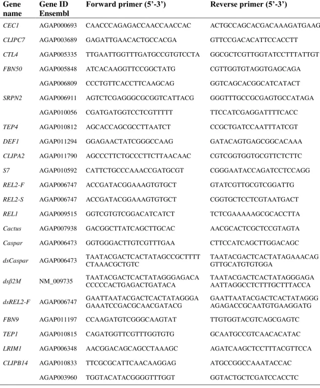
transcription PCR (qRT-PCR) as described below: AGAP000693, AGAP003689,
AGAP005335, AGAP005848, AGAP006809, AGAP006911, AGAP010056,
AGAP010812, AGAP011294 and AGAP011790. The sequences of primers used for
amplification can be found in Table 1.
2.6 Stimulation of
A. gambiae
Cells
Immortalized A. gambiae hemocyte-like cells Sua 5.1 were cultured as in
(Akman-Anderson et al., 2007) and incubated at 28 ºC. One to 1.5 x 10⁶ cells/well were seeded in
24-well plates and left to grow overnight. Cells were stimulated with different concentrations of sHz or endotoxin-free PBS as a control and incubated at 28 ºC for 24 h or 48 h. Three independent experiments were performed.
2.7 Tissue Collection Before and After Blood Ingestion
Mosquitoes were inoculated with 69 nl of a 200 µg/ml solution of sHz or with the same volume of endotoxin-free PBS, as described above. Batches of circa 30 mosquitoes were
dissected 24 h after sHz inoculation (immediately before feeding) and fat bodies collected as before. The remaining mosquitoes were fed with either a P. berghei-infected or a