Duodenal gastric metaplasia and
Helicobacter pylori
infection in patients
with diffuse nodular duodenitis
1Department of Gastroenterology, 2Department of Pathology, Renji Hospital,
Shanghai Institute of Digestive Diseases,
Medical College of Shanghai Jiaotong University, Shanghai, China X.B. Li1,
Z.Z. Ge1,
X.Y. Chen2
and W.Z. Liu1
Abstract
Whether the regression of gastric metaplasia in the duodenum can be achieved after eradication of Helicobacter pylori is not clear. The aim of the present study was to investigate the relationship between H. pylori infection and gastric metaplasia in patients with endoscopic diffuse nodular duodenitis. Eighty-six patients with endoscopically confirmed nodular duodenitis and 40 control patients with normal duodenal appearance were investigated. The H. pylori-positive pa-tients with duodenitis received anti-H. pylori triple therapy (20 mg omeprazole plus 250 mg clarithromycin and 400 mg metronidazole, all twice daily) for one week. A control endoscopy was performed 6 months after H. pylori treatment. The H. pylori-negative patients with duodenitis received 20 mg omeprazole once daily for 6 months and a control endoscopy was performed 2 weeks after treatment. The preva-lence of H. pylori infection was 58.1%, and the prevalence of gastric metaplasia was 57.0%. Seventy-six patients underwent endoscopy again. No influence on the endoscopic appearance of nodular duodenitis was found after eradication of H. pylori or acid suppression therapy. However, gastric metaplasia significantly decreased and complete regression was achieved in 15/28 patients (53.6%) 6 months after eradication of H. pylori, accompanied by significant improvement of other histological alterations. Only mild chronic inflammation, but not gastric metaplasia, was found in the control group, none with H. pylori infection in the duodenal bulb. Therefore, H. pylori infection is related to the extent of gastric metaplasia in the duodenum, but not to the presence of diffuse nodular duodenitis.
Correspondence
X.B. Li
Shandong Zhong Road, 145 200001, Shanghai China
Fax: +86-021-56338496 E-mail: lxb1969@gmail.com Research supported by Shanghai leading academic discipline project (No. Y0205).
Received April 28, 2006 Accepted April 26, 2007
Key words
•Nodular duodenitis •Helicobacter pylori •Gastric metaplasia
Introduction
Helicobacter pylori infection is a major cause of many gastroduodenal diseases, es-pecially chronic gastritis, duodenal ulcer,
and duodenitis. In previous studies,the preva-lence of GM was 72 to 90% in patients with duodenal ulcer, 40 to 95% in patients with duodenitis, and 0 to 33% in patients with normal endoscopic duodenal appearance (1,2).
GM is generally believed to occur as a non-specific response to acid/peptic dam-age. It has been speculated that H. pylori infection induces a high level of acid burden in the duodenum by increasing gastrin secre-tion, which may influence the development of GM. It has been shown that the amount of H. pylori in the duodenal bulb may be re-lated to the extent of GM (3). Theoretically, regression of GM may be induced by eradi-cation of H. pylori or acid-suppression thera-py. However, published results are conflict-ing and few studies have focused on the relationship between H. pylori infection and GM in the pathogenesis of duodenitis (4-11).
Clinically, we observed quite a few pa-tients with endoscopic diffuse nodularity, which is independent of duodenal ulceration, in the duodenal bulb. This alteration may be a special type of chronic duodenitis. The purpose of the present prospective study was to investigate the association between H. pylori infection and GM in patients with diffuse nodular duodenitis.
Patients and Methods
Patients
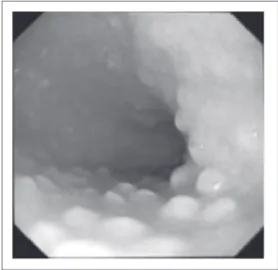
Between January 2002 and June 2005, 86 patients with endoscopically confirmed diffuse nodular duodenitis (Figure 1) were enrolled, including 50 patients (22 males; median age 49.0 years, range 23-75 years) with H. pylori infection and 36 patients (13 males; median age 52.6 years, range 22-76 years) without H. pylori infection. Forty pa-tients (14 males; median age 51.6 years, range 20-78 years) with normal appearance of the duodenum were used as controls.
Patients were excluded who had 1) evi-dence of gastroduodenal malignancies, gas-tric ulcer, duodenal ulcer, or scar on gastros-copy; 2) liver, biliary, or pancreatic diseases on ultrasound examination; 3) received pro-ton pump inhibitors (PPIs, such as omepra-zole, lansopraomepra-zole, rabepraomepra-zole, and panto-prazole, etc.), bismuth, antibiotics, aspirin, or other non-steroidal anti-inflammatory drugs (NSAID) in the preceding 2 weeks; 4) Zollinger-Ellison syndrome or Crohn’s dis-ease involving the duodenum; 5) previous upper gastrointestinal surgery; 6) pregnancy, or 7) renal insufficiency or other severe con-comitant illnesses. This study was approved by the Medicine Ethics Committee of Shang-hai Renji Hospital and signed informed con-sent was obtained from all patients.
Clinical procedures
Patients with endoscopically confirmed diffuse nodular duodenitis were treated with anti-H. pylori triple therapyconsisting of 20 mg omeprazole twice daily, 250 mg clari-thromycin twice daily, and 400 mg metroni-dazole twice daily for one week when H. pylori infection was diagnosed either in the duodenal bulb or in the gastric antrum. A control endoscopy was performed 6 months after the anti-H. pylori treatment. The H. pylori-negative (both in the duodenal bulb
and in the gastric antrum) patients with dif-fuse nodular duodenitis received 20 mg ome-prazole once daily for 6 months and a con-trol endoscopy was performed 2 weeks after treatment. During the follow-up period after eradication treatment, intermittent short courses of hydrotalcite or sucralfate were prescribed to avoid symptoms and PPIs were avoided when possible.
Endoscopy and biopsy sampling
Endoscopic findings of duodenal mor-phology were defined according to the crite-ria of endoscopic duodenitis described by the Sydney classification (12), including the subjective assessment of severity as mild, moderate or severe. During each endoscopic examination, three antral biopsies were taken from the lesser and the greater curvature within 2-3 cm from the pylorus, and two duodenal biopsies were taken from the dif-fuse nodular lesion in the duodenal bulb. Among these biopsies, one antral specimen was used to detect H. pylori by the rapid urease test, and the others were used for histological assessment.
Histology
The biopsy specimens for histological examination were fixed in 10% formalin, embedded in paraffin on the oriented edge, and cut into 4-µm thick sequential sections. The tissue sections were then stained with hematoxylin and eosin for histological ex-amination, with Alcian blue periodic acid-Schiff (AB/PAS) for identifying and assess-ing the extent of GM, and with Giemsa for H. pylori assessment.
The slides were evaluated by an experi-enced pathologist who was blind to clinical, endoscopic and other tests for H. pylori. Gastric antral and duodenal biopsy speci-mens were assessed according to the up-dated Sydney System(12). In each duodenal biopsy specimen, the infiltration of the
duode-nal mucosa by mononuclear cells (chronic inflammation) and polymorphonuclear leu-kocytes (neutrophilic activity) was graded as follows: 0, none; 1, mild; 2, moderate; 3, severe. GM was defined by the presence of surface epithelial cells containing PAS-posi-tive neutral mucin in the duodenal bulb. The extent of gastric type epithelium in duodenal biopsy specimens was arbitrarily graded on a scale of 0-3 as follows: grade 0, no GM observed; grade 1, GM involving a few villi; grade 2, GM involving several villi, and grade 3, GM involving almost all villi (12). The lymphoid follicles were considered to be positive when aggregates of lymphocytes with a germinal center (secondary lymphoid follicles) or accumulations of lymphocytes and plasma cells without a germinal center (primary lymphoid aggregates) were ob-served. Villus atrophy was defined as flat-tening or widening of villi. H. pylori infec-tion was diagnosed when the rapid urease test, hematoxylin and eosin stain, and Gi-emsa stain were all positive.
Statistical analysis
Data are reported as means ± SD. Differ-ences were evaluated by the Fisher exact test, t-test, or Wilcoxon signed rank test. P values of <0.05 were considered significant.
Results
Helicobacter pylori status
Fifty patients (50/86, 58.1%) were H. pylori positive. Among the 76 patients who completed the study, 48 were H. pylori posi-tive. After eradication treatment, 9 patients were still H. pylori positive.
Pre-treatment endoscopic appearance and histological findings
infestation, were present in patients with nodular duodenitis. There was no significant difference in the severity of endoscopic nodu-lar duodenitis between the H. pylori -posi-tive and -nega-posi-tive groups (P = 0.144, Table 1).
GM was detected in 49 of the 86 patients (57.0%). The prevalence of GM, villus atro-phy, goblet cell reduction, and lymphoid follicles was significantly higher in patients with H. pylori infection than in patients with-out H. pylori infection (P < 0.05 for all). The extent of GM was also greater in H. pylori-positive patients than that in H. pylori- nega-tive patients (P = 0.032, Table 2). Further-more, different degrees of chronic duodenal inflammation (average score, 3.4 ± 1.3) and neutrophilic activity (average score, 2.3 ± 1.9) were found in most of the patients with nodular duodenitis.
Only mild duodenal chronic inflamma-tion (average score 0.8 ± 1.0) was found in 30 of 40 control patients with normal ap-pearance of the duodenum. The values were significantly lower than those in H. pylori-positive or -negative patients in the treat-ment group (both P < 0.0001). No other histological alterations, including GM, were found in this group.
Post-treatment endoscopic appearance and histological changes
Seventy-six patients completed the study, including 48 patients with H. pylori eradica-tion treatment (successful in 39) and 28 pa-tients with PPI treatment. Neither the suc-cessful eradication treatment nor PPI treat-ment could improve the endoscopic appear-ance of nodular duodenitis (both P > 0.05, Table 1).
Six months after successful eradication of H. pylori, both the values of chronic in-flammation and neutrophilic activity were significantly decreased (2.2 ± 1.2 vs 4.0 ± 1.5, P < 0.0001, and 0.1 ± 0.3 vs 3.8 ± 1.5, P < 0.0001, respectively). Furthermore, the
Table 1. Severity of diffuse nodular duodenitis in Helicobacter pylori-positive and H. pylori-negative groups before and after treatment.
Severity of diffuse nodular duodenitis
None Mild Moderate Severe
H. pylori-positive (N = 50)+ 0 10 18 22
H. pylori-negative (N = 36) 0 10 16 10
Successful eradication of H. pylori (N = 39)
Before eradication* 0 6 14 19
After eradication 5 5 13 16
PPI treatment (N = 28)
Before treatment** 0 7 12 9
After treatment 4 8 8 8
+P > 0.05 compared with the H. pylori-negative group; *P > 0.05 compared with the
group after H. pylori eradication; **P > 0.05 compared with the group after treatment with proton pump inhibitors (PPI;Wilcoxon signed rank test).
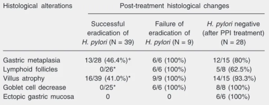
Table 3. Post-treatment histological changes of diffuse nodular duodenitis in the Helicobacter pylori-positive and H. pylori-negative groups.
Histological alterations Post-treatment histological changes
Successful Failure of H. pylori negative eradication of eradication of (after PPI treatment) H. pylori (N = 39) H. pylori (N = 9) (N = 28)
Gastric metaplasia 13/28 (46.4%)+ 6/6 (100%) 12/15 (80%)
Lymphoid follicles 0/26* 6/6 (100%) 5/8 (62.5%)
Villus atrophy 16/39 (41.0%)* 9/9 (100%) 14/15 (93.3%)
Goblet cell decrease 0/25* 6/6 (100%) 8/8 (100%)
Ectopic gastric mucosa 0 0 6/6 (100%)
PPI = proton pump inhibitors.
+P = 0.033, *P< 0.001 compared with the H. pylori-negative group (Fisher exact test).
Table 2.Extent of duodenal gastric metaplasia in the Helicobacter pylori-positive and H. pylori-negative groups before and after treatment.
Extent of duodenal metaplasia
Grade 0 Grade 1 Grade 2 Grade 3
Successful eradication of H. pylori (N = 39)
Before eradication+* 11 4 10 14
After eradication 26 6 4 3
PPI treatment (N = 28)
Before treatment** 13 5 6 4
After treatment 16 7 5 0
Grade 0, no gastric metaplasia observed; grade 1, gastric metaplasia involving a few villi; grade 2, gastric metaplasia involving several villi; grade 3, gastric metaplasia involving almost all villi.
+P = 0.032 compared with the group before proton pump inhibitors (PPI) treatment; *P
extent of duodenal GM was significantly reduced (P = 0.0001, Table 2), and regres-sion of histological alterations was achieved in most or all patients (Table 3).
However, no significant change in histo-logical findings, including GM, was found in H. pylori-negative patients on PPI treatment or in patients with persistent H. pylori infec-tion after eradicainfec-tion treatment (Table 3).
Discussion
An association between GM, H. pylori infection and duodenitis has been reported (13-15). GM of the duodenum is character-ized by the metaplastic replacement of the normal duodenal epithelial cells with those displaying a phenotype similar to that of mucus-secreting cells of the gastric mucosa. It resembles gastric foveolar epithelium in many respects, including H. pylori coloniza-tion (16,17). It has been suggested that the presence of GM may create a suitable envi-ronment for H. pylori colonization in the duodenum, resulting in chronic duodenitis (13,14,17,18). The development of meta-plastic tissue is a necessary prerequisite for H. pylori colonization because the bacte-rium may survive in the gastric mucosa but it cannot grow on normal duodenal epithelium (13,16,18). When spread to the duodenal mucosa, H. pylori is believed to exert a cytotoxic effect on mucosal cells. The in-flammatory injury to duodenal mucosa by H. pylori may eventually lead to the devel-opment of further GM (13,14,19). H. pylori infection was confirmed to be one of the independent risk factors of GM (20), and an inductive role of H. pylori in the develop-ment of GM was suggested recently (21).
However, there are conflicting reports concerning the relationship between the ex-tent of GM and H. pylori infection. It has been reported that neither the prevalence nor the extent of GM was affected by H. pylori status (5,6,9,22). In contrast, some studies found such a relationship between the extent
of GM in the duodenum and the amount of H. pylori in the gastric antrum or in the duodenal bulb (3,4,13,17). Our study showed that both the presence and the extent of GM in nodular duodenitis are related to the pres-ence of H. pylori infection.
There are also conflicting reports as to whether the extent of GM decreases after eradication of H. pylori. Some studies have shown no decrease in the extent of GM after eradication of H. pylori (5-7,9-11), whereas a significant reduction in the extent of GM was demonstrated in others (4,8). In agree-ment with the latter reports, the results of the present study showed that the extent of GM significantly decreased and complete regres-sion occurred in more than half (15/28) the patients 6 months after eradication of H. pylori. In addition, the patients with success-ful eradication of H. pylori show a signifi-cantly higher prevalence of GM regression than those with no eradication. These find-ings further confirm the important contribu-tory role of H. pylori infection in the devel-opment and persistence of GM.
In addition to H. pylori infection, it was speculated that the presence of duodenal GM is most likely caused by duodenal injury or high acidity (14,15), and is considered to be a protective mechanism to excess acid (23). But long-term acid inhibition treatment with H2-receptor antagonists could not
de-crease the extent of GM (5). It was shown in another study that acid suppression alone produced a 43% reduction in GM, similar to the effect of H. pylori eradication. The au-thors concluded that the extent of GM was partly due to H. pylori and partly to acid (4). In the present study, only 20% regression in GM could be achieved with long-term acid inhibition treatment alone in patients with-out H. pylori infection. However, the results of PPI treatment should be interpreted with caution because the acidity levels were not evaluated. This aspect deserves further in-vestigation.
type of chronic duodenitis. Up to now, we know little about its pathogenesis. It was speculated that the appearance of endoscopic findings of duodenitis may result from H. pylori infection or from an acid-pepsin at-tack and is usually accompanied with some histopathologic abnormalities, including GM (17). However, few studies had focused on the association between the appearance of duodenitis and H. pylori infection. It has been reported that no significant difference exists in the severity of endoscopic duodeni-tis between H. pylori-positive and -negative
patients, and successful eradication of H. pylori did not lead to any change in endo-scopic appearance (9). In agreement with this finding, we found that neither the eradi-cation of H. pylori nor the acid suppression treatment could change the appearance of endoscopic nodular duodenitis in a signifi-cant manner.
In conclusion, the present results suggest that H. pylori infection is related to the ex-tent of GM in the duodenum, but not to the presence of diffuse nodular duodenitis.
References
1. Heikkinen M, Pikkarainen P, Vornanen M, Hollmen S, Julkunen R. Prevalence of gastric metaplasia in the duodenal bulb is low in Helicobacter pylori positive non-ulcer dyspepsia patients. Dig Liver Dis 2001; 33: 459-463.
2. Chen XY, Shi Y, Peng YS, Ma YZ, Xiao SD. Relationship between Helicobacter pylori infection and gastric metaplasia in the mucosa of duodenal bulb. Chin J Dig 2004; 9: 519-522.
3. Futami H, Takashima M, Furuta T, Hanai H, Kaneko E. Relationship between Helicobacter pylori infection and gastric metaplasia in the duodenal bulb in the pathogenesis of duodenal ulcer. J Gastroenter-ol HepatGastroenter-ol 1999; 14: 114-119.
4. Khulusi S, Badve S, Patel P, Lloyd R, Marrero JM, Finlayson C, et al. Pathogenesis of gastric metaplasia of the human duodenum: role of Helicobacter pylori, gastric acid, and ulceration. Gastroenterology 1996; 110: 452-458.
5. Noach LA, Rolf TM, Bosma NB, Schwartz MP, Oosting J, Rauws EA, et al. Gastric metaplasia and Helicobacter pylori infection. Gut 1993; 34: 1510-1514.
6. Harris AW, Gummett PA, Walker MM, Misiewicz JJ, Baron JH. Relation between gastric acid output, Helicobacter pylori, and gas-tric metaplasia in the duodenal bulb. Gut 1996; 39: 513-520. 7. Savarino V, Mela GS, Zentilin P, Mele MR, Bisso G, Pivari M, et al.
Effect of Helicobacter pylori eradication on 24-hour gastric pH and duodenal gastric metaplasia. Dig Dis Sci 2000; 45: 1315-1321. 8. Ciancio G, Nuti M, Orsini B, Iovi F, Ortolani M, Palomba A, et al.
Regression of duodenal gastric metaplasia in Helicobacter pylori positive patients with duodenal ulcer disease. Dig Liver Dis 2002; 34: 16-21.
9. Urakami Y, Sano T. Endoscopic duodenitis, gastric metaplasia and Helicobacter pylori. J Gastroenterol Hepatol 2001; 16: 513-518. 10. Urakami Y, Sano T. Long-term follow-up of gastric metaplasia after
eradication of Helicobacter pylori. J Med Invest 2003; 50: 48-54. 11. Bago J, Strinic D, Halle ZB, Jandric D, Tomic M, Bilic A, et al. Effect
of Helicobacter pylori eradication on extent of duodenal gastric metaplasia and grade of gastritis. Coll Antropol 2002; 26: 557-563. 12. Tytgat GN. The Sydney System: endoscopic division. Endoscopic
appearances in gastritis/duodenitis. J Gastroenterol Hepatol 1991;
6: 223-234.
13. Madsen JE, Vetvik K, Aase S. Helicobacter-associated duodenitis and gastric metaplasia in duodenal ulcer patients. APMIS 1991; 99: 997-1000.
14. Satoh K, Kimura K, Yoshida Y, Kasano T, Kihira K, Taniguchi Y. Relationship between Helicobacter pylori colonization and acute inflammation of the duodenal mucosa. Am J Gastroenterol 1993; 88: 360-363.
15. Fitzgibbons PL, Dooley CP, Cohen H, Appleman MD. Prevalence of gastric metaplasia, inflammation, and Campylobacter pylori in the duodenum of members of a normal population. Am J Clin Pathol 1988; 90: 711-714.
16. Carrick J, Lee A, Hazell S, Ralston M, Daskalopoulos G. Campylo-bacter pylori, duodenal ulcer, and gastric metaplasia: possible role of functional heterotopic tissue in ulcerogenesis. Gut 1989; 30: 790-797.
17. Wyatt JI, Rathbone BJ, Sobala GM, Shallcross T, Heatley RV, Axon AT, et al. Gastric epithelium in the duodenum: its association with Helicobacter pylori and inflammation. J Clin Pathol 1990; 43: 981-986.
18. Johan G, Offerhaus A, Molyvas EN, Hoedemaeker PJ. Helicobacter pylori infection of gastric mucin cell metaplasia: the duodenum revisited. J Pathol 1990; 162: 239-243.
19. Walker MM, Dixon MF. Gastric metaplasia: its role in duodenal ulceration. Aliment Pharmacol Ther 1996; 10 (Suppl 1): 119-128. 20. Voutilainen M, Juhola M, Farkkila M, Sipponen P. Gastric
metapla-sia and chronic inflammation at the duodenal bulb mucosa. Dig Liver Dis 2003; 35: 94-98.
21. Van De Bovenkamp JH, Korteland-Van Male AM, Buller HA, Einerhand AW, Dekker J. Metaplasia of the duodenum shows a Helicobacter pylori-correlated differentiation into gastric-type pro-tein expression. Hum Pathol 2003; 34: 156-165.
22. Suriani R, Venturini I, Actis GC, Rocca G, Rizzetto M, Cerutti E, et al. Effect of Helicobacter pylori eradication on bulbitis and duodenal gastric metaplasia. Hepatogastroenterology 2004; 51: 176-180. 23. James AH. Gastric epithelium in the duodenum. Gut 1964; 5: