Newborn screening for biotinidase
deficiency in Brazil: biochemical
and molecular characterizations
1LaboratórioNobel RIE, Porto Alegre, RS, Brasil 2Centro de Triagem Neonatal, Porto Alegre, RS, Brasil
3Serviço de Genética Médica, Hospital de Clínicas de Porto Alegre,
and Departamento de Genética, Universidade Federal do Rio Grande do Sul, Porto Alegre, RS, Brasil
4General Clinical Research Center, Core Laboratory, and
5Department of Pediatrics, University of Connecticut School of Medicine, Farmington, CT, USA
6Department of Research, Connecticut Children’s Medical Center, Hartford, CT, USA E.C. Neto1,2, J. Schulte1,
R. Rubim1, E. Lewis2, J. DeMari2, C. Castilhos2, A. Brites2, R. Giugliani3, K.P. Jensen4 and B. Wolf5,6
Abstract
Biotinidase deficiency is an inherited metabolic disorder character-ized by neurological and cutaneous symptoms. Fortunately, it can be treated and the symptoms prevented by oral administration of the vitamin biotin. Using dried blood-soaked filter paper cards, biotinidase activity was determined in the sera of 225,136 newborns in Brazil. Mutation analysis performed on DNA from 21 babies with low serum biotinidase activity confirmed that 3 had profound biotinidase defi-ciency (less than 10% of mean normal sera biotinidase activity), 10 had partial biotinidase deficiency (10 to 30% of mean normal serum activity), 1 was homozygous for partial biotinidase deficiency, 4 were heterozygous for either profound or partial deficiency, and 3 were normal. Variability in serum enzyme activities and discrepancies with mutation analyses were probably due to inappropriate han-dling and storage of samples sent to the laboratory. Obtaining an appropriate control serum at the same time as that of the suspected child will undoubtedly decrease the false-positive rate (0.09%). Muta-tion analysis can be used to confirm the genotype of these children. The estimated incidence of biotinidase deficiency in Brazil is about 1 in 9,000, higher than in most other countries. Screening and treatment of biotinidase deficiency are effective and warranted. These results strongly suggest that biotinidase deficiency should be included in the newborn mass screening program of Brazil.
Correspondence
E.C. Neto
Centro de Triagem Neonatal Av. Ipiranga, 5000 90610-000 Porto Alegre, RS Brasil
Fax: +51-3339-5000 E-mail: [email protected] Research supported in part by the Safra Research Fund (B.W.) at Connecticut Children’s Medical Center and by a General Clinical Research Center Grant to the University of Connecticut Health Center (MO1RR06192) from the National Institutes of Health.
Received March 11, 2003 Accepted October 24, 2003
Key words
•Newborn screening •Biotinidase deficiency •Biotinidase
•Mutations •Enzyme assay
Biotinidase deficiency is an autosomal recessive inherited disorder of the recycling of biotin, an essential water-soluble vitamin. Biotin is the coenzyme of four carboxylases involved in amino acid catabolism, fatty acid synthesis, and gluconeogenesis (1). Deficient activity of this enzyme is the primary defect
vision problems, skin rash and alopecia (3). These children can develop metabolic ke-toacidosis, hyperammonemia, and organic aciduria (3,4). These features are caused by secondary deficiencies of the activities of the biotin-dependent enzymes, propionyl-CoA carboxylase, pyruvate-propionyl-CoA carboxy-lase, and ß-methylcrotonyl-CoA carboxyl-ase (1). If not treated with biotin, children with the enzyme deficiency may become comatose and die. Symptoms can be pre-vented if biotin therapy is initiated early (1). Because biotinidase deficiency meets the criteria for inclusion in newborn screening programs, at least 25 countries have included the disorder in their programs (5). Brazil is a country with multiple ethnic groups and the incidence of biotinidase deficiency is un-known. We now report our experience with screening newborns for biotinidase deficiency from October 1995 to November 1999 in a private neonatal screening program in South-ern Brazil.
Some private programs in Brazil offer a great number of tests for genetic and infec-tious diseases. Based on the high incidence of biotinidase deficiency found through the currently available private screening pro-grams in Brazil, more widespread screening will undoubtedly identify many more chil-dren with the disorder. Compared to other disorders that are currently screened coun-try-wide through public supported programs (phenylketonuria, congenital hypothyroid-ism and hemoglobinopathies), screening for biotinidase deficiency would be cost-effec-tive.
Blood-soaked filter paper cards (filter paper SS 903 from Schleicher and Schuell, Keene, NH, USA) were obtained from 225,136 babies between the ages of 2 and 30 days (median age: 13 days) from throughout Brazil. To identify potentially biotinidase-deficient children, biotinidase activity was determined in punched disks from the cards using a qualitative colorimetric assay in which biotinyl-p-aminobenzoate is the substrate (6).
Confirmation of biotinidase activity in se-rum was obtained for babies suspected of having the deficiency and for their parents by a quantitative colorimetric assay using biotinyl-p-aminobenzoate as described pre-viously (2). Normal serum enzyme activity ranged from 4 to 10 µmol p-aminobenzoate formed ml-1 min-1. Blood for DNA was ob-tained from 21 children with serum enzyme activity below 30% of the mean activity of normal children to confirm and identify mu-tations by direct sequencing (7,8). Mutation analyses were performed by direct sequenc-ing of the codsequenc-ing regions of the biotinidase gene including exon/intron boundaries. Novel mutations were confirmed by sequencing the entire gene and by identifying the alter-ations in the DNA of the parents when avail-able.
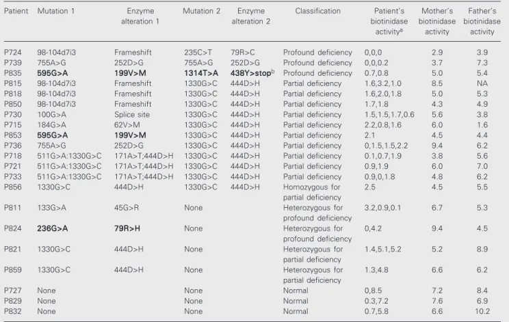
parents are presented in Table 1. None of the families were consanguineous. Clinical fol-low-up was performed by telephone with the physicians and parents of the children. All children on biotin therapy have remained asymptomatic.
As can be seen in Table 1, there were discrepancies in the serum enzyme activities of some of the children. Because there are multiple factors that can result in falsely low enzyme activity, the highest enzyme activity of a child was likely to represent the true activity. Based on these enzyme results, it would be expected that some of the children initially thought to have profound or partial biotinidase deficiency would be heterozy-gous for the disorder or would not have a mutation at all. Mutational analyses
con-firmed or excluded the diagnosis of biotin-idase deficiency in all children tested. Based on these mutation studies, of the 21 children tested, 3 were confirmed to have profound biotinidase deficiency, 10 were confirmed to have partial biotinidase deficiency, 1 was found to be homozygous for partial biotin-idase deficiency, 4 were heterozygous for either profound or partial deficiency, and 3 were found to be normal. Only the 14 chil-dren confirmed to have profound or partial biotinidase deficiency required continued bi-otin treatment.
We identified four novel mutations in our population (1314T>A, 133G>A, 595G>A and 236G>A). All of these mutations appear to cause profound biotinidase deficiency based on serum enzyme activity. The 1314T>A
Table 1. Biochemical and molecular characterization of children suspected of having biotinidase deficiency by newborn screening.
Patient Mutation 1 Enzyme Mutation 2 Enzyme Classification Patient’s Mother’s Father’s
alteration 1 alteration 2 biotinidase biotinidase biotinidase
activitya activity activity
P724 98-104d7i3 Frameshift 235C>T 79R>C Profound deficiency 0,0,0 2.9 3.9
P739 755A>G 252D>G 755A>G 252D>G Profound deficiency 0,0,0.2 3.7 7.3
P835 595G>A595G>A595G>A595G>A595G>A 199V>M199V>M199V>M199V>M199V>M 1314T>A1314T>A1314T>A1314T>A1314T>A 438Y>stop438Y>stop438Y>stop438Y>stop438Y>stopb Profound deficiency 0.7,0.8 5.0 5.4
P815 98-104d7i3 Frameshift 1330G>C 444D>H Partial deficiency 1.6,3.2,1.0 8.5 NA
P818 98-104d7i3 Frameshift 1330G>C 444D>H Partial deficiency 1.6,2.0,1.8 5.0 5.3
P850 98-104d7i3 Frameshift 1330G>C 444D>H Partial deficiency 1.7,1.8 4.3 4.9
P730 100G>A Splice site 1330G>C 444D>H Partial deficiency 1.5,1.5,1.7,0.6 5.6 3.8
P715 184G>A 62V>M 1330G>C 444D>H Partial deficiency 2.2,0.8,1.6 6.0 1.6
P853 595G>A595G>A595G>A595G>A595G>A 199V>M199V>M199V>M199V>M199V>M 1330G>C 444D>H Partial deficiency 2.1 4.5 4.4
P736 755A>G 252D>G 1330G>C 444D>H Partial deficiency 0,1.5,1.5,2.2 9.4 6.2
P718 511G>A:1330G>C 171A>T;444D>H 1330G>C 444D>H Partial deficiency 0.1,0.7,1.9 3.8 5.6
P721 511G>A:1330G>C 171A>T;444D>H 1330G>C 444D>H Partial deficiency 0.9,1.9 6.0 7.0
P733 511G>A:1330G>C 171A>T;444D>H 1330G>C 444D>H Partial deficiency 0.9,0,1.8 4.8 6.2
P856 1330G>C 444D>H 1330G>C 444D>H Homozygous for 2.5 4.5 5.5
partial deficiency
P811 133G>A 45G>R None Heterozygous for 3.2,0.9,0.1 6.7 5.3
profound deficiency
P824 236G>A236G>A236G>A236G>A236G>A 79R>H79R>H79R>H79R>H79R>H None Heterozygous for 0,4.2 9.4 4.5 profound deficiency
P821 1330G>C 444D>H None Heterozygous for 1.4,5.1,5.2 5.2 8.9
partial deficiency
P859 1330G>C 444D>H None Heterozygous for 1.3,4.8 6.6 6.2
partial deficiency
P727 None None Normal 0,8.5 7.2 8.4
P829 None None Normal 0.3,7.2 7.6 6.9
P832 None None Normal 0.7,5.8 6.6 10.2
aMultiple serum enzyme determinations. Units of activity are reported as nmol min-1 ml serum-1. Normal activity is 4.4 to 12. bAlso has 1413T>C
mutation results in an alteration of a tyrosine residue to a termination codon in the latter portion of the enzyme protein. The 133G>A mutation is a subtle mutation resulting in an alteration of a glycine to alanine. This muta-tion has been observed in an allelic double mutation previously, but not alone (9). The 595G>A mutation was observed in two chil-dren and results in a change from a valine to methionine. This mutation resides in a region where multiple other missense mutations have been found. The 236G>A mutation results in a change of an arginine to a histidine and is located in a region of biotinidase that is homologous with the active site of bacterial aliphatic hydrolases (10).
Biotinidase deficiency is diagnosed by demonstrating deficient enzyme activity in serum. Children with profound biotinidase deficiency have serum enzyme activity less than 10% of mean normal activity (1). These children are most likely to become sympto-matic if they are not treated with biotin. Children with partial biotinidase deficiency have 10 to 30% of mean normal serum biotinidase activity. These children may de-velop symptoms if they are stressed by an infection (11). Both groups of children are now routinely treated with biotin. In our study of 21 children suspected of having either profound or partial biotinidase defi-ciency, 14 or 66.7% were confirmed to have the disorder based on mutation analysis. The other children were shown to be carriers of mutations causing either profound or partial deficiency or did not have any mutations at all. In addition, the screening revealed four novel mutations, three missense mutations, and one that resulted in premature termina-tion.
The incidence of profound and partial biotinidase deficiency worldwide is estimated at about 1 in 60,000 (5). If we consider 22 cases in the present adjusted sample size of 198,694 newborns, then the incidence of biotinidase deficiency in Brazil is about 1 in 9,000 births. Either estimation indicates that
the incidence of biotinidase deficiency in Brazil is one of the highest known.
Our recall rate for second screening samples of 0.12% is considerably higher than reported by most other laboratories (5,12). This problem is usually attributable to poor impregnation of blood on filter pa-per, increased humidity, failure to allow the filter paper to dry sufficiently before pack-ing in plastic bags, or heat exposure. The problem with discrepancies in quantitative enzyme determinations is usually failure to rapidly freeze and ship the samples on dry ice or failure to store the samples in the laboratory at -80°C if not immediately as-sayed. As stated above, samples were not usually frozen when received by the labora-tory. Unfortunately, most samples were shipped at room temperature and were two to five days old when they arrived. Rou-tinely, samples were assayed immediately after they were received. The inadequate handling and shipping of the samples are undoubtedly the cause for the high false-positive rate and the disparity in reproduc-ibility in serum enzyme activities. These fac-tors also explain the discordance between the low serum biotinidase activities and the failure to find mutations in some of the children who initially had low enzyme ac-tivities.
confirma-tion of biotinidase deficiency can be obtained using mutation analysis, but this is an expen-sive, inconvenient and time consuming pro-cedure.
Parental testing was not helpful in dis-cerning which children were likely to have biotinidase deficiency as it usually is when samples are frozen on arrival to the labora-tory. Although only 1 of the 6 parents of children who was found to be normal had serum biotinidase activity in the heterozy-gous range, 3 of 6 parents of children con-firmed to have profound biotinidase defi-ciency had normal enzyme activities. Per-haps the parental enzyme activities would be helpful in deducing the genotype if a simul-taneously obtained normal control was in-cluded for comparison.
Despite the high number of samples with false-positive results in filter paper when compared with the true positive cases, new-born screening for biotinidase deficiency appears to be reasonable and effective. Al-terations in obtaining appropriate controls should help to eliminate or minimize dis-crepancies and variability in determining true deficiencies. Final confirmation can be achieved by mutation analysis that does not depend on freezing the samples on ship-ment. The preliminary estimated incidence of biotinidase deficiency in Brazil is rela-tively high compared to most other countries and warrants inclusion of biotinidase defi-ciency in the group of disorders screened for in Brazil.
References
1. Wolf B (2001). Disorders of biotin metabolism. In: Scriver CR, Beaudet AL, Sly WS & Valle D (Editors), The Metabolic and Molecu-lar Bases of Inherited Disease. 8th edn. McGraw-Hill, New York, 3935-3962.
2. Wolf B, Grier RE, Allen RJ, Goodman SI & Kien CL (1983). Biotinidase deficiency: The enzymatic defect in late-onset multiple carboxylase deficiency. Clinica Chimica Acta, 131: 273-281.
3. Wolf B, Grier RE, Allen RJ, Goodman SI, Kien CL, Parker WD, Howell DM & Hurst DL (1983). Phenotypic variation in biotinidase deficiency. Journal of Pediatrics, 103: 233-237.
4. Wolf B, Grier RE, Secor McVoy JR & Heard GS (1985). Biotinidase deficiency: A novel vitamin recycling defect. Journal of Inherited Metabolic Disease, 8 (Suppl 1): 53-58.
5. Wolf B & Heard GS (1990). Screening for biotinidase deficiency in newborns: Worldwide experience. Pediatrics, 85: 512-517. 6. Heard GS, Secor McVoy JR & Wolf B (1984). A screening method
for biotinidase deficiency in newborns. Clinical Chemistry, 30: 125-127.
7. Pomponio RJ, Reynolds TR, Cole H, Buck GA & Wolf B (1995). Mutational “hotspot” in the human biotinidase gene as a cause of
biotinidase deficiency. Nature Genetics, 11: 96-98.
8. Pomponio RJ, Hymes J, Reynolds TR, Meyers GA, Fleischhauer K, Buck GA & Wolf B (1997). Mutations in the human biotinidase gene that cause profound biotinidase deficiency in symptomatic children: Molecular, biochemical and clinical analysis. Pediatric Research, 42: 840-848.
9. Hymes J, Stanley CM & Wolf B (2001). Mutations in BTD causing biotinidase deficiency. Human Mutation, 200: 375-381.
10. Swango KL, Hymes J, Brown P & Wolf B (2000). Amino acid homologies between human biotinidase and bacterial aliphatic ami-dase: putative identification of the active site of biotinidase. Molec-ular Genetics and Metabolism, 69: 111-115.