ISSN 1414-431X
BIOMEDICAL SCIENCES
www.bjournal.com.br
www.bjournal.com.br
Volume 45 (10) 875-994 October 2012
Braz J Med Biol Res, October 2012, Volume 45(10) 977-981
doi: 10.1590/S0100-879X2012007500092
A simple test of muscle coactivation estimation using
electromyography
U.F. Ervilha, T. Graven-Nielsen and M. Duarte
Institutional Sponsors
The Brazilian Journal of Medical and Biological Research is partially financed by
Faculdade de Medicina de Ribeirão Preto Campus
Ribeirão Preto
Explore High - Performance MS Orbitrap Technology In Proteomics & Metabolomics
A simple test of muscle coactivation
estimation using electromyography
U.F. Ervilha
1, T. Graven-Nielsen
2and M. Duarte
31Laboratório de Biodinâmica do Movimento Humano, Escola de Educação Física, Universidade São Judas Tadeu, São Paulo, SP, Brasil 2Center for Sensory-Motor Interaction, Aalborg University, Aalborg, Denmark 3Programa de Engenharia Biomédica, Universidade Federal do ABC, Santo André, SP, Brasil
Abstract
In numerous motor tasks, muscles around a joint act coactively to generate opposite torques. A variety of indexes based on electromyography signals have been presented in the literature to quantify muscle coactivation. However, it is not known how to estimate it reliably using such indexes. The goal of this study was to test the reliability of the estimation of muscle coactivation using electromyography. Isometric coactivation was obtained at various muscle activation levels. For this task, any coactiva-tion measurement/index should present the maximal score (100% of coactivacoactiva-tion). Two coactivacoactiva-tion indexes were applied. In the first, the antagonistic muscle activity (the lower electromyographic signal between two muscles that generate opposite joint torques) is divided by the mean between the agonistic and antagonistic muscle activations. In the second, the ratio between antagonistic and agonistic muscle activation is calculated. Moreover, we computed these indexes considering different elec-tromyographic amplitude normalization procedures. It was found that the first algorithm, with all signals normalized by their respective maximal voluntary coactivation, generates the index closest to the true value (100%), reaching 92 ± 6%. In contrast, the coactivation index value was 82 ± 12% when the second algorithm was applied and the electromyographic signal was not normalized (P < 0.04). The new finding of the present study is that muscle coactivation is more reliably estimated if the EMG signals are normalized by their respective maximal voluntary contraction obtained during maximal coactivation prior to dividing the antagonistic muscle activity by the mean between the agonistic and antagonistic muscle activations.
Key words: Muscle coactivation; Electromyogram; Muscle cocontraction; EMG index
Introduction
Correspondence: U.F. Ervilha, Laboratório de Biodinâmica do Movimento Humano, Escola de Educação Física, Universidade São Judas Tadeu, Rua Taquari, 546, 03166-000 São Paulo, SP, Brasil. E-mail: prof.ulysses@saojudas.br
Received August 15, 2011. Accepted May 17, 2012. Available online June 1, 2012. Published September 3, 2012. Muscle coactivation, the simultaneous activation of
agonist and antagonist muscle groups around a joint (1,2), is an important and common strategy for the control of voluntary movement in humans and has been experimen-tally observed during a wide variety of conditions including locomotion (3), isometric and functional activities (4), low and high accurate pointing tasks (2,5), upright standing (6), central nervous system impairment (7,8), and lumbopelvic stabilization (9), among others. In the control of movements, it has been suggested that muscle coactivation modulates the impedance of a joint, mainly stabilizing the joint (2).
In experimental conditions, coactivation is most often estimated by comparing the amplitude of the myoelectric activity of muscles that generate opposite torques during a task. Despite the fact that a muscle can be active more than another and yet generate less force (or vice versa) due to a number of factors, including pennation angle and
fiber length, and also considering that caution should be
used if the relationship between force and electromyo-graphic (EMG) signal is not established for all the muscles investigated, various indexes of coactivation based solely on the EMG signal have been proposed (10-17). However, it is not known how coactivation based on EMG is reliably estimated with such indexes. The goal of this study was to test the reliability of the estimation of muscle coactivation using EMG.
Subjects and Methods
Subjects
978 U.F. Ervilha et al.
with the Helsinki Declaration and was approved by the Aalborg University Ethics Committee (VN2003/61). The volunteers received information about the experiment and gave written informed consent to participate.
Indexes of coactivation
Two indexes were selected to represent most of the coactivation indexes (CI) based on the amplitude of the
EMG signal employed in the literature (10-17). In the first
index (CI1), the antagonistic muscle activity (the lower EMG
amplitude between two muscles that generate opposite joint torques) is divided by the mean between the agonistic
(EMGAG) and antagonistic (EMGANT) muscle activations
(10). The second index (CI2) is obtained by calculating the
ratio between antagonistic and agonistic muscle activation (11). The formulas for these indexes are shown below:
1= ∗2 ∗100
+
ANT
AG ANT
EMG CI
EMG EMG (1)
2= ∗100
ANT AG EMG CI EMG (2)
The method for EMG amplitude normalization used when coactivation is calculated varies in the literature. The EMG signals have often been normalized by the maximum voluntary isometric contraction (MVIC) (13,18), the maxi-mum voluntary isometric coactivation value (MVICa) (10), or have not been normalized (19,20). Here we will investigate how these three methods affect the CI.
Protocol
Coactivation around a joint that does not move and with no external moments implies that the net joint moment is equal to zero. Such task can be performed at any level of muscle activation, as long as the joint does not move (full coactivation). Thus, the volunteers were instructed to
acti-vate simultaneously elbow extensor and flexor muscles in
order to achieve full coactivation at various levels of muscle activation. In order to allow the volunteers to maintain a targeted level of muscle activation, established for that task, the EMG linear envelope from the medial belly of the biceps brachii muscle was shown in real time on an oscilloscope. The volunteers practiced the task prior to data collection by performing 3 to 4 times 2 s of full coactivation at various muscle activation levels. The data for 1 volunteer was not
includedin the study because she was unable to perform
the task. The subjects rested for 1 min between two prac-ticing trials. A full coactivation was then carried out for 4 s, at different biceps muscle activation levels (25, 50, and 75%) of the biceps muscle EMG activity achieved during a maximal effort muscle activation (100%) keeping a full coactivation. At the beginning of the session, the volunteers performed a coactivation at a maximum effort so the EMG activity from the medial head of the biceps brachii muscle could be used as the reference for the biofeedback and the peak EMG from the biceps and triceps brachii muscles
could be used for EMG normalization. For a second EMG normalization procedure, the peak EMG from a maximal
isometric voluntary contraction for elbow flexors and elbow
extensors was used.
Setup
The volunteers were comfortably seated with the arm
supported at 90° abduction and 90° elbow flexion. A pair
of surface electrodes (Medicotest 72001-k, Denmark) was
placed in the direction of the muscle fibers (2-cm apart) on
shaved, cleaned skin. On the biceps brachii muscle, the electrodes were placed on the medial and lateral head, on the lead-line between the acromion and the fossa cubit at 1/3 from the fossa cubit. On the triceps brachii muscle, the electrodes were placed on the lateral and medial head - 1 cm lateral to the lead-line just on the midpoint between the acromion and the olecranon process. The EMG signals
were bandpass filtered (2nd order, 20 to 500 Hz), amplified
1000 times (CounterPoint MK2, Dantec, Denmark) and sampled at 1024 Hz.
Data analysis and statistics
The digital EMG signal was band-pass filtered (2nd
order, zero-phase-lag Butterworth, 20 to 400 Hz), full wave
rectified, and smoothed (low-pass, 4th order, zero-phase-lag Butterworth filter with a 3-Hz cutoff frequency). From the
4 s of coactivation, a 1-s interval was extracted in which the squared difference between the acquired EMG and the targeted EMG intensity was the lowest. This procedure al-lowed us to select the 1-s window where the subject’s EMG activation was closest to the target displayed on the oscil-loscope. The difference between the target versus the actual EMG activation level was calculated as percent error.
Data are reported as means ± SD. Three-way repeated analyses of variance (ANOVA) were used to examine the
effects of CI (CI1 and CI2), normalization procedures
[non-normalization (NN), [non-normalization by the MVIC (NMVIC), and normalization by the MVICa (NMVICa)], and muscle activation level (25, 50, 75, 100%) on the coactivation index.
When ANOVA was found to be significant, the Student-New
-man-Keuls post hoc test was used for multiple comparisons.
The level of significance was set at P< 0.05.
Results
to. Although the variability throughout the 4-s contractions was quite large, as shown in Figure 1, it is important to emphasize the fact that the volunteers were requested to maintain the contraction at the target for 2 s only in order to avoid fatigue (as instructed in the familiarization training). Moreover, the fact that a 1-s window was selected makes the behavior during the other 3 s less important for the present investigation.
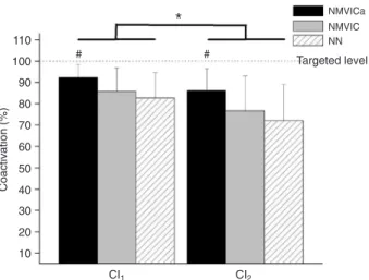
The mean values and standard deviation of the two coactivation indexes considering the different normalization methods applied are shown in Figure 2.
ANOVA for the factors algorithm (CI1 and CI2),
normal-ization procedures (NN, NMVIC, NMVICa), and muscle activation levels (25, 50, 75, and 100% MVIC) revealed a main effect of algorithm (F(1,9) = 137, P < 0.001) and a main effect of normalization procedure (F(2,18) = 3.9, P =
0.04). The post hoc analyses revealed that the CI calculated
by applying Equation 1 (CI1) had a higher value than that
calculated by applying Equation 2 (CI2) for all normalization
procedures, with respective pooled means ± SD values of 87 ± 10 and 78 ± 15% (P < 0.03). Moreover, the CI was
higher when the EMG signal was normalized by the NMVICa compared to the CI obtained when the EMG was not nor-malized for both indexes (NMVICa > NN; P < 0.04); pooled means ± SD respectively equal to 89 ± 6 and 77 ± 12%. No interactions among factors were found by ANOVA.
Discussion
In this study, we verified the accuracy of CI based on
EMG recordings. Our results showed that none of the in-vestigated CI was able to accurately estimate the level of coactivation, which was theoretically known. The present data shows that muscle coactivation is more reliably esti-mated if the EMG signals are normalized by their respective maximal voluntary contraction obtained during maximal coactivation, prior to dividing the antagonistic muscle ac-tivity by the mean between the agonistic and antagonistic
muscle activations.The inability of surface EMG electrodes
to accurately record all motor units equally, the fact that not all muscles involved are recorded, and data process-ing limitations certainly contributed to coactivation values
980 U.F. Ervilha et al.
different from the theoretical expected ones. Furthermore, several muscles act at a joint and it seems to be crucial to consider the contribution of all muscles involved, as well as possible nonlinearities to reliably calculate coactivation. Note, however, that these very same limitations are present in the great majority of studies that employ EMG signals to estimate muscle coactivation (e.g., 2, 10-12, 3, 13, 14, 7, 15, 8, 16, 17, 6, 5, 4, 9). Our rationale is that the low ac-curacy of the CI we found, which certainly resulted from the far from perfect methods employed here (and elsewhere), exposes the limitation of such CI.
When muscles that produce torque in opposite directions are simultaneously activated, they limit the net moment generated at that joint. If the resultant contraction is an ac-tive maintenance of a static joint positioning, the muscles surrounding the joint are in full coactivation at any point from the minimum to the maximum muscle activation. However, any estimation of coactivation at that point must show that the net torque around that joint is zero, which is represented by the highest score when coactivation is measured.
In the present study, there was no joint movement or resultant external torques, implying that the volunteers had their muscles around the elbow in full coactivation, meaning
zero net torque. In that case, any elbow flexor muscle is an
antagonist to an elbow extensor and vice versa. In order to have a reference, the higher and the lower EMG inten-sity levels were considered for agonistic and antagonistic muscle activation, respectively. In theory, a CI at 100% would be expected in all conditions studied in the present investigation. It was shown that a CI that accounts for the antagonist torque generated at the joint and also for the additional agonist torque required to compensate for it, as
proposed in CI1, is more reliable because the index value
reaches values closer to the maximum score. Moreover,
there was no significant difference in the CI calculated for
different EMG levels, indicating that neither CI is affected by the muscle activation level.
In order to minimize inter-subject EMG differences, the EMG signal amplitude was normalized. Various EMG amplitude normalization methods have been used in the lit-erature when a CI is calculated (10,12,16,17). In the present study, it was shown that the EMG normalization procedure
also influences the CI outcome. When the EMG amplitude
was normalized by the peak EMG signal obtained during a maximal full coactivation, the CI approached the expected values for that motor task better than when the EMG am-plitude was not normalized. Possibly, this is because this
normalization procedure is related to the targeted task. That is, full coactivation at various muscle activation levels.
The present results were derived from a controlled task designed to validate the different methods to quantify coacti-vation. The task was always performed in a static condition
and at the same position (90° elbow flexion). Since muscle
force is affected by its length and velocity (21) and during dynamic tasks the length and velocity of the muscles vary differently, these two factors might limit even more the use of CI based on the EMG signals.
Furthermore, the low accuracy of the CI suggests that coactivation indexes should be interpreted with caution and any methodological difference in the calculation should be considered before a comparison of different studies is performed.
Acknowledgments
M. Duarte was supported by a research grant from FAPESP (#08/10461-07).
References
1. Levine MG, Kabat H. Cocontraction and reciprocal inner-vation in voluntary movement in man. Science 1952; 116: 115-118.
2. Hogan N. Adaptive control of mechanical impedance by
coactivation of antagonist muscles. IEEE Transactions on
Automatic Control 1984; 29: 681-690.
3. Collins JJ. The redundant nature of locomotor optimization laws. J Biomech 1995; 28: 251-267.
4. Busse ME, Wiles CM, van Deursen RW. Co-activation: its association with weakness and specific neurological pathol-ogy. J Neuroeng Rehabil 2006; 3: 26.
5. Ervilha UF, Arendt-Nielsen L, Duarte M, Graven-Nielsen T. The effect of muscle pain on elbow flexion and coactivation tasks. Exp Brain Res 2004; 156: 174-182.
6. Benjuya N, Melzer I, Kaplanski J. Aging-induced shifts from a reliance on sensory input to muscle cocontraction during balanced standing. J Gerontol A Biol Sci Med Sci 2004; 59: 166-171.
7. Damiano DL, Martellotta TL, Sullivan DJ, Granata KP, Abel MF. Muscle force production and functional performance in spastic cerebral palsy: relationship of cocontraction. Arch
Phys Med Rehabil 2000; 81: 895-900.
8. Chae J, Yang G, Park BK, Labatia I. Muscle weakness and cocontraction in upper limb hemiparesis: relationship to mo-tor impairment and physical disability. Neurorehabil Neural
Repair 2002; 16: 241-248.
9. Belavy DL, Richardson CA, Wilson SJ, Rittweger J, Felsen-berg D. Superficial lumbopelvic muscle overactivity and decreased cocontraction after 8 weeks of bed rest. Spine
2007; 32: E23-E29.
10. Falconer K, Winter DA. Quantitative assessment of co-contraction at the ankle joint in walking. Electromyogr Clin
Neurophysiol 1985; 25: 135-149.
11. Osternig LR, Hamill J, Lander JE, Robertson R. Co-activa-tion of sprinter and distance runner muscles in isokinetic exercise. Med Sci Sports Exerc 1986; 18: 431-435. 12. Hammond MC, Fitts SS, Kraft GH, Nutter PB, Trotter MJ,
Robinson LM. Co-contraction in the hemiparetic forearm: quantitative EMG evaluation. Arch Phys Med Rehabil 1988; 69: 348-351.
13. Unnithan VB, Dowling JJ, Frost G, Volpe Ayub B, Bar-Or O.
Cocontraction and phasic activity during GAIT in children with cerebral palsy. Electromyogr Clin Neurophysiol 1996; 36: 487-494.
14. Frost G, Dowling J, Dyson K, Bar-Or O. Cocontraction in three age groups of children during treadmill locomotion. J
Electromyogr Kinesiol 1997; 7: 179-186.
15. Lamontagne A, Richards CL, Malouin F. Coactivation during gait as an adaptive behavior after stroke. J Electromyogr
Kinesiol 2000; 10: 407-415.
16. Macaluso A, Nimmo MA, Foster JE, Cockburn M, McMillan NC, De Vito G. Contractile muscle volume and agonist-antagonist coactivation account for differences in torque between young and older women. Muscle Nerve 2002; 25: 858-863.
17. Kellis E, Arabatzi F, Papadopoulos C. Muscle co-activation around the knee in drop jumping using the co-contraction index. J Electromyogr Kinesiol 2003; 13: 229-238.
18. Hurd WJ, Chmielewski TL, Snyder-Mackler L. Perturbation-enhanced neuromuscular training alters muscle activity in female athletes. Knee Surg Sports Traumatol Arthrosc 2006; 14: 60-69.
19. Hopkins JT, Ingersoll CD, Sandrey MA, Bleggi SD. An elec-tromyographic comparison of 4 closed chain exercises. J
Athl Train 1999; 34: 353-357.
20. Heise CO, Goncalves LR, Barbosa ER, Gherpelli JL. Botulinum toxin for treatment of cocontractions related to obstetrical brachial plexopathy. Arq Neuropsiquiatr 2005; 63: 588-591.
