DOI 10.1007/s11064-017-2379-5
ORIGINAL PAPER
Neuroprotective Activities of Spirulina platensis in the 6-OHDA
Model of Parkinson’s Disease Are Related to Its
Anti-Inlammatory Efects
Francisco Arnaldo Viana Lima1 · Ivan Pinheiro Joventino3 · Francisca Pinheiro Joventino3 · Aline Cordeiro de Almeida1 · Kelly Rose Tavares Neves1 · Marta Regina do Carmo1 ·
Luzia Kalyne Almeida Moreira Leal1 · Geanne Matos de Andrade1 · Glauce Socorro de Barros Viana1,2,3
Received: 19 March 2017 / Revised: 2 August 2017 / Accepted: 8 August 2017 / Published online: 31 August 2017 © Springer Science+Business Media, LLC 2017
characterizing the neuroprotective potential of Spirulina and stimulating translational studies focusing on its use as an alternative treatment for PD.
Keywords Spirulina platensis · Parkinson’s disease · Neuroinlammation · Oxidative stress
Introduction
Spirulina platensis (Nordest.) Geitler or Arthrospira platen-sis (Nordest.) Gomont is a multicellular ilamentous cyano-bacterium, presenting high contents of protein, along with high amounts of essential fatty acids and amino acids, min-erals, vitamins, antioxidant pigments and polysaccharides [1, 2]. Although S. platensis is used in many countries as a nutritional supplement, based on its potential as a source of protein and vitamins, recently more attention has been paid to its therapeutic potential. S. platensis shows potent anti-inlammatory and antioxidant efects in rodents, among several other biological efects [3, 4].
A neurodegenerative process, leading to a slow and pro-gressive dysfunction and loss of neurons and axons in the central nervous system (CNS), is a primary pathological fea-ture of chronic neurodegenerative pathologies. Parkinson’s disease (PD) is a common, chronic, progressive, neurode-generative disease with a multifactorial etiology, character-ized by bradykinesia, rigidity, tremor and postural instabil-ity. The economic burden of the disease is substantial, but efective management of PD can minimize the disability and long-term health care costs [5].
Chronic inlammation is a major feature of PD and this neurodegenerative pathology not only encompasses deregu-lation of inlammatory pathways, resulting from genetic vul-nerability, but also immune alterations associated with aging
Abstract Spirulina platensis (SPI) is a cyanobacterium, presenting anti-inflammatory and antioxidant actions. Considering the importance of inlammation and oxida-tive stress in Parkinson’s disease (PD), SPI neuroprotecoxida-tive efects were evaluated in a model of PD. Male Wistar rats were divided into: sham-operated (SO), untreated 6-OHDA and 6-OHDA treated with SPI (25 and 50 mg/kg, p.o.). The 6-OHDA was injected into the right striata and SPI treat-ments started 24 h later for 2 weeks. The SO and untreated 6-OHDA-lesioned groups were administered with distilled water, for the same period. Afterwards, the animals were subjected to the apomorphine-induced rotational test and euthanized for striatal measurements of DA and DOPAC, nitrite and TBARS and immunohistochemistry assays for TH, DAT, iNOS and COX-2. SPI reduced the apomorphine-induced rotational behavior, DA and DOPAC depletions and nitrite and TBARS increases, at its high dose. Furthermore, TH and DAT immunoreactivities in the lesioned striatum of the untreated 6-OHDA-lesioned group were attenuated by SPI. Similarly, immunoreactivities for iNOS and COX-2 were also decreased after SPI treatments. In conclusion, we showed that behavioral and neurochemical alterations in hemiparkinsonian rats were partly reversed by SPI,
* Glauce Socorro de Barros Viana gbviana@live.com
1 Department of Physiology and Pharmacology, Faculty of Medicine of the Federal University of Ceará (UFC), Rua Cel. Nunes de Melo, 1000, Fortaleza CEP 60416-000, Ceará, Brazil
2
Department of Biophysiology, Faculty of Medicine Estácio of Juazeiro do Norte (Estácio/FMJ), Avenida Tenente Raimundo Rocha, 555, Juazeiro do Norte 63040-360, Ceará, Brazil
and activation of glia, as the result of neuronal injury [6]. Clinical post mortem studies show evidence of increased lev-els of pro-inlammatory molecules in brains of PD patients, as well as in the cerebrospinal luid [7–10]. In addition, results from experimental PD models indicate that dopamine neurons are particularly vulnerable to both oxidative stress and inlammation [11, 12].
Furthermore, oxidative stress has been largely accepted to play a fundamental role in neurodegenerative diseases as PD, where aging is the most important risk factor, and cumu-lative oxidative stress leads to mitochondrial dysfunction and oxidative damage [13, 14]. Oxidative stress causes ROS production that can chemically interact with biological mol-ecules, giving as a consequence changes in cell function and also cell death. Thus, neuroinlammation and mitochondrial dysfunction are common features of chronic neurodegenera-tive diseases. Both conditions can lead to increased oxidaneurodegenera-tive stress by excessive release of ROS and RNS that further pro-motes neuronal damage and subsequent inlammation [15]. Considering that S. platensis presents anti-inlammatory and antioxidant properties, the objectives were to evaluate its efects on an experimental model of PD, in rats. For that, behavioral (apomorphine-induced rotation), neurochemical (DA, DOPAC, nitrite and lipid peroxidation measurements), as well as immunohistochemical (TH, DAT, iNOS and COX-2) assays in 6-OHDA-lesioned striata of animals subjected to this hemiparkinsonian model were also performed.
Materials and Methods
Drugs and Reagents
Ascorbic acid, 6-hydroxydopamine (6-OHDA) and HPLC standards were from Sigma-Aldrich (St. Louis, MO, USA); apomorphine was purchased from Tocris Bioscience (Bris-tol, UK); ketamine and xylazine were from Konig do Brasil (Santana do Parnaíba, São Paulo, Brazil). Rabbit polyclonal antibodies for immunohistochemistry assays were from Abcam (Cambridge, MA, USA). All other reagents were of analytical grade.
Cultivation of Spirulina platensis (SPI)
The innocula came from Antenna Technologies Foundation (Genève, Switzerland) where it was previously identiied. Received as a gift from Dr. Denis von der Weid, it was cul-tivated in the laboratory, utilizing the Zarrouk media, and aerated with a 3000 lx illumination for a 12-h photo-period. Physical, chemical and microbiologic parameters were always determined, with a procedure aiming to control the light intensity and to decrease the rate of evaporation. After iltration, the material was weighed for determining the wet
biomass and submitted to desiccation for 5 h. The dried material was weighed again to measure the dried biomass and, then, kept at −20 °C until use. It was suspended in dis-tilled water for oral administration to the animals.
Preparation of the Aqueous Extract from Spirulina platensis for Total Phenol and Protein Contents Determinations
The 10% (w/v) aqueous Spirulina extract was prepared from a dried sample. The extraction was achieved with the aid of ultrasound for 40 min at room temperature (25 °C), and the solid residue content determined according to the Brazilian Pharmacopeia (2010) was 17.8 ± 0.98 mg/mL.
Total Phenol Contents of the Aqueous Extract from S. platensis
For the analyses of phenol contents, an aliquot (1 mL) of the 10% extract was transferred to a volumetric lask of 25 mL for dilution. Samples of the diluted extract (4 mL) were transferred to a 10 mL volumetric lask containing 0.25 mL of Folin–Ciocalteu and 4 mL of Milli-Q water. After alka-linization of the medium (3 mL 10% Na2CO3 solution), the volume was completed to 10 mL with Milli-Q water and readings performed spectrophotometrically at 785 nm. The calibration curve was prepared with standard gallic acid at concentrations ranging from 1 to 6 µg/mL. The results showed 16.5 mg/g of total phenols, expressed as gallic acid equivalent per gram of dry weight Spirulina.
Total Proteins Content of the Aqueous Extract from S. platensis
The quantiication of total proteins was determined by col-orimetric assay using a DC protein determination kit™ from Bio Rad (California, USA). The samples (5 µL of S. platen-sis extract) were transferred to a 96 well plate, followed by the addition of 25 µL of reagent A (alkaline copper tartrate) and 200 µL of reagent B (Folin) to each well, protected from light.). After 15 min at rest at room temperature (25 °C) in the dark, the total protein was quantiied spectrophoto-metrically at 750 nm in a microplate reader. The calibration curve for the determination of proteins was prepared using bovine serum albumin (0.5; 1.0; 1.5; 2.0; 2.5, 3.0, 4.0 mg/ mL).The results showed 455 mg/g (45.5%) of total protein in the aqueous extract of S. platensis.
Animals
Ethical Committee for Animal Experimentation of the Fac-ulty of Medicine of the Federal University of Ceará (Brazil) and was approved under the number 23/2010. All experi-ments followed the ethical principles established in the Guide for the Care and Use of Laboratory Animals, USA, 2011.
The 6-OHDA Model of PD and the Experimental Protocol
The unilateral nigrostriatal 6-OHDA lesion is a very popular model of Parkinson’s disease [16]. This classical method of intracerebral infusion of 6-OHDA involves a massive destruction of nigrostriatal dopaminergic neurons and is largely used to investigate motor and biochemical dys-functions in Parkinson’s disease [17]. Although this neu-rotoxin selectively and rapidly destroys catecholaminergic neurons there are some diferences between this model and the human pathology since in this last case its pathogen-esis follows a progressive course and presents Lewy bodies [18]. The animals were anesthetized with the association of xylazine (20 mg/kg, i.p.) and ketamine (100 mg/kg, i.p.), submitted to shaving of the head superior region, and ixed to the stereotaxic frame by their ear canals. A longitudinal midline incision was made and the tissues were separated for bregma visualization. Then, a thin hole was performed in the skull, over the target area, and a 1 µL solution containing 6 µg 6-OHDA was injected into three diferent points. The following coordinates were used: 1st point: AP, +0.5; ML, −2.5; DV, +5.0; 2nd point: AP, −0.5; ML, −3.0; DV, +6.0 and 3rd point: AP, −0.9; ML, −3.7; DV, +6.5. The syringe stayed in place for 5 min for assuring the solution difusion and, then, the incision was sutured. The sham-operated (SO) animals were subjected to all procedures, except that saline was injected into the three points. Afterwards, the animals returned to their cages for recovering. They were divided into the following groups: SO (treated by gavage with saline), 6-OHDA-lesioned (orally administered with saline), 6-OHDA-lesioned + SPI25 and 6-OHDA-lesioned + SPI50 (these last groups were orally treated by gavage with SPI, at the doses of 25 or 50 mg/kg). All treatments started 2 h before the surgical procedure and continued for 14 days, with drug volumes of 0.2 mL/100 g body weight. Then, after treatments and 1 h after the last drug administration, the animals were submitted to the behavioral tests. At the next day, they were euthanized (decapitation) and their striata removed for neurochemical studies. Four animals of each group were transcardially perfused with paraformaldehyde and processed for histological and immunohistochemical assays.
Rotational Behavior
The apomorphine induction of rotational (circling) behav-ior is widely used for assessing the efects of lesions to the dopaminergic system and the success of treatment strate-gies, in rat models of Parkinson’s disease. The number of rotations under apomorphine is related to the extent of dopamine depletion after the unilateral 6-hydroxydopamine lesion. The contralateral rotations (opposite to the lesioned right-side) induced by apomorphine (3 mg/kg, s.c.) were monitored for 1 h. The cause for this apomorphine-induced rotational behavior is related to the unbalance, in the nigros-triatal dopaminergic pathways, between the right (lesioned) and left (unlesioned) brain hemispheres. This asymmetric circling behavior, after the apomorphine administration, is a quantiiable motor deicit and an important paradigm in this model of PD [19, 20]. At day 14 and 1 h after the last SPI administration, the animals (6–13 per group) were subjected to the apomorphine-induced circling behavior test.
Neurochemical Determinations of DA and DOPAC by HPLC
The striatal contents of DA and DOPAC were determined by HPLC. Homogenates (from striatal tissue of 5–9 ani-mals) were prepared in 10% HClO4 and centrifuged at 4 °C (20,817×g, 15 min). The supernatants were iltered and 20 µL injected into the HPLC column. For that, an electro-chemical detector (model L-ECD-6A from Shimadzu, Japan) coupled to a column (Shim-Pak CLC-ODS, 25 cm) with a lux of 0.6 mL/min were employed. A mobile phase was prepared with monohydrated citric acid (150 mM), sodium octyl sulfate (67 mM), 2% tetrahydrofuran and 4% acetoni-trile, in deionized water. The mobile phase pH was adjusted to 3.0 with NaOH (10 mM). Monoamines were quantiied by comparison with standards, processed the same manner as the samples. The results are expressed as ng/g tissue.
Determination of Nitrite Contents
Determination of Lipid Peroxidation by Tiobarbituric Acid Reactive Substances (TBARS)
Lipid peroxidation expresses oxidative stress induced by ROS reactivity. A largely used method for measuring it is the determination of malondihaldehyde (MDA) in biological samples [22]. Striatal homogenates (10%) prepared from 4 to 5 animals in 1.15% KCl were added (250 μL) to 1 mL 10% TCA, followed by addition of 1 mL 0.6% thiobarbituric acid. After agitation, this mixture was maintained in a water-bath (95–100 °C) for 15 min. Then, the mixture was cooled on ice and centrifuged (2655×g/5 min). The TBARS content was determined in a plate reader, at 540 nm, with results expressed in μmol MDA per g tissue. A standard curve with several MDA concentrations was also performed.
Immunohistochemistry Assays
Four rats from each group were anesthetized with sodium pentobarbital (50 mg/kg, i.p.) and the tissues ixed by tran-scardial perfusion with 0.1 M phosphate-bufered saline (PBS, pH 7.2), followed by 4% paraformaldehyde (PAF) in PBS. The brains were removed, post-ixed in 4% PAF, for 24 h, and cryoprotected with 30% sucrose/0.1 M phosphate bufer. The brains were embedded in Tissue-Tek (Sakura-Americas, USA), frozen at −21 °C and cut into 50 μm coronal sections, using a cryostat. Nigral (substantia nigra pars compacta) and striatal slices were collected in series (300 μm interval) and the slices stored in 24-well plates, as free-loating sections in PBS with 0.01% NaN3. The sections were rinsed three times for 5 min in PBS and the endog-enous peroxidase was inhibited, by incubating them in 3% H2O2 in PBS, for 1 h at RT. Slices were permeabilized and blocked with PBS, containing 1% Triton X-100 and 10% normal goat serum (NGS), for 1 h at RT. The sections were incubated with the appropriate primary antibody (anti-TH, anti-DAT, anti-iNOS and anti-COX-2) diluted according the manufacturers’ instructions), for 48 h at 4 °C, rinsed three times for 10 min in PBS and subsequently incubated with avidin–biotin horseradish peroxidase conjugate, for 30 min. After washing, the slices were incubated with biotinylated goat anti-rabbit secondary antibody, diluted in blocking solution (1:500). The color was developed using DAB as a chromogen. The sections were mounted in Entellan® (Merck, Germany), cover slipped and visualized under a microscope. Eight sections per animal (Olympus BX41 microscope equipped with an Olympus DP71 camera) were analyzed by the ×10 or ×40 objectives, for obtaining a ros-trocaudal sampling of the striatum, and the intensity of the immunoreactivity was measured by semiquantitative den-sitometric analysis. The results were expressed as relative optical density by using an image analysis program (Image J software, NIH, MD, USA).
Statistical Analyses
For statistical analyses, One-way ANOVA, followed by Tukey as the post hoc test, were used for multiple compari-sons. Whenever needed, the two-tailed paired or unpaired Student’s t-test was used for comparisons between two means. The photomicrographs’ data were quantiied in rela-tive optical density by the Image J software (NIH, USA). Diferences were considered signiicant at p < 0.05.
Results
Apomorphine-Induced Rotational Behavior
The untreated 6-OHDA-lesioned group increased by more than 250-fold the number of contralateral rotations/h, related to the SO group (SO: 1.23 ± 0.871; untreated 6-OHDA-lesioned: 309.1 ± 42.33). On the other hand, treatments of the 6-OHDA-lesioned group with SPI (25 and 50 mg/ kg) decreased dose-dependently this parameter (SPI25: 218.9 ± 70.83; SPI50: 157.8 ± 19.71), suggesting a neuro-protective efect (Fig. 1).
Determinations of Striatal DA and DOPAC Concentrations by HPLC
The untreated 6-OHDA-lesioned group showed around a 90% decrease of DA contents in the ipsilateral striatum, relatively to the ipsilateral side of the SO group and to its own contralateral side. A smaller decrease in DA was pre-sented by the 6-OHDA-lesioned group, after treatments
SO
6-O
HD
A
6-O
HD
A+
SP
I 2
5
6-O
HD
A+
SP
I 50
0 100 200 300 400
a,b
c
d
N
o
. of rotations/h
Fig. 1 Spirulina (SPI, 25 and 50 mg/kg) treatments reverse partly the
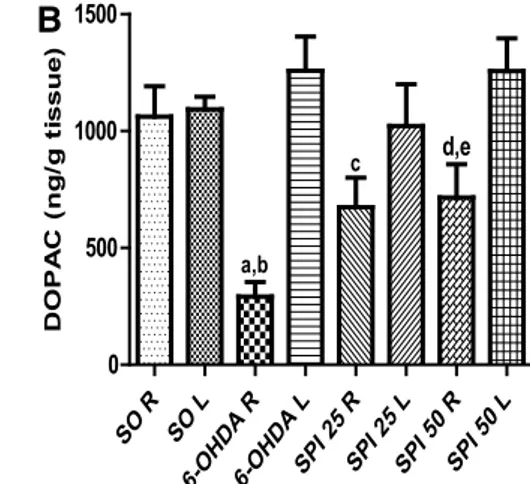
with SPI25 and SPI50 (comparatively to the ipsilateral side of the SO group or to the contralateral side of these two groups) (Fig. 2a). As far as DOPAC levels are concerned, the untreated 6-OHDA-lesioned group showed a 72% decrease, as related to the right side of the SO group, as well as to its own contralateral side. The 6-OHDA-lesioned group pre-sented smaller decreases after SPI treatments comparatively to the ipsilateral side of the untreated 6-OHDA-lesioned group and to its own contralateral side (Fig. 2b).
Nitrite Determinations and Lipid Peroxidation in the Rat Striata
The right (lesioned) striata from the untreated 6-OHDA-lesioned group presented a 3 times increase in nitrite con-tents, related to the same side of the SO group. On the other hand, values even lower than those showed by the SO group were demonstrated after SPI treatments, suggesting an anti-oxidant efect (Fig. 3a). In addition, a 2 times increase in MDA contents, related to the SO group, was observed in the right (lesioned) striata from the untreated 6-OHDA-lesioned group. This efect was completely reversed after SPI treat-ments, supporting the results seen in nitrite determination, above (Fig. 3b).
SO R SO L 6-O HD
A R
6-O HD
A L
SP I 25
R
SP I 25
L
SP I 50
R
SP I 50
L 0 1000 2000 3000 4000 5000 a,b c,d e,f DA (n g/ g ti ssue) SO R SO L 6-O HD
A R
6-O HD
A L
SP I 25
R
SP I 25
L
SP I 50
R
SP I 50
L 0 500 1000 1500 a,b c d,e DOPA C (n g/ g ti ssue )
A
B
Fig. 2 Spirulina (SPI, 25 and 50 mg/kg) treatments reverse partly
DA and DOPAC depletions of the untreated 6-OHDA-lesioned groups. DA: a vs SO R, q = 6.041, p < 0.01; b vs 6-OHDA L, q = 6.970, p < 0.001; c vs 6-OHDA R, t = 7.762, df = 9, p = 0.0101; d vs 6-OHDA + SPI 25 L, q = 6.068, p < 0.01; e vs 6-OHDA R, t = 3.524, df = 11, p = 0.0485; f vs 6-OHDA + SPI50 L, q = 7.441,
p < 0.001. DOPAC: a vs SO R, q = 5.099, p < 0.05; b vs 6-OHDA L, q = 6.610, p < 0.001; c vs 6-OHDA R, p = 0.0315, t = 2.544, df = 9; d vs 6-OHDA R, p = 0.0264, t = 2.715, df = 8; e vs 6-OHDA + SPI50 L, p = 0.034, t = 2.911, df = 5. R right side (lesioned), L left side (One-way ANOVA and Tukey as the post hoc test and paired or unpaired two-tailed Student’s t test)
Fig. 3 Spirulina (SPI)
treat-ments (50 mg/kg) partially reversed the increased nitrite contents (μM) and lipid peroxidation, expressed as MDA (μg/g tissue) observed in the striatal lesioned right side from the untreated 6-OHDA group. Nitrite: a vs SO, q = 6.137, p < 0.001; b vs 6-OHDA + SPI50, q = 6.299, p < 0.001. MDA: a vs SO, q = 6.381, p < 0.001; b vs 6-OHDA + SPI50, q = 6.816, p < 0.001 (One way ANOVA
and Tukey as the post hoc test) SO
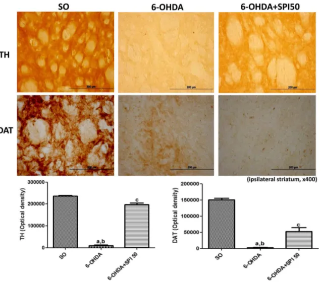
Immunohistochemistry for Tyrosine Hydroxylase (TH) and Dopamine Transporter (DAT) in Rat Striata
The immunohistochemical data for TH and DAT were quantiied by the Image J software. The results showed a 99% decrease in the TH immunoreactivity, on the lesioned striatal right side of the untreated 6-OHDA group related to the SO group. On the other hand, only a 42% decrease in TH immunostaining was observed after treatment of the 6-OHDA-lesioned group with SPI50. Similarly to the TH indings, a drastic decrease in DAT immunoreactivity (98%) was demonstrated in the lesioned right striatum of the untreated 6-OHDA-lesioned group. This reduction was partially blocked (55%) in the 6-OHDA-lesioned group after SPI treatment with the dose of 50 mg/kg (Fig. 4).
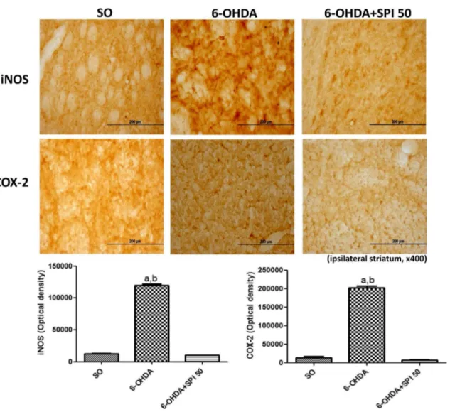
Immunohistochemistry for the Inducible Nitric Oxide Syntase (iNOS) and Cyclooxygenase-2 (COX-2) in Rat Striata
The immunohistochemical data for iNOS and COX-2 were quantiied by the Image J software. In the present study, while a 9 times increase was observed for iNOS immunore-activity in the untreated 6-OHDA-lesioned group striatum, this went down to values even lower than those of the SO group, after the treatment with SPI (50 mg/kg). In addition, a 15 times higher immunoreactivity for COX-2 was demon-strated in the untreated 6-OHDA-lesioned group. Similarly, these values went down after SPI50, thus the immunostain-ing intensity was lower than that presented by the SO group (Fig. 5).
Fig. 4 Representative photomicrographs (×400) of tyrosine
hydrox-ylase (TH) and dopamine transporter (DAT), showing a decreased immunoreactivity in the lesioned striatum from the untreated 6-OHDA group, what was highly attenuated after the Spirulina treat-ment. TH: a vs SO, q = 62.05, p < 0.001; b vs 6-OHDA + SPI50,
q = 26.13, p < 0.001; c vs SO, q = 35.92, p < 0.001. DAT: a vs SO, q = 23.47, p < 0.001; b vs 6-OHDA + SPI50, q = 7.525, p < 0.001; c vs SO, q = 14. 60, p < 0.001. The data were quantiied by the Image J software, NIH, USA (One-way ANOVA followed by Tukey as the
Discussion
Spirulina platensis is a rich source of protein, used as a nutritional supplement especially in low income countries. In the present study the Spirulina standardization shows 16.5 mg/g of total phenols and 45.5% of protein. This cyano-bacterium besides being a rich source of protein, is known to present many properties, including anti-inlammatory, antioxidant and neuroprotective ones, generally needed for the treatment of chronic neurodegenerative pathologies, as Parkinson’s disease.
Furthermore, the presence of neuroinlammation and mitochondrial dysfunction, common features of these neu-rologic conditions, can lead to increased oxidative stress by excessive release of reactive oxygen and nitrogen species. These last alterations will further promote neuronal damage
and subsequent inlammation, resulting in a feed-forward loop of neurodegeneration (15). In the present work, we evaluated the neuroprotective properties of S. platensis (SPI) in the hemiparkinsonism model, resulting from 6-OHDA striatal lesion.
In the present work, the potential neuroprotective activ-ity of SPI was supported by the decrease of apomorphine-induced rotational behavior, related to the untreated 6-OHDA-lesioned group. Rats with unilateral 6-OHDA lesions of the nigrostriatal DA pathway are known to exhibit a contralateral rotational behavior, when challenged with DA agonists, such as apomorphine. This rotational response to apomorphine is used as a behavioral index of DA receptor supersensitivity, after unilateral 6-OHDA lesion [19], as well as for evaluating the extent of unilateral nigros-triatal denervation [23].
Fig. 5 Representative photomicrographs (×400) of
immunohisto-chemistry for iNOS and COX-2, showing an increased immunore-activity in the lesioned striatum from the untreated 6-OHDA group, what was highly attenuated after the Spirulina treatment. iNOS: a vs SO, q = 100.4, p < 0.001; b vs 5-OHDA + SPI50, q = 103.0, p < 0.001.
COX-2: a vs. SO, q = 47.68, p < 0.001; b vs 6-OHDA + SPI50,
We showed that SPI partly recovered the DA and DOPAC depletions observed in the striatum of untreated 6-OHDA-lesioned animals. The most important neuropathological feature of Parkinson’s disease is the loss of dopaminergic neurons of the substantia nigra, leading to a severe depletion of striatal DA [24]. The striatum has a population of dopa-minergic neurons that act as a source of DA. This neuronal population increases in size, in animal models of Parkinson’s disease where striatal DA levels are low [25]. The dopa-minergic deiciency in Parkinson’s disease patients causes abnormalities of movement, behavior, learning and emotions [26]. Thus, SPI by partly reversing DA depletion would ofer some protection for PD patients.
SPI signiicantly reversed the increased nitrite contents and lipid peroxidation observed in the right (lesioned) stria-tum of the 6-OHDA group. It is largely accepted that oxida-tive stress, leading to ROS production, is present in neuro-degenerative diseases [27–30]. Previously, we demonstrated increased lipid peroxidation and nitrite levels in brains from AD patients [31]. These two parameters are good markers of oxidative stress and were blocked by SPI, indicating its potential as an antioxidant drug. Previously [32] the protean extract from S. platensis was shown to present a poweful antioxidant activity on SH-SY5Y neuroblastoma cells. Other results [33] observed similar efects on some cell lines, with the S. platensis water extract. In addition, a recent study [34] demonstrated that S. platensis reduced the kainic acid neuronal death in mice.
Furthermore, the striatal injection of 6-OHDA was shown [35] to lead to a massive disappearance of TH immunore-active terminals, in a deined area within the striatum sur-rounding the injection site. These authors also observed a disappearance of DA cell bodies, in a small region of the ipsilateral substantia nigra pars compacta and concluded that the striatal injection of 6-OHDA leads to retrograde degeneration, as well as glial activation in the nigostriatal dopamine pathway. TH is the rate-limiting enzyme for brain catecholamine synthesis; and reduction of TH expression results in diminished DA synthesis, leading to PD, what makes TH essential in the pathogenesis of PD [36]. Thus, PD can be considered as a TH-deiciency syndrome of the striatum [37]. We showed that SPI treatment of 6-OHDA-lesioned animals produced an almost complete recovery of the drastic decrease in TH immunoreactivity. Considering the great importance of TH activity in PD, a logical and eicient therapeutic strategy for its treatment could be based on correcting TH deiciency.
We also demonstrated a drastic decrease in DAT immu-noreactivity in the untreated 6-OHDA-lesioned animals, what was completely recovered after SPI treatment. The dopamine transporter (DAT) is a transmembrane protein, responsible for the reuptake of DA from the synaptic cleft and termination of dopaminergic transmission. Although
it is usually accepted that parkinsonian symptoms develop when approximately 70–80% of DA neurons are lost, imag-ing studies have shown that a loss around 50% of DA ter-minals is required for the onset of symptoms in PD patients [38]. There is a decline of DAT, as the disease progresses in an exponential process, that can be revealed by DAT imag-ing [39]. Thus, this parameter can be considered as a marker for diferentiating PD patients from healthy individuals [40].
Neuroinlammation may contribute to a variety of neu-rodegenerative diseases; and ROS are major regulators of this process, exerting a direct damaging efect on neurons. Nitric oxide is a highly reactive chemical messenger, play-ing an important role in mediatplay-ing physiological as well as pathological pathways [41]. The expression of iNOS is linked to the generation of ROS which afects various cel-lular components and preferentially damages midbrain dopa-minergic neurons in PD [42]. We showed a ninefold increase in the striatal immunoreactivity for iNOS in the untreated 6-OHDA-lesioned group, as related to the SO group. On the other hand, after SPI50 treatment, this value was even lower than that of the SO group.
An upregulation of inlammatory molecules, as iNOS and COX-2, has been suggested to play an important role in Parkinson’s disease, leading to dopaminergic neuron loss [43]. COX-2 is the enzyme responsible for the irst com-mitted step in the synthesis of several important mediators involved in the initiation and resolution of inlammation. The subsequent generation of PGs, upon activation of this inlammatory pathway, has been shown to participate in neurodegenerative processes, including those present in PD [44]. Furthermore, COX-2 is rapidly expressed in sev-eral cell types, in response to cytokines, growth factors and pro-inlammatory molecules [45]. In the present work, we demonstrated that the large increase in immunostaining for COX-2, in the striatum from the untreated 6-OHDA-lesioned group, was completely reversed after SPI treatments (50 mg/ kg).
A recent study showed the neuroprotective properties of some other Spirulina species (S. fusiform) on a 6-OHDA model of PD. Spirulina improved locomotor activity and oxidative stress, as well as DA contents, in the lesioned rats [46]. Another study [47] demonstrated the protective efect of Spirulina treatment on the locomotor function and mor-phological damage, in the injured spinal cord model in rats. Garbuzova-Davis et al., 2010 [48], showed that a Spirulina supplemented diet signiicantly maintained body weight and extension relex, and also reduced inlammatory markers and motor neuron degeneration, in a mouse model of amyo-trophic lateral sclerosis.
cells were observed in the substantia nigra. Spirulina also decreased the numbers of activated microglial cells. Although these indings corroborate with ours and point out to the Spirulina potential in neurodegeneration, the authors used a diferent protocol: irstly, the administration of a diet containing 0.1% Spirulina, for a 30-day period (pretreatment) and, secondly, continuing the treatment in the presence of synuclein for 2 weeks, 1 or 4 months.
Our results suggest that Spirulina is neuroprotective in the model of 6-OHDA-induced hemiparkinsonism. This natural product presents potent anti-inlammatory and antioxidant activities, as demonstrated in acute and chronic experimental models of inlammation [50–53].
Interestingly, Spirulina was recently shown to inhibit LPS-induced inflammation, through the repression of HDAC in macrophages [54]. HDACs enzymes are known to play a role in homeostasis of protein acetylation and to regulate cellular activities, such as transcription, and their dysfunction is associated with a wide variety of brain disorders [55]. Thus, HDAC inhibitors, besides exerting anti-inlammatory efects also present a potential as neuroprotective drugs [56–59]. Recently, we demon-strated that valproic acid, in the 6-OHDA model of PD, probably exerts a neuroprotective action, in part through HDAC inhibition [60]. Valproic acid has been reported to upregulate the expression of neurotrophic factors, what could contribute to its neuroprotective efects on DA neu-rons [61–64]. HDAC inhibition has been hypothesized as cooperating to neuroprotection, through gene expression regulation in glial cells [65]. Furthermore, we believe that Spirulina antioxidant and anti-inlammatory efects would certainly contribute to its neuroprotective actions, as shown in the present work, suggesting this drug as a potential candidate to be included in the clinic for PD treatment.
Acknowledgements The authors are grateful to the inancial
sup-port of the Foundation for Development of Research and Innovation of the State of Ceará, Brazil (FUNCAP) and to the Brazilian National Research Council (CNPq). They thank Prof. M.O.L. Viana for the orthographic revision of the manuscript.
Author Contributions FAVL carried most of the behavioral and
neurochemical experiments; IPJ and FPJ were responsible for the cul-tivation of S. platensis; KRTN and MRC carried out all immunohisto-chemical assays; GMA performed the statistical analyses and GSBV coordinated and wrote the work. All authors read and approved the manuscript.
Compliance with Ethical Standards
Conlict of interest The authors declare no conlict of interests.
References
1. Belay A, Ota Y, Miyakawa K, Shimamatsu H (1993) Current knowledge on potential health beneits of Spirulina. J Appl Phy-col 5:235–241
2. Vonshak A (1997) Spirulina platensis (Arthrospira): physiology, cell biology and biotechnology. Taylor and Francis, London 3. Patro N, Sharma A, Kariaya K, Patro I (2011) Spirulina
plat-ensis protects neurons via suppression of glial activation and peripheral sensitization leading to restoration of motor function in collagen-induced arthritic rats. Indian J Exp Biol 49:739–748 4. Ismail M, Hossain MF, Tanu AR, Shekhar HU (2015). Efect
of Spirulina intervention on oxidative stress, antioxidant sta-tus, and lipid proile in chronic obstructive pulmonary disease patients. Biomed Res Int 2015:486120
5. Weintraub D (2008) Dopamine and impulse control disorders in Parkinson’s disease. Ann Neurol 64(Suppl 2):S93–S100 6. Herrero M-T, Estrada C, Maatouk L, Vyas S (2015)
Inlam-mation in Parkinson’s disease: role of glucocorticoids. Front Neuroanat 9:32. doi: 10.3389/fnana.2015.00032
7. Mogi M, Harada M, Riederer P, Narabyashi H, Fujita J, Nagatsu T (1994) Interleukin-1 beta growth factor and transforming growth factor-alpha are elevated in the brain from Parkinsonian patients. Neurosci Lett 180:147–150
8. Dobbs RJ, Charlett A, Purkiss AG, Dobbs SM, Weller C, Peter-son DW (1999) Association of circulating TNF-alpha and IL-6 with ageing and Parkinsonism. Acta Neurol Scand 100:34–41 9. Reale M, Greig NH, Kamal MA (2009) Peripheral
chemo-cytokine proiles in Alzheimer’s and Parkinson’s diseases. Mini Rev Med Chem 9:1229–1241
10. Reale M, Iarlori C, Thomas A, Gambi D, Perfetti B, Di Nicola M, Onofrj M (2009) Peripheral cytokines proile in Parkinson’s disease. Brain Behav Immun 23:55–63
11. McGeer PL, McGeer EG (2008) The alpha-synuclein burden hypothesis of Parkinson disease and its relationship to Alzhei-mer disease. Exp Neurol 212:235–238
12. Pott Godoy MC, Tarelli R, Ferrari CC, Sarchi MI, Pitossi FJ (2008) Central and systemic IL-1 exacerbates neurodegenera-tion and motor symptoms in a model of Parkinson’s disease. Brain 131:1880–1894
13. Gandhi S, Abramov AY (2012). Mechanism of oxidative stress in neurodegeneration. Oxidat Med Cell Longev 2012:11.
doi:10.1155/2012/428010
14. Lin MT, Beal MF (2006) Mitochondrial dysfunction and oxida-tive stress in neurodegeneraoxida-tive diseases. Nature 443:787–795 15. Fischer R, Maier O (2015). Interrelation of oxidative stress and
inlammation in neurodegenerative disease: role of TNF. Oxidat Med Cell Longev 2015:18. doi:10.1155/2015/610813
16. Jackson-Lewis V, Blesa J, Przedborski S (2012) Animal mod-els of Parkinson’s disease. Parkinsonism Relat Disord 18(Suppl 1):S183–S185
17. Simola N, Morelli M, Carta AR (2007) The 6-hydroxydopamine model of Parkinson’s disease. Neurotox Res 11:151–167 18. Tieu K (2011) A guide to neurotoxic animal models of
Parkin-son’s disease. Cold Spring Harb Perspect Med 1:a009316 19. Waddington JL, Cross AJ, Longden A, Owen F, Poulter
M (1979) Apomorphine-induced rotation in the unilateral 6-OHDA-lesioned rat: relationship to changes in striatal ade-nylate cyclase activity and 3H-spiperone binding. Neurophar-macology 18:643–645
21. Green LC, Wagner DA, Glogowski J, Skipper PL, Wishnok JS, Tannenbaum SR (1982) Analysis of nitrate, nitrite, and [15N] nitrate in biological luids. Anal Biochem 126(1):131–138 22. Draper HH, Hadley M (1990) Malondialdehyde determination as
index of lipid peroxidation. Methods Enzymol 186:421–431 23. Hudson JL, van Horne CG, Stromberg I, Brock S, Clayton J,
Masserano J, Hofer BJ, Gerhardt GA (1993) Correlation of apo-morphine- and amphetamine-induced turning with nigrostriatal dopamine content in unilateral 6-hydroxydopamine lesioned rats. Brain Res 626:167–174
24. Hornykiewicz O, Kish SJ (1986) Biochemical pathophysiology of Parkinson’s disease. Adv Neurol 45:19–34
25. Huot P, Lévesque M, Parent A (2007) The fate of striatal dopa-minergic neurons in Parkinson’s disease and Huntington’s chorea. Brain 130(Pt 1):222–232
26. Rodriguez-Oroz MC, Jahanshahi M, Krack P, Litvan I, Macias R, Bezard E, Obeso JA (2009) Initial clinical manifestations of Parkinson’s disease: features and pathophysiological mechanisms. Lancet Neurol 8:1128–1139
27. Chen X, Guo C, Kong J (2012) Oxidative stress in neurodegenera-tive diseases. Neural Regen Res 7(5):376–385
28. Kim GH, Kim JE, Rhie SJ, Yoon S (2015) The role of oxida-tive stress in neurodegeneraoxida-tive diseases. Exp Neurobiol 24(4):325–340
29. Sultana R, Perluigi M, Butterield DA (2013) Lipid peroxidation triggers neurodegeneration: a redox proteomics view into the Alz-heimer disease brain. Free Radic Biol Med 62:157–169
30. Uttara B, Singh AV, Zamboni P, Mahajan RT (2009) Oxidative stress and neurodegenerative diseases: a review of upstream and downstream antioxidant therapeutic options. Curr Neuropharma-col 7:65–74
31. DiCiero MM, de Bruin VM, Viana GS (2000) Lipid peroxidation and nitrite plus nitrate levels in brain tissues from patients with Alzheimer’s disease. Gerontology 46(4):179–184
32. Bermejo-Bescos P, Pinero-Estrada E, Villar del Fresno AM (2008) Neuroprotection by Spirulina platensis protean extract and phyco-cyanin against iron-induced toxicity in SH-SY5Y neuroblastoma cells. Toxicol In Vitro 22:1496–1502
33. Zaid AAA, Hammad DM, Sharaf EM (2015) Antioxidant and abticancer activity of Spirulina platensis water extracts. Int J Phar-macol 11:846–851
34. Pérez-Juárez A, Chamorro G, Alva-Sánchez C, Paniaqua-Castro N, Pacheco-Rosado J (2016) Neuroprotective efect of arthrospira (Spirulina)-platensis against kainic acid-neuronal death. Pharm Biol 54(8):1408–1412
35. Rodrigues RWP, Gomide VC, Chadi G (2003) Striatal injection of 6-hydroxydopamine induces retrograde degeneration and glial activation in the nigrostriatal pathway. Acta Cirúrgica Brasileira 18:272–282
36. Zhu Yanzhen Zhang, Jing Zeng Yanjun (2012) Overview of tyros-ine hydroxylase in Parkinson’s disease. CNS Neurol Disord Drug Targets 11:350–358
37. Tabrez S, Jabir NR, Shakil S, Greig NH, Alam Q, Abuzenadah AM, Damanhouri GA, Kamal MA (2012) A synopsis on the role of tyrosine hydroxylase in Parkinson’s disease. CNS Neurol Dis-ord Drug Targets 11:395–409
38. Varrone A, Halldin C (2010) Molecular imaging of the dopamine transporter. J Nucl Med 51:1331–1334
39. Antonini A, Biundo R (2014) Parkinson disease: can dopamine transporter imaging deine early PD? Nat Rev Neurol 10:432–433 40. Bor-Seng-Shu E, Felicio AC, Braga-Neto P, Batista IS, Paiva WS,
Andrade DC, Teixeira MJ, Andrade LAF, Barsottini OGP, Shih MC, Bressan RA, Ferraz HB (2014) Dopamine transporter imag-ing usimag-ing 99mTc-TRODAT-1 SPECT in Parkinson’s disease. Med Sci Monit 20:1413–1418
41. Kavya R, Dikshit M (2015) Role of nitric oxide/nitric oxide syn-thase in Parkinson’s disease. Ann Neurosci (India) 12:1–5 42. Koppula S, Kumar H, Kim IS, Choi D-K (2012). Reactive oxygen
species and inhibitors of inlammatory enzymes, NADPH oxidase, and iNOS in experimental models of Parkinson’s disease. Med Inlamm 2012, Article ID 823902
43. Okuno T, Nakatsuji Y, Kumanogoh A, Moriya M, Ichinose H, Sumi H, Fujimura H, Kikutani H, Sakoda S (2005) Loss of dopa-minergic neurons by the induction of inducible nitric oxide syn-thase and cyclooxygenase-2 via CD 40: relevance to Parkinson’s disease. J Neurosci Res 81(6):874–882
44. Teismann P (2012) COX-2 in the neurodegenerative process of Parkinson’s disease. Int Un Biochem Mol Biol Inc 38:395–397 45. Bartels AL, Leenders KL (2010) Cyclooxygenase and
neuroin-lammation in Parkinson’s disease neurodegeneration. Curr Neu-ropharmacol 8:62–68
46. Chattopadhyaya I, Gupta S, Mohammed A, Mushtag N, Chau-han S, Ghosh S (2015) Neuroprotective effect of Spirulina fusiform and amantadine in the 6-OHDA induced parkinson-ism in rats. BMC Complem Altern Med 15:296. doi:10.1186/ s12906-015-0815-0
47. Aziz I, Ramli MDC, Zain NSM, Sanusi J (2014). Behavioral and histopathological study of changes in spinal cord injured rats sup-plemented with Spirulina platensis. Evid-Bas Complem Altern Med 2014:8. doi:10.1155/2014/871657
48. Garbuzova-Davis S, Bickford PC (2010) Neuroprotective efect of Spirulina in a mouse model of ALS. Open Tissue Eng Regen Med J 3:36–41
49. Pabon MM, Jernberg JN, Morganti J, Contreras J, Hudsen CE, Klein RL, Bickford PC (2012) A Spirulina-enhanced diet provides neuroprotection in an alpha-synuclein model of Parkinson’s dis-ease. PLoS ONE 7(9):e45256
50. Pham TX, Kim B, Lee J (2013) Spirulina platensis inhibits lipopolysaccharide-induced inlammation through the repression of histone deacetylases in RAW 264.7 macrophages. FASEB 27:247.1
51. Yogianti F, Kunisada M, Nakano E, Ono R, Sakumi K, Oka S, Nakabeppu Y, Nishigori C (2014) Inhibitory efects of dietary
Spirulina platensis on UVB-induced skin inlammatory responses and carcinogenesis. J Investig Dermatol 134:2610–2619 52. Muga MA, Chao Jane C-J (2014) Efects of ish oil and
Spir-ulina on oxidative stress and inflammation in hypercholes-terolemic hamsters. BMC Complement Altern Med 14:470.
doi:10.1186/1472-6882-14-470
53. Gutiérrez-Rebolledo GA, Galar-Martínez M, García-Rodríguez RV, Chamorro-Cevallos GA, Hernández-Reyes AG, Martínez-Galero E (2015) Antioxidant efect of Spirulina (Arthrospira) maxima on chronic inlammation induced by Freund’s complete adjuvant in rats. J Med Food 18:865–871
54. Chuang D-M, Leng Y, Marinova Z, Kim H-J, Chiu C-T (2009) Multiple roles of HDAC inhibition in neurodegenerative condi-tions. Trends Neurosci 32:591–601
55. Halili MA, Andrews MR, Sweet MJ, Fairlie DP (2009) Histone deacetylase inhibitors in inlammatory disease. Curr Top Med Chem 9:309–319
56. Grabiec AM, Tak PP, Reedquist KA (2011) Function of his-tone deacetylase inhibitors in inlammation. Crit Rev Immunol 31:233–263
57. Lu J, Frerich JM, Turtzo LC, Li S, Chiang J, Yang C, Wang X, Zhang C, Wu C, Sun Z, Niu G, Zhuang Z, Brady RO, Chen X (2013) Histone deacetylase inhibitors are neuroprotective and preserve NGF-mediated cell survival following traumatic brain injury. Proc Natl Acad Sci USA 110:47–52
59. Kim HJ, Rowe M, Ren M, Hong JS, Chen PS, Chuang DM (2007) Histone deacetylase inhibitors exhibit anti-inlammatory and neuroprotective efects in a rat permanent ischemic model of stroke: multiple mechanisms of action. J Pharmacol Exp Ther 321:892–901
60. Ximenes JC, Neves KR, Leal LK, do Carmo MR, Brito GA, Naffah-Mazzacoratti MG, Cavalheiro EA, Viana GS (2015). Valproic acid neuroprotection in the 6-OHDA model of Par-kinson’s disease is possibly related to its anti-inlammatory and HDAC inhibitory properties.J Neurodegener Dis 2015:313702.
doi:10.1155/2015/313702
61. Chen P-S, Peng G-S, Li G, Yang S, Wu X, Wang C-C, Wilson B, Lu R-B, Gean P-W, Chuang D-M, Hong J-S (2006) Valproate protects dopaminergic neurons in midbrain neuron/glia cultures by stimulating the release of neurotrophic factors from astrocytes. Mol Psychiatry 11:1116–1125
62. Chen P-S, Wang C-C, Bortner CD, Peng G-S, Wu X, Pang H, Lu R-B, Gean P-W, Chuang D-M, Hong J-S (2007) Valproic acid and other HDAC inhibitors induce microglial apoptosis and attenuate lipopolysaccharide-induced dopaminergic neurotoxicity. Neurosci 149:203–212
63. Leng Y, Wang J, Wang Z, Liao H-M, Wei M, Leeds P, Chuang D-M (2016) Valproic acid and other HDAC inhibitors upregu-late FGF21 gene expression and promote process elongation in glia by inhibiting HDAC2 and 3.. Int J Neuropsychopharmacol 19(8):pyw035
64. Chiu C-T, Wang Z, Hubsberger JG, Chuang D-M (2013) Thera-peutic potential of mood stabilizers lithium and valproic acid: beyond bipolar disorder. Pharmacol Rev 61(1):105–142 65. Wu X, Li S, Wu Q, Peng Y, Yu D, Wang H, Chui D, Zhao J (2013)