139 Kaposi sarcoma related to AIDS
Radiol Bras. 2008 Mar/Abr;41(2):139–140 Case Report • Relato de Caso
Kaposi sarcoma related to acquired immunodeficiency
syndrome: hepatic findings on computed tomography
and magnetic resonance imaging*
Sarcoma de Kaposi relacionado à síndrome da imunodeficiência adquirida: características do comprometimento hepático na tomografia computadorizada e na ressonância magnética
Daniel Nobrega da Costa1, Publio Cesar Cavalcante Viana1, Rosângela Pereira Maciel2, Eloisa Maria Mello Santiago Gebrim3, Manoel de Souza Rocha2
Kaposi sarcoma is a neoplasm associated with immunosuppressive conditions, and involving blood and lymphatic vessels. It is the most frequent intrahepatic neoplasm in patients with acquired immunodeficiency syndrome. Computed tomography and magnetic resonance imaging demonstrate multiple small nodules, prominence and contrast-enhancement of periportal branches due to the presence of the neoplastic tissue. The authors report a case of a 47-year-old male patient with acquired immunodeficiency syndrome presenting disseminated Kaposi sarcoma.
Keywords: Kaposi sarcoma; Acquired immunodeficiency syndrome; Liver disease; Cancer; Computed tomog-raphy; Magnetic resonance imaging.
Sarcoma de Kaposi é uma neoplasia associada a condições de imunossupressão que acomete os vasos lin-fáticos e sanguíneos. É a neoplasia intra-hepática mais comum na síndrome da imunodeficiência adquirida. A tomografia computadorizada e a ressonância magnética revelam múltiplos pequenos nódulos, proeminên-cia e realce dos planos periportais, devido à presença de tecido neoplásico. Os autores descrevem um caso de paciente masculino, de 47 anos de idade, com síndrome da imunodeficiência adquirida e sarcoma de Kaposi disseminado.
Unitermos: Sarcoma de Kaposi; Síndrome da imunodeficiência adquirida; Doença hepática; Câncer; Tomo-grafia computadorizada; Imagem por ressonância magnética.
Abstract
Resumo
* Study developed at Instituto de Radiologia do Hospital das Clínicas da Faculdade de Medicina da Universidade de São Paulo (InRad/HC-FMUSP), São Paulo, SP, Brazil.
1. MDs, Residents in Radiology and Imaging Diagnosis at Ins-tituto de Radiologia do Hospital das Clínicas da Faculdade de Medicina da Universidade de São Paulo (InRad/HC-FMUSP), São Paulo, SP, Brazil.
2. PhDs, MDs, Radiologist Assistants at Instituto de Radiologia do Hospital das Clínicas da Faculdade de Medicina da Universi-dade de São Paulo (InRad/HC-FMUSP), São Paulo, SP, Brazil.
3. PhD, MD, Radiologist Assistant Responsible for the Division of Computed Tomography at Instituto de Radiologia do Hospital das Clínicas da Faculdade de Medicina da Universidade de São Paulo (InRad/HC-FMUSP), São Paulo, SP, Brazil.
Mailing address: Dr. Daniel Nóbrega da Costa. Rua Dona Adma Jafet, 91, Bela Vista. São Paulo, SP, Brasil, 05411-001. E-mail: [email protected]
Received November 22, 2006. Accepted after revision Janu-ary 31, 2007.
DISCUSSION
KS is a low-grade mesenchymal neo-plasm initially described by Moritz Kaposi in 1872. Generally, this disease affects blood and lymphatic vessels, most fre-quently on the skin, although frefre-quently may be found in other organs.
Presently four variants of the disease are found with different clinical presentations: 1) classic (sporadic or Mediterranean) KS; 2) endemic (African) KS; 3) iatrogenic (or-gan transplant-related) KS; 4) AIDS-re-lated KS.
For decades before the AIDS breakout and the increase in the number of organ transplants, KS used to be viewed as a very rare disease. Although its etiology is still to be fully understood, the AIDS- and trans-plant-related forms of the disease are clearly associated with immunosuppres-sion or immunodeficiency. Infection by the
Costa DN, Viana PCC, Maciel RP, Gebrim EMMS, Rocha MS. Kaposi sarcoma related to acquired immunodeficiency syn-drome: hepatic findings on computed tomography and magnetic resonance imaging. Radiol Bras. 2008;41(2):139–140.
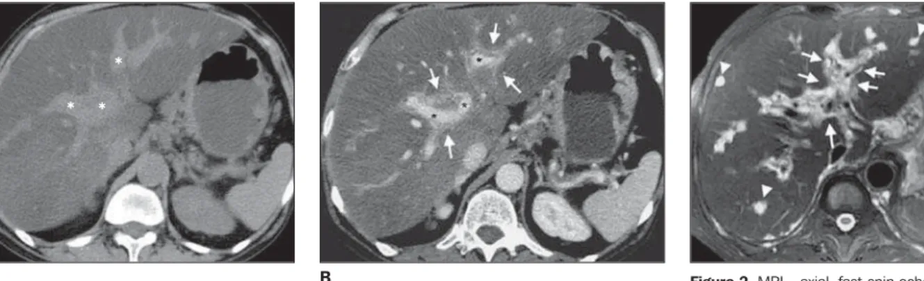
demonstrated nodules and disseminated violaceous plaques on the skin, predomi-nantly in the lower limbs. Based on this clinical context, the diagnosis of AIDS-re-lated Kaposi sarcoma (KS) has been sug-gested and later confirmed by skin biopsy. The patient underwent upper digestive endoscopy and colonoscopy to investigate the melena, and gastroesophageal involve-ment was detected. Additionally, chest/ab-dominal computed tomography (CT) and magnetic resonance imaging (MRI) have been requested to rule out visceral involve-ment. Chest CT has demonstrated pro-nounced hepatic steatosis and prominence of periportal spaces (Figure 1A), with exu-berant periportal contrast-enhancement (Figure 1B). Abdominal MRI has clearly demonstrated the presence of tissue sur-rounding the portal branches (arrows on the Figure 2) and the overall nodular involve-ment of the hepatic parenchyma.
0100-3984 © Colégio Brasileiro de Radiologia e Diagnóstico por Imagem CASE REPORT
140
Costa DN et al.
Radiol Bras. 2008 Mar/Abr;41(2):139–140 human herpes virus 8 – HHV8 and other
factors have been implied in the develop-ment of the disease. The HHV8 transmis-sion mechanism also is still to be under-stood, but epidemiological studies indicate the male-to-male sexual contact as an im-portant transmission route to be consid-ered(1). Generally, the disease affects indi-viduals with low CD4 lymphocytes counts (< 150–200 cell/mm³). Most frequent sites of visceral involvement in AIDS-related KS are lymphonodi (72% of cases), lungs (51%), gastrointestinal tract (48%), liver (34%), and spleen (27%)(2).
AIDS-related KS is the most frequent hepatic neoplasm in AIDS patients. Cuta-neous involvement is present in the major-ity of cases and represents a useful infor-mation in the diagnostic approach in cases where other hepatic conditions must be taken into consideration in the differential diagnosis. It is important to note that, in these cases, no specific treatment is re-quired, and the condition alone does not affect the clinical progression of the
pa-tients(1). Macroscopically, multiple brown-violaceous, spongiform nodules are ob-served, diffusely scattered over the peripor-tal conjunctive tissue(2).
Ultrasonography demonstrates multiple hyperechogenic bands and nodules adja-cent to the portal branches, besides the heterogeneous texture of the hepatic paren-chyma(2,3). CT demonstrates multiple, small nodules diffusely distributed throughout the liver or predominantly found adjacent to the periportal branches, prominence and contrast-enhancement of periportal and hilar planes secondary to the presence of neoplastic tissue(3,4).
In cases of predominantly nodular he-patic presentation of KS, metastatic disease, fungal microabscesses and multiple he-mangiomas should be taken into consider-ation as differential diagnoses(3,4). The exu-berant periportal hepatic contrast-enhance-ment identified at CT and MRI also can be found in schistosomal patients with peri-portal metastatic infiltration and in cases of diffuse, infiltrating cholangiocarcinoma(4).
Although the hepatic involvement re-sulting from AIDS-related KS is rarely di-agnosed or symptomatic, infrequently con-tributing to the morbimortality in these patients, it is important to recognize and differentiate this condition from other he-patic diseases.
Considering that percutaneous biopsies may result in massive hemorrhage and he-moperitoneum, the radiologist must be fa-miliarized with the diagnosis to assist in avoiding this complication.
REFERENCES
1. Tappero JW, Conant MA, Wolfe SF, et al. Kaposi’s sarcoma. Epidemiology, pathogenesis, histology, clinical spectrum, staging criteria and therapy. J Am Acad Dermatol. 1993;28:371–95.
2. Restrepo CS, Martinez S, Lemos JA, et al. Imaging manifestations of Kaposi sarcoma. Radiographics. 2006;26:1169–85.
3. Luburich P, Bru C, Ayuso MC, et al. Hepatic Kaposi sarcoma in AIDS: US and CT findings. Radiology. 1990;175:172–4.
4. Hammerman AM, Kotner LM Jr, Doyle TB. Peri-portal contrast enhancement on CT scans of the liver. AJR Am J Roentgenol. 1991;156:313–5. Figure 2. MRI – axial, fast-spin echo, T2-weighted image better demonstrates the tissue character-ized by hyperintense signal surrounding the peri-portal planes (arrows), and manifesting as nod-ules in some regions (arrowheads).
Figure 1. CT – axial, pre-contrast- (A) and post-contrast-enhancement (B) images demonstrating promi-nence of periportal branches (asterisks on A) and contrast-enhancement of this region (arrows on B). In addition, a pronounced hypoattenuation of the hepatic parenchyma compatible with steatosis is ob-served, probably secondary to chronic disease and/or drugs use.
A B