LFBT 0890-5436 1532-4249
Food Biotechnology, Vol. 23, No. 1, January 2009: pp. 1–38 Food Biotechnology
Partial Characterization of Nine
Bacteriocins Produced by
Lactic Acid Bacteria Isolated
from Cold-Smoked Salmon
with Activity against Listeria
monocytogenes
LAB’s Bacteriocins CharacterizationE. Tomé et al.
Elisabetta Tomé
1,2, Svetoslav D. Todorov
3, Paul A. Gibbs
1,4,
and Paula C. Teixeira
11Escola Superior de Biotecnologia, Universidade Católica Portuguesa, Porto, Portugal 2Instituto de Ciencias y Tecnología de Alimentos, Escuela de Biología, Universidad
Central de Venezuela, Caracas, Venezuela
3Department of Microbiology, Stellenbosch University, Stellenbosch, South Africa 4Leatherhead Food International, Surrey, UK
Nine LAB bacteriocin-producers, isolated from vacuum-packaged cold-smoked salmon (CSS), were phenotypically and genotypically identified as Lactobacillus curvatus,
Lactobacillus delbrueckii, Lactobacillus fermentum, Enterococcus faecium, and Pedio-coccus acidilactici. Their bacteriocins were partially characterized. The antimicrobial
spectrum was determined against Listeria monocytogenes, E. faecalis, E. faecium, and
Staphylococcus aureus. The molecular size of bacteriocins ranged from 2.8 to 4.5 kDa.
They were inactivated by treatment with proteolytic enzymes but not by lipolytic or glycolytic enzymes. Maximal activity against L. monocytogenes ranged between 800 and 10000 AU/mL at pH 6.5. Most of the bacteriocins maintained full activity in a pH range of 2.0 to 8.0 but were partially or completely inactivated at pH 10.0. After heating at 60°C and 100°C, only two bacteriocins from Lb. curvatus strains partially lost activity. All bacteriocins showed a narrow spectrum of activity and a high anti-listerial activity, which is characteristic of the class IIa bacteriocins. Isolated bacteriocin-producing LAB could be used successfully in the bio-preservation of CSS and develop-ment of new potential bio-preservatives for CSS active against L. monocytogenes.
Address correspondence to Prof. Paula C. Teixeira, Escola Superior de Biotecnologia, Universidade Católica Portuguesa, R. Dr. António Bernardino de Almeida, 4200-072 Porto, Portugal; Tel.: +351 22 5580001; Fax: +351 22 5090351; E-mail: pcteixeira@ esb.ucp.pt
Key Words: cold smoked fish; bacteriocins; lactic acid bacteria; salmon; Listeria monocytogenes
INTRODUCTION
The increasing consumer demand for natural food additives has focused interest on bacteriocins. They are ribosomally synthesized peptides of 30 to less than 60 amino acids, with a narrow to wide antibacterial spectrum against Gram-positive bacteria (Savadogo et al., 2006). The antibacterial compound is heat stable, and a producer strain displays a degree of specific self-protection against its own antibacterial peptide. Bacteriocins of lactic acid bacteria (LAB) are con-sidered biopreservatives, as it is assumed that bacteriocins are degraded by the proteases of the gastrointestinal tract and most of the LAB are considered as GRAS (Generally Recognized as Safe) microorganisms (Holzapfel et al., 1995).
Although by definition all bacteriocins have a protein or peptide compo-nent that is essential for their bactericidal function, some have been reported to consist of combinations of different proteins or are composites of proteins together with lipid or carbohydrate moieties (Jimenez-Diaz et al., 1993). Improved protein purification protocols have shown that some bacteriocins previously considered high-molecular-weight protein aggregates may be small peptides that, because of their highly hydrophobic nature, had previously copurified with some other cellular components (Sahl, 1994). Although some Gram-positive bacteria have been shown to synthesize relatively high-molecular-weight, heat-labile bacteriocin-like substances (Vaughan et al., 1994), most of those described to date have been small, heat-stable cationic peptides.
A wide variety of bacterial products of Gram-positive bacteria have been referred to as bacteriocins, and various attempts have been made to classify these agents. Klaenhammer (1993) defined four distinct classes of lactic acid bacterial bacteriocins:
1. Class I, lantibiotics, are small (<5 kDa) peptides containing the unusual
amino acids lanthionine (Lan), -methyllanthionine (MeLan), dehydroalanine, and dehydrobutyrine;
2. Class II, small (<10-kDa), relatively heat-stable, non-lanthionine-containing
membrane-active peptides, subdivided into Listeria-active peptides with the N-terminal consensus sequence -Tyr-Gly-Asn-Gly-Val-Xaa-Cys- (Class IIa), poration complexes requiring two different peptides for activity (Class IIb), and thiol-activated peptides requiring reduced cysteine residues for activity (Class IIc);
3. Class III, large (>30-kDa), heat-labile proteins; and
4. Class IV, complex bacteriocins that contain essential lipid or carbohydrate
In the last decade many bacteriocins from LAB belonging to different groups have been characterized and purified, including nisin, diplococcin, acidophilin, bulgarican, helveticins, lactacins, sacakins, pediocin PA-1, and plantaricins (Nettles and Barefoot, 1993). Of these, nisin produced by
Lacto-coccus lactis subsp. lactis, has been the most extensively characterized
(Moreno et al., 2000). At present, nisin is the only bacteriocin commercially available and marketed because it has been conferred GRAS status by the Food and Drug Administration (FDA, 1988; FAO/WHO, 2006).
Only bacteriocins well characterized could be considered as potential natural food preservatives (De Vuyst and Vandamme, 1994). Therefore, the aim of this study was to isolate and identify potential bacteriocin producing LAB from CSS and to characterize the inhibitory activities of these bacteriocins.
MATERIAL AND METHODS
Fish Source
Fresh-gutted farmed salmon from Norway (Salmo salar) were acquired at Matosinhos’ Doca (Porto, Portugal). Salmon arrived by lorry (72 h travel) in a chilled container with the temperature controlled between 0°C and 4°C inside polystyrene boxes (two layers of fish between two layers of ice). The fish were transported to the ESB/UCP, under chilled conditions, and submitted to a cold-smoking process (filleted, salted by brining or dry salt method, rinsed, smoked and vacuum-packaged).
Smoking Process
During salting, fillets were placed in a chilled chamber at 5°C. For dry-salting, the salt and sugar added corresponded to one third of the weight of fillet. For brining, 80% of saturated NaCl solution was used (brine/fish: 1/1). Draining was done overnight at 5°C. Smoking was done by two different pro-cesses. The first process consisted of drying for 2 h in the smoke chamber but without smoke at less than 30°C, followed by smoking for 6 h at less than 30°C. The second process involved drying for 6 h in the smoke chamber but without smoke at less than 30°C and smoking for 2 h. According to Tomé et al. (2007), these drying/smoking conditions enhance growth of total LAB and lactobacilli during the storage period in these products.
Two batches were processed for each smoking process. Each batch consisted of three salmon and three fillets processed in each smoking process.
The smoked samples were cooled overnight at 5°C. The following day, lug and pin bones and belly flaps were removed and the fillets were then sliced and vacuum packed in a Multivac-Gastrovac (Multivac Sepp Haggenmüller KG, A300/41/42, Germany) 1 mbar/10 s. The permeability of the packs to O2,
CO2 and N2 were 4 mol/m2.d.bar., 13 mol/m2.d.bar, and 4 mol/m2.d.bar, respec-tively (Vaz-Velho, 2000). Packs were stored for 3 weeks at 5°C and analyzed at the beginning (t0) and at the end of the storage period (t1).
Isolation and Phenotypic Characterization of Microorganisms
At the beginning of storage, t0, and after three weeks, t1, fillets from each smoking process were cut into small pieces and mixed. Ten grams of this mix were picked randomly and homogenized in 90 mL of sterile ¼ -strength Ringer’s solution (Lab M, Bury, UK) for 2 min in a Stomacher 400 Lab Blender (Seward Medical, London, UK). Serial decimal dilutions in ¼ -strength Ringer’s solution were prepared. Three independent samples per batch/smoking pro-cess were analyzed at each time interval. Total LAB were enumerated by pour-plating in nitrite actidione polymyxin (NAP) agar, pH 6.7 (Davidson and Cronin, 1973). All colonies were counted as presumptive LAB after 5 days of anaerobic incubation at 25°C. At t0 and t1, 10% of colonies overall were picked randomly from NAP plates containing 10–100 colonies. Presumptive LAB were subcultured on NAP agar without selective agents or on All-Purpose Tween (APT, Difco Laboratories, Detroit, Mich., USA) agar, examined for purity and characterized using Gram stain, cytochrome oxidase, and catalase tests. Organisms that were Gram-positive, cytochrome oxidase negative, and catalase negative were stored in APT broth with glycerol (30% v/v) at −80°C until further use.
Anti-listerial Activity of LAB, Nature of the Inhibition
and Titre Determination
The antimicrobial activity of 614 presumptive LAB colonies picked from NAP plates, previously isolated from vacuum-packaged CSS was investigated against L. monocytogenes 54 (culture collection from ESB/UCP, Porto, Portugal) and L. innocua 2030c (Central Public Health Laboratory, Colindale, London, UK), a tetracycline resistant strain, using the spot method described by Tomé et al. (2006). Then, a 48 h-old culture in APT broth at 25°C was made of each LAB culture showing defined inhibition zones, ≥4 mm zone width. Cultures were adjusted to pH 6.5 with NaOH (1N), and cells were collected by centrifugation (7500 x g, 10 min, 4°C) followed by filtration of the supernatant through a 0.22 μm pore size membrane filter (Millipore Co., Bedford, Mass., USA). This solution was designated as a crude filtrate supernatant fluid (CFSF). The nature of the inhibition was assessed by treating the CFSF with the enzymes catalase and trypsin (both from Sigma–Aldrich Chemie GmbH, Steinheim, Germany) sepa-rately at a final concentration of 500 IU/mL and 0.1 mg/mL, respectively, for 2 h at 37°C. Cell-free supernatant (CFSF), CFSF treated with catalase and trypsin were spotted against L. monocytogenes 54 and L. innocua 2030c. Untreated samples were used as positive controls while enzyme solutions were used as
negative controls. The anti-listerial titre of each CFSF was determined against
L. monocytogenes 54 by the serial twofold dilution assay. It was defined as the
reciprocal of the highest dilution showing a distinct inhibition of the target strain and expressed in terms of arbitrary units per milliliter (AU/mL).
Strain Identification
The carbohydrate fermentation pattern of 9 bacteriocin-producing LAB strains was determined by using the API 50 CHL kit (BioMérieux, Mercy-l’Etoile, France). Coccoid strains, were submitted to some characteristic tests for
Enterococcus spp. for example, the ability to grow at 10°C and 45°C in
Tryptone Soy Broth (TSB, Lab M), growth at pH 9.0 and growth in TSB in the presence of 6.5% (w/v) NaCl at 30°C. The Vitek System (BioMérieux) was used for species identification of those strains which were able to grow at 10°C and 45°C and in the presence of 6.5% (w/v) of NaCl, using the Gram-Positive Identification Cards (GPI) according to the manufacturer’s instructions.
Identification of all nine strains was genotypically confirmed by using genus/species specific PCR amplification. Each strain was cultured overnight in de Man, Rogosa, and Sharpe (MRS) broth (Merck, Darmstadt, Germany) supplemented with 0.4% DL-threonine (Sigma) at 30°C, and then the DNA was extracted according to Dellaglio et al. (1973).
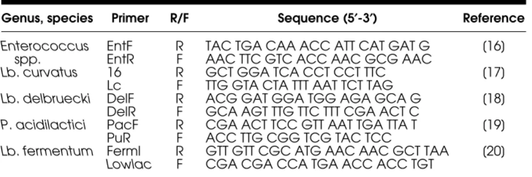
Strains were further identified using specific-species primers (Table 1). The PCR reactions were performed in a thermal cycler (My Cycler TM Thermal Cycler Firware, Bio-Rad, Richmond, Calif., USA) in a total volume of 20 μL according Ke et al. (1999), Berthier and Ehrlich (1988), Tilsala-Timisjarvi and Alantossova (1997), Mora et al. (1997), and Chagnand et al. (2001). PCR products were separated by electrophoresis (100 V) in agarose gel (2% agarose gel for
Enterococcus spp. and Lb. curvatus, and 0.8% agarose gel for Pediococcus spp.
and Lb. fermentum) (Qbiogene, Illkirch, France) in 2M Tris-Acetate-0.05M
Table 1: PCR primers used for genus/species specific PCR amplification.
Genus, species Primer R/F Sequence (5¢-3¢) Reference Enterococcus
spp. EntFEntR RF TAC TGA CAA ACC ATT CAT GAT GAAC TTC GTC ACC AAC GCG AAC [16]
Lb. curvatus 16 R GCT GGA TCA CCT CCT TTC [17]
Lc F TTG GTA CTA TTT AAT TCT TAG
Lb. delbruecki DelF R ACG GAT GGA TGG AGA GCA G [18]
DelR F GCA AGT TTG TTC TTT CGA ACT C
P. acidilactici PacF R CGA ACT TCC GTT AAT TGA TTA T [19]
PuR F ACC TTG CGG TCG TAC TCC
Lb. fermentum Ferml R GTT GTT CGC ATG AAC AAC GCT TAA [20]
Lowlac F CGA CGA CCA TGA ACC ACC TGT
EDTA buffer and then stained with 0.5 μg/mL of ethidium bromide in deionized water. A 50-bp DNA or 100-bp DNA ladder (only for Lb. curvatus strains; JulesTM, Qbiogene) was used as molecular size marker. Strains E. mundtii PTA-7278 (American Type Culture Collection) and E. faecium HKLHS (CCSU); Lb.
curvatus NCFB 2739T (National Collection of Food Bacteria); Lb. delbrueckii
subsp. delbrueckii ATCC 9649; P. acidilactici ATCC 12697; Lb. fermentum ATCC 8289 were used as positive controls in specific genus/species PCR amplifi-cation reaction for Enterococcus strains (ET05, ET12, ET88), Lb. curvatus strains (ET06, ET30; ET31); Lb. delbrueckii (ET32); P. acidilactici (ET34) and
Lb. fermentum (ET35) identification, respectively. Lb. brevis ATCC 14869, and
Lb. plantarum ATCC 14917T were used as negative control strains.
Molecular Size of the Bacteriocins
The molecular size of the bacteriocins from LAB ET05, ET06, ET12, ET30, ET31, ET32, ET34, ET35, and ET88 was determined by tricine-SDS-PAGE according the method described by Schagger and Von Jagow (1987). All strains were grown in MRS broth for 20 h at 30°C. The cells were harvested by centrifugation (8000 x g, 10 min, 4°C) and the bacteriocin precipitated from the cell-free supernatant with 70% saturated ammonium sulphate. The pre-cipitate was resuspended in one tenth of the initial volume, in 25 mM ammonium acetate buffer (pH 6.5), desalted by using a 1000 Da cut-off dialysis membrane (Spectrum Inc., Oakland, Calif., USA) and separated by tricine–SDS–PAGE. A low molecular weight marker with sizes ranging from 2.5 to 4.5 kDa (Amersham International, Amersham, UK) was used. The gels were fixed and one half of each was stained with Coomassie Blue R250 (Saarchem, Krugersdorp, South Africa), and the other half remained unstained. The position of the active pep-tide band in the gel was determined by overlaying an unstained gel with cells of L. innocua 2030c (106 CFU/mL) suspended in Brain Heart Infusion (BHI,
Oxoid Ltd., Cambridge, UK) agar and incubated at 30°C for 24 h.
Characterization of Crude Filtrate Supernatant Fluid (CFSF)
Sensitivity to Enzymes
To test the sensitivity of the CFSF to the enzymes, samples of 1 mL were treated for 2 h at 37°C with the following filter-sterilized enzyme solutions, at a final concentration of 0.1 mg/mL, (v/v, 1/1): proteinase K in 20 mM Tris - HCl, pH 7.0; trypsin in 40 mM Tris-HCl, pH 8.2; protease E in 20 mM Tris-HCl, pH 7.4; pepsin A in 20 mM buffer Na2HPO4, pH 2.8; lysozyme in 20 mM Tris-HCl, pH 7.8, lipase in 0.1 M potassium phosphate, pH 6.0 and α - amylase in 20 mM Tris-HCl, pH 7.0 (with the exception of proteinase K supplied by Boehringer Mannheim GmbH, Germany, all the other enzymes were supplied
by Sigma). After, residual anti-listerial activity against L. monocytogenes 54 (ESB/UCP) was determined. Untreated samples were used as positive con-trols while enzymes solutions were used as negative concon-trols.
Sensitivity to Different pH Values
The sensitivity of the CFSF from isolates ET05, ET06, ET12, ET30, ET31, ET32, ET34, ET35, and ET88 to different pH values was estimated by adjusting each sample to pH 2.0 to 10.0 (2.0, 4.0, 8.0, 10.0) with 1N NaOH or 1N HCl. After incubation at 10°C for 24 h, samples were re-adjusted to pH 6.5, filtered through a 0.22 μm pore size filter (Millipore Co). Serial twofold dilutions of each CFSF were spotted (10 μL) onto fresh APT plates seeded with L.
monocy-togenes 54 and the antimicrobial titres were calculated. Untreated samples
were used as control.
Sensitivity to Heat Treatment
The effect of temperature on the CFSF from isolates ET05, ET06, ET12, ET30, ET31, ET32, ET34, ET35, and ET88 was ascertained by heating each of the CFSFs to 60°C for 0 (control), 10, 15, 30, and 60 min, or 100°C for 0 (control), 5, 10, 15, and 20 min. Residual activity was tested against L. monocytogenes 54 and titres were determined at each temperature and time interval.
Spectrum of Antimicrobial Activity
The spectrum of activity of the CFSF was tested against a wide range of target strains (Table 2) as well as against the bacteriocin-producing strains and 60 LAB isolated from CSS. These cultures were maintained at 4°C on agar slants. The following culture media and growth conditions used were: TSB supplemented with 0.6% (w/v) Yeast Extract (TSYE) for Listeria spp. (30°C), BHI for S. aureus and Escherichia coli (both at 30°C), TSB for Salmonella sp. (30°C), MRS medium for Enterococcus spp. (30°C), Columbia Agar containing 5% (v/v) of sheep blood (BioMerieux, for Campylobacter jejuni (37°C, microaerophilic atmosphere), and Thiosulfate Citrate Bile Sucrose (TCBS) agar (Merck) for Vibrio parahaemolyticus (25°C). The determination of the antimicrobial spectrum was performed quantitatively against Listeria spp. and qualitatively against the other microorganisms.
RESULTS
Anti-listerial Activity, Nature of the Inhibition
and Titre Determination
From 614 presumptive LAB colonies isolated from NAP plates, 93 showed anti-listerial activity. Of these, two LAB cultures were inhibitory by organic
Ta ble 2: Orig in of microorganisms u sed in determining th e antimic robial spectrum of activit y of CF SFs. S p ec ies S e ro typ e O ri gim S pe cies S e ro ty p e S o ur ce L. monocytogen e s 11994 4b NCT C L. innocua 11288 6a N C TC L. monocytogen e s 4031T 1a CECT L. innocua 2030c PHLS L. monocytogen e s 911 1/2 c CECT E. faecalis -A TCC29212 L. monocytogen e s 934 4a CECT E. faecalis -E S B /U C P L. monocytogen e s 936 1/2 b CECT E. faecalis E88 -CCSU L. monocytogen e s 937 3b CECT E. faecium HK LHS -CCSU L. monocytogen e s 78.39 4c CIP S. a u reus 8532 -N CTC L. monocytogen e s 104794 1/2 a CIP S. a u reus 1803 -N CTC L. monocytogen e s S c ottA 4b PHLS/HA P S. a u reus 25923 -A TCC L. monocytogen e s 18 1/2 a ESB/UCP S. a u reus 29213 -A TCC L. monocytogen e s 54 4b ESB/UCP C. jejuni -C linical, ESB/UCB L. monocytogen e s 211 3b ESB/UCP E. coli 9001 -N CTC L. monocytogen e s A92 1 /2 c or 3c E S B/ UCP E. coli O157 non pathogenic -E SB /UCP L. monocytogen e s B15 4d, 4b or 4e ESB/UCP Pse u dom o na s a e rug inosa -E S B /U C P L. monocytogen e s B19 4b, 4d or 4e ESB/UCP Salmonella ent e riti dis 05188 -N CTC L. monocytogen e s G12 4 b, 4d or 4e ESB/UCP S. typhimu rium ESB/ UCP L. monocytogen e s L7 4b, 4d or 4e ESB/UCP V. par a ha emo ly ticu s -INIAP/IPIM A R-CR IPN ATC C : A m er ican Type Culture C o lle c tion ; CE CT: Co le cción Españo la
de Cultivos tipo; CIP: Pas
te u r Institute Col lection; CCSU: Cul tur e C o llection Ste lle nbosh Un ive rsity ; De partme nt of Micro b iology, So uth Africa; ES B/U C P: E scol a S u peri or de Bi otecno lo gía/Un iversid a de C a toli c a Port uguesa; INIA P/I P IM AR-CRIPN – Instituto Na cion al de Inves ti g ação
Agrária e das Pescas, Port
ugal; NCTC
: National Co
llection of Type Cultu
res, UK; PHLS , Central Publ ic Health Labo ratory, Lon d on UK.
acid production and nine were bacteriocinogenic. The CFSF of these nine bacteriocin-producing strains were able to inhibit L. monocytogenes 54 and
L. innocua 2030c in an in vitro assay performed at 25°C. Anti-listerial
activity was not lost in any of the CFSF either after adjusting their pH to 6.5 or by catalase treatment. Anti-listerial activity was lost after incuba-tion with trypsin, indicating the proteolytic nature of the nine inhibitory compounds.
The lowest titres were calculated for ET12 and ET88, 800 AU/mL. ET06, ET31, ET34, and ET35 had titres of 1000 AU/mL. ET30 and ET32 had titres of 2000 AU/mL whilst the highest titre was of 10000 AU/mL, recorded for CFSF ET05.
Strain Identification
All strains considered as LAB and bacteriocin-producers isolated from vacuum-packaged CSS fillets, were Gram-positive, catalase, and oxidase neg-ative. Isolates ET05, ET12, ET34, and ET88 were cocci, whereas ET06, ET30, ET31, ET32, and ET35 were rods. According to the carbohydrate fermentation patterns of the isolates carried out with the API 50 CHL system (results not shown), all the strains could be successfully identified: ET06, ET30, and ET31 as Lb. curvatus (% Id: 98.3; 92.7; 93.3, respectively); ET32 as Lb. delbrueckii (% Id: 99.6); ET34 as P. pentosaceus (% Id: 99.9%); ET35 as Lb. cellobiosus (% Id: 99.5%; this species was reclassified recently as Lb. fermentum). Strains ET05, ET12, and ET88 were identified as members of the genus Enterococcus based on morphology (cocci in pairs), ability to grow in broth at 10°C and at 45°C, at pH 9.0 and in the presence of 6.5% of NaCl, and by PCR with genus specific primers (results not shown). Further identification to species level, E. faecium, was based on the Gram-Positive Identification Cards, (GPI) from the Vitek System. Identification obtained with API 50 CHL coincided with those obtained by PCR with specific primers for Lb. curvatus (ET06, ET30, ET31, results not shown), Lb. delbrueckii (ET32) and Lb. fermentum (ET35), whilst isolate ET34 identified as P. pentosaceus by API 50 CHL, instead was identi-fied as P. acidilactici by PCR (results not shown).
Molecular Size of the Bacteriocins
The molecular size of the nine bacteriocins produced by the LAB strains isolated from vacuum-packaged CSS, ranged from 2.8 to 4.5 kDa. Bacterio-cins ET05, ET12, ET32, and ET34 from E. faecium strains Lb. delbrueckii strain and P. acidilactici strain, respectively, are peptides with molecular weights of approximately 3.5 kDa (Fig. 1A and B). Bacteriocins ET88 and ET35 from E. faecium and Lb. fermentum have molecular weights of approx-imately 3.7 kDa and 4.0 kDa, respectively (Fig. 1C). Bacteriocins ET30 and ET31 from Lb. curvatus strains had molecular weights of 3.1 kDa and
Figure 1: Tricine-SDS-PAGE. (A) Lanes 1–2 and 5–7 = zone of growth inhibition corresponding to the positions of bacteriocins ET05 and ET12, respectively. Lanes 3 and 4 = peptide bands stained with Coomassie Blue R250 of bacteriocins ET05 and ET12, respectively. (B) Lanes 1–2 and 5–7 = zone of growth inhibition corresponding to the positions of bacteriocins ET32 and ET34, respectively. Lanes 3 and 4 = peptide bands stained with Coomassie Blue R250 of bac-teriocins ET32 and ET34, respectively. (C) Lanes 1–2 and 5–7 = zone of growth inhibition corre-sponding to the positions of bacteriocins ET35 and ET88, respectively. Lanes 3 and 4 = peptide bands stained with Coomassie Blue R250 of bacteriocins ET35 and ET88, respectively. (D) Lanes 1–2 and 5–7 = zone of growth inhibition corresponding to the positions of bacteriocins ET30 and ET31, respectively. Lanes 3 and 4 = peptide bands stained with Coomassie Blue R250 of bacteriocins ET30 and ET31, respectively. (E) Lanes 2–3 = zone of growth inhibition corre-sponding to the positions of bacteriocin ET06. Lane 1 = peptide bands stained with Coo-massie Blue R250 of bacteriocin ET06. Lane M = molecular-mass marker (range 2.5 to 45.0 kDa; Amersham). The gel was overlaid with L. innocua 2030c (approximately 106 CFU mL−1
sus-pended in BHI agar).
2.5kDa 3.5kDa 6.5kDa 14.3kDa 21.1kDa 30.0kDa 45.0kDa 2.5kDa 3.5kDa 6.5kDa 14.3kDa 21.1kDa 30.0kDa 45.0kDa (A) (B)
3.5kDa 3.5kDa 3.5kDa
3.5kDa (C) (D) 2.5kDa 3.5kDa 6.5kDa 14.3kDa 21.1kDa 30.0kDa 45.0kDa (E) 1 2 3 M 4 5 6 7 1 2 3 M 4 5 6 7 1 2 3 M 4 5 6 7 1 2 3 M 4 5 6 7 M 1 2 3 2.5kDa 3.5kDa 6.5kDa 14.3kDa 21.1kDa 30.0kDa 45.0kDa 2.5kDa 3.5kDa 6.5kDa 14.3kDa 21.1kDa 30.0kDa 45.0kDa 4.0kDa 3.7kDa 2.8kDa 3.1kDa 4.5kDa
4.5 kDa respectively (Fig. 1D). Finally bacteriocin ET06, also from a Lb.
curvatus strain, was the smallest peptide with a molecular mass of about
2.8 kDa (Fig. 1E).
Characterization of Crude Filtrate Supernatant Fluid (CFSF)
a. Sensitivity to Enzymes
The anti-bacterial activities of all preparations were not affected by catalase, lipase, α-amylase or lysozyme but were completely inactivated by the proteolytic enzymes protease E, trypsin and proteinase K indicating that anti-bacterial activity was associated with proteinaceous sustances. The enzyme pepsin A also inactivated supernatants from Lb. curvatus strains.
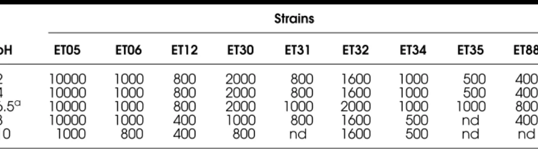
b. Sensitivity to Different pH Values
The stability of CFSFs in a pH range from 2.0 to 10.0 is shown in Table 3. They differed with regard to their sensitivity to inactivation by changes in pH. Many were stable only in acidic and neutral conditions and were even inacti-vated at pH 8.0, for example, CFSF from Lb. fermentum ET35. Although the nine CFSFs tested were stable at pH 6.5, only CFSF from E. faecium ET05 and Lb. curvatus ET06 strains remained constant in a wide range of pH from 2.0 to 8.0. All the other bacteriocins were partly inactivated in the extremes of the pH range. CFSF activity of Lb. curvatus ET31, Lb. fermentum ET35 and
E. faecium ET88 was completely lost at an alkaline pH while the maximal
activity of the others CFSF was reduced to a half.
c. Sensitivity to Heat Treatment
All CFSFs maintained their antimicrobial activity even at 100°C for 20 min. Only CFSFs titres from Lb. curvatus ET30 and ET31 lost 20% and 50% of their activities after 10 min and 30 min of heating at 60°C, respectively, and
Table 3: Effect of pH on bacteriocin activity (AU mL−1) produced by the test isolates.
pH
Strains
ET05 ET06 ET12 ET30 ET31 ET32 ET34 ET35 ET88
2 10000 1000 800 2000 800 1600 1000 500 400 4 10000 1000 800 2000 800 1600 1000 500 400 6.5a 10000 1000 800 2000 1000 2000 1000 1000 800
8 10000 1000 400 1000 800 1600 500 nd 400
10 1000 800 400 800 nd 1600 500 nd nd
after 10 min and less than 5 min of heating at 100°C, respectively (titre declined from 2000 AU/mL to 1000 AU/mL for the former and from 1000 AU/mL to 800 AU/mL for the last one). Then, both maintained their stability during the remaining time of the heating period.
Spectrum of Antimicrobial Activity
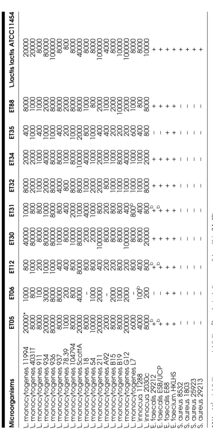
The antagonistic effect of the neutralized and filtered culture supernatants on various Gram-positive bacteria was tested and titres were calculated for each one (Table 4). The inhibition of the various target microorganisms was compared with the inhibitory activity of the nisin-producing Lc. lactis subsp.
lactis ATCC 11454. CFSF from this strain presented a wide range of
inhibi-tory spectrum affecting different nontaxonomically related genera like
Listeria, Staphylococcus and Enterococcus. Although CFSF from the other
bacteriocin-producing strains inhibited a similar but limited range of target microorganisms, there was considerable variability of sensitivities of
L. monocytogenes strains inter and intra the nine CFSFs tested. The highest
activities (>10000 AU/mL) were obtained from E. faecium (ET05),
Lb. curvatus (ET30) and Lc. lactis subsp. lactis ATCC 11454 against
practi-cally the same Listeria strains, i.e. L. monocytogenes 11994, L.
monocytoge-nes 934, L. monocytogemonocytoge-nes 936, L. monocytogemonocytoge-nes Scott A, L. monocytogemonocytoge-nes
211, L. monocytogenes G12 and L. innocua 2030c. No inhibitory activity was recorded for CFSF from Lb. curvatus ET06 against L. monocytogenes 18, L. monocytogenes A92 and L. monocytogenes L7. No relationship seems to exist among the several serotypes of Listeria spp. tested and the level of inhibition obtained. Only CFSFs from Lb. curvatus ET06 and from Lb.
fermentum ET35 were not able to inhibit E. faecalis 29212 and E. faecalis
(ESB/UCP) while partial or complete inhibition was observed for E. faecalis 29212, E. faecalis (ESB/UCP), E. faecalis E88, E. faecium HKLHSE with the others CFSFs. On the other hand, just Lc. lactis subsp. lactis ATCC 11454 showed antimicrobial properties against S. aureus strains. None of the CFSFs inhibited any of the Gram-negative strains assessed (results not shown).
The antagonistic activity exhibited by each CFSF against the other bacteriocin-producing strains is recorded in Table 5. Bacteriocin ET31 (Lb. curvatus) showed inhibitory activity against the E. faecium strains (weak and reversible against E. faecium ET12), while bacteriocins ET06 and ET30 from Lb. curvatus strains did not show anti-bacterial activities against the same LAB tested. Inhibition by bacteriocins from E. faecium was restricted to closely related bacteria, although the antimicrobial com-pound inhibited Lc. lactis subsp. lactis ATCC 11454. CFSF from Lb.
del-brueckii ET32 exhibited activity only against E. faecium ET05 while
Ta ble 4: Inhibitory spec trum of bac teriocins agains t several Gram-positive bacteria. Micr oor g a nis m s E T0 5 E T0 6 E T1 2 E T3 0 E T3 1 E T3 2 E T3 4 E T3 5 E T8 8 L. la ctis lactis AT CC 114 54 L. monocytogen e s 11994 20000* 1000 800 40000 1000 8000 2000 400 8000 20000 L. monocytogen e s 4031T 8000 800 1000 80000 800 2000 2000 1000 1000 20000 L. monocytogen e s 911 8000 100 200 8000 800 1000 1000 400 1000 8000 L. monocytogen e s 934 20000 3000 1000 80000 1000 2000 4000 1000 2000 80000 L. monocytogen e s 936 80000 8000 1000 80000 8000 8000 8000 1000 8000 100000 L. monocytogen e s 937 8000 8000 400 10000 800 4000 8000 400 8000 8000 L. monocytogen e s 78.39 1000 200 400 8000 400 800 1000 200 2000 800 L. m o nocy toge nes 104794 8000 800 800 10000 2000 8000 8000 1000 8000 8000 L. monocytogen e s ScottA 20000 4000 8000 80000 1000 8000 10000 2000 8000 40000 L. monocytogen e s 18 8000 – 800 2000 4000 1000 4000 800 1000 8000 L. monocytogen e s 54 10000 1000 800 2000 1000 2000 1000 1000 800 8000 L. monocytogen e s 211 20000 2000 400 100000 800 2000 2000 400 2000 100000 L. monocytogen e s A92 2000 – 200 8000 200 800 1000 400 1000 4000 L. monocytogen e s B15 8000 2000 200 8000 800 1000 1000 200 2000 8000 L. monocytogen e s B19 8000 1000 800 8000 800 1000 8000 200 1 0000 10000 L. monocytogen e s G12 20000 2000 200 100000 400 1000 4000 200 2000 100000 L. monocytogen e s L7 6000 – 800 8000 800 b 1000 1000 600 1000 8000 L. innocua 11288 4000 100 b 400 8000 400 1000 1000 400 800 8000 L. innocua 2030c 8000 200 800 20000 800 8000 2000 800 8000 10000 E. faecalis 29212 + b − + b ++ b ++ − ++ E. faecalis ESB/ UCP + b − + b ++ b ++ − ++ E. faecalis E 8 8 + + + + + + +++ + E. faecium H K LH S + + + + + + +++ + S. a u reus 8532 −− − − − − − − − + S. a u reus 1803 −− − − − − − − − + S. a u reus 25923 −− − − − − − − − + S. a u reus 29213 −− − − − − − − − + *A U m L − 1 +: in h ib iti on − : no in hibition bbacteriostatic, rev e rsi b le w ithin 24–4 8 h.
bacteria tested. Activity of CFSFs was also screened against 60 LAB iso-lated from vacuum-packaged CSS; the growth of LAB tested was not inhib-ited by any of the CFSFs tested.
DISCUSSION
Strain Identification
Our results indicated phenotypic heterogeneity and genetic diversity among the vacuum-packaged CSS bacteriocin-producing isolates, with a good correlation between the phenotypic and genetic identification of the strains. As sugar fermentation patterns are not considered to be a reliable method of distinguishing among Enterococcus spp. due to the heterogeneous and atypical profiles displayed by this genus (Pérez et al., 2000), further identification to species level was carried out with the Vitek system, which classified all the enterococcal isolates as E. faecium in accordance with the genotypic results at genus level performed by PCR reaction. The fermentation activities recorded with API galleries agreed with those registered with VITEK; however, API 50 CH reactions were not enough to identify microorganisms belonging to Enterococcus genus. On the other hand, the biochemical tests carried out with the GPI cards showed that all the Enterococcus isolated belonged to the same genus/ species E. faecium. The genus/species specific PCR identification assay allowed the proper identification of all LAB.
The species identified in our study had been previously isolated from vacuum-packaged CSS or fish products. For instance, González-Rodriguez
Table 5: Antimicrobial spectrum of activity of CFSFs from LAB against the bacteriocin-producing strains.
Target strains
Crude Filtrate Supernatant Fluid (CFSF)
ET05 ET06 ET12 ET30 ET31 ET32 ET34 ET35 ET88
L.lactis ATCC 11454 E. faecium ET05 – 0* 3 0 5 3b 5b 0 4 10 Lb. curvatus ET06 0 – 0 0 0 0 0 0 0 0 E. faecium ET12 11b 0 – 0 5b 0 0 0 2b 6 Lb. curvatus ET30 0 0 0 – 0 0 0 0 0 0 Lb. curvatus ET31 0 0 0 0 – 0 0 0 0 0 Lb. delbrueckii ET32 0 0 0 0 0 – 0 0 0 0 P. acidilactici ET34 0 0 0 0 0 0 – 0 0 0 Lb. fermentum ET35 0 0 0 0 0 0 0 – 0 0 E. faecium ET88 10 0 2b 0 8 0 0 0 – 10 Lc. lactis ATCC11454 8 0 10 0 0 0 0 0 4 –
et al. (2002), in a study conducted to assess the microbiological quality of vacuum-packaged CSS produced by various processors in Spain, isolated eight species of Lactobacillus that included a high percentage of Lb. curvatus subsp.
curvatus as well as Lb. delbrueckii subsp. delbrueckii, although this last was a
minor proportion. Several strains of Enterococcus spp. were also isolated. Lyhs et al. (1999) isolated Lb. curvatus together with Leuconostoc mesenteroides subsp. mesenteroides, Lb. citreum, Lb. sakei, from spoiled vacuum-packaged, cold-smoked rainbow trout. In contrast, the occurrence of Lb. curvatus in non-spoiled vacuum-packaged salmon has been reported previously by Truelstrup-Hansen (1995), who identified from 168 LAB, 50% as Lb. curvatus.
Few reports have recorded the presence of Lb. fermentum in fish. According to Sharpe and Pettipher (1993), this heterofermentative Lactobacillus specie has predominated in different kinds of spoiled herring. Also, it has been frequently identified in chickens (Reque et al., 2000), a Balinese sausage (Antara et al., 2002), or boza, a traditional Bulgarian fermented beverage (von Mollendorff et al., 2006).
The importance of P. acidilactici strains in the food industry is related to their use as starter cultures in fermented meat and vegetable products (Amézquita and Brashears, 2002). This species, like most other LAB species, is involved in extending the shelf life and improving the hygienic quality of various fermented products, via the production of lactic acid and/or the secretion of anti-bacterial compounds such as bacteriocins (Stiles, 1996). The association of pediococci with proteinaceous foods such as fresh and cured meat, and raw sausages has frequently been reported (Holzapfel et al., 2005), and particularly for P. acidilactici in fermented sausages (Parente et al., 2001). Pediococcus spp. also have been reported in fresh and marinated fish (Paludan-Muller et al., 2002).
Molecular Size of the Bacteriocins
Although tricine-SDS-PAGE is not an accurate technique to calculate the molecular mass of molecules, it gives valuable information about the presence of either one or two peptides (Moreno et al., 2002). A single inhibition zone was seen for all the bacteriocins assessed by this technique in this study, although the samples precipitated by ammonium sulphate and analyzed by tricine-SDS-PAGE contained more than one protein band.
Bacteriocins ET05 and ET12 from E. faecium strains ET05 and ET12, respectively, possessed a molecular mass near 3.5 kDa whereas bacteriocin ET88 from E. faecium ET88 has a molecular mass slightly higher. Other enterocins from E. faecium strains with close molecular masses have been reported (Moreno et al., 2002) as well as others with higher masses such as enterocin 012 (Jennes et al., 2000). Moreover, strains of E. faecium capable of producing two kinds of bacteriocins with different molecular weights have
been described, that is, enterocin A, pediocin-like bacteriocin with a molecular weight of 4.8 kDa (Aymerich et al., 1996), and enterocin B, a small nonlantibi-otic bacteriocin not belonging to the pediocin group and with a molecular weight of 5.5 kDa (Nilsen et al., 1998).
All the three bacteriocins isolated from Lb. curvatus strains, named ET06, ET30, and ET31, had different molecular masses. While bacteriocins ET30 and ET06 migrated upon SDS-PAGE gel electrophoresis as small peptides of approximately 3.1 kDa and 2.8 kDa (Fig. 1E), respectively, bacteriocin ET31 had a molecular mass slightly higher (4.5 kDa, Fig. 1D). The molecular weight of curvacin A, the first bacteriocin identified and characterized from a strain of Lb. curvatus, is around 3.0 – 5.0 kDa (Messens et al., 2002). Like the other bacteriocins assessed here, ET32 from Lb. delbrueckii ET32 is a small peptide with an apparent molecular size of approximately 3.5 kDa (as estimated by tricine-SDS-PAGE analysis). Although bacteriocins from Lb. delbrueckii have been rarely described, Boris et al. (2001) characterized a bacteriocin UO004 produced by Lb. delbrueckii subsp. lactis UO004 with a molecular weight near 6.0 kDa by SDS-PAGE analysis. The molecular mass of the pediocin ET34 was approx. 3.5 kDa. Its size is similar to pediocin PD-1 from P. damnosus and does not correspond with the molecular mass of the pediocin PA-1 of 4.629 kDa from P. acidilactici PAC 1.0 mainly found in fermented sausages and other meat and vegetable fermentations (Jager and Harlander, 1992). Different elec-trophoretic mobilities of bacteriocins of Lb. fermentum have been reported; >1.0 kDa to <5.0 kDa for Fermenticin B produced by Lb. fermentum Beijerinck CCRC 14018 (Yan and Lee, 1997), 2.3 kDa – 3.0 kDa (von Mollendorff et al., 2006). Taking into account their bactericidal activity, proteinaceous nature, heat resistance, and low molecular weight, bacteriocins ET05, ET06, ET12, ET32, ET34, ET35, and ET88 can be classified as small, heat-stable Listeria-active peptides possibly belonging to class IIa according to the definition given by Klaenhammer (1993).
Characterization of Crude Filtrate Supernatant Fluid (CFSF)
a. Sensitivity to Enzymes
The sensitivity of the inhibitors to enzymes was tested to gain insight into their chemical structure. Results demonstrated that the active moiety of the entire inhibitory substances was not hydrogen peroxide, a lipid, or a glucan, respectively. The loss of anti-bacterial activity of the CFSFs upon treatment with trypsin, protease E, and proteinase K permits their classification as bacteriocins or/and bacteriocin-like inhibitory substances. The susceptibility of bacteriocins to enzymatic degradation suggests that these peptides will be degraded in the intestinal tract and so will be easily digested without affecting the intestinal flora. From this point of view, Lb. curvatus strains and their
bacteriocins could be of great interest as bioprotective cultures, because their bacteriocins will be more quickly digested than the other bacteriocins (they are digested by pepsin A, at pH 2). De Martinis et al. (2003) reported similar bacteriocin sensitivity to these enzymes for the bacteriocins of two strains of
Lb. curvatus isolated from sausages.
The bacteriocins assessed in this study consisted of pure peptides, as their activities were inactivated by treatment with proteolytic enzymes, while lipolytic and glycolytic enzymes had no effect on activity. All of them shared their sensitivity to treatment with proteolytic enzymes but insensitivity to lipolytic and glycolytic enzymes. According to Piard and Desmazeaud (1992), LAB synthesize many bactericidal agents, some of which are bacteriocins with a proteinaceous active moiety and others are nonprotein agents.
b. Sensitivity to Different pH Values
Bacteriocins differ greatly with regard to their sensitivity to inactivation by changes in pH and temperature. Many are stable only in acid and neutral conditions, and are even inactivated at pH 8.0, for example, lactostrepcins, or pH 10.0, like nisin and pediocin PA-1 (Moreno et al., 2000; Chien-Wei et al., 2004). Most of the bacteriocins maintained full activity over a pH range of 2.0–8.0 and were partially or completely inactivated at pH 10.0. In this case, the loss of the activity was irreversible and could not be regained upon lowering the pH to 6.5. Irreversible inactivation can result from a combina-tion of denaturacombina-tion and chemical modificacombina-tions of the molecule. The loss of activity can be related to the solubility of the bacteriocins. In the case of enterocins, the isoelectric point of all known enterocins was around 8.3–10.7, which implies that the solubility increases at pHs below the pI. All the nine inhibitory substances showed maximal activity at pH 6.5. This pH value is very close to the normal pH of CSS fillets (6.0) (Tomé et al., 2006), which is a positive aspect for the addition of these anti-listerial peptides (or their pro-ducing bacteria) into this product.
c. Sensitivity to Heat Treatment
Bacteriocins were similar with regard to their sensitivity to inactivation by temperature. Like most of the known bacteriocins, they were mainly heat-tolerant at pH 6.5 (Todorov et al., 1999) after 60 min of treatment at 60°C and 20 min of treatment at 100°C. Hill (1994) and Jennes et al. (2000) also reported the heat stability of enterocins. Only bacteriocins ET30 and ET31 were moderately heat-stable at 60°C and 100oC, thus resembling nisin pro-duced by Lc. lactis WNC20 which was inactivated after 15 min at 121°C at pH 7.0 (Noonpakdee et al., 2003) or the pediocin PA-1 showing about 40% activity lost after 15 min of heating at 121°C in the pH range pH 2.5–9.0 (Ray, 1994). The moderate stability of the inhibitory peptide produced by Lb. curvatus strains was previously reported (De Martinis et al., 2003).
Spectrum of Antimicrobial Activity
The assessment of the inhibitory spectrum is an important characteristic in order to evaluate the possibility of using the bacteriocin-producing strains or the bacteriocins alone as an additional barrier against spoilage and/or food-borne pathogens in food. Only 2 (22%) out of 9 neutralized culture filtrates displayed activity toward all the Gram-positive microorganisms examined. One characteristic of classical bacteriocins is a narrow spectrum of activity (Tagg et al., 1976). CFSFs of ET05 and ET88 of E. faecium demonstrated the widest antimicrobial spectrum of activity. Moreover it was similar to the inhibitory spectrum of the nisin of Lc. lactis subsp. lactis ATCC 11454, which suppressed the growth of all the Listeria spp. as well as E. faecalis strains,
E. faecium and S. aureus strains. CFSF from P. acidilactici ET34 showed an
inhibitory spectrum slightly different from those exhibited by the antimicrobial peptide called pediocin F and pediocin A-1 produced by P. acidilactici (Osmanagaoglu et al., 1998), which is inhibitory to a variety of LAB often encountered in foods, such as Enterococcus, as well as spoilage and pathogenic strains i.e. E. coli and S. aureus. Fimland et al. (2000) showed that the C-terminal disulfide bridge in pediocin-like bacteriocins contributes to widening of the antimicrobial spectrum.
All bacteriocins inhibited strains of L. monocytogenes, a pathogen often isolated from a variety of foods. This activity is quite interesting, especially taking into account that many reports showed the occurrence and growth of
L. monocytogenes in fish. L. monocytogenes serovar 4b, which is involved in
human infections has been isolated from fresh trout samples (Hangard-Vidaud et al., 1989) as well as smoked salmon. Among the 19 Listeria spp. strains tested, 84.2% were inhibited by bacteriocin ET06. The high anti-listerial activity displayed by the nine bacteriocins is characteristic of the class IIa bacteriocins (Klaenhammer, 1993; Ennahar et al., 2000). It is interesting to note the resistance of L. monocytogenes 18, L. monocytogenes A92 and
L. monocytogenes L7 to bacteriocin ET06. It has been a common observation by
bacteriocin researchers that resistance to bacteriocin action is not only species or strain-specific but also dose-dependent. Strains that have the receptors and relevant characteristics of cytoplasmic membrane for proper attachment and sensitivity to the bacteriocin, are generally inhibited (Ennahar et al., 2000).
Our study confirms that L. innocua 2030c resembles L. monocytogenes in sensitivity toward LABs ET05, ET06, ET12, ET30, ET31, ET32, ET34, ET35, ET88 bacteriocins and is thus a suitable organism when extensive work with the pathogen is undesirable. On the other hand, the sensitivity differences of each Listeria spp. against the nine CFSFs studied is notable. The highest anti-listerial activity was recorded for CFSF ET05, ET30 and nisin from
Lc. lactis subsp lactis ATCC 11454. The dissimilar responses of Listeria spp.
of the organisms, nor by the growth phase of Listeria strains; all were tested in the stationary phase. As mentioned by Jydegaard et al. (2000), the bacterio-cin inactivation of L. monocytogenes 412 by nisin and low pediobacterio-cin concentra-tions are growth phase dependent, with exponentially growing cells being more susceptible than stationary phase cells. The sensitivity differences of
Listeria spp., as well other food-borne and spoilage microorganisms toward
diverse bacteriocins have been documented previously by several researchers (Moreno et al., 2000; Coventry et al., 1997; Østergaard et al., 1998). As expected, none of the CFSFs inhibited any of the Gram-negative bacteria eval-uated. Activity of bacteriocins against Gram-negative bacteria is unusual and has only been reported for a few bacteriocins of LAB (Caridi, 2002; Todorov and Dicks, 2004a; Todorov and Dicks LMT, 2004b).
Well-defined bacteriocins produced by lactobacilli usually have inhibitory activities restricted to closely related species. However, it is worth noting that CFSFs showed limited activity against other bacteriocin-producer strains (Table 5); just CFSF ET05, ET12 and ET88 exhibited antimicrobial properties toward E. faecium strains and Lc. lactis subsp. lactis ATCC11454, although in some cases this inhibitory activity disappeared within 48 h. CFSF from
Lc. lactis subsp. lactis ATCC 11454 such as CFSF ET31 showed limited
activ-ity toward bacteria belonging to the genus Enterococcus and no activactiv-ity at all against the Lactobacillus strains tested. Contrary to what was expected, most of the bacteriocins did not kill species of bacteria that are known to have the same ecological niche. Similar results were reported for pediocin PD-1 produced by P. damnosus as it was not active against other pediococci. In that respect, Coventry et al. (1997) pointed out that the sensitivity of Listeria species as well as LAB to bacteriocins may be influenced by the content or type of agar in the growth media. The different inhibition patterns registered for neutralised and filtered culture supernatants of Lb. curvatus strains, against the LAB tested, could suggest that the inhibitory compound produced by Lb. curvatus ET31 is different from those produced by the other Lb. curvatus strains. These results are promising in view of a recent investigation into the use of combinations of LAB bacteriocins or their producing strains in order to broaden the spectrum to a wide variety of pathogens and food spoilage organ-isms, and avoid the phenomenon of bacteriocin resistance development, which is the main limiting factor for bacteriocin effectiveness. It is generally admitted that each bacteriocin-sensitive bacterial population includes potentially tolerant and/or resistant cells with structural modifications or at least with a high pre-disposition to such modifications, which would allow them to spontaneously emerge during exposure to the bacteriocin (Hanlin et al., 1993). In particular, modifications in the cytoplasmic membrane composition are often investi-gated to explain bacteriocin resistance, considering the key role of the mem-brane in the activity of bacteriocins (Mazzotta and Montville, 1997). On the other hand, no activity of CFSFs was obtained in the screening assay against
60 LAB isolated from vacuum-packaged CSS (results not shown). This speci-ficity could be useful for suppressing growth of Listeria in lightly preserved seafood where it could be a serious problem, without interfering with other desirable lactic flora. These results resemble the activity spectrum of the divergicin M35 from Carnobacterium divergens M35, which did not inhibit bacteria belonging to the genera Lactobacillus, Lactococcus, Enterococcus,
Pediococcus, Propionibacterium, and Bifidobacterium (Tahiri et al., 2004).
ACKNOWLEDGMENTS
Financial support to author Tomé was provided by a PhD fellowship issued by Consejo de Desarrollo Científico y Humanístico de la Universidad Central de Venezuela.
REFERENCES
Amézquita, A., Brashears, M.M. (2002). Competitive inhibition of Listeria
monocyto-genes in ready-to-eat meat products by lactic acid bacteria. J. Food Protect.
65:316–325.
Antara, N.S., Sujaya, I.N., Yokota, A., Asano, K., Aryanta, W.R., Tomita, F. (2002). Identification and succession of lactic acid bacteria during fermentation of“urutan,” a Balinese indigenous fermented sausage. World J. Microbiol.
Biotech-nol. 18:255–262.
Aymerich, T., Holo, H., Håvarstein, L.S., Hugas, M., Garriga, M., Nes, I.F. (1996). Biochemical and genetic characterization of enterocin A from Enterococcus
faecium, a new antilisterial bacteriocin in the pediocin family of bacteriocins. Appl. Environ. Microbiol. 62:1676–1682.
Berthier, F., Ehrlich, D.S. (1988). Rapid species identification within two groups of closely related lactobacilli using PCR primers that target the 16S/23S rRNA spacer region. FEMS Microbiol. Lett. 161:97–106.
Boris, S., Jiménez-Díaz, R., Caso, L.J., Barbés, C. (2001). Partial characterization of a bacteriocin produced by Lactobacillus delbrueckii subsp. lactis UO004, an intestinal isolate with probiotic potential. J. Appl. Microbiol. 91:328–333.
Caridi, A. (2002). Selection of Escherichia coli-inhibiting strains of Lactobacillus
para-casei subsp. parapara-casei. J. Ind. Microbiol. Biotechnol. 29:303–308.
Chagnaud, P., Machinis, K., Coutte, L.A., Marecat, A., Mereenier, A. (2001). Rapid PCR-based procedure to identify lactic acid bacteria: application to six common
Lactobacillus species. J. Microbiol. Meth. 44:139–148.
Chien-Wei, W., Li-Jung, Y., Shann-Tzong, J. (2004). Purification and characterization of bacteriocin from Pediococcus pentosaseus ACCEL. J. Agric. Food. Chem. 52:1146–1151. Coventry, M.J., Gordon, J.B., Wilcock, A.H., Davidson, B.E., Hickey, M.W., Hiller, A.J., Wan, J. (1997). Detections of bacteriocins of lactic acid bacteria isolated from foods and comparison with pediocin and nisin. J. Appl. Microbiol. 83:248–258.
Davidson, C., Cronin, F. (1973). Medium for the selective enumeration of lactic acid bacteria from foods. J. Appl. Microbiol. 26:439–440.
De Martinis, E.C.P., Santarosa, P.R., Freitas, F. (2003). Caracterização preliminar de bacteriocinas produzidas por seis cepas de bactérias láticas isoladas de produtos cárneos embalados a vácuo. Ciênc. Tecnol. Alimen. 23:195–199.
De Vuyst, L., Vandamme, E.J. (1994). In: Bacteriocins of Lactic Acid Bacteria;
Microbi-ology, Genetics and Applications. London: Blackie Academic and Professional.
Dellaglio, F., Bottazzi, V., Trovatelli, L.D. (1973). Deoxyribonucleic acid homology and base composition in some thermophilic lactobacilli. J. Gen. Microbiol. 74:289–297. Ennahar, S., Sashihara, T., Sonomoto, K., Ishizaki, A. (2000). Class IIa bacteriocins:
biosynthesis, structure and activity. FEMS Microbiol. Rev. 24:85–106.
[FAO/WHO] Food and Agriculture Organization/World Health Organization . (2006). Summary of Evaluations Performed by the Joint FAO/WHO Expert Committee on Food Additives (JECFA 1956–2005). WashingtonDC: ILSI Press International Life Sciences Institute, 20036–4810.
[FDA] Food and Drug Administration. (1988). Federal Register. Nisin preparation: affir-mation of GRAS status as a direct human food ingredient, Fed. Reg. 53, 11247–11251. Fimland, G., Johnsen, L., Axelsson, L., Brurberg, M.B., Nes, I.F., Eijsink, V.G., Nissen-Meyer, J. (2000). A C-terminal disulfide bridge in pediocin-like bacteriocins ren-ders bacteriocin activity less temperature dependent and is a major determinant of the antimicrobial spectrum. J. Bacteriol. 182:2643–2648.
González-Rodríguez, M., Sanz, J., Santos, J., Otero, A., García López, M.L. (2002). Numbers and types of microorganisms in vacuum-packed cold-smoked fresh water fish at the retail level. Int. J. Food Microbiol. 36:1–29.
Hangard-Vidaud, N., Nicolas, J.A., Bosgiraud, C., Cormuejols, M.J. (1989). Recherche de Listeria chez les poisons d’eau douce. Microbiol. Alim. Nutr. 7:421–423.
Hanlin, M.B., Kalchayanand, N., Ray, P., Ray, B. (1993). Bacteriocins of lactic acid bac-teria in combination have greater antibacbac-terial activity. J. Food Prot. 54: 252–255. Hill, C. (1994). Enterocin 1146: a bacteriocin produced by Enterococcus faecium DPC1146. In: De Vuyst, L. and Vandamme, E.J., eds. Bacteriocins of Lactic Acid Bacteria. Microbiology, Genetics and Applications. London: Chapman & Hall, pp. 515–527. Holzapfel, W.H., Franz, C.M.A.P., Ludwing, W., Back, W., Dicks, L.M.T. (2005). The
Genera Pediococcus and Tetragenococcus. In: Dworkin, et al., eds. The Prokaryotes:
An Evolving Electronic Resource for the Microbiological Community, third ed.,
release 3.2. New York: Springer-Verlag.
Holzapfel, W.H., Geisen, R., Schillinger, U. (1995). Biological preservation of foods with reference to protective cultures, bacteriocins and food-grade enzymes. Int. J. Food.
Microbiol. 24:343–362.
Jager, K., Harlander, S. (1992). Characterization of a bacteriocin from Pediococcus
acidilactici PC and comparison of bacteriocin-producing strains using molecular
typing procedures. Appl. Microbiol. Biotechnol. 37:631–637.
Jennes, W., Dicks, L.M.T., Verwoerd, D.J. (2000). Enterocin 012, a bacteriocin produced by Enterococcus gallinarum isolated from the intestinal tract of ostrich.
J. Appl. Microbiol. 88:349–357.
Jimenez-Diaz, R., Rios-Sanchez, R.M., Dezmazeaud, M., Ruiz-Barba, J.L., Piard, J.C. (1993). Plantaricins S and T, two new bacteriocins produced by Lactobacillus
plan-tarum LPCO10 isolated from a green olive fermentation. Appl. Environ. Microbiol.
59:1416–1424.
Jydegaard, A.M., Gravesen, A., Knochel, S. (2000). Growth conditions-related response of
Ke, D., Picard, F.J., Martineau, F., Ménard, C., Roy, P.H., Onellette, M., Bergeron, M.G. (1999). Development of a PCR assay for rapid detection of Enterococci. J. Clin.
Microbiol. 37:3497–3503.
Klaenhammer, T.R. (1993). Genetics of bacteriocins produced by lactic acid bacteria.
FEMS Microbiol. Rev. 12:39–86.
Lyhs, U., Björkroth, J., Korkeala, H. (1999). Characterization of lactic acid bacteria from spoiled, vacuum-packaged, cold-smoked rainbow trout using ribotyping. Int.
J. Food Microbiol. 52:77–84.
Mazzotta, A.S., Montville, T.J. (1997). Nisin induces changes in membrane fatty acid composition of Listeria monocytogenes nisi-resistant strains at 10°C and 30°C.
J. Appl. Microbiol. 82:32–38.
Messens, W., Verluyten, J., Leroy, F., De Vuyst, L. (2002). Modelling growth and bacte-riocin production by Lactobacillus curvatus LTH 1174 in response to temperature and pH values used for European sausage fermentation processes. Int. J. Food
Microbiol. 81:41–52.
Mora, D., Fortina, M.G., Parini, C., Manachini, P.L. (1997). Identification of Pediococcus
acidilactici and Pediococcus pentosaceus based on 16S rRNA and ldhD gene-targeted
multiplex PCR analysis. FEMS Microbiol. Lett. 151:231–236.
Moreno, I., Lerayer, A.L.S., Baldini, V.L.S., Leitaõ, M.F. (2000). Characterization of bacteriocins produced by Lactococcus lactis strains. Braz. J. Microbiol. 31:84–192. Moreno, M.R., Leisner, J.J., Tee, L.K., Ley, C., Radu, S., Rusul, G., Vancanney, M., De Vuyst,
L. (2002). Microbial analysis of Malaysian tempeh, and characterization of two bacterio-cins produced by isolates of Enterococcus faecium. J. Appl. Microbiol. 92:147–157. Nettles, C.G., Barefoot, S.F. (1993). Biochemical and genetic characteristics of
bacterio-cins of food-associated lactic acid bacteria. J. Food Prot. 56:338–356.
Nilsen, T., Nes, I.F., Holo, H. (1998). An exported inducer peptide regulates bacteriocin production in Enterococcus faecium CTC492. J. Bacteriol. 174:5686–5692.
Noonpakdee, W., Santivarangkna, C., Jumriangrit, P., Sonomoto, K., Panyim, S. (2003). Isolation of nisin-producing Lactococcus lactis WNC20 strain from nham, a traditional Thai fermented sausage. Int. J. Microbiol. 81:137–145.
Osmanagaoglu, O., Gündüz, U., Beyatly, Y., Çökmüs, C. (1998). Purification and characterization of Pediocin F, a bacteriocin produced by Pediococcus acidilactici F. Turk. J. Biol. 22:217–228.
Østergaard, A., Embarek, P.K.B., Wedell-Neergaard, C., Huss, H.H., Gram, L. (1998). Characterization of anti-listerial lactic acid bacteria isolated from Thai fermented fish products. Food Microbiol. 15:223–233.
Paludan-Muller, C., Madsen, M., Sophanodora, P., Gram, L., Moller, P.L. (2002). Fer-mentation and microflora of plaa-som, a Thai fermented fish product prepared with different salt concentrations. Int. J. Food Microbiol. 73:61–70.
Parente, E., Grieco, S., Crudele, M.A. (2001). Phenotypic diversity of lactic acid bacte-ria isolated from fermented sausages produced in Basilicata (Southern Italy).
J. Appl. Microbiol. 90:943–952.
Pérez, G., Cardell, E., Zarate, V. (2000). Protein fingerprinting as a complementary analysis to classical phenotyping for the identification of lactic acid bacteria from Tenerife cheese. Lait 80:589–600.
Piard, J.C., Desmazeaud, M. (1992). Inhibiting factors produced by lactic acid bacteria. Part II. Bacteriocins and other antibacterial substances. Lait 72:113–142.
Ray, B. (1994). Pediocins of Pediococcus species. In: De Vuyst, L., Vandanme, E.J., eds.
Bacteriocins of Lactic Acid Bacteria: Microbiology, Genetics and Applications.
London: Chapman and Hall, pp 465–495.
Reque, E., Pandey, A., Franco, S., Soccol, C. (2000). Isolation, identification and physi-ological study of Lactobacillus fermentum LPB for use as a probiotic in chickens.
Braz. J. Microbiol. 31:303–307.
Sahl, H.G. (1994). Staphylococcin 1580 is identical to the lantibiotic epidermin. Implications for the nature of bacteriocins from gram-positive bacteria. Appl.
Environ. Microbiol. 60:752–755.
Savadogo, A., Outara, C.A., Bassole, I.H., Traores, A. (2006). Bacteriocins and lactic acid bacteria - a minireview. Afric. J. Biotechnol. 5:678–683.
Schagger, H., Von Jagow, G. (1987). Tricine-sodium dodecyl sulfate–polyacrylamide gel electrophoresis for the separation of proteins in the range from 1 to 100 kDa. Anal.
Biochem. 166:368–379.
Sharpe, M.E., Pettipher, G.L. (1983). Food spoilage by lactic acid bacteria. Ecol. Microbiol. 8:199–223.
Stiles, M.E. (1996). Biopreservation by lactic acid bacteria. Antonie van Leeuw. 70:331–345. Tagg, J.R., Dajani, A.S., Wannamaker, L.W. (1976). Bacteriocins of Gram-positive
bacteria. Bacteriol. Rev. 40:722–756.
Tahiri, I., Desbiens, M., Benech, R., Kheadr, E., Lacroix, C., Thibault, S., Oullet, D., Fliss, I. (2004). Purification, characterization and amino acid sequencing of diver-gicin M35: a novel class IIa bacteriocin produced by Carnobacterium divergens M35. Int. J. Food Microbiol. 97:123–136.
Tilsala-Timisjarvi, A., Alatossava, T. (1997). Development of ologonucleotide primers from 16S-23S rRNA intergenic sequence for identifying different dairy and probi-otic lactic acid bacteria. Int. J. Food Microbiol. 35:49–56.
Todorov, S., Onno, B., Sorokin, O., Chobert, J.M., Ivanova, I., Dousset, X. (1999). Detection and characterization of a novel antibacterial substance produced by Lactobacillus
plantarum ST31 isolated from sourdough. Int. J. Food Microbiol. 48:167–177.
Todorov, S.D., Dicks, L.M.T. (2004a). Effect of medium components on bacteriocin pro-duction by Lactobacillus pentosus ST151BR, a strain isolated from beer produced by the fermentation of maize, barley and soy flour. World J. Microbiol. Biotechnol. 20:643–650.
Todorov, S.D., Dicks, L.M.T. (2004b). Screening of lactic-acid bacteria from South African barley beer for production of bacteriocin-like compounds. Folia Microbiol. 49:406–410.
Tomé, E., Teixeira, P., Gibbs, P.A. (2006). Anti-listerial inhibitory lactic acid bacteria isolated from commercial cold smoked salmon. Food Microbiol. 23:399–405. Tomé, E., Gibbs, P.A., Teixeira, P. (2007). Could modifications of processing
parame-ters enhance the growth and selection of lactic acid bacteria in cold smoked salmon to improve preservation by natural means? J. Food Prot. 70(7):1007–1014. Truelstrup-Hansen, L. (1995). Quality of chilled, vacuum-packed of cold-smoked
salmon. PhD Thesis, Danish Institute of Fisheries. Research, Department of Seafood Research, Technical University Denmark.
Vaughan, E.E., Caplice, E., Looney, R., O’Rourke, N., Coveney, H., Daly, C., Fitzgerald, G.F. (1994). Isolation from food sources, of lactic acid bacteria that produced anti-microbials. J. Appl. Bacteriol. 76:118–123.
Vaz-Velho, M. (2000). Cold-smoked fish processing and safety. Ph.D Thesis, Escola Superior de Biotecnologia, Universidade Católica PortuguesaPorto, Portugal. Von Mollendorff, J.W., Todorov, S.D., Dicks, L.M.T. (2006). Comparison of bacteriocins
produced by lactic acid bacteria isolated from boza, a cereal-based fermented beverage from the Balkan Peninsula. Curr. Microbiol. 53:209–216.
Yan, T.R., Lee, C.S. (1997). Characterization of a partially purified bacteriocin, Fermentcin B, from Lactobacillus fermentum. Biotechnol. Lett. 19:741–744.