PABLO JAVIER GONZÁLEZ
P
ERIPLASMIC
N
ITRATE
R
EDUCTASES
:
S
TRUCTURAL AND
S
PECTROSCOPIC
S
TUDIES
PABLO JAVIER GONZÁLEZ
P
ERIPLASMIC
N
ITRATE
R
EDUCTASES
:
S
TRUCTURAL AND
S
PECTROSCOPIC
S
TUDIES
DISSERTAÇÃO APRESENTADA PARA A OBTENÇÃO DO GRAU DE DOUTOR EM BIOQUIMICA, ESPECIALIDADE BIOQUIMICA-FISICA, PELA FACULDADE DE CIÊNCIAS E TECNOLOGIA DA
UNIVERSIDADE NOVA DE LISBOA.
- Nº DE ARQUIVO
…no hay asunto, por complicado que sea, que estudiado con paciencia e inteligencia no se complique aun más...
I
A
CKNOWLEDGEMENTS
I would like to express firstly my gratefulness to Prof. José J.G. Moura, my advisor, for receiving me and accepting me to carry out my PhD work in his research group and for his unconditional support and confidence in the development of the work performed during these four years. I would like to thanks also for his friendship and encouragement to pursue the objectives of this research work.
Prof. Isabel Moura, very special thanks for let me work at your lab and for teaching me all the secrets about protein purification. This work would not have been possible without your support and motivation to face the hard work. The discussions, encouragement and critiques were of essence to the progress of this work.
Prof. Carlos Brondino (El Jefe), it is difficult to emphasize my gratitude taking into account how bárbaro you are… you did not introduce me in the field… but you were my mentor-like. I thank your great efforts to explain the things beyond my understanding but more important to study the subjects beyond yours! …that helped to make this to progress. I acknowledge you for guiding me through the course of this thesis work and the writing of the published papers, and for the critiques (constructives and destructives also!) which improve all the work I have made this last years.
Prof. María João Romão, thank you for your help and constant interest with the work developed with the nitrate reductases and for invite me and trust me the work with the Nap from Ralstonia.
Many many thanks the people who “tilt the scale”: Alberto Rizzi, Laura Felice (por la paciencia), Sofia Pauleta, Ana Teresa Lopes, Jorge Dias, Marta Carepo, Pedro Cabrito, Jorge Martinho, Simone Dell’acqua (and Francesca), Ines Cabrito, Teresa SSS, Joana, Cristina Correia, Stephane Besson (Katy, Prisca and Tom), Celia Silveira, Cristiano Mota…I hope did not forgot someone.
I am totally indebted to Maria João Baleizão, who performed the tough work of molecular biology with the nap operon of Desulfovibrio desulfuricans. All these results, although not conclusive yet, will be very useful for the future work.
II
I would like to thank Dr. Sergey Bursakov for the help at the starting of this PhD work and the tips for the purification of Nap from Desulfovibrio desulfuricans. In this line I would like to thank also Sandro Soares, Ana Sofia Cruz and Angelo.
My special gratitude goes to Roeland Boer, who starts the crystallization of the reacted samples of Dd NapA, and obviously to Shabir Najmudin who crystallized, solved the structures reported in this work, and helped me in the writing of the crystallography sections. I would like to thank also Jose Trincão and Catarina Coelho for the collaboration with the Nap from Ralstonia.
Many thanks to Mario Passeggi for his friendship and help with “dark side” of the EPR spectroscopy.
Jose Luis Capelo and Carlos Lodeiro, thank you very much for your friendship and for all the help and advices…the time spent in Portugal would have been very different without you.
Thanks the colleagues of Bioin, Bioprot, and the people from X-tal.
Thanks Eng. Lucia, Isabel Rodrigues and Maria Jose Carapinha for all the help with the burocracy.
Thanks Fundação para a Ciência e a Tecnologia (SFRH/BD/10825/2002) for financial support.
Finally, I would like to thank those closest to me; they are very important to me and their presence helped to complete every goal I planned.
My family, Gonza, Cristy, Malu, Diego y Sergio, I am grateful for your absolute support and confidence in every moment. My grandmother Sarita, thank you for giving me a home in Santa Fe and for being the person you are. Mabel, Alicia, Florencia, Ale, Marcelo, Ruben y Juan…you have been there since I remember…
My best best friends Cano y Nilson, your unconditional friendship is one of my treasures in this life.
III
you give me love, friendship, total support, encouragement and patient companionship since the time of the university.
V
R
ESUMO
A redução do nitrato é um processo levado a cabo nas células com o objectivo de incorporar azoto em biomoléculas (amonificação asimilatoria), consumir os equivalentes de redução no crescimento anaeróbio das bactérias (desnitrificação/ amonificação disimilatoria), e/ou eliminar o excesso de energia produto do metabolismo duma determinada fonte de carbono (amonificação disimilatoria). As redutases do nitrato são enzimas que catalisam a conversão do nitrato ao nitrito. Estas enzimas são proteínas que contem um centro mononuclear de molibdénio e outros cofactores redox tais como centros ferro-enxofre e hemos do tipo b ou c, que medeiam a transferência de electrões desde o doador até o nitrato. As redutases do nitrato foram classificadas em quatro grupos: (1) redutases do nitrato de eucariontes (Euk-NR), (2) redutases do nitrato asimilatorias (Nas), (3) redutases do nitrato respiratórias (Nar), (4) redutases do nitrato periplasmáticas (Nap). As redutases do nitrato periplasmáticas isoladas de Desulfovibrio desulfuricans (Dd), Paracoccus pantotrophus (Pp) e Rhodobacter sphaeroides (Rs) constituem as Nap melhor estudadas na actualidade. A estrutura cristalográfica da Nap de Dd em condições aeróbias foi a primeira estrutura publicada destas enzimas. O sítio activo no estado oxidado está formado pelo ion de molibdénio (Mo6+) hexacoordenado aos quatro átomos de enxofre dos dois cofactores pterínicos, o enxofre gamma da Cys140, e um Hidroxilo/Agua na sexta posição de coordenação.
Nesta tese são reportados estudos realizados nas redutases do nitrato periplasmáticas isoladas a partir das bactérias Desulfovibrio desulfuricans ATCC 27774 e Cupriavidus necator H16. Ambas enzimas foram purificadas até homogeneidade electroforética.
A Nap purificada a partir de células de D. desulfuricans ATCC 27774 crescidas em anaerobiose e em presença de nitrato foi isolada como uma proteína monómerica solúvel de ~80 kDa. Estudos cinéticos indicam que, a diferença com as Naps diméricas, esta enzima possui promiscuidade pelo substrato devido a que pode reduzir não só o nitrato como também outros aniões de estrutura similar. As titulações redox monitorizados por espectroscopia de EPR realizadas na ausência e presença do nitrato na escala de +200 a -500 mV (vs. NHE), e os estudos de EPR da enzima em condições catalíticas ou em presença de inibidores, revelam diversas espécies de Mo(V) activas no EPR, o que sugere que o sitio activo possui uma importante flexibilidade de coordenação. Estes estudos demonstram também que o substrato modula as propriedades redox do Mo mas não o do centro ferro-enxofre.
VI
nitrato foi isolada como uma proteína heterodimérica solúvel com subunidades grande e pequena de ~90 e ~15 kDa, respectivamente. Estudos preliminares são reportados para esta proteina. As propriedades bioquímicas, estruturais e espectroscópicas de ambas enzimas são discutidas em comparação com outras proteínas relacionadas.
VII
A
BSTRACT
Nitrate reduction occurs in the cell in order to incorporate nitrogen into biomolecules (assimilatory ammonification), as the final electron acceptor when bacteria are grown in anaerobic conditions (denitrification/dissimilatory ammonification) and to eliminate energy excess generated by the cell metabolism (dissimilatory ammonification). Nitrate reductases are enzimes that catalize the conversion of nitrate to nitrite. Most nitrate reductases are mononuclear molybdenum-containing enzymes that have, besides the Mo-pterin cofactor, additional metallic centers such as iron-sulfur clusters and b- or c-type hemes that mediate electron transfer reactions between the electron donor and the nitrate. Nitrate reductases have been classified into four groups: (1) eukaryotic nitrate reductases (Euk-NR), (2) assimilatory nitrate reductases (Nas), (3) respiratory nitrate reductases (Nar) and (4) periplasmic nitrate reductases (Nap). Naps isolated from Desulfovibrio desulfuricans (Dd), Paracoccus pantotrophus (Pp) and Rhodobacter sphaeroides (Rs) constitute the best-characterized Naps, so far. The 3D X-ray structure of Dd NapA in its oxidized form was the first reported structure for these enzymes. The active site in the oxidized state is made up by a distorted Mo6+ ion hexacoordinated by four sulfurs from the two pterin cofactors, the γ-sulfur from a cysteine (Cys140), and a hydroxo/water ligand.
This thesis deals with the study of the nitrate reductases isolated from Desulfovibrio desulfuricans ATCC 27774 and Cupriavidus necator H16. Both enzymes were purified up to electrophoretic homogeinity.
Nap purified from D. desulfuricans ATCC 27774 cells grown in anaerobeosis and in the presence of nitrate was isolated as a soluble monomeric protein of ~80 kDa. Kinetic studies show that, in contrast to dimeric Naps, this enzyme presents substrate promiscuity since it is able to reduce not only nitrate but also other anions of similar structure. EPR-monitored redox titrations, carried out with and without nitrate in the potential range from 200 to -500 mV (vs. NHE), and EPR studies in both catalytic and inhibiting conditions, reveal distinct types of Mo(V) EPR-active species, which indicates that the Mo site shows a high coordination flexibility. These studies show that nitrate modulates the redox properties of the active site, but not those of the iron-sulfur cluster.
Nap purified from Cn H16 cells grown in aerobic conditions in the absence of nitrate was isolated as a soluble heterodimeric protein with large and small subunits of ~90 and ~15 kDa, respectively. Preliminary results are reported for this protein. The biochemical,
VIII those of other closely related proteins.
IX
A
BBREVIATIONS
A value of the hyperfine coupling Å Angstrom Aav average of the mean A-values
AMP adenosine monophosphate ANAMOX anaerobic ammonium oxidation
APS adenylyl-phospho sulfate APSr APS reductase
ATCC American Type Culture Collection ATP adenosine triphosphate ATPs ATP sulfurylase
B. Bradyrhizobium C. Cupriavidus
ccNir/NrfAH multihemic nitrite reductase
Cn Cupriavidus necator
Cu-Nir cooper-containing nitrite reductase Cyt cd1 Cytochrome cd1 nitrite reductase
D axial distortion of the ZFS
D. Desulfovibrio
Dd Desulfovibrio desulfuricans
DEAE diethyl-aminoethyl DFT density functional theory
Dg Desulfovibrio gigas
DMSO dimethylsulfoxide Dsr dissimilatory sulfite reductase E rhombic distortion of the ZFS
E. Escherichia
Ec Escherichia coli
Ecat midpoint redox potential of the catalytic current EDTA ethylenediamine tetra-acetic acid
Eº standard midpoint redox potential
EPR electron paramagnetic resonance Euk-NR eukaryotic nitrate reductase
EXAFS extended X-ray absorption fine structure Fdh formate dehydrogenase Fdh-H Fdh of the formate-hydrogen lyase complex Fdh-N membrane anchored Fdh induced by nitrate g g-factor
G Gauss gav average of the mean g-values
GOGAT glutamine-oxoglutarate aminotransferase GS glutamine synthase
I nuclear spin
K kelvin
K. Klebsiella
MAD multi-wavelength anomalous dispersion MCD molybdopterin cytosine dinucleotide
MES morpholine-ethano-sulphonic acid MGD molybdopterin guanosine dinucleotide MQH2 menaquinone
mV millivolts MV methyl viologen
X
N2H4 hydrazine N2O nitrous oxide Nap periplasmic nitrate reductase
Nar respiratory nitrate reductase Nas assimilatory nitrate reductase
NH2OH hydroxilamine
NH4+ ammonium
NHE normal hydrogen electrode
NirB NADH-dependent nitrite reductase
NO nitric oxide
NO2- nitrite NO3- nitrate
NR nitrate reductase
P. Paracoccus
PAPS phospho-adenylyl-phospho sulfate PEG polyethyleneglycol
PMF proton motive force
Pn Pseudomonas nautica
Pp Paracoccus pantotrophus
QH2 quinone
R. Rhodobacter
RMSD root-mean square deviation
Rs Rhodobacter sphaeroides
S total spin
S2- sulfide
S2O32- thiosulfate
SAD single-wavelength anomalous dispersion Sn elemental sulfur
SO sulfite oxidase SO32- sulfite
SO42- sulfate
SOR sulfur oxigenase-reductase Sox multienzymatic sulfur oxidizing system SRB sulfate-reducing bacteria
UQH2 ubiquinone
v rhombic distortion
W. Wolinella
ZFS zero field splitting Δ axial distortion ε UV-Vis extinction coefficient
T
ABLE OF
C
ONTENTS
ACKNOWLEDGEMENTS I RESUMO V ABSTRACT VII ABBREVIATIONS IX CHAPTER I: INTRODUCTION 1I.1 Molybdenum in nature 2
I.2 The N-cycle 6
I.3 The sulfur metabolism in SRB: the Desulfovibrio genus 8 I.4 The nitrogen metabolism in D. desulfuricans ATCC 27774 and C. necator
H16 11
I.5 Nitrate reductases: classification and structural properties 14
I.5.1 Respiratory Nitrate Reductases (Nar) 15 I.5.1.1 Molecular and spectroscopic properties 15 I.5.1.2 Gene organization, expression control, and mechanism of action 18
I.5.2 Periplasmic Nitrate Reductases 20 I.5.2.1 Molecular and spectroscopic properties 20
I.5.2.2 Genes organization, expression control, and mechanism of action 22
I.5.3 Assimilatory Nitrate Reductases 26 I.5.3.1 Molecular and spectroscopic properties 26
I.5.3.2 Genes organization, expression control, and mechanism of action 28
I.6 Electronic configuration of the metal cofactors of NapA and its detection by
EPR spectroscopy 30
I.6.1 Electronic properties of the Mo ions 30 I.6.2 Electronic properties of the [4Fe-4S] clusters 33
CHAPTER II: MATERIALS AND METHODS 35 II.1 Desulfovibrio desulfuricans ATCC 27774 cultures conditions and media
composition 36 II.2 Cultures and media composition of Cupriavidus necator H16 37
II.3 Desulfovibrio desulfuricans NapA purification protocol 38
II.4 Preparation of periplasmic extract from C. necator 41 II.5 Cupriavidus necator NapAB purification protocol 41
II.6.2 Continuous method 42
II.7 Protein quantification 43
II.8 Metal quantification 43
II.9 UV-Vis spectroscopy 43
II.10 EPR spectroscopy 43
II.11 Spectro-potentiometric redox titrations 44
II.12 Crystallization of native and reacted Dd NapA samples 44 II.13 Data collection and processing of crystallized samples of Dd NapA 45
II.14 Cupriavidus necator NapAB Crystallization 46
CHAPTER III: RESULTS 49 III.1.1 UV-Vis spectroscopy in Dd NapA: electronic spectrum and epsilons 50
III.1.2 Kinetic studies of Dd NapA 51
III.1.3 X-band Electron paramagnetic resonance of the Mo(V) species 54
III.1.4 Spectropotentiometric redox titration 59
III.1.5 The Turnover signal 62
III.1.6 Spectropotentiometric redox titration using Zn-reduced Methyl
Viologen as reducing agent 64
III.1.7 X-ray crystallography studies in Dd NapA 65
III.2.1 UV-Vis spectroscopy of Cn NapAB 73
III.2.2 X-ray crystallography studies in Cn NapAB 74
CHAPTER IV: DISCUSSION 77 IV.1 Dd NapA presents substrate promiscuity like the membrane bound nitrate
reductases 78 IV.2 The nitrate, low-potential, and high-potential Mo(V) species 82
IV.3 Inhibition of Dd NapA by cyanide and perchlorate 84 IV.4 The redox potential modulation of the metal centers in Dd NapA 86 IV.5 The turnover signal and the mechanism of nitrate reduction 90 IV.6 New structural evidences raise questions about the nature of the OH/OH2
ligand of Dd NapA. 93
CHAPTER V: CONCLUDING REMARKS 95
F
IGURES
I
NDEX
Figure I.1: The active sites of Mo-containing enzymes. Heteroatoms are depicted as balls and
aminoacid sidechains as stick. Element color code: grey: carbon, red: oxygen, blue: nitrogen, yellow: sulfur, orange: selenium, violet: molybdenum, cyan: copper, brown: iron. FMF stands for
Formylmethanofuran. 3
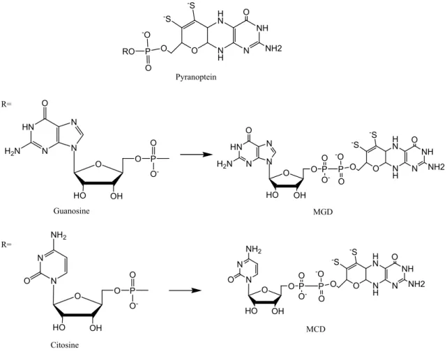
Figure I.2: The pterin cofactor present in mononuclear Mo-containing enzymes and CO
dehydrogenase. The pyranopterin molecule (upper) can attach either a guanosine (middle) or a cytosine (lower) nucleoside via a pyrophosphate link. Adapted from reference [5] 4
Figure I.3: General mechanism for oxidative (left) and reductive (right) reactions catalyzed by
mononuclear Mo-containing enzymes. Adapted from reference [5] 5
Figure I.4: The inorganic nitrogen cycle including the enzymes responsible for each step. The
oxidation state of each compound is indicated between parentheses. The pathways are identified as follow: black arrows, respiratory pathway (denitrification); yellow arrows, dissimilatory and assimilatory ammonification (note that nitrate reduction is indicated only as black arrow); green arrow, nitrogen fixation; red arrows, nitrification; blue arrows, ANAMOX. Adapted from reference [17] 7
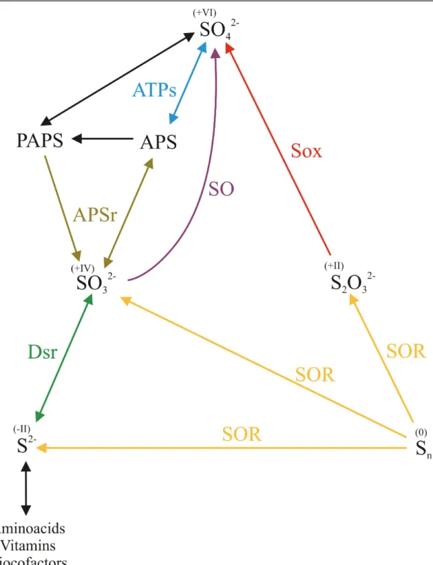
Figure I.5: Simplified biogeochemical S-cycle, including the enzymes responsible for each step. The
oxidation state of each compound is indicated between parentheses. The pathways are identified as follow: yellow arrows, elemental sulfur disproportionation; red arrow, thiosulfate oxidation; purple arrow, sulfite oxidation; blue, brown and green arrows, sulfate-reducing and sulfide-oxidizing pathways. There are many other sulfur species that comprise the cycle of sulfur which were omitted for
simplicity 9
Figure I.6: Left: Active site structure of the three families of mononuclear of mononuclear
Mo-containing enzymes. Right: active site structure of the periplasmic Dd NapA and Rs NapAB enzymes, and membrane-bound Ec NarGH and Ec NarGHI. X and Y represent ligands such as oxygen (oxo, hydroxo, water, serine, aspartate), sulfur (cysteine) and selenium (selenocysteine) found in the several enzymes of the dimethylsulfoxide reductase family. Reproduced from reference [17] 14
Figure I.7: Overall three-dimensional structure of NarGHI from E. coli K12. The names of the
respective subunits together with the metal cofactors are indicated. Reproduced from reference [17] 16
Figure I.8: A) Respiratory nitrate reduction by Nar enzymes. B) Gene organization of the nar gene
clusters. Note the symbols “-” (short distance in the DNA sequence) and “-//-” (long distance in the DNA sequence). Reproduced from reference [17] 19
Figure I.9: Overall three-dimensional structure of NapA from Desulfovibrio desulfuricans ATCC
27774. Reproduced from reference [17] 21
Figure I.10: A) Dissimilatory nitrate reduction by Nap enzymes. B) Gene organization of the nap gene
clusters. Reproduced from reference [17] 23
Figure I.11: A) Pathway for nitrate assimilation by Nas enzymes. The figure depicts the enzymes of the
Klebsiella/Rhodobacter (left) and Cyanobacteria (right) groups. B) Organization of the gene clusters from the discussed examples. The meaning of the symbols “-” and “-//-” are given in the caption to Fig.
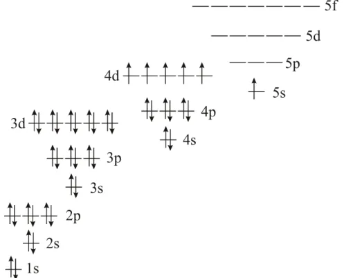
state 30
Figure I.13: relative order of the energy levels of, Left: the five 4d orbitals for cubic and two common
distorted octahedral geometries, Right: energy levels of the three lowest 4d orbitals under axial and two degrees of rhombic distortions. Energy levels were extrapolated from former calculations of the 3d orbitals of Ni(I), Ni(III) and Cu(II) [126] , and DFT calculations of reference [125] 31
Figure I.14: Electron spin energy levels behavior for S=1 in a tetragonal (a-without ZFS, b-weak ZFS
and c-strong ZFS) and S=1/2 with BL parallel to the z-axis. Allowed transitions are depicted in blue. The forbidden transitions (by the EPR selection rule Δms=±1) are in red. Note that a single EPR line should be observed in all cases with the only exception of the triplet state with D~hν 32
Figure I.15: 3D structure of the 4Fe-4S cluster from Dd NapA. Element color code, brown: iron,
yellow: sulfur, grey: carbon. Heteroatoms are represented as balls and cysteine sidechains as sticks 34
Figure II.1: a) Purification flowchart and elution order of known proteins. b) SDS-PAGE of the pools
having nitrate reductase activity after each chromatographic step 40
Figure II.2: SDS-PAGE of the pools having nitrate reductase activity after each chromatographic step 42
Figure III.1: UV-Vis absorption spectrum of pure Dd NapA 50
Figure III.2: Plot of specific activity (left axis, open circles) and total units (right axis, filled circles) as
a function of the enzyme concentration. One unit of nitrate reductase activity (U) corresponds to one
μmol of nitrite formed per minute 51
Figure III.3: Plot of specific activity as a function of the pH 52
Figure III.4: Plot of enzyme inhibition by azide (up-left panel) and cyanide (up-right panel),
perchlorate (down-left panel) and tiocyanate (down-right panel). The red lines are arbitrary fittings
included to guide the eye 53
Figure III.5: Time-scan of methyl viologen oxidation recorded at 600 nm. Red and blue lines
correspond to nitrate and chlorate reductase activity, respectively 54
Figure III.6: Mo(V) EPR spectra obtained in Dd NapA at 100 K [93] together with simulation (grey
lines). a) as-prepared sample, b) as-prepared sample reduced with 5 mM sodium dithionite, 4 c) idem b but after addition of 100 mM sodium nitrate, d) idem c but oxidized with air, and 5 e) idem b but added 20 mM cyanide and followed by air oxidation. EPR parameters used in simulations are given in Table
III.1 56
Figure III.7: Low temperature (25 K) EPR spectra of Dd NapA [93] samples together with simulation
(grey lines) a) sample reduced with 5 mM sodium dithionite, b) idem a after adding 100 mM sodium nitrate, and c) idem a but containing 20 mM cyanide. The EPR parameters used in simulation of the FeS center signal were g1=2.049 (12), g2=1.952 (12) and g3=1.906 (21) for spectra a and b (Linewidths in Gauss between parenthesis). The parameters for spectrum c were the same except g3=1.903 (23). The parameters used for Mo(V) signals are given in Table III.1 57
Figure III.8: Redox titrations of Dd NapA at room temperature monitored by EPR. Upper panel: low
temperature spectra recorded at the different electrochemical potentials. Lower panel: Circles: FeS signal; Triangles: low potential Mo(V) signal. See II.11 for details. In the lower panel, the inset shows the same data but in a different scale 60
mM of potassium nitrate. Panel a) and b) show the spectra recorded at 100 K and 25 K, respectively 61
Figure III.10: Mo(V) species obtained in turnover conditions. a) Enzyme solution with 5 mM
Zn-reduced methyl viologen, b) idem a) reacted with 100 mM potassium nitrate, c) idem b) but in D2O
exchanged solutions 63
Figure III.11: nitrate species 3D representation of the Mo-site of Dd NapA superimposed with the
electron density map contoured at 3.0 σ 67
Figure III.12: cyanide species 3D representation of the Mo-site of Dd NapA as balls and sticks and
superimposed with the electron density map contoured at 3.0 σ. The sixth coordination position of the Mo ion was modeled with: a) oxygen, b) sulfur, c) cyanide bound through the nitrogen, and d) cyanide bound through the carbon. Green and red surfaces in a) represent positive and negative FO-FC peaks,
respectively 68
Figure III.13: a) representation of the ClO4- anions superimposed with the electron density map contoured at 3.0 σ, b) representation of the ClO4- anions superimposed with the FO-FC map before modeling the ClO4-, and c) representation of the global structure of Dd NapA with ClO4- anions
blocking the funnel-like cavity 70
Figure III.14: Three-dimensional representation of the Mo-site of Dd NapA superimposed with the
sulfur K-edge SAD signals peaks (upper panel) and the final total electron density map (lower panel) 71
Figure III.15: UV-Vis absorption spectrum of Cn NapAB 73
Figure III.16: single-crystals obtained from as-prepared samples of Cn NapAB. Arrows indicate
crystals that were measured. Crystallization conditions: a) 0.1M succinate pH 7.0, 25% PEG 3350, b) 0.1M bis-Tris pH 5.5, 25% PEG 3350, and c) 0.1M bis-Tris pH 6.5, 25% PEG 3350 74
Figure III.17: Overall three-dimensional structure of NapAB from Cupriavidus necator H16. The large
and small subunits are represented as blue and red ribbons, respectively 75
Figure IV.1: primary sequence alignment of periplasmic nitrate reductases from Rhodobacter
sphaeroides (Rs), Paracoccus pantotrophus (Pp), Cupriavidus necator (Cn), Escherichia coli (Ec) and Desulfovibrio desulfuricans (Dd). Residues conservation: Red: 100%, Blue: 75%, Dark green: 50%,
Green: not conserved amino acids only in Dd NapA. Amino acids highlighted in cyan are located at the funnel cavity and potentially involved in substrate promiscuity differences 80
Figure IV.2: a) front and b) top views of the amino acids sidechains from the highly conserved residues
of dimeric Naps that are different in Dd NapA. Red lines envelop the amino acids that interact directly with the funnel cavity. The 3D coordinates of the sidechains correspond to the structure of Rs NapAB
[84] 81
Figure IV.3: superimposition of the amino acids sidechains potentially involved in substrate specificity
that are highlighted in cyan in figure IV.1. Color code: sticks in CPK depict the amino acids of Rs NapAB and in dark red the amino acids of Dd NapA. The conserved cysteine and methionine are also shown sticks in CPK color code. The backbone shown in wires corresponds to Rs NapA 82
Figure IV.4: Structure of the Mo site of a) as-prepared, b) nitrate species, c) cyanide species, d)
Dd NapA. Plot adapted from [84] shows the midpoint redox potentials of the metal cofactors in both the
free (red and blue are NapB and NapA, respectively) and complexed forms (black). In yellow is the Ecat value reported in [143]. In green are represented the potentials of the Dd NapA cofactors. Arrows depict the redox potential modulation produced for enzyme- partner and enzyme-substrate interactions 88
Figure IV.6: mechanism of nitrate reduction proposed on the basis of the 3D crystallographic structure
[80] 90
Figure IV.7: new proposal of the mechanism of nitrate reduction on the basis of the EPR results. In red
I
NTRODUCTION
I.1 Molybdenum in nature 2
I.2 The N-cycle 6
I.3 The sulfur metabolism in SRB: the Desulfovibrio genus 8 I.4 The nitrogen metabolism in D. desulfuricans ATCC 27774 and C. necator
H16 11
I.5 Nitrate reductases: classification and structural properties 14
I.5.1 Respiratory Nitrate Reductases (Nar) 15 I.5.1.1 Molecular and spectroscopic properties 15 I.5.1.2 Gene organization, expression control, and mechanism of action 18 I.5.2 Periplasmic Nitrate Reductases 20 I.5.2.1 Molecular and spectroscopic properties 20 I.5.2.2 Genes organization, expression control, and mechanism of action 22 I.5.3 Assimilatory Nitrate Reductases 26 I.5.3.1 Molecular and spectroscopic properties 26 I.5.3.2 Genes organization, expression control, and mechanism of action 28
I.6 Electronic configuration of the metal cofactors of NapA and its detection by
EPR spectroscopy 30
I.6.1 Electronic properties of the Mo ions 30 I.6.2 Electronic properties of the [4Fe-4S] clusters 33
- 2 - I.1 Molybdenum in nature
Molybdenum (Mo, element 42) is a transition metal belonging to the group 6 of the periodic table of the elements located at the 5th period. Molybdenum does not occur free in nature and is usually found in molybdenite (MoS2) and wulfenite (PbMoO4) ores or recovered as a by-product of copper and tungsten mining. Molybdenum has seven naturally occurring isotopes, which are listed in table I.1 together with their respective abundances and nuclear spins. As shown in this table, only the isotopes 95Mo and 97Mo have non-zero nuclear spins.
Table I.1: naturally occurring isotopes of Mo together with their atomic masses, natural abundances, nuclear
spins, and magnetic moments.
Molybdenum is relevant in biology because of its presence in the active site of a wide number of enzymes with key roles in the metabolism of both organic and inorganic compounds [1, 2]. The best characterized examples of Mo-containing proteins correspond to the enzyme nitrogenase [3], CO dehydrogenase [4] and several enzymes which have been grouped in the big family of the mononuclear Mo-enzymes [1, 2].
Molybdenum is present in the active site of the nitrogenase as part of a complex heterometalic cluster (Figure I.1, bottom). In the case of the mononuclear enzymes, Mo is bonded to one or two pyranopterin molecules and to a variable number of ligands such as oxygens (oxo, hydroxo, water, serine, and aspartate), sulfur (sulfido and cysteine) and selenium (selenocysteine) atoms (Figure I.1).
Isotope Atomic mass (m
a/u) Natural abundance (atom %) Nuclear spin (I) Magnetic moment (μ/μN) 92 Mo 91,9068090 14.84 0 94Mo 93,9050853 9.25 0 95Mo 94,9058411 15.92 5/2 -0.9142 96 Mo 95,9046785 16.68 0 97Mo 96,9060205 9.55 5/2 -0.9335 98 Mo 97,9054073 24.13 0 100Mo 99,9074770 9.63 0
- 3 -
Figure I.1: The active sites of Mo-containing enzymes. Heteroatoms are depicted as balls and aminoacid
sidechains as stick. Element color code: grey: carbon, red: oxygen, blue: nitrogen, yellow: sulfur, orange: selenium, violet: molybdenum, cyan: copper, brown: iron. FMF stands for Formylmethanofuran.
Xantine oxidase Aldehyde oxidoreductase Isoquinoline oxidoreductase FMF dehydrogenase Isonicotinate dehydrogenase Arsenite oxidase Piridoxal oxidase Sulfite oxidase
Eukaryotic Nitrate reductase
Formate dehydrogenase (from Escherichia coli) Respiratory Nitrate
reductase (from NarGHI)
Respiratory Nitrate reductase (from NarGH)
Formate dehydrogenase (from D. gigas and E. coli) Periplasmic Nitrate reductase
Chlorate reductase Perchlorate reductase Nitrite oxidase Biotin-sulfoxide reductase Polysulfide reductase Selenate reductase Formate dehydrogenase Ethylbenzene dehydrogenase DMSO reductase TMAO reductase Carbon monoxide dehydrogenase Nitrogenase
The active sites of mononuclear Mo-enzymes
- 4 -
The pyranopterin molecule is an organic ligand that can be either in the monophosphate form (R=H) or with a nucleotide molecule attached by a pyrophosphate link (Figure I.2). This type of Mo-site is also present in CO dehydrogenase but, in addition, the Mo is bound to a Cu ion via a sulfur bridge (Figure I.1, bottom). There are other examples of proteins containing Mo included in heterometalic cluster whose role and structures has not been established yet.
Figure I.2: The pterin cofactor present in mononuclear Mo-containing enzymes and CO dehydrogenase. The
pyranopterin molecule (upper) can attach either a guanosine (middle) or a cytosine (lower) nucleoside via a pyrophosphate link. Adapted from reference [5].
All the Mo-containing enzymes shown in Figure I.1 catalyze a huge variety of both oxidative and reductive reactions which are crucial in many biological processes occurring in nature such as the nitrogen, sulfur and carbon metabolisms. Most of these reactions are catalyzed by mononuclear Mo-enzymes. With a few exceptions, these enzymes catalyze the transfer of an oxygen atom from water to the substrate (or vice versa) in reactions that imply a
O N H H N N NH O NH2 -S -S O P O RO -O O HO N N OH O P O O -O NH2 HN N N N O H2N O HO OH O P O O -Pyranoptein Guanosine Citosine O N H H N N NH O NH2 -S -S O P O -O HN N N N O H2N O HO OH O P O O -O N H H N N NH O NH2 -S -S O P O -O O HO N N OH O P O O -O NH2 MGD MCD R= R=
- 5 -
net exchange of two electrons between the enzyme and the substrate and in which the metal ion cycles between the redox states +4 and +6 (Figure I.3).
Figure I.3: General mechanism for oxidative (left) and reductive (right) reactions catalyzed by mononuclear
Mo-containing enzymes. Adapted from reference [5].
This work will be devoted to the study of the mononuclear Mo-containing enzyme nitrate reductase isolated from the periplasm of Desulfovibrio desulfuricans ATCC 27774 and Cupriavidus necator H16. Desulfovibrio desulfuricans is a sulfate reducing bacterium that produces a periplasmic nitrate reductase when grown in denitrifying conditions. Cupriavidus necator is a chemolithotrophic facultative bacterium that produces a periplasmic nitrate reductase when aerobic cultures reach the late death-phase of growth. In the following pages, it will be discussed briefly the main metabolic routes in which these enzymes participate, the molecular and structural properties of the best characterized nitrate reductases, including the current knowledge on molecular biology aspects which are essential to understand their specific function in the cell. Since one of the NRs studied here belong to a sulfate reducing organism, the biogeochemical cycle of the sulfur is also outlined despite this enzyme is not directly involved in this cycle. Furthermore, as an important part of this work is pointed to the
Oxidative Reactions
Substrate
Product
Mo(VI)
Mo(IV)
Mo(IV)
Mo(VI)
Electron flow Electron flow Reductases OxidasesAcceptor
(Oxidized)
Donor
(Oxidized)
Donor
(Reduced)
Acceptor
(Reduced)
+2 e -+2 e --2 e --2 e-Substrate
Product
Reductive reactions
Oxidative Reactions
Substrate
Product
Mo(VI)
Mo(IV)
Mo(IV)
Mo(VI)
Electron flow Electron flow Reductases OxidasesAcceptor
(Oxidized)
Donor
(Oxidized)
Donor
(Reduced)
Acceptor
(Reduced)
+2 e -+2 e --2 e --2 e-Substrate
Product
Reductive reactions
- 6 -
characterization of the Mo ion and 4Fe-4S cluster by EPR spectroscopy, it is briefly analyzed the electronic configuration of both metallic centers in the relevant redox states necessary to understand this work.
I.2 The N-cycle
Nitrogen is a vital component of essential biomolecules such as proteins and nucleic acids. In the biosphere, nitrogen cycles between the oxidation states +V and –III producing many species that constitute the biogeochemical cycle of nitrogen (N-cycle). This cycle involves a number of redox reactions in which prokaryotes play the main role since only they have the enzymes carrying out these processes [6].
The N-cycle involves a number of redox processes, which are shown schematically in Figure I.4. These processes are completely independent in some cases but share some steps in others.
The dissimilatory processes involve the conversion of nitrate into N2 (respiration/denitrification) or into ammonia (respiration/ammonification) [6-10]. Denitrification is performed by a group of enzymes used by the cell to generate the proton motive force (PMF) across the cytoplasmic membrane [11]. Dissimilatory ammonification is also started with the reduction of nitrate to nitrite, but then nitrite is reduced to ammonia. Both denitrification and dissimilatory ammonification are energy conserving and can be used as an electron sink, i.e., to remove the excess of reducing power in the cell. In addition, ammonification may play an important role in cell detoxification.
The assimilatory process, which also involves the conversion of nitrate to ammonia, starts with the reduction of nitrate in the cytoplasm and is used by the cell to incorporate nitrogen into biomolecules [6, 10, 12-14]. The process called nitrification is the only one that involves oxidative reactions, and is attributed exclusively to bacteria from the Nitrosomonas and Nitrobacter genus [15, 16].
- 7 -
Figure I.4: The inorganic nitrogen cycle including the enzymes responsible for each step. The oxidation state of
each compound is indicated between parentheses. The pathways are identified as follow: black arrows, respiratory pathway (denitrification); yellow arrows, dissimilatory and assimilatory ammonification (note that nitrate reduction is indicated only as black arrow); green arrow, nitrogen fixation; red arrows, nitrification; blue
arrows, ANAMOX. Adapted from reference [17].
The N-cycle is completed with nitrogen fixation, in which the enzyme nitrogenase reduces nitrogen from both the atmosphere and denitrification to ammonia [14]. A fifth less characterized process named ANAMOX (anaerobic ammonium oxidation), which involves
- 8 -
both oxidative and reductive reactions, is used for bacteria to grow in chemolithoautotrophic conditions (i.e. organisms that use carbon dioxide from the environment as carbon source for metabolic processes and use inorganic compounds such as nitrogen, iron, or sulfur for the energy to power these processes) using ammonia as electron donor and nitrite as electron acceptor [6, 18, 19].
I.3 The sulfur metabolism in SRB: the Desulfovibrio genus
The Sulfur, an essential component of life, is a ubiquitous element of the earth that cycles between the +VI and -II oxidation states, and is mostly present as elemental sulfur, sulfide and sulfate salts. The various inorganic and organic states of the sulfur in nature constitute the biogeochemical sulfur cycle (S-cycle). This cycle includes reductive and oxidative reactions, which are mainly carried out by microorganisms of archaea and bacteria, and much less by eukaryotes. A simplified version of the S-cycle is schematized in Figure I.5.
The oxidative reactions of sulfur are carried out by microorganisms such as Acidianus, Paracoccus, Thiobacillus, green and purple sulfur bacteria, among others [20]. These microorganisms use the reduced sulfur species (sulfide, elemental sulfur, thiosulfate) to gain electrons for energetic purposes and produce sulfate as the major final product. These reactions are catalyzed by a huge variety of enzymes. For example, the thermo- and acidophilic archaeon Acidianus ambivalens [21], can metabolize elemental sulfur in a reaction catalyzed by the sulfur oxygenase-reductase (SOR), leading to its disproportionation to sulfide, sulfite and thiosulfate (Figure I.5, yellow arrows). Thiosulfate can be further oxidized to sulfate by the multienzymatic system called Sox (Figure I.5, red arrow), an enzyme that was already isolated from microorganisms such as Paracoccus pantotrophus, green, and purple sulfur bacteria [20].
- 9 -
Figure I.5: Simplified biogeochemical S-cycle, including the enzymes responsible for each step.
The oxidation state of each compound is indicated between parentheses. The pathways are identified as follow: yellow arrows, elemental sulfur disproportionation; red arrow, thiosulfate oxidation; purple arrow, sulfite oxidation; blue, brown and green arrows, sulfate-reducing and sulfide-oxidizing pathways. There are many other
- 10 -
Another oxidative reaction is carried out by the sulfite oxidase (SO) [22]. This is a molybdenum- and heme-containing enzyme belonging to the mononuclear Mo-enzymes (Figure I.2) that catalyzes the oxidation of sulfite to sulfate (Figure I.5, purple arrow). In mammals, this is the terminal step in the oxidative degradation of cysteine, methionine, and membrane components such as sulfatides. Deficiency of this enzyme in humans leads to the accumulation of sulfite producing major neurological abnormalities and early death [23]. Bacterial sulfite oxidases have been purified from Thiobacillus, Starkeya novella, among others and the main difference with respect to the eukaryotic enzymes is that they lack the heme cofactor [24]. Direct oxidation of sulfite to sulfate occurs in various photo- and chemotrophic sulfur oxidizing microorganisms as the final step in the oxidation of reduced sulfur compounds to obtain reducing equivalents for energetic balance [25].
The reductive pathway (reduction of sulfate to sulfide) is carried out by the sulfate reducing bacteria, yeasts and green plants. They can use sulfate as sulfur source (assimilatory process) to synthesize amino acids, vitamins, and cofactors, among others. Furthermore, sulfate reducing bacteria of genus such as Desulfobacter, Desulfotomaculum and Desulfovibrio utilize sulfate as the acceptor of the reducing equivalents generated during metabolism. This means that sulfate is used as an oxidizing agent for the dissimilation of the organic matter (dissimilatory process), as oxygen does in aerobic organisms during the respiration process.
As sulfate is a molecule with low reactivity, the first step of the dissimilatory sulfate reduction is the activation of the sulfate anion by ATP in a reaction catalyzed by the ATP sulfurylase (ATPs, Figure I.5, blue arrow). The product of this reaction is the Adenylyl-Phospho-Sulfate (APS), which is the substrate of the APS reductase (APSr), which catalyzes the two-electron reduction of APS to sulfite and releasing AMP. In the assimilatory process, the bi-phosphorilated form, Phospho-Adenylyl-Phospho-Sulfate (PAPS), is formed instead of APS, which is the substrate of an APS reductase as well (brown arrows). The sulfite thus
- 11 -
produced is directly reduced to sulfide in a six-electron reaction catalyzed by the dissimilatory sulfite reductase (Dsr - Desulfoviridin, Desulforubidin, Desulfofuscidin and P-582 - green arrows). Dsr can also produce trithionate and thiosulfate; however, the production of these intermediates is an irreversible process and may not ocurr in intact cells. In contrast, the assimilatory sulfite reductases do not produce these free intermediates [26].
ATPs, APSr and Dsr can catalyze also the inverse reactions, and are used by chemolithotrophic aerobic bacteria to oxidize sulfide to sulfate.
The best characterized sulfate reducing bacteria belong to the Desulfovibrio genus [27]. About 60 Desulfovibrio species have been identified and the genomes of D. vulgaris Hildenborough and D. desulfuricans G20 (to be reclassified as D. alaskensis) have been already annotated [28, 29]. All the strains of the Desulfovibrio genus are gram negative bacteria that grow under anaerobic conditions using lactate, pyruvate and/or formate as carbon source. As explained above, the reducing equivalents produced during the metabolism of carbon sources are transferred to the electron acceptors using the Cobalt/Zinc-containing ATPs [30], the heterodimeric APSr that contains a Flavin and two 4Fe-4S cofactors [31], and the tetrameric Dsr (dimer of dimers - Desulfoviridin), which holds a FAD and two sirohemic cofactors [32].
I.4 The nitrogen metabolism in D. desulfuricans ATCC 27774 and C. necator H16 Formerly, Desulfovibrio species and SRB in general were assumed to be microorganisms using only a limited spectrum of organic substrates, in which sulphate is used as terminal electron acceptor. Currently, SRB appear to be the microorganisms that reduce the largest number of electron acceptors, including inorganic sulfur compounds and various other organic and inorganic compounds like nitrate and nitrite [33-35]. Specifically, Desulfovibrio desulfuricans ATCC 27774 can grow using nitrate/nitrite as the electron acceptors under anaerobic conditions. The dissimilatory reduction of nitrate and nitrite (called dissimilatory
- 12 -
ammonification) can function as the sole energy-conserving process in a few Desulfovibrio species such as D. desulfuricans, D. furfuralis, D. profundus, D. oxamicus, D. simplex, D. multispirans and D. termitidis [34, 36-43]. A dissimilatory nitrate reduction has been also reported more recently with Desulfotomaculum thermobenzoicum [44], Desulfobacterium catecholicum [45], Desulforhopalus singaporenssi [46], Thermodesulfovibrio islandicus [47], Thermodesulfobium narugense [48] and Desulfobulbus propionicus [45]. Dissimilatory nitrite reduction by SRB is widespread, but strains capable of nitrate ammonification are far less common [40, 41].
Desulfovibrio desulfuricans subsp. desulfuricans ATCC 27774 (DSM 6949) is the best studied ammonifying strain of a sulfate reducer from a physiological and a biochemical point of view. The growth yield of D. desulfuricans ATCC 27774 grown in lactate/nitrate medium is higher when compared with cultures where sulfate is the terminal electron acceptor [33]. The free energy change per hydrogen oxidized is about four times higher with nitrate than with sulfate. Desulfovibrio desulfuricans ATCC 27774 is also active in the bidirectional transformation of aromatic aldehydes (such as benzaldehyde, 3-hydroxybenzaldehyde) under nitrate-respiring conditions and the direction of transformation (i.e. oxidation or reduction) is mainly regulated by reductant availability [49].
In D. desulfuricans ATCC 27774, nitrate reduction is carried out exclusively by the soluble enzyme periplasmic nitrate reductase (NapA). The reduction of nitrite to ammonia is carried out by the multihemic cytochrome c Nir (NrfAH) which is a heterodimeric membrane-bound enzyme. These processes would be coupled to the translocation of protons to the periplasm, generating an electrochemical gradient for the synthesis of ATP.
In Cupriavidus necator H16, three types of prokaryotic nitrate reductases were found (Nas, Nar and Nap), which are expressed under different culture conditions. Nas is expressed in the presence of nitrate independently of the presence of oxygen, and is repressed when the intracellular ammonia level is high, which is in agreement with its assimilative role. However,
- 13 -
expression of Nar is induced in the presence of nitrate under strict anaerobic conditions. The enzymes Cu-Nir, Nor and Nos, needed to complete the denitrification pathway (see Figure I.4), are also expressed under this conditions. Unexpectedly, Nap expression is not directly linked to the nitrogen metabolism since Nar defective mutants of Cn H16 are unable to grow under denitrifying conditions. Nap activity is detected when Cn H16 cells cultured in aerobic conditions reach stationary phase of growth, and is maximally accumulated in the late death phase [50]. Maximal expression of Nap, as judged by the purification yield, is achieved when cultures are carried out in chemolithotrophic conditions, using fructose and ammonia as carbon and nitrogen sources, respectively. This indicates that the role of Cn Nap is different to that attributed to the Dd NapA as explained in the next paragraphs.
- 14 -
I.5 Nitrate reductases: classification and structural properties
All the reductive branches of the N-cycle involve the conversion of nitrate to nitrite. This step is performed by distinct enzymes that catalyze the unique reaction
NO3¯ + 2 H+ + 2e- → NO2¯ + H2O Eº = 420 mV vs. NHE
Nitrate reductases (EC 1.7.99.4) have been classified by taking into consideration source, localization of the enzyme in the cell, molecular properties of the catalytic center, and function. Several nitrate reductases have been obtained from both prokaryotic and eukaryotic organisms.
Figure I.6: Left: Active site structure of the three families of mononuclear of mononuclear Mo-containing
enzymes. Right: active site structure of the periplasmic Dd NapA and Rs NapAB enzymes, and membrane-bound
Ec NarGH and Ec NarGHI. X and Y represent ligands such as oxygen (oxo, hydroxo, water, serine, aspartate),
sulfur (cysteine) and selenium (selenocysteine) found in the several enzymes of the dimethylsulfoxide reductase family. Reproduced from reference [17].
- 15 -
All of them are mononuclear Mo-containing enzymes that, according to the Hille´s classification [1], belong to the dimethyl sulfoxide (DMSO) reductase family with the only exception being eukaryotic nitrate reductases, which belong to the sulfite oxidase (SO) family (Figure I.6). The prokaryotic enzymes can be also sub-grouped as respiratory nitrate reductase (Nar), periplasmic nitrate reductases (Nap) and assimilatory nitrate reductases (Nas) [51]. Their characterization has allowed gaining considerable information on the molecular basis of nitrate reduction involved in all the branches of the N-cycle.
I.5.1 Respiratory Nitrate Reductases (Nar)
I.5.1.1 Molecular and spectroscopic properties. The Nar enzyme has been isolated
from many nitrate-respiring and denitrifying bacteria [52-58]. All the Nars isolated so far are heterotrimeric enzymes composed of the subunits NarG (112-140 kDa), NarH (52-64 kDa) and NarI (19-25 KDa). NarG and NarH are placed in the cytoplasm anchored to the membrane through NarI (Figure I.7). The crystal structures of the oxidized forms of NarGH [59] and NarGHI [60] were reported at resolutions of 2.0 Å and 1.9 Å, respectively. NarGHI has a flower-like arrangement with dimensions of 90×128×70 Å3. The NarI subunit, which is completely immersed in the membrane, is associated with the NarGH dimer through a hydrophobic patch present in NarH. The global arrangement of the catalytic subunit NarG is similar to those from other enzymes belonging to the DMSO reductase family [61], having four domains with α/β type folding. In this sense, it presents high homology with Dd Nap, Ec Fdh-N, Ec Fdh-H and Dg Fdh [59-61]. As shown in Figure I.7, NarG contains the active site, a Mo-bisMGD cofactor, and an iron-sulfur center of the [4Fe-4S] type (FS0). NarH contains one [3Fe-4S] (FeS4) and three [4Fe-4S] clusters (FeS1-3), whereas NarI contains two b-type hemes responsible for the QH2 oxidation and proton translocation. These eight redox centers are separated by 12-14 Å giving an electron transfer pathway of about 90 Å.
- 16 -
Furthermore, all these metal cofactors are present in all the Nars isolated so far with the only exception of the membrane bound Nar from Pseudomonas nautica 617 (Pn NarGHI), in which biochemical studies suggest that one of the haems b of the NarI subunit is replaced by a c-type heme (Correia et al., to be published).
Figure I.7: Overall three-dimensional structure of NarGHI from E. coli K12. The names of the respective
subunits together with the metal cofactors are indicated. Reproduced from reference [17].
The structure of NarGHI reveals that the active site is composed of a molybdenum atom coordinated to four sulfur atoms from two pterin cofactors, as found in all the members of the DMSO reductase family, and a bidentate coordination with the oxygen atoms from the
NarI
Cytoplasm
Membrane
Periplasm
Mo-
bis
MGD
NarG
NarH
FeS0
FeS3
FeS4
FeS2
FeS1
Heme b
PHeme b
D- 17 -
side chain carboxylate group of an aspartate molecule (Asp222) (Figure I.6) [60], which had been not observed in a mononuclear Mo-enzyme. The soluble NarGH shows a similar active site structure with two pterin molecules but, in contrast, the coordination with aspartic acid is through only one oxygen with the sixth coordination position being occupied by an oxo group at 1.8 Å (Figure I.6) [59]. These differences cannot be attributed to the redox state of the sample since both NarGHI and NarGH were crystallized in aerobic conditions and therefore the reported structures must represent oxidized forms of the enzymes. Another novel aspect of Nar is the unusual coordination of FeS0 by one histidine and three cysteines, which was found only once before in Ni-Fe Hydrogenase from Desulfovibrio gigas [59, 60, 62].
The redox and EPR properties of Nar from several sources have been the subject of several studies [52, 54, 58, 63-65]. Nars show two types of pH-dependent Mo(V) ion EPR signals, which show, in addition, resonance lines split by a solvent exchangeable proton. The Mo(V) ion species associated with these signals were named the low-pH (gav=1.984, Aav=9.3G) and the high-pH (gav=1.977, Aav=3.4 G) forms. The molecular structure of the active site of NarGHI does not show an hydroxyl/water ligand [60], as one would expect from the EPR data, and the coordination of the Mo site in NarGH presents an Oxo ligand and one oxygen from a carboxylate (Figure I.6) [59]. These results indicate that the bidentate coordination is open upon reduction to Mo(V) giving rise to the solvent exchangeable low- and high-pH forms of the enzymes. Recent studies performed in Pn NarGHI shows, besides the low and high pH signals, two novel EPR signals with no differences in D2O exchanged samples that are obtained upon nitrate oxidation of reduced enzyme (Correia et al, to be published). These two species might be associated, in principle, with Mo(V) species having the molecular structure seen in both crystal structures of Ec Nar. The catalytic involvement of all these Mo(V) species is far from being elucidated and additional work is necessary to clarify their roles [52, 63].
- 18 -
EPR studies in Ec Nar detected the presence of only four FeS centers while the structures of NarGH and NarGHI demonstrated the existence of five FeS centers. The EPR signal of the [3Fe-4S] cluster (gav=2.00) and one additional signal with two components (major and minor) was attributed to a single [4Fe-4S] center in different conformations (major, g1=2.049, g2=1.947, g3=1.870 and minor, g1=2.010, g2=1.885, g3=1.871) [64-66]. The EPR properties of the remaining EPR-detected two centers could not be determined precisely because of magnetic interaction between centers, which broaden the spectra making their deconvolution unfeasible. Recently, EPR studies by Rothery et al. performed in a NarG mutant that lacks the molybdenum site showed that the fifth FeS (FeS0) center is a [4Fe-4S] cluster in a high-spin configuration (g1=5.556, g2=5.023) [67].
I.5.1.2 Gene organization, expression control, and mechanism of action. In E. coli,
the genes that code nitrate reductase are grouped in the operon narGHJI , which is located in the chlC locus of the chromosome. NarJ is a polypeptide that participates in NarGH assembly prior to its attachment to NarI in the membrane [68]. The expression of nar genes are nitrate/nitrite regulated by a signal transduction system of kinases-phosphatases, involving the operons narXL and narQP, (Figure I.8) [69]. The operon narXL is located in the chlC locus but narQP is coded out. NarX and NarQ are homologous membrane proteins that detect periplasmic nitrate and nitrite and then activate the NarL and NarP regulators by phosphorylation [70-73]. In this conformation, the regulators bind specific DNA consensus sequences called NarL heptamers [74]. The operons narGHJI (respiratory nitrate reductase), frdABCD (fumarate reductase) and narK are only under the control of NarL, while both NarL and NarP regulate nrfABCDEFG (nitrite reductase) and napFDAGHBC (periplasmic nitrate reductase) [71]. This is consequently, a complex regulatory system that detects the nitrate:nitrite ratio, inactivating NarL when nitrate is in low levels, and favoring nitrite consumption.
- 19 -
E. coli shows an additional Nar coded in the narZWYV operon in the chlZ locus, which is expressed constitutively at low levels, and its proposed role is to cushion the bacterium respiration to switch from oxygen to nitrate respiration [69].
Figure I.8: A) Respiratory nitrate reduction by Nar enzymes. B) Gene organization of the nar gene clusters.
Note the symbols “-” (short distance in the DNA sequence) and “-//-” (long distance in the DNA sequence). Reproduced from reference [17].
Nars in other bacteria such as Paracoccus denitrificans and Paracoccus pantotrophus are coded by operons with the same composition (Figure I.8). These operons code a Nar having basically the same molecular structure, but that differ in respect to those in E. coli in
NO3 -NO3 -H+ H+ NO2 -NO2 -Mo bisMGD FeS0 FeS1 FeS2 FeS3 FeS4 bP bD MQH2 MQ+ 2xH+ 2xH+ NO3 -Cell Wall Periplasm Cytoplasm NarG NarH NarI NarK1 NarK2
A)
B)
Escherichia coli
narLX-//-K-//-GHJI
narU-//-ZYWV
Paracoccus denitrificans
narK-//-GHJI
Paracoccus pantotrophus
narK-//-GHJI
- 20 -
how they control the expression system, with the transcription being under the control of the FNR-like regulator NarR [75].
The proposed mechanism for the reduction of nitrate to nitrite in E. coli K12 is schematized in Figure I.8. The MQH2 situated in the outer part of the cytoplasmic membrane are oxidized by NarI translocating 2 protons into the periplasm. The two electrons are conducted through the electron transfer pathway of the NarGHI complex to the Mo-bisMGD site, where nitrate is reduced consuming two cytoplasmic protons. As the catalytic subunit of Nar is cytoplasmic, nitrate must be introduced into the cell. This is performed by two transporters. The proton:nitrate symporter NarK1 is activated at the beginning of the nitrate respiration and the co-transported protons are consumed in the nitrate reduction. Once the reaction accumulates nitrite, the function of NarK1 is substituted by NarK2, which acts as nitrate:nitrite antiporter, allowing the maintenance of the steady state [76]. This membrane process, when coupled to formate oxidation by the Fdh-N, generates the proton motive force (PMF) necessary for ATP synthesis [11, 77].
I.5.2 Periplasmic Nitrate Reductases
I.5.2.1 Molecular and spectroscopic properties. NapA from D. desulfuricans (Dd
NapA) was the first reported structure for a periplasmic nitrate reductase (Figure I.9) [78, 79]. Dd NapA is a monomeric protein of ellipsoidal shape with an α/β type fold organized in four domains, all involved in cofactor binding. The active site is a hexa-coordinated Mo ion with four sulfur atoms from two pterin ligands, one hydroxo/water group and sulfur from a cysteine as ligands (Figure I.6). In addition, the protein has an iron-sulfur cluster of the type [4Fe-4S], which is supposed to be involved in electron transfer.
A funnel-like cavity is formed from the surface to the catalytic site (~15 Å) due to the spatial arrangement of domains II and III. The hydroxo/water ligand of the molybdenum atom, which is supposed to be in the position where the substrate binds the active site, points into
- 21 -
this channel, suggesting that nitrate entrance and nitrite exit would be via this channel. In contrast, Naps obtained from other sources such as Rhodobacter sphaeroides (Rs Nap) [80], Cupriavidus necator (Cn Nap, formerly Alcaligenes eutrophus, Ralstonia eutropha, Wautersia eutropha) [50] and Paracoccus pantotrophus (Pp Nap) [81, 82] are heterodimeric proteins with a large (80-90 kDa) and small (~15-17 kDa) subunits.
Figure I.9: Overall three-dimensional structure of NapA from Desulfovibrio desulfuricans ATCC 27774.
Reproduced from reference [17].
The only crystal structure reported for an heterodimeric Nap belongs to Rs NapAB which was determined at a resolution of 3.2 Å [83, 84]. The arrangement of the catalytic subunits of Dd NapA and Rs NapA are very similar in terms of metal cofactor content, global fold and domain organization, with a RMSD of 1.26 Å for the 683 Cα atoms. However, their primary sequences reveal a low identity (~35%), although the Mo-bisMGD catalytic site and
Mo-
bis
MGD
- 22 -
[4Fe-4S] cluster binding residues are conserved along with the amino acids proposed to mediate the electron flow from the [4Fe-4S] center to the molybdenum [85, 86]. The structural comparison between all these proteins was recently reported by Moura et al. [61]. The small subunit NapB, which is present in most Naps but absent in Dd Nap, was recently determined at a higher resolution (1.25 Å) using the recombinant NapB protein from Haemophilus influenzae through MAD (multiwavelenght anomalous dispersion) methods [87]. EPR studies performed in Dd NapA and Pp NapAB indicate a Mo-site with high coordination flexibility as shown by the different EPR active species obtained with the enzymes poised at different redox potentials and in the presence of different inhibitors [88-93]. The Pp NapA enzyme in the as-prepared form shows a Mo(V) EPR signal (high g resting) typical of Mo(V) ion species (g1=1.998, g2=1.990, g3=1.981) split by two non exchangeable species with nuclear spin I=1/2, presumed to be the β-methylene protons from the coordinated cysteine seen in the crystal structure of Dd NapA (Aav1= 5.5 G, Aav2=2.8 G). A similar resting EPR signal was detected in Rs NapAB [83]. Under slow-turnover conditions, the resting signal is replaced with a new Mo(V) ion signal (high g [nitrate]) showing a hyperfine split with a single non exchangeable proton (g1=1.999, g2=1.989, g3=1.981, Aav=5.1 G), which was suggested to be a catalytic intermediary. This signal is similar to that obtained in Dd NapA when reacted in the same conditions [94]. The results in Dd NapA also show similar EPR signal in some cases in the presence of inhibitors [93]. The data obtained in both Dd NapA and Rs NapAB suggests that the active sites of the three enzymes are similar. Nevertheless, the coordination sphere deduced from EXAFS studies performed in Pp NapAB for the several Mo(V) species are in contradiction with the results presented in this work, which indicates that besides the bi-pterin coordination, the active site presents three extra ligands. This point will be discussed in this thesis.
I.5.2.2 Genes organization, expression control, and mechanism of action. The nap
- 23 -
contrast to the nar operon, the nap operons present heterogeneity both in gene composition and ordering. Eleven different genes have been identified as components for operons that code Naps in different organisms.
Figure I.10: A) Dissimilatory nitrate reduction by Nap enzymes. B) Gene organization of the nap gene clusters.
Reproduced from reference [17].
Mo bisMGD 4Fe4S NO3 -NO2 -MQH2 MQ+ 2xH+ 2xH+ NO3 -Cell Wall Periplasm Cytoplasm UQ+ UQH2 2xH+ 2xH+ 4Fe4S Mo bisMGD 2xH+ 2xH+ C C C C C C 4Fe4S 4Fe4S 4Fe4S 4Fe4S 4Fe4S 4Fe4S NapA NapB NapC NapG NapH NapA A) B)
Desulfovibrio desulfuricans napCMADGH
Escherichia coli napFDAGHBC Campylobacter jejunii napAGHBLD Wollinella succinogenes napAGHBFLD Haemophylus influenzae napFDAGHBC Shewanella oneidensis napDAGHB Desulfitobacterium hafniense napDGAH
K12
MR1
Cupriavidus necator napEDABC
Paracoccus pantotrophus napEDABC Rhodobacter sphaeroides napKEFDABC Bradyrhizobium japonicum napEDABC Pseudomonas napEFDABCG-179
- 24 -
The napA gene codes the catalytic subunit NapA that contains the Mo-bisMGD active site and a FeS center. The assembling of the apo-protein with the metal cofactors is carried out in the cytoplasm. Then, the folded holo-protein is transported to periplasm by the TAT (Twin Arginine Translocator) system by recognizing the signal-peptide present in NapA [100]. However, this is not valid for all the catalytic subunits since although NapA from Pseudomonas G-179 is found in the periplasm, it lacks this signal peptide. Once in the periplasm, NapA and the product of napB gene are assembled to give the heterodimeric NapAB. NapB is secreted into periplasm by the general secretory pathway Sec [101]. This process occurs for all the Naps except for Dd NapA, which is obtained in monomeric form [94]. This should also be the case for D. hafniense DCB-2 NapA, whose nap operon and whole shotgun genomic sequence does not show the presence of the napB gene.
Almost all the nap operons code the protein NapC (Figure I.10), which is a c-type tetra-hemic membrane-anchored protein (~25 kDa) belonging to the NapC/NirT family [102-104]. This protein is involved in the electron transfer from the quinol pool to periplasmic reductases. When NapC is not coded in the nap operon, its function is substituted by another protein of the NapC/NirT family which is coded out of the operon and is expressed under the required conditions. Furthermore, the operons of E. coli K12 and D. desulfuricans code the ferredoxins NapG and NapH. NapG is a periplasmic protein having 4×[4Fe-4S] clusters. NapH is an integral membrane protein with 2×[4Fe-4S] exposed to the cytoplasm [105, 106]. The nap operon of D. desulfuricans codes for an additional protein called NapM. This is a putative soluble tetrahemic c-type cytochrome with periplasmic localization, and its proposed function is to transfer electrons from NapC to NapA. The remaining genes, napD, napE, napF, napK and napL code for different proteins that are not directly involved in the nitrate reduction.
NapD is a cytoplasmic protein that belongs to the TorD family, which act as chaperones and are always present in operons that code molybdo-enzymes [107]. NapF is a