UNIVERSIDADE DE LISBOA
Faculdade de Medicina Veterinária
RETROSPECTIVE STUDY OF THE ASSOCIATION BETWEEN NEUTERING STATUS AND CHANGES SECONDARY TO DEGENERATIVE MITRAL VALVE DISEASE
ANA MARGARIDA RIBEIRO DA SILVA
CONSTITUIÇÃO DO JÚRI ORIENTADOR
Doutor José Paulo Pacheco Sales Luís Doutora Joanna Dukes-McEwan
Doutora Luísa Maria Freire Leal Mateus
CO-ORIENTADOR Doutor Rodolfo Assis Oliveira Leal Doutor Rodolfo Oliveira Leal
2019 LISBOA
UNIVERSIDADE DE LISBOA
Faculdade de Medicina Veterinária
RETROSPECTIVE STUDY OF THE ASSOCIATION BETWEEN NEUTERING STATUS AND CHANGES SECONDARY TO DEGENERATIVE MITRAL VALVE DISEASE
DISSERTAÇÃO DE MESTRADO INTEGRADO DE MEDICINA VETERINÁRIA
ANA MARGARIDA RIBEIRO DA SILVA
CONSTITUIÇÃO DO JÚRI ORIENTADOR
Doutor José Paulo Pacheco Sales Luís Doutora Joanna Dukes-McEwan
Doutora Luísa Maria Freire Leal Mateus
CO-ORIENTADOR Doutor Rodolfo Assis Oliveira Leal Doutor Rodolfo Oliveira Leal
2019 LISBOA
ACKNOWLEDGEMENTS
O meu primeiro agradecimento é dirigido aos meus pais por todo o apoio que sempre me deram e por me tornarem na pessoa que sou hoje. Sem vocês eu não teria chegado aqui. Aos meus amigos que estiveram sempre lá para mim durante todos estes anos. À Joana Bastos, João Mendes, Gonçalo Reis, Sandrine Silva, Miguel Serra, Inês Pinto e Mariana Abreu. Obrigada por todos os momentos e por serem tão espetaculares! Este percurso não teria sido o mesmo sem vocês.
Ao Chico, por estar sempre do meu lado, por me ajudar a acreditar em mim mesma e, sobretudo, pela paciência para me aturar.
À VETuna e ao 55 por serem a minha segunda família e a minha maior alegria nesta faculdade. Nunca vos esquecerei. Aos meus queridos estandartes, obrigada por todo o amor e dedicação que partilharam comigo, e por nos terem dado o prémio antes de eu sair desta casa. Foi a melhor prenda que me podiam ter dado.
Ao GOTA FMV-ULisboa que me ensinou a crescer e que me deu amigos para a vida.
A todos aqueles que me acompanharam nas noites intermináveis no Santa Maria e na Cantina Velha, obrigada por terem tornado as épocas de exame suportáveis e por me ajudarem a não desistir.
Ao meu co-orientador, Professor Rodolfo Leal, por todo o apoio que me deu e por tudo o que me ensinou.
I would also like to thank Jo, my supervisor and mentor that gave me this wonderful opportunity to join the Liverpool cardio team. To Liz, thank you for all the support you gave me and for being such a role model! This thesis wouldn’t have happened without your help.
To all the Liverpool cardio team, Hannah, Fabio, Julie, Sid, Cat, Vicky and Nikki. Thank you for all your kindness and for being wonderful teachers. You always made me feel like part of the team and I couldn’t be more grateful to you guys.
To Marie-Suzanne and Ivo, for being my Erasmus family and my partners in crime. Thank you for making this experience unforgettable and for being such good friends. I’m sure we will meet again!
ABSTRACT
Retrospective study of the association between neutering status and changes secondary to degenerative mitral valve disease
Myxomatous mitral valve disease is the most common cardiovascular disease reported in dogs. Although many patients may remain asymptomatic, some of them progress to left-sided congestive heart failure and develop clinical signs. Little has yet been published regarding the possible influence of the neutering status on changes secondary to myxomatous mitral valve disease. This study aims to assess a possible correlation between neutering status and myxomatous mitral valve disease.
A retrospective study was conducted and included all the cases diagnosed with myxomatous mitral valve disease, consulted at the Cardiology service of the University of Liverpool. To help assess the association between neutering status and cardiac remodelling, dogs were categorized into four groups: FE (female entire), FN (female neutered), ME (male entire) and MN (male neutered).
Retrospective review of echocardiographic data, signalment, and underlying diseases were performed. Echocardiographic measurements were made through offline analysis. Echocardiographic measurements were then compared between groups.
Five hundred and eighty-two dogs (n = 582) were included: female entire (n = 24), female neutered (n = 235), male entire (n = 115) and male neutered (n = 208).
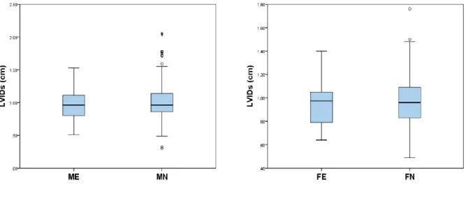
Left ventricular internal diameter at end diastole (LVIDd), left atrial dimension to the aortic root diameter (LA:Ao) and left atrium maximal dimension to the aortic root dimension (LAmax:Ao) were significantly different between ME and MN, with ME dogs presenting higher mean values for LVIDd and higher median LA:Ao and LAmax:Ao measurements. Left ventricular internal diameter at end systole (LVIDs) was not significantly different between ME and MN. There were no significant differences between FE and FN groups.
This study shows that neutering status may influence the development of myxomatous mitral valve disease in male dogs and that entire male dogs could be at higher risk of developing cardiac remodelling secondary to myxomatous mitral valve disease. On the other hand, neutering status doesn’t seem to have an influence on disease progression in female dogs.
Keywords: degenerative myxomatous mitral valve disease, dog, cardiac remodelling,
RESUMO
Estudo retrospetivo da associação entre a esterilização e alterações secundárias à doença mixomatosa da válvula mitral
A doença mixomatosa da válvula mitral é a doença cardiovascular mais prevalente em cães. Apesar da maior parte dos pacientes permanecerem assintomáticos, alguns podem progredir para insuficiência cardíaca esquerda e desenvolver sinais clínicos. Até hoje, existem poucas publicações sobre o possível efeito que a esterilização possa ter no desenvolvimento da doença mixomatosa da válvula mitral. O objetivo deste estudo é avaliar se existe de facto uma relação entre a esterilização e a doença mixomatosa da válvula mitral.
Foi realizado um estudo retrospetivo que incluiu todos os casos diagnosticados com doença mixomatosa da válvula mitral consultados no serviço de Cardiologia da Universidade de Liverpool. Para avaliar a relação entre a esterilização e a presença de remodelação cardíaca, os cães foram categorizados em quatro grupos: FE (fêmeas inteiras), FN (fêmeas esterilizadas), ME (machos inteiros) e MN (machos castrados).
A história pregressa e os dados ecocardiográficos dos animais foram revistos retrospetivamente e as medidas ecocardiográficas foram obtidas por medição offline. Estas medições foram depois comparadas entre os diferentes grupos.
Quinhentos e oitenta e dois cães (n = 582) foram incluídos: fêmeas inteiras (n = 24), fêmeas esterilizadas (n = 235), machos inteiros (n = 115) e machos castrados (n = 208).
Nos resultados obtidos, o diâmetro interno do ventrículo esquerdo no final da diástole (LVIDd), o rácio átrio esquerdo-aorta (LA:Ao) e o rácio diâmetro máximo do átrio esquerdo-aorta (LAmax:Ao) foram estatisticamente significativos entre machos inteiros e machos castrados. Os machos inteiros apresentaram não só um LVIDd médio superior, como também uma mediana de LA:Ao e LAmax:Ao superior aos machos castrados.
Ao mesmo tempo, o diâmetro interno do ventrículo esquerdo no final da sístole (LVIDs) não mostrou ser estatisticamente significativo entre machos inteiros e machos castrados e nenhum dos parâmetros ecocardiográficos anteriormente referidos revelou ser estatisticamente significativo entre fêmeas inteiras e fêmeas esterilizadas.
Este estudo demonstra que a esterilização poderá influenciar o desenvolvimento da doença mixomatosa da válvula mitral e que os cães machos inteiros poderão apresentar um maior risco de desenvolver remodelação cardíaca secundária a esta doença. No entanto, a esterilização não aparenta influenciar a progressão desta doença em cadelas.
Palavras-chave: doença mixomatosa da válvula mitral, cão, remodelação cardíaca,
TABLE OF CONTENTS
ACKNOWLEDGEMENTS ... i ABSTRACT ... ii RESUMO ...iii TABLE OF CONTENTS ... iv LIST OF FIGURES ... vi LIST OF TABLES... vi LIST OF GRAPHICS ... viLIST OF ABBREVIATIONS ... vii
I INTRODUCTION ... 2
II BIBLIOGRAPHIC REVIEW ... 3
1. Myxomatous mitral valve disease ... 3
1.1. The normal mitral valve complex ... 3
1.2. Macroscopic lesions and classification ... 4
1.3. Histopathologic features ... 5
1.4. Mitral regurgitation and cardiac remodelling ... 6
1.5. Cardiovascular sequalae secondary to myxomatous mitral valve disease ... 7
1.6. Activation of neurohormonal pathways and heart failure ... 8
1.7. Classification of heart disease and heart failure ... 9
2. Signalment ... 10
3. History, clinical signs and physical examination ... 10
4. Diagnostic testing ... 12
4.1– Cardiac biomarkers ... 12
4.2– Echocardiography ... 14
4.3– Radiography ... 20
4.4– Electrocardiography ... 22
4.5– Further diagnostic recommendations ... 23
5. Treatment ... 23
6. Prognosis ... 26
III INTRODUCTION AND OBJECTIVES ... 27
IV MATERIALS AND METHODS ... 28
1. Inclusion criteria ... 28 2. Clinical data ... 28 3. Echocardiographic data ... 28 4. Statistical analysis ... 31 V RESULTS ... 32 1. Study population ... 32
2. Echocardiographic findings... 32
VI DISCUSSION ... 35
VII CONCLUSION... 39
REFERENCES ... 40
LIST OF FIGURES
Figure 1. Right parasternal long axis four-chamber view (A) and left ventricular outflow view
(B) (original) ... 15
Figure 2. Right parasternal short axis left ventricle view at the level of the papillary muscles
(A), heart base aorta and left atrium view (B) and heart base pulmonary artery view (C) (original). ... 16
Figure 3. Left parasternal five-chamber (A) and four chamber view (B) (original) ... 17 Figure 4. Left ventricular M-mode image (original) ... 17 Figure 5. Representation of a right parasternal short-axis view used for measuring the LA:Ao
ratio (original) ... 29
Figure 6. Representation of a right parasternal long-axis four chamber view used for
measuring the LAmax:Ao ratio (original) ... 30
Figure 7. Representation of a M-mode image obtained from the right parasternal short axis
view at the level of the papillary muscles used for measuring the LVIDd and the LVIDs (original) ... 30
LIST OF TABLES
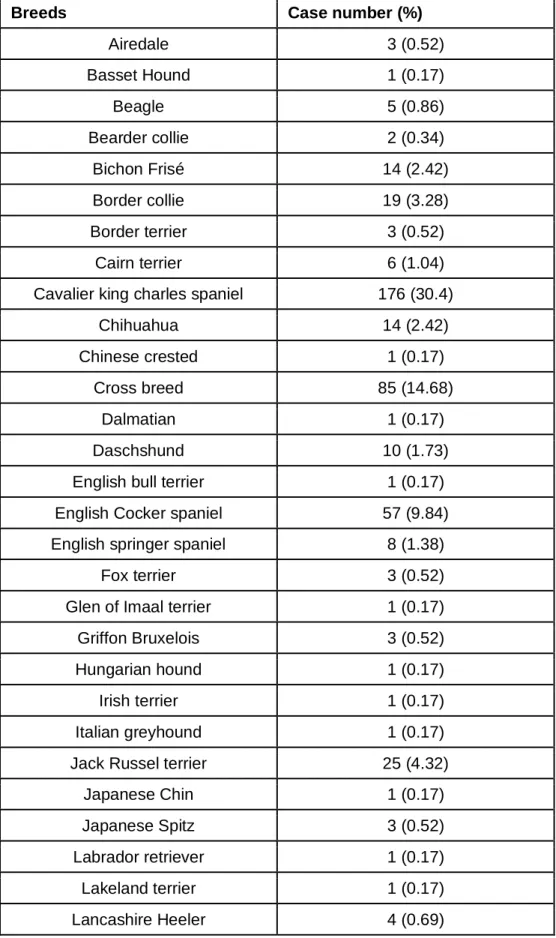
Table 1. Breed prevalence among 582 dogs diagnosed with MMVD ... 49
LIST OF GRAPHICS
Graphic 1. Box-and-whiskers plots of LVIDd between Male Entire (ME) vs Male Neutered
(MN) and Female Entire (FE) vs Female Neutered (FN). ... 33
Graphic 2. Box-and-whiskers plots of LVIDs between Male Entire (ME) vs Male Neutered (MN)
and Female Entire (FE) vs Female Neutered (FN). ... 33
Graphic 3. Box-and-whiskers plots of LA:Ao between Male Entire (ME) vs Male Neutered (MN)
and Female Entire (FE) vs Female Neutered (FN). ... 34
Graphic 4. Box-and-whiskers plots of LAmax:Ao between Male Entire (ME) vs Male Neutered
LIST OF ABBREVIATIONS
MMVD Myxomatous mitral valve disease
VEC Valve endothelial cell
VIC Valve interstitial cell
RAAS Renin-angiotensin-aldosterone system
ANP Atrial natriuretic peptide
BNP Brain natriuretic peptide
NT-proANP Amino-terminal atrial natriuretic peptide NT-proBNP Amino-terminal brain natriuretic peptide
ACVIM American College of Veterinary Internal Medicine
CKCS Cavalier King Charles spaniel
cTnI Cardiac troponin I
LAmax:Ao Ratio of the long axis left atrium maximal dimension to the aortic root dimension
LA:Ao Ratio of the short axis left atrial dimension to the aortic root diameter
ASE American Society of Echocardiography
LVIDd M-mode left ventricular internal dimension at end diastole
LVIDs M-mode left ventricular internal dimension at end systole
FS Fractional shortening
TDI Tissue doppler imaging
VLAS Vertebral left atrial size
VHS Vertebral heart score
ACEI Angiotensin converting enzyme inhibitors
IV Intravascular
CRI Constant rate infusion
PO Per os
FE Female entire
FN Female neutered
ME Male entire
MN Male neutered
TRAINEESHIP REPORT
During my final year as a Veterinary Medicine student from the Faculty of Veterinary Medicine of the University of Lisbon, I carried out a curricular traineeship at the Cardiology service of the University of Liverpool Small Animal Teaching Hospital, United Kingdom. This traineeship had a 4-month duration and began on the 10th September and lasted until the 21st of December.
During this time I was able to follow the cardiology team on consultations, diagnostic techniques – such as bronchoscopy, advanced echocardiography and electrocardiography – patient care and management and also several procedures – thoracocentesis, pericardiocentesis, transcatheter patent ductus arteriosus occlusion and balloon valvuloplasty. I also had the chance to participate in the weekly cardiology journal clubs, weekly radiology rounds and staff seminars. Furthermore, I was also given the chance to attend tutorials from different services of the Small Animal Teaching Hospital – such as Cardiology, Diagnostic Imaging, Dermatology, Oncology and Orthopaedics.
I had the opportunity to develop clinical skills on physical examination and identification of heart murmurs, collection of blood samples and recording of blood pressure, interpretation of thoracic radiographs, Computed Tomography (CT) scans, electrocardiograms and echocardiographic images. Moreover, I was able to gain experience in transthoracic echocardiography and transoesophageal echocardiography, under the supervision of different members of the cardiology team.
While assisting the staff members of the cardiology service, I was also able to interact with members of other departments and follow different diagnostic procedures – such as CT-scan and abdominal imaging. I managed to improve my clinical knowledge, my ability to develop differential diagnosis lists and to establish treatment strategies.
To complete my training, I also carried out six weeks of clinical rotations in Cardiology, Oncology and Neurology in the Hospital for Small Animals, University of Edinburgh, United Kingdom, from the 3rd February until the 15th of March. Additionally, I completed an externship
at the Hospital Veterinário de Oeiras, in Oeiras, Portugal from the 1st of April until the 11th of
I INTRODUCTION
Myxomatous mitral valve disease (MMVD) is the most common cardiovascular disease reported in dogs and a high prevalence of this disease has been reported among dogs attending primary-care practices in England (Sisson et al., 1999; Mattin et al., 2015). It is characterized by progressive degenerative lesions of the left atrioventricular valve complex, including the valve annulus, leaflets, chordae tendineae and papillary muscles. This myxomatous degeneration often results in mitral regurgitation, causing an increased volume load on the left side of the heart (Kogure, 1980; Boswood et al., 2016).
The disease is known to primarily affect small breed dogs, with Cavalier King Charles spaniels being the most prevalent breed (Pedersen, Lorentzen, & Kristensen, 1999). Although the prevalence seems to be much lower in larger breeds, these dogs may also develop mitral valve disease (Mattin et al., 2015). It has also been reported that males tend to develop this disease at a younger age compared with females (Fox, 2012). Although some animals end up developing left-sided congestive heart failure, most of the dogs will remain asymptomatic throughout their lives (Borgarelli & Haggstrom, 2010).
Standard transthoracic echocardiographic examination is currently the method of choice for visualization and diagnosis of the mitral valve lesions and degree of cardiac remodelling. Nevertheless, other diagnostic tools are also recommended in order to assess the hemodynamic significance of the disease, such as thoracic radiographs and blood pressure measurements (Chetboul & Tissier, 2012; Keene et al., 2019).
Guidelines have been published, providing a form of categorization of patients according to the course of their degenerative mitral valve disease and presence of cardiac enlargement. These guidelines are commonly used as a classification method that aids clinicians in therapeutic decisions and prognosis (Keene et al., 2019)
The physiopathology of valvular degeneration in dogs is yet not fully understood (Fox, 2012). Nonetheless, neuroendocrine regulation seems to play an important role in the progression of the disease (Orton, Lacerda, & MacLea, 2012). Although many risk factors have been identified (Mattin et al., 2015) there are currently no publications regarding the possible effects that neutering status could have on the development of mitral valve disease. Thus, the aim of this study is to determine if there is, in fact, an association between the neutering status and cardiac changes secondary to mitral valve degeneration in female and male dogs.
II BIBLIOGRAPHIC REVIEW
1. MYXOMATOUS MITRAL VALVE DISEASE
1.1 - THE NORMAL MITRAL VALVE COMPLEX
The mitral valve complex includes several components: the posterior left atrial wall, the mitral valve annulus, the mitral valve leaflets, chordae tendineae, the left ventricular papillary muscles and associated left ventricular wall (Fox, 2012). During diastole, the blood in the left atrium flows through the mitral valve and into the left ventricle, while during systole, the contraction of the left ventricle allows the ejection of blood through the aorta. Normally in systole, the atrioventricular valves remain closed, allowing a direct blood flow from the ventricle into the aorta, without regurgitation (Lincoln, Lange & Yutzey, 2006; Vlaming et al., 2015). The mitral valve comprises the anterior or septal leaflet and the posterior or parietal leaflet. These two leaflets are supported by the chordae tendineae that insert into the papillary muscles. All of these structures help prevent mitral valve prolapse and regurgitation (Lincoln et al., 2006; Fox, 2012). Normally, the mitral valve leaflets appear as thin, translucent structures, without any nodules or thickening of the valve’s margins (Fox, 2012).
Chordae tendineae are fibrous chords, and can be classified as first-order chordae, which are thin and anchor the free edges of the leaflets to the papillary muscles; second-order chordae, which are larger than the previous ones, arise from the papillary muscles and insert beyond in the midventricular surface of the leaflets; and finally, the third-order chordae, that arise from the ventricular wall and insert similarly to second-order chordae. Third-order chordae tendineae are uncommonly found in dogs (François et al., 2007; Fox, 2012).
Histologically, the mitral valve has four distinct layers, from the atrial to the ventricular aspect: the atrialis, the spongiosa, the fibrosa and the ventricularis (Lincoln et al., 2006; Fox, 2012; Orton et al., 2012). The atrialis is composed of a thin layer of endothelial cells overlying elastic fibres, collagen fibres and smooth muscle cells. The spongiosa consists of loose collagen fibres and is rich in proteoglycans and glycosaminoglycans. The fibrosa is formed by densely organized collagen bundles, that help countering the tensile forces placed on the closed valve. This fibrosa layer branches off to form the central core of the chordae tendineae. The ventricularis is the thin layer on the ventricular side, similar to the atrialis, except that it does not have smooth muscle cells (Fox, 2012; Orton et al., 2012; Markby, Summers, MacRae, Del-Pozo, & Corcoran, 2017).
The cellular component of the heart valves is composed predominantly of two major cellular types: the valve endothelial cells (VEC) and the valve interstitial cells (VIC). Both these cells are believed to help maintain valve homeostasis (Aupperle & Disatian, 2012). The VECs form
a single cell layer that covers the surface of both leaflets’ sides. This layer functions as a barrier between the valve leaflets and the blood (Aupperle & Disatian, 2012; Orton et al., 2012). Valve interstitial cells are the most common cells in the heart valves and they can be found scattered throughout the atrialis, spongiosa and fibrosa layers (Liu, Joag, & Gotlieb, 2007; Markby et al., 2017). They are thought to have a major role in the maintenance of the extracellular matrix, through continuous production and degradation of the matrix components, preserving the normal valve structure and function (Aupperle & Disatian, 2012; Orton et al., 2012). Given that myxomatous mitral valve disease is primarily a dysregulation of the extracellular matrix, these cells are believed to play an important role in the pathogenesis of the disease (Orton et al., 2012). Moreover, it seems that VICs undergo a phenotypic transformation in diseased valves, which corroborates their importance in the genesis of mitral valve degeneration (Liu et al., 2007).
1.2 – MACROSCOPIC LESIONS AND CLASSIFICATION
The predominant feature of MMVD is the nodular thickening and deformation of the valve leaflets, that result in mitral insufficiency (Kogure, 1980). Lesions typically occur in the mitral or left atrioventricular valve, however, sometimes lesions can be seen in the tricuspid valve. The aortic and pulmonary valve are rarely affected. Nevertheless, lesions in the mitral valve have the greatest frequency and severity (Whitney, 1974).
Early valvular changes are mostly found on the free edge of the valve leaflet, particularly where the first-order chordae tendineae attach (Corcoran et al., 2004; Fox, 2012). The normal thin translucent leaflets become opaque and thickened. With disease progression, they begin to exhibit a more diffuse valve nodularity and deformation (Fox, 2012). In advanced cases, the chordae tendineae are also affected, with thickening and shortening of the chordae that can later result in their rupture (Detweiler, Patterson, Hubben, & Botts, 1961; Corcoran et al., 2004). The marked deformity of the valve allows the leaflets to bulge convexly towards the atrium, resulting in mitral valve prolapse (Detweiler et al., 1961; Fox, 2012).
The macroscopic lesions secondary to MMVD can be described according to the Whitney (1974) classification system:
Type I lesions: a few small oedematous nodules can be found on the free edge of the valve leaflets. Chordae tendineae are unaffected and there is no valvular incompetence during this stage.
Type II lesions: the oedematous nodules become larger and more numerous and they may exhibit a greyish-white colour. Some nodules may start to coalesce with the surrounding nodules. Chordae tendineae remain unaffected during this stage and there is no valvular incompetence.
Type III lesions: the greyish-white nodules become even larger and plaque-like elevations can also be seen. The chordae tendineae are now affected and they appear thickened and irregular
in their proximal portion, near their insertion in the valve leaflets. There can be evidence of valvular incompetence.
Type IV lesions: there are large greyish-white nodules and plaque-like elevations that result in severe distortion and thickening of the valve. The chordae tendineae appear thickened and irregular, and some may even be ruptured. Most cases present valvular incompetence. These grades are strongly associated with advancing age and intensity of cardiac murmurs (Han, Black, Culshaw, French, & Corcoran, 2010).
1.3 – HISTOPATHOLOGIC FEATURES
Histopathologic changes in MMVD assume a focal distribution (Han et al., 2008). As expected, changes occur mainly in the free edge of the leaflets, although some changes can also be seen towards the midzone. The base typically remains unaffected. Both the anterior and posterior leaflets appear to be altered. However, the posterior leaflet is usually slightly thicker than the anterior one. (Han et al., 2010; Markby et al., 2017).
The most prominent microscopic feature in myxomatous degeneration is the destruction of collagen bundles and deposition of glycosaminoglycans (Kogure, 1980). The connective tissue of the fibrosa layer becomes markedly disorganized, with reduction of the collagen density and alterations in fibril alignment. This decrease in collagen seems to gradually progress with the disease (Han et al., 2010). There is also a progressive expansion of the spongiosa, with accumulation of glycosaminoglycans and proliferation of oedematous ground substance within the layer. The expansion of the spongiosa eventually results in the thickness of the valve (Han et al., 2010; Fox, 2012).
The lesions assume a patchy distribution and, in more advanced cases, there is a loss of distinction between the fibrosa and spongiosa layers (Corcoran et al., 2004; Fox, 2012). Myxomatous areas also have fewer cells than normal valve tissue. However, cell numbers seem to remain constant throughout the disease progression, while the remaining cells seem to become more elongated (Han et al., 2010). Myxomatous degeneration can also affect chordae tendineae, which may lead to chordal rupture (Corcoran et al., 2004; Fox, 2012). Valvular interstitial cells are believed to play an important role in the pathophysiology of MMVD (Han et al., 2008). VICs exist in their quiescent form in the adult valve and are believed to undergo a phenotypic transformation and become activated in response to valve injury and disease (Liu et al., 2007; Markby et al., 2017). It does seem that quiescent cells predominate in the mitral valves of clinically normal dogs, while valves with myxomatous degeneration suffer an increase in the activated VICs population (Han et al., 2008). Activated cells are involved in extracellular remodelling, and they can be found among the areas of loose collagen bundles. The activation of quiescent VICs is associated with an increase in secretion of extracellular matrix. It is believed that this phenotypic alteration accounts for the alterations in the extracellular matrix that are found in MMVD (Han et al., 2008; Markby et al., 2017).
Valve leaflets alterations also include endothelial damage (Fox, 2012). Scanning electron microscopy has revealed areas of endothelial loss and increase of surface microappendages. These areas of endothelial injury seem to be more prominent at the valve’s free edge, both on the ventricular and atrial side. Platelets have also be found adhered to areas of endothelial damage (Corcoran et al., 2004).
It has been speculated that platelets could adhere to the damaged endothelium in affected valves, and release a serotonin signal which could potentially lead to the activation of the quiescent VICs (Han et al., 2008). Indeed, in vitro models have shown that VICs can migrate and proliferate in response to endothelial injury. (Lester, Damji, Tanaka, & Gedeon, 1992). Furthermore, adhesion molecules are upregulated in MMVD. Injured endothelial cells can express these adhesion molecules, which might facilitate platelet adhesion (Oyama & Chittur, 2006). These findings seem to support the theory that serotonin release in response to a primary injury of unknown aetiology might act as a perpetuating mechanism for MMVD (Oyama & Levy, 2010; Orton et al., 2012). Nevertheless, more studies are needed to determine the primary cause of this valve injury.
1.4 – MITRAL REGURGITATION AND CARDIAC REMODELLING
The degenerative changes that occur in the mitral valve are responsible for mitral regurgitation, which can lead to congestive heart failure (Richards, Farrar, Kornreich, Moise, & Butcher, 2012). The valve becomes weakened by the degeneration of the collagen bundles, resulting in mechanical failure of the valve (Kogure, 1980; Markby et al., 2017). Additionally, chordal elongation and rupture also contribute to valvular insufficiency (Fox, 2012).
The deformation of the valve structure that occurs with MMVD, combined with the elongation of the chordae tendineae, contribute to reduced coaptation, which in turn results in mitral regurgitation (Richards et al., 2012). Regurgitation volume is influenced by the severity of the valve damage, the integrity of the chordae tendineae, and the degree of left atrial dilation, annular dilation and ventricular dilation (Sisson, Kvart, & Darke, 1999).
Valvular regurgitation can lead to left ventricular volume overload, inducing ventricular remodelling with eccentric hypertrophy (Keene et al., 2019). In eccentric hypertrophy, the left ventricular lumen appears dilated, while the left ventricular free wall is either normal or slightly thickened (Sisson, Kvart, & Darke, 1999). This volume overload contributes to the increase in cardiomyocyte length. Remodelling of the cardiomyocytes can also result in left atrial dilation and this can eventually culminate in left-sided congestive heart failure (Lord, Hansson, Kvart, & Haggstrom, 2010).
1.5 – CARDIOVASCULAR SEQUALAE SECONDARY TO MYXOMATOUS MITRAL VALVE DISEASE
Firstly, the most important consequence of MMVD is heart failure. Heart failure can be described as a clinical syndrome that can be caused by multiple heart diseases, such as MMVD (Keene et al., 2019). When there is a sufficient rise in the left atrial pressure, pulmonary congestion develops, accompanied with left-sided congestive heart failure (Sisson et al., 1999). Impaired cardiac function can result in increased venous and capillary pressures, which cause congestion and oedema (Hamlin, 1999).
MMVD is also an important cause of pulmonary hypertension (Kellihan & Stepien, 2012). A previous study with 54 dogs reported that 41% of cases of pulmonary hypertension were due to MMVD (Pyle, Abbott, & Maclean, 2004). An increase in left atrial pressure is responsible for the development of pulmonary hypertension, and pulmonary hypertension is in the continuum of progression of heart disease to heart failure (Stepien, 2009; Kellihan & Stepien, 2012). The severity of pulmonary hypertension increases with the progression of MMVD. In more advanced cases the increased pulmonary pressure may induce right ventricular hypertrophy and right atrial dilation, culminating in right-sided heart failure (Pyle et al., 2004; Serres et al., 2006; Kellihan & Stepien, 2012). Nevertheless, mild pulmonary hypertension is identified in most dogs with severe MMVD (Serres et al., 2006). Echocardiographic imaging is the most common, non-invasive method used to diagnose pulmonary hypertension (Kellihan & Stepien, 2012).
It has been well described that MMVD can affect chordae tendineae and, in more advanced cases, can lead to chordal rupture (Whitney, 1974). A previous study reported that from a population of 114 dogs with MMVD, 16% presented with chordal rupture, and 90% of the dogs with ruptured chordae had severe regurgitation jets assessed by colour Doppler echocardiography (François et al., 2007). Rupture most commonly affects first-order chordae and they usually occur in the proximal third of the chordae, close to their insertion to the valve’s leaflets (Fox, 2012). Clinical signs associated with the rupture of first-order chordae are usually more severe, particularly with the first-order chordae of the anterior mitral valve leaflet. Rupture of second or third-order chordae may result in minimal or no clinical signs (Sisson et. al., 1999). Chordal rupture can lead to acute heart failure with associated pulmonary oedema and respiratory distress (Yun, Niczyporuk, Sarris, Fann, & Miller, 1991; Sisson et al., 1999; François et al., 2007). Nevertheless, chordal rupture can also be identified in asymptomatic patients with MMVD through echocardiographic examination. Some of these dogs may even show no signs of cardiac enlargement, despite their marked mitral valve regurgitation (François et al., 2007).
Lastly, left atrial endocardial tear is another possible, though uncommon, complication of MMVD (Sisson et al., 1999). The high-velocity jets of mitral regurgitation can sometimes traumatize the left atrial endocardium, inflicting focal endocardial lesions, which can eventually
perforate (Fox, 2012). This complication seems to be more prevalent in male dogs (Buchanan, 1972). Atrial tears can range from non-perforating atrial tears to perforated left atrium with hemopericardium. Non-perforating atrial tears seem to be the most prevalent type (Buchanan, 1972; Fox, 2012).
1.6 – ACTIVATION OF NEUROHORMONAL PATHWAYS AND HEART FAILURE
Heart failure is the outcome of a cascade of events that involves activation of the sympathetic system and of the renin-angiotensin-aldosterone system (RAAS) (Hasenfu, 1998). In MMVD, mitral regurgitation often leads to a decrease in cardiac output. Decreased cardiac output leads to a reduction in systemic arterial pressure. This reduction stimulates the baroreceptors located in the aorta and carotid sinus, which in turn promote generalized sympathetic stimulation (Hamlin, 1999; Sisson, 2004). The elevated sympathetic system increases heart rate and promotes constriction of arterioles and veins (Hamlin, 1999).
The reduction in cardiac output also leads to a reduction in the renal blood flow and stimulation of the kidney’s juxtaglomerular apparatus. This stimulation promotes the activation of RAAS via the release of renin. Activation of RAAS culminates with the conversion of angiotensin II, which is responsible for multiple events, such as: the constriction of arteries, arterioles and veins; the release of the antidiuretic hormone vasopressin; the increase of water consumption and production of aldosterone (Hamlin, 1999). Vasopressin and aldosterone result in sodium and water retention, leading to the development of oedema (Riegger & Liebau, 1982; Hamlin, 1999).
Elevation of angiotensin II and sympathetic system activation are important in the restoration of arterial blood pressure in the early stages of the disease, but heightened neurohormonal activity may eventually lead to heart failure (Cant et al., 2008). Venous constriction helps to increase the venous return or preload, increasing cardiac output, but the resulting high venous and capillary pressures can culminate in the development of congestion. Angiotensin II and aldosterone induce detrimental ventricular remodelling processes, that further accelerates clinical deterioration (Sisson, 2004). Vasopressin and aldosterone also contribute to the development of ascites and pulmonary oedema (Hamlin, 1999).
In response to the increasing effects of the RAAS in heart diseases, there is an increased production of natriuretic peptides. Natriuretic peptides are produced in response to myocardial overload and they antagonize RAAS’ effects: by promoting natriuresis, diuresis, blood flow in the kidneys and vasodilation; and by increasing the heart’s diastolic function (Lima & Ferreira, 2017). Natriuretic peptides are hormones that are synthetized essentially by the cardiomyocytes in response to cardiac wall stretch. These include the atrial natriuretic peptide (ANP) and the brain natriuretic peptide (BNP) (Biondo et al., 2003).
ANP and BNP are produced as peptide sequences, pro-ANP and pro-BNP, which are stored in membrane-bound granules. Pro-ANP is produced in the atria, while pro-BNP is mainly
formed in the ventricles, but can also be produced in the atria. (Sisson, 2004; Chetboul et al., 2009). These are later cleaved into the circulating biologically active forms, ANP and BNP, and into the inactive ones, the amino-terminal fragments, also termed NT-proANP and NT-proBNP. Both active and inactive molecules are released into the circulation. A sudden rise in ANP and BNP plasma levels occurs secondary to their release from the storage granules, in response to cardiac wall stress (Sisson, 2004). ANP (and NT-proANP) increases secondary to atrial wall stress, while BNP’s (and NT-proBNP’s) plasma levels rise chiefly due to ventricular dysfunction and dilation (Haggstrom et al., 2000). A previous study demonstrated that ANP and BNP’s plasma levels were higher in dogs with experimental mitral regurgitation and also that plasma levels were higher in dogs with heart failure than in dogs with subclinical disease (Asano, Masuda, Okumura, Kadosawa, & Fujinaga, 1999). Thus, natriuretic peptides have gained importance as cardiac biomarkers, aiding both in the diagnosis and monitoring of heart diseases (Lima & Ferreira, 2017).
1.7 – CLASSIFICATION OF HEART DISEASE AND HEART FAILURE
The American College of Veterinary Internal Medicine (ACVIM) adapted the American Heart Association classification system for the treatment of heart disease and failure in humans to canine MMVD (Keene et al., 2019). In this statement, they propose the following classification system:
Stage A: dogs at high-risk of developing MMVD - like Cavalier King Charles spaniels (CKCS) and other predisposed breeds - that still have not developed any cardiac remodelling or mitral regurgitation;
Stage B1: asymptomatic dogs with heart disease that still present no echocardiographic or radiographic evidence of cardiac remodelling, and also the ones who present cardiac remodelling, but still do not meet the criteria outlined below;
Stage B2: asymptomatic patients that have severe hemodynamic mitral regurgitation, which can be evidenced by echocardiographic and radiographic findings of left-sided heart enlargement that meet the following criteria:
1. Murmur intensity III/VI or bigger;
2. Echocardiographic left atrial dimension to the aortic root dimension (LA:Ao) ratio ≥ 1.6, which should be measured in the right-sided short axis view in early diastole; 3. Left ventricular internal diameter in early diastole (LVIDd) ≥ 1.7, which is normalized
for body weight;
4. Radiographic vertebral heart score ≥ 10.5;
The presented criteria were elaborated to help clinicians on the identification of dogs that should start medical treatment in order to delay the onset of heart failure;
Stage C: dogs with past or current clinical signs of congestive heart failure. This stage can be further differentiated between dogs with acute heart failure that require hospital care and dogs with heart failure that can be managed as outpatients;
Stage D: patients with heart failure refractory to standard treatment. As with stage C, this stage can be further differentiated between dogs in need of hospital care and dogs that can be treated on an outpatient basis.
2. SIGNALMENT
Myxomatous mitral valve disease is the most common cardiovascular disease found in the canine population, representing 75% of all cardiovascular diseases in this species (Detweiler & Patterson, 1965). It is also the main cause of left apical systolic murmurs in small-breed dogs (Serfass et al., 2006).
It has been well established that the prevalence and severity of the disease increase with age (Pedersen et al., 1999; Pedersen & Haggstrom, 2000). MMVD is rarely found in dogs less than 5 years of age (Whitney, 1974), except in Cavalier King Charles spaniels (CKCS), in which the disease can appear at younger ages (Chetboul et al., 2004). Indeed CKCS have a much higher predisposition to MMVD compared with other breeds and MMVD seems to have a polygenic inheritance in this breed ( Swenson, Haggstrom, Kvart, & Juneja, 1996; Pedersen et al., 1999; Serfass et al., 2006). In one study, that included a population of 451 CKCS, the prevalence of the disease reached 100% in dogs over 11 years of age (Chetboul et al., 2004). Hence, CKCS are considered to be particularly at risk of developing MMVD (Serfass et al., 2006).
Other breeds also seem to be predisposed, such as Dachshunds, miniature and toy Poodles, Yorkshire terriers, Chihuahuas and Shih Tzus (Serfass et al., 2006; Borgarelli et al., 2012; Mattin et al., 2015; Keene et al., 2019). The disease also seems to be inherited in Dachshunds (Olsen, Fredholm, & Pedersen, 1999). Overall, MMVD affects primarily small-breed dogs and less commonly larger breeds. German Shepherd dogs seem to be over-represented among large-breeds (Borgarelli et al., 2004; Borgarelli et al., 2012).
Even though both genders may develop MMVD, male dogs seem to develop the disease at younger ages, which means that at a given age the prevalence is higher in males than in females (Egenvall, Bonnett, & Haggstrom, 2006; Ljungvall & Haggstrom, 2017). Moreover, a previous study that analysed the prevalence of MMVD in dogs attending primary-care practices in England reported that MMVD was more prevalent in dogs compared to bitches (Mattin et al., 2015).
3. HISTORY, CLINICAL SIGNS AND PHYSICAL EXAMINATION
Mitral regurgitation results in a left-apical systolic murmur, and this is the most important clinical manifestation in dogs with MMVD (Haggstrom, Kvart, & Hansson, 1995; Serfass et al., 2006;
Borgarelli et al., 2012). The clinician can suspect MMVD when this auscultatory finding is found in a dog with typical signalment (Borgarelli & Haggstrom, 2010). The intensity of the heart murmur seems to increase with the severity of the disease and with the age of the patient (Haggstrom et al., 1995; Serfass et al., 2006). However, large breed dogs affected with MMVD seem to have less intense murmurs compared with small breed dogs (Borgarelli et al., 2004). The clinical presentation of dogs with MMVD can be rather variable. Some patients may remain asymptomatic throughout their lives, while others may develop signs of pulmonary oedema (Borgarelli & Haggstrom, 2010). Patients with MMVD may also exhibit cough (Borgarelli et al., 2012). Cough has been thought to be a sign of developing congestive heart failure in dogs. However, patients with pulmonary oedema should have a significant amount of fluid flooding the upper airways in order to develop a cough, which is generally described as a moist cough (Ware & Bonagura, 1999; Ferasin, Crews, Biller, Lamb, & Borgarelli, 2013). Therefore, coughing is now believed to be primarily caused by cardiomegaly and, particularly, left atrial enlargement. It is thought that left atrial enlargement may cause mechanical compression of both the trachea and mainstem bronchi, leading to the stimulation of cough receptors (Ferasin et al., 2013; Ferasin & Linney, 2019). Coughing dogs with MMVD can show radiographic signs of compression of the left mainstem bronchus by the dilated left atrium, which is consistent with the previous statement. Nevertheless, it is important to note that old, small-breed dogs, which are more commonly affected by MMVD are also predisposed to respiratory disorders that may lead to coughing, such as chronic bronchitis and tracheal collapse. Thus, in some cases, coughing may be of both cardiac and respiratory origin (Guglielmini, Diana, Pietra, Di Tommaso, & Cipone, 2009).
Patients can also present with a history of syncope and progressive exercise intolerance, especially when left ventricular output is impaired. When patients progress to left-sided congestive heart failure, they start exhibiting clinical signs of respiratory distress due to pulmonary oedema, such as dyspnoea and tachypnoea (Armstrong, Terrance, Stopps, Ford, & De Bold, 1986; Sisson et al., 1999; Kellihan & Stepien, 2012). A previous study characterized the sleeping respiratory rates of 190 dogs with subclinical left-sided heart disease. The results of this study suggest that in patients with subclinical heart disease, their sleeping respiratory rate should be equal or lower than 25 breaths/minute. A persistent sleeping respiratory rate of 30 breaths/minute or higher may indicate that the patient is progressing to left-sided heart failure and further investigations should be pursued. The authors recommend the use of this parameter as a means of helping owners to monitor patients at home (Ohad, Rishniw, Ljungvall, Porciello, & Haggstrom, 2013). When patients develop right-sided CHF secondary to pulmonary hypertension, they can also exhibit ascites and pleural effusion (Chetboul et al., 2009).
A previous study reported that several dogs without an audible murmur had mild mitral regurgitation secondary to MMVD, assessed by echocardiographic examination. This is
probably because minimal leakages may not be capable of inducing audible murmurs. Therefore, it is important to point out that the lack of an audible murmur cannot rule out mild mitral regurgitation (Ljungvall et al., 2009). Furthermore, these results seem to be in agreement with a previous study, which suggests that echocardiography is a more sensitive method of diagnosing dogs with very mild MMVD (Borgarelli et al., 2012).
4. DIAGNOSTIC TESTING
4.1– CARDIAC BIOMARKERS
A biological marker, or biomarker, can be described as “a characteristic that is objectively measured and evaluated as an indicator of normal biological processes, pathogenic processes, or pharmacologic responses to a therapeutic intervention” (Atkinson et al., 2001, p. 91). Biomarkers can then be used as diagnostic tools for identification of disease processes, as a mean to determine disease extension, as an indicator of treatment response and as a predictor of disease prognosis (Atkinson et al., 2001). Although cardiac biomarkers have not yet been included in the general veterinary biochemical profile, along with renal and hepatic biomarkers, they are gaining importance in the clinical work-up in small animal practice (Langhorn & Willesen, 2016). Presently, in veterinary medicine, the most reliable biomarkers for cardiac diseases are cardiac troponin I (cTnI) and amino-terminal brain natriuretic peptide (NT-proBNP) (Baisan, De Rosa, Di Loria, Vulpe, & Piantedosi, 2016).
Troponin can be found in the cardiac muscle cells – the cardiomyocytes. It mediates the interaction between the actin and myosin filaments within the sarcomeres, regulating cardiac muscle contraction, through the presence of calcium. Troponin consists of three subunits: troponin T, troponin I and troponin C. Cardiac troponin C has a skeletal isoform that is completely homologous to it, which is why this subunit cannot be used as a cardiac biomarker. Furthermore, troponin T is considered less sensitive than troponin I and is only released in more severe cardiac injuries. Hence, troponin I is the most used cardiac troponin in the veterinary field (Langhorn & Willesen, 2016).
Given that troponins are exclusively found intracellularly, their presence in circulation only occurs when there is cardiomyocyte destruction and membrane rupture, resulting in a quick rise in the serum troponin levels. The continuous breakdown of the contractile apparatus will culminate with a sustained increase of troponin concentrations that can last for days, allowing its measurement (Baisan et al., 2016; Langhorn & Willesen, 2016).
This biomarker is a very sensitive tool in the detection of acute myocardial injury, although it is not specific for any of the possible underlying causes. After myocardial injury, cardiac troponin’s serum concentration increases in 3 to 8 hours and reaches a peak concentration in 12 to 24 hours. Most of the troponin in the cell is bound to its contractile apparatus, also known
as the structural pool, while a smaller portion of troponin is found free in the cytosol, which constitutes the cytosolic pool. When cardiomyocyte injury occurs, the cytosolic pool is quickly released into circulation, which results in a rapid rise in troponin levels. On the other hand, the structural pool is released into circulation as the contractile apparatus is damaged, resulting in a much slower release. Troponin levels can then remain elevated for days to weeks following the initial injury, which provides a good method for the diagnosis of myocardial damage (Langhorn & Willesen, 2016).
On the other hand, patients with mild cardiac disease often have normal cTnI concentrations (Oyama, 2013). In fact, in dogs with MMVD, the use of high sensitivity assays is needed in order to detect the low cTnI levels. Nevertheless, it has been shown that circulating cTnI concentrations increase with the severity of MMVD. It was hypothesized that this increase was caused by a continuous release of cTnI secondary to the ongoing cardiac remodelling, which suggests that cTnI analysis may provide a better awareness of the degree of remodelling in dogs with MMVD (Ljungvall et al., 2010). Another study showed that high cTnI concentrations are associated with lower survival time, indicating that this diagnostic method could potentially be used as a prognostic tool in MMVD (Hezzell et al., 2012).
From another side, natriuretic peptides have recently gained importance as cardiac biomarkers (Lima & Ferreira, 2017). In veterinary medicine, the NT-proBNP is the natriuretic peptide most commonly used to evaluate the presence of heart disease. BNP is not useful has a biomarker because it has a very short half-life (about 90 seconds). On the other hand, NT-proBNP has a longer half-life, of approximately 120 minutes, and also seems to be more stable during sample collection (Lima & Ferreira, 2017).
Increased plasma levels of natriuretic peptides are found in patients with heart disease (Haggstrom et al., 2000). Indeed, it has been shown that in dogs with signs of respiratory distress and cough, NT-proBNP measurements help determine whether those signs are caused by a primary pulmonary disease or whether they are due to congestive heart failure (Fine, Declue, & Reinero, 2008). These findings suggest that NT-proBNP is a good diagnostic tool to consider in dogs with respiratory signs when routine diagnostic tests are unclear (Oyama et al., 2013). The consensus statement issued by the ACVIM recommends the use of NT-proBNP in all symptomatic dogs, along with other diagnostic tests, in order to determine whether or not these signs are caused by heart failure (Keene et al., 2019).
The usefulness of NT-proBNP measurements in the prediction of heart failure onset in dogs with MMVD has been reported. In a former study, 55 dogs with preclinical MMVD were monitored for 12 months, after which it was detected that the NT-proBNP concentrations were significantly higher in dogs that developed heart failure, while dogs that remained preclinical presented lower concentrations (Chetboul et al., 2009). Another study with 39 dogs with mitral regurgitation attributed to MMVD stated that dogs with severe mitral regurgitation had higher NT-proBNP levels than dogs with less severe mitral regurgitation (Tarnow et al., 2009). Both
these studies show that NT-proBNP can be used in the detection of worsening preclinical MMVD. In fact, some authors recommend the use of NT-proBNP in asymptomatic dogs with MMVD with evidence of cardiac enlargement on echocardiography or radiography, as a means to determine the likelihood of development of heart failure (Oyama et al., 2013).
Specific guidelines regarding the use of cTnI and NT-proBNP in the cardiac workup are still not available (Oyama, 2013). It is important to attain that the measurement of these biomarkers is not necessary to achieve a diagnosis of MMVD. In comparison, the presence of a left-apical systolic heart murmur is a more reliable sign of this disease (Oyama et al., 2013; Baisan et al., 2016).
4.2– ECHOCARDIOGRAPHY
Echocardiography is a non-invasive diagnostic ultrasound exam that uses sound waves that are sent from a transducer and reflected from soft tissue structures, allowing the visualization of the ventricles and atria, the aorta and pulmonary artery, the auricular appendages and all of the cardiac valves. Furthermore, this diagnostic method grants an assessment of valvular lesions, cardiac chamber dimensions and cardiac function, and an evaluation of blood flow and myocardial motion (Boon, 2011).
There are several known imaging ultrasound modes as will be further explained:
2D echocardiography
Two-dimensional echocardiography produces an image of the heart that has depth and width, resulting in a two-dimensional image. When the sound waves are uninterruptedly fired and received from the transducer, they generate multiple two-dimensional images, originating a real-time moving image of the heart – also called B-Mode ultrasound (Boon, 2011). This provides an observation of all cardiac structures and possible lesions, abnormal cardiac chamber dimensions, anomalous cardiac wall motion and presence of pleural and pericardial effusions (Fuentes, 2015). In MMVD patients, 2D echocardiography allows the identification of valvular lesions, mitral valve prolapse and rupture of the chordae tendineae. Furthermore, when mitral regurgitation is significant enough to cause volume overload, we may visualize left atrial enlargement and left atrial rupture, an uncommon but possible complication of MMVD that can also result in pericardial effusion (Chetboul & Tissier, 2012).
Different imaging planes or views can be obtained with 2D echocardiography and the names of the planes are related to their orientation towards the left side of the heart. Therefore, a plane that transects the left ventricle parallel to the long axis of the heart is called a long axis plane or view. In contrast, a plane that transects the left ventricle perpendicular to the long axis of the heart is known as a short axis view (Thomas et al., 1994).
Right parasternal long axis four-chamber view
The right parasternal long axis four-chamber view is usually the first image to be obtained in the echocardiographic examination (Thomas et al., 1994). This view allows good visualization of the four heart chambers and both the tricuspid and mitral valve. The four chamber view also allows the determination of the left atrium maximal dimension by drawing a line that divides the left atrium into two identical halves, parallel to the mitral valve annulus (Figure 1A) (Boon, 2011).
Right parasternal long axis left ventricular outflow view
This second view is usually obtained by doing a slight clockwise rotation of the transducer from the four-chamber view (Thomas et al., 1994). This will reveal the aorta, aortic valve and left ventricular outflow tract, while the left atrium can be seen below the aorta, on the right side of the image (Figure 1B). The right atrium and tricuspid valve can also be seen in this view, but the four-chamber view allows a better assessment of these structures. This view is usually used to determine the aortic root dimension. This dimension is obtained by drawing a line delineating the aortic annulus (Boon, 2011).
Figure 1. Right parasternal long axis four-chamber view (A) and left ventricular outflow view
(B). Courtesy of Elizabeth Bode, University of Liverpool.
Right parasternal short axis left ventricle views
On these views, the right ventricle appears at the top of the image, while the left ventricle appears at the bottom, below the interventricular septum (Figure 2A) (Boon, 2011). These planes can be obtained by doing a 90º clockwise rotation of the transducer from the long axis views. The ventricles should be visualized through different planes, namely at the level of the mitral valve, chordae tendineae, papillary muscles and of the left ventricular apex (Thomas et al., 1994).
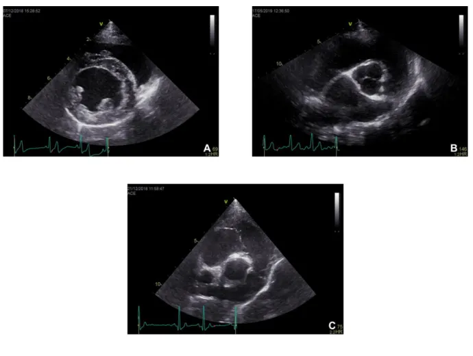
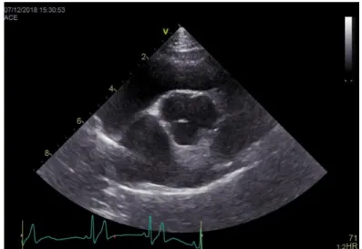
Right parasternal short axis view of the heart base
At the level of the heart base, we can acquire two different planes: the aorta and left atrium view (Figure 2B) and the pulmonary artery view (Figure 2C). In the aortic view, the aorta and its three cusps are seen as a circle in the middle of the image, creating the “Mercedes sign”. This is the most commonly used view to assess the left atrial dimension to the aortic root diameter (LA:Ao). To measure this ratio, the frame just after the aortic valve closure is selected, and both the aortic and left atrial diameter are measured. With the pulmonary artery view, the pulmonary artery can be visualized in its length. (Boon, 2011).
Figure 2. Right parasternal short axis left ventricle view at the level of the papillary muscles
(A), heart base aorta and left atrium view (B) and heart base pulmonary artery view (C). Courtesy of Elizabeth Bode, University of Liverpool.
Left parasternal five-chamber and four-chamber views
On both the five-chamber (Figure 3A) and four-chamber (Figure 3B) views the ventricles are seen on top of the image, while the atria appear at the bottom, with the tricuspid and mitral valve. On the five-chamber view, the aorta can also be visualized in its length, descending between the two atria (Boon, 2011).
Figure 3. Left parasternal five-chamber (A) and four chamber view (B). Courtesy of Elizabeth
Bode, University of Liverpool.
M-mode echocardiography
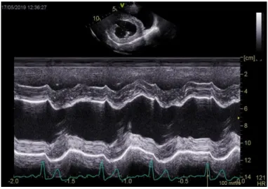
M-mode echocardiography, also known as motion mode, displays the cardiac structures in a one-dimensional image. In order to create a M-mode image, we select one of the sound beams used to generate a two-dimensional image, so that only the structures struck by that one sound beam are seen. The image has the right ventricle at the top, followed by the interventricular septum, the left ventricular chamber and the left ventricular free wall at the bottom (Figure 4) (Boon, 2011).
Figure 4. Left ventricular M-mode image. Courtesy of Elizabeth Bode, University of Liverpool.
The resulting image appears as a distance-time graph, in which the vertical axis represents depth and time is seen along the horizontal axis. This mode is commonly used for measuring chamber dimensions and detecting slight changes in wall and valve motion. M-mode is mostly used for accurate assessment of size and function at the level of the left ventricle, mitral valve and aortic root (Boon, 2011; Fuentes, 2015).
To achieve genuine measurements using M-mode, the cursor must be perpendicular to the cardiac structures measured. In M-mode, we can visualize the movement of the interventricular septum and the left ventricular free wall through the cardiac cycles; in systole, the interventricular septum and the left ventricular free wall move towards each other, while in diastole they move away from one another (Madron, 2016b). In dogs, left ventricular M-mode can be obtained from the right parasternal long axis left ventricular outflow view (Figure 1B) or from the right parasternal short axis view (Figure 2A). In the right parasternal long axis left ventricular outflow view, the M-mode cursor is placed perpendicular to the interventricular septum and left the ventricular free wall, between the tips of the mitral valve leaflets and the left ventricular papillary muscles. Alternatively, when the right parasternal short axis view is used, the cursor is positioned over the interventricular septum and the free wall at the level of the chordae tendineae, bisecting the left ventricle in two identical halves (Boon, 2011). Both these views allow the measurement of two important parameters: the left ventricular internal diameter at end diastole (LVIDd) and the left ventricular internal diameter at end systole (LVIDs) (Madron, 2016b).
LVIDd reflects the maximal ventricular filling when the heart is relaxed and is used to determine whether there is left ventricular volume overload. On the other hand, LVIDs reflects the heart’s systolic function (Boon, 2011). A previous study reported that both LVIDd and LVIDs increase in MMVD prior to the development of left-sided heart failure, suggesting progression to myocardial failure before the onset of heart failure (Lord et al., 2010). Both these measurements are made from the top of the endocardial surface of the interventricular septum to the top of the left ventricular free wall.
Another important parameter that is obtained with M-mode is Fractional Shortening (FS), which is a measurement of left ventricular function. FS is calculated with the mathematical formula
𝐹𝑆 =
𝐿𝑉𝐼𝐷𝑑−𝐿𝑉𝐼𝐷𝑠𝐿𝑉𝐼𝐷𝑑
× 100
, in which the percentage obtained represents the change in leftventricular size between its filling and emptying (Boon, 2011). FS often increases with mitral regurgitation due to increased left ventricular emptying during systole. This is related to the fact that in MMVD there is lower afterload as part of the left ventricular stroke volume goes to the left atrium before the opening of the aortic valve (Madron, 2016a).
Electrocardiography should be used for measuring purposes, to assure the reliability of the results and to guarantee consistency between different operators (Boon, 2011). The American Society of Echocardiography (ASE) has issued recommendations for M-mode measurements in human patients and the same guidelines are used for veterinary medicine (Sahn, DeMaria, Kisslo, & Weyman, 1978; Boon, 2011). The ASE recommends that LVIDd should be measured at the beginning of the QRS complex, which corresponds to the maximal ventricular filling after atrial contraction. On the other hand, LVIDs should be measured at the nadir of interventricular septal motion (Sahn et al., 1978; Madron, 2016b). Different dog breeds can differ a lot in
weight, which is why these parameters should take into account body weight (Cornell et al., 2004).
Doppler echocardiography
Doppler echocardiography analyses the direction, velocity and timing of blood flow and myocardial motion based on the Doppler effect. The Doppler effect is known as the change in frequency, in this case of a sound wave, in relation to a target. When both the source of the sound wave and the target are stationary, the frequency of the transmitted and reflected sound waves is the same. When the target is moving towards the source it reflects an increased number of sound waves, so the frequency of the reflected sound waves will be superior to the frequency of the transmitted waves. On the contrary, when the target moves away from the source, it will reflect fewer sound waves, so that the frequency of the reflected wave will be inferior (Boon, 2011). This change in frequency that occurs when a sound wave hits a moving target is known as Doppler shift and is proportional to the speed at which the target is moving. This helps to determine the velocity of the target, which in echocardiography represents the blood cells, if the frequency of the transmitted wave and the reflected wave is known (Fuentes, 2015; Madron, 2016c).
Doppler examination studies the characteristics of the intracardiac blood flows and detects the presence of abnormal flows. An abnormal direction of blood flow is usually associated with valvular insufficiency, while abnormal flow velocities result from abnormal chamber pressure (Fuentes, 2015). There are different types of Doppler echocardiography as will be further discussed (Madron, 2016c).
Pulsed wave Doppler examines blood flows at very specific sites (Boon, 2011). In Pulsed wave Doppler the sound beams are sent as pulses of waves while the transducer acts as a transmitter or a receiver at different times. The main limitation of this Doppler mode is the fact that it cannot accurately determine the maximal velocity of the blood flow (Fuentes, 2015). Continuous wave Doppler analyses the blood flow all along the sound beam (Boon, 2011). In this Doppler mode, the transducer receives and transmits sound waves at the same time. This allows the detection of higher velocity values (Fuentes, 2015). However, Continuous wave Doppler cannot localize the exact origin of the blood flow (Madron, 2016c).
Colour flow Doppler is a form of Pulsed wave Doppler in which the blood flow is visible in colour on 2D images. It uses a colour code for different flow velocities and directions; when the flow moves towards the transducer it appears in red, while if the flow moves away from the transducer it appears in blue. Colour flow Doppler can detect the presence of turbulent flow, which can be secondary to valvular insufficiency, in which the flow is displayed as a mosaic of colours that ranges between the red and blue spectrum (Boon, 2011; Madron, 2016c). Semi-quantification of the degree of mitral regurgitation is also possible with Colour flow Doppler,
which provides a quick assessment of the severity of mitral regurgitation (Chetboul & Tissier, 2012).
Finally, tissue Doppler imaging (TDI) records the velocity of myocardial motion and is applied to assess myocardial synchronicity and systolic and diastolic function (Boon, 2011). TDI is mostly used to detect diastolic dysfunction, therefore its relevance in MMVD cases is limited (Fuentes, 2015).
The ACVIM consensus guidelines recommends echocardiographic examination for all stage B and stage C patients. Echocardiography identifies the presence of MMVD and quantifies the degree of chamber remodelling and cardiac function. Furthermore, it identifies the presence of increased left atrial pressures and pulmonary hypertension, which can ultimately result in left-sided heart failure. Echocardiography also aids in the recognition of stage B2 dogs, in which the degree of cardiac remodelling is enough to start medical therapy. Patients with a LA:Ao ratio ≥ 1.6 in the right parasternal short axis view in early diastole and with a LVIDd ≥ 1.7 measured with M-mode echocardiography meet the stage B2 criteria (Keene et al., 2019).
4.3– RADIOGRAPHY
Thoracic radiographs are an essential tool for monitoring patients with MMVD (Borgarelli & Haggstrom, 2010). The ACVIM consensus statement recommends the use of thoracic radiography in all dogs with MMVD in order to assess the hemodynamic significance of the disease. In stage B patients, thoracic radiographs are also important to acquire a baseline radiograph of the asymptomatic patient, as this will allow close monitoring of disease progression. Care should be taken when assessing thoracic radiographs, so that other possible concomitant primary respiratory diseases can be excluded, which could contribute to the clinical signs, such as the cough (Keene et al., 2019).
Thoracic radiographs allow an assessment of the severity of mitral regurgitation by visualization of left atrial enlargement, general heart enlargement and signs of left-sided congestive heart failure (Lord & Suter, 1999). Left atrial enlargement is the earliest radiographic sign and is considered more reliable for indirect estimation of mitral regurgitation than general heart enlargement (Lord & Suter, 1999; Hansson, Haggstrom, Kvart, & Lord, 2009). Left atrial enlargement is usually associated with dorsal displacement of the trachea and left mainstem bronchus on the lateral view, a dorsocaudal bulging of soft tissue opacity on the lateral view and an area of increased opacity superimposed to the cardiac silhouette on the ventrodorsal view (Hansson et al., 2009). Dogs with MMVD normally do not develop signs of heart failure without some degree of left atrial enlargement, except on those rare cases when a rupture of a first-order chordae tendineae leads to acute heart failure. Therefore, assessment of left atrial size, either by radiography or echocardiography, is very important to identify patients at risk of developing heart failure (Malcom, Visser, Phillips, & Johnson, 2018).
A recent study described an alternative method to estimate left atrial size radiographically, known as vertebral left atrial size (VLAS). To determine the VLAS, a line is drawn on the lateral view, starting from the centre of the most ventral aspect of the carina and finishing in the most caudal aspect of the left atrium where it intersects with the dorsal border of the caudal vena cava. Afterwards, a second line with the same length as the first is drawn, beginning on the cranial edge of the 4th thoracic vertebra. A VLAS cut-off value of 2.3 has been suggested as
an indicator of left atrial enlargement. Results suggest that this new method is a good predictor of left atrial enlargement in dogs with MMVD and that it has a good correlation with the echocardiographic measurements of the left atrium (Malcom et al., 2018).
General heart enlargement can be judged by a subjective increase of the cardiac silhouette’s length and width, by a dorsal displacement of the trachea on the lateral view, by observation of a rounder cardiac silhouette and by the measurement of the Vertebral Heart Score (VHS) (Hansson et al., 2009). The VHS allows an objective evaluation of the dimensions of the cardiac silhouette, by measuring the cardiac long axis and short axis on the lateral view. These two axes are then summed up and compared with the length of the thoracic spine, starting on the cranial border of the 4th thoracic vertebra (Guglielmini et al., 2009). A VHS value of 10.5
has been suggested as the upper limit to a normal heart size, but since some dog breeds are known to have variations in thoracic conformation and cardiac dimensions, normal VHS values have been proposed for other breeds (Lamb, Wikeley, Boswood, & Pfeiffer, 2001; Buchanan, 2015). This is considered a good method to evaluate cardiomegaly associated with eccentric hypertrophy secondary to volume overload, which is the case of MMVD (Nakayama, Nakayama, & Hamlin, 2001; Guglielmini et al., 2009). Nevertheless, this technique should be used in combination with other radiographic signs (Guglielmini et al., 2009). The inclusion criteria to decide whether or not a stage B dog should start medical treatment with pimobendan has been established (Boswood et al., 2016). Because these criteria rely on echocardiography, and since this diagnostic method may not be available for all veterinary clinical practices, it has been proposed that patients with subclinical MMVD and a VHS > 11.7 would benefit from starting therapy with pimobendan (Vitt et al., 2017).
The determination of the presence of left-sided heart failure is based on the observation of radiographic signs of pulmonary oedema, which include the dilation of the pulmonary veins and an increased opacity of the pulmonary field. (Hansson et al., 2009). The elevation of left-sided filling pressures in the left atrium can cause pulmonary venous hypertension, which will, in turn, increase the width and opacity of the pulmonary veins. These veins become then more evident than the corresponding pulmonary arteries (Ware & Bonagura, 1999; Rosenkranz et al., 2016). Pulmonary venous hypertension is not necessarily a sign of heart failure, but it does precede the onset of heart failure, hence, it can be used to identify at-risk patients (Bahr, 2018). Radiographically, the increased lung opacity can be observed as an interstitial pattern, an alveolar pattern or a mixed interstitial-alveolar pattern (Diana et al., 2009; Hansson et al.,