DOUTORAMENTO
EM
QUÍ
MI
CA
(
Quí
mi
c
a
Or
g
â
ni
c
a
)
Ana
Ca
t
a
r
i
na
de
Ar
a
új
o
Si
l
v
a
POL
YFUNCTI
ONALI
ZED
CARBOHYDRATE-
DERI
VED
SCAFF
OLDS
F
OR
THE
PRODUCTI
ON
OF
LI
BRARI
ES
OF
BI
OACTI
VE
COMPOUNDS
F
ACULDADE
DE
CI
ÊNCI
AS
DOUTORAMENTO
EM
QUÍ
MI
CA
(
Quí
mi
c
a
Or
g
â
ni
c
a
)
(
Pr
of
e
s
s
or
a
As
s
oc
i
a
da
c
om
Agr
e
ga
ç
ã
o,
De
pa
r
t
a
me
nt
o
de
Quí
mi
c
a
e
Bi
oquí
mi
c
a
,
F
a
c
ul
da
de
de
Ci
ê
nc
i
a
s
da
Uni
v
e
r
s
i
da
de
de
Li
s
boa
)
(
Pr
of
e
s
s
or
e
Or
di
na
r
i
o
di
Chi
mi
c
a
Or
ga
ni
c
a
,
Di
pa
r
t
i
me
nt
o
di
Bi
ot
e
c
nol
ogi
e
e
Bi
os
c
i
e
nz
e
,
Uni
v
e
r
s
i
t
à
de
gl
i
St
udi
di
Mi
l
a
no-
Bi
c
oc
c
a
)
Ana
Ca
t
a
r
i
na
de
Ar
a
új
o
Si
l
v
a
POL
YFUNCTI
ONALI
ZED
CARBOHYDRATE-
DERI
VED
SCAFF
OLDS
F
OR
THE
PRODUCTI
ON
OF
LI
BRARI
ES
OF
BI
OACTI
VE
COMPOUNDS
F
ACULDADE
DE
CI
ÊNCI
AS
To my parents…
The most beautiful experience we can have is the mysterious –
the fundamental emotion which stands at the cradle of true art and true
science.
Living Philosophies, 1931
Albert Einstein
ACKNOWLEDGMENTS
“A adversidade desperta em nós capacidades que, em circunstâncias favoráveis, teriam ficado adormecidas.”
Quintus Horatius Flaccus
De uma forma pouco convencional quero iniciar este texto agradecendo a todos aqueles que de modo directo ou indirecto contribuíram para as minhas desventuras científicas. Obrigada aos ‘obstáculos’ que ao longo deste percurso académico se fizeram notar, para eles uma mensagem:
“Pedras no caminho?
Guardo todas, um dia vou construir um castelo...”
Fernando Pessoa
A nível académico os meus agradecimentos vão, em primeiro lugar, para os meus orientadores, Professora Amélia Pilar Rauter e Professore Francesco Nicotra. Tendo sido este doutoramento muito mais do que uma experiência académica, tenho a agradecer não só o contributo científico mas todo o carinho e inspiração que recebi de ambos. Professora Amélia, obrigada pela sua acurada orientação e afectuosa dedicação.
Um agradecimento aos meus colegas de laboratório (FCUL), Nuno, Joana, Susana, Caio, Miguel e Rui, que com extrema paciência me apoiaram e contribuíram de modo sempre espirituoso para o ‘desenrascar’ à Catarina. Ao meu casal target, João e Filipa, quero agradecer a disponibilidade constante para as minhas pequenas/grandes dúvidas.
Ao meu colega de laboratório (UNIMIB) e amigo Marcos quero agradecer todo o apoio nos primeiros Km desta aventura. Uns anitos mais tarde tive a oportunidade de trabalhar com uma menina muito especial, a Íris. Obrigada pela tua doçura e a tua sincera disponibilidade em passar a ferro as minhas roupas (Incrível!).
OBRIGADA Anabela, Ana Elisa, Isabel e Verinha (por ordem alfabética); vocês são as
gaijias, mulheris, químicas da FCUL, doceiras, mães, voluntárias, e outras
características in press…. do meu coração. Devo salientar contudo que contribuíram de modo significativo na minha incessante busca de novos psicofármacos as noitadas de poker, o cheesecake (ou bolo de laranja), aquele filme na casa da Vera (cabos para quê?!), os petiscos da Anabela (e a piscina…não esquecer!), os croissants quentes de Famalicão, as latas de feijão e grão, os telefonemas ao domingo de manhã, e muitos, muitos outros momentos. A vossa amizade forneceu-me os sacos para guardar todas as pedras que fui encontrando ao longo deste caminho, e o vosso apoio e estímulo deram-me as forças para construir as paredes do meu castelo. Muito obrigada.
Isabel, agradeço te a ti em especial todo o apoio científico e emocional que me deste nesta aventura. Lado a lado iniciámos este percurso, e muitas foram as aventuras e desventuras. A tua sensatez foi muitas vezes uma espécie de barómetro na minha vida, ao apaziguar muitos dos meus impulsos irracionais, típicos da minha essência taurina. Obrigada pela tua amizade.
Um especial agradecimento ao meu amigo Rodrigo que generosamente realizou a capa desta tese. Para além do seu contributo gráfico, agradeço a sua protecção quase ‘paternal’ e enorme afecto. Sei que poderei contar sempre contigo para aliviar o peso de algumas pedras.
Tânia e Susana, OBRIGADA migas! O meu trabalho por vezes torna-se a minha vida e nem sempre consigo estar presente como gostaria nos vossos projectos de vida, mas mesmo assim a vossa compreensão é total. Agradeço-vos por não deixarem a minha vida ser normal.
Para ti Miguel sei que palavras valem muito pouco, mas nestas páginas não posso colar actos. Obrigada pelo teu amor….
PedeSenos…. ou é do nome ou realmente sou mesmo eu que sou CHATA. Porque será que estou sempre a PEDIR-te favores?? Não há palavras que descrevam o teu contributo nesta tese, foi só PEDE, PEDE e tu sempre disponível para as inúmeras tarefas que te solicitava. Ora foram correcções, ora boleias à FCUL, ora foi imprimir a tese, ‘arranjar’ material informático vital para o desenvolvimento do projecto, etc. Tens um segundo emprego e ele é: fazer-me FELIZ. Obrigada por tornares as minhas pedras mais leves!
….And the winner is: Francisco Cadorna!! O que este rapaz aturou, eu não devia
agradecer, devia pedir desculpa, no mínimo… Admiro a tua perseverança e determinação, mas adoro quando me deixas levar a bicicleta para casa. Sabemos ambos que nem uma página chega para agradecer TUDO o que fizeste por mim. A aprendizagem no laboratório foi importante mas cruciais foram as experiências de vida. Aspirar ou varrer podem colocar a minha integridade moral em risco! Obrigada por me aturares.
To Oscar…. tack min älskade för ditt ovärderliga stöd.
Por fim, agradeço aos pilares da minha vida, os meus pais. OBRIGADA pelo infinito apoio. O vosso amor incondicional permitiu-me construir os alicerces do meu castelo e viver nele como uma princesa. A vocês dedico esta tese, porque sem o vosso afecto nada seria possível.
“Sono buona fino a che non divento cattiva”.
A Milano ho cominciato questo progetto di ricerca e sono tante le persone con cui ho lavorato o che ho avuto l’opportunità di conoscere. GRAZIE a tutti, è stata fino ad ora una dell’esperienza più bella della mia vita.
Il mio primo ringraziamento va al Professore Nicotra, che più che un professore di chimica organica è stato un professore di filosofia di vita. Lei mi ha insegnato che ‘abbinare’ è possibile anche nel modo di vivere. Il suo carattere mi è stato di ispirazione. Grazie per il buon vino e la piscina sempre disponibile…
Ringrazio Cristina Redaelli, la mia prima ‘supervisora’ i primi tempi in laboratorio, che in modo sempre dolce mi ha insegnato a fare le prime reazioni. Alla mia seconda ‘supervisora’, Ingrid, grazie per l’amicizia e tutto l’appoggio scientifico che mi hai dato. Voglio ringraziare Laura Cipolla per il suo atteggiamento, che mi ha sempre spinto a fare più e più ricerca. Un’enorme riconoscenza anche a Barbara Costa, Pietro e Prof. Gabriella Giagnoni per tutta l’assistenza nel lavoro e per l’affetto.
I ‘mie’ studenti e amici: Krama, Betta, Rossella e Junior. A loro voglio esprimere gratitudine per tutto l’aiuto che mi hanno dato. Siete un contributo molto importante della mia tesi. Krama, a te più che per le sintesi delle benzo, ti ringrazio per le lezioni di italiano.
In laboratorio 4014, Barbara, ‘Autista’, Dario, Cristiano, ‘Brasuca’, in laboratorio 4018, Simone (VIVA il trenino), Paolino, Nasrin, Elena e in laboratorio 4048, Peri, Sissi, Square e Luca, a tutti voi GRAZIE per tutto.
Laboratorio 4054: ‘Paradise Girl’, Erika e ‘Ragazzino’, voglio dirvi: MI MANCATE tanto, comunque RITORNERÒ…
Chiara e Maria grazie per il viaggio della mia vita: I LOVE NY.
Ale (o Alexandre per il volo RYANAIR), senza di te non riuscivo a sopravvivere neanche cinque minuti a Milano. Grazie per tutte le volte che mi hai aiutato….sono state TANTISSIME. Sei un bravo ragazzo!
Cristina e Russo, GRAZIE amiche mie! Quella serata MERAVIGLOSA non la SCORDERÒ mai… perciò (Attenzione!) possiamo cancellare il video? Grazie Cristina per la vera amicizia e per l’aiuto nella tesi. Russo…. FA MALEEEEEEEEEE ma vicino a te il dolore diminuisce. Grazie per le belle serate!
‘Tesoro’ mio e Paty, a voi voglio dire che siete stati la mia luce in quegli ultimi mesi a Milano, in cui io non ce la facevo più. Vi ADORO (Viva Partanna).
ABSTRACT
Inspired by the role of carbohydrates as natural scaffolds, we exploited the sugar skeleton to generate new libraries of polyfunctionalized compounds as GABAA
receptor ligands. Hybrids of benzodiazepines, γ-butyrolactone and -lactam derivatives, and a GABA analogue were developed. The incorporated sugar moiety offered the possibility of diverse and controlled functionalization, modulating physicochemical and structural properties, namely solubility and rigidity, and consequently biological activity of the synthesized scaffolds.
1,4-Benzodiazepine-2,5-dione scaffolds (i and iv) derived from spiro bicyclic D- or L
-proline analogues (ii and v), containing a D- or a L-fructose moiety were synthesized in the present work. The D-proline analogue ii was prepared in a sixteen-step synthesis in 24% overall yield, adopting a methodology which used D-fructose as starting material and 3-C-(3,4,6-tri-O-benzyl-α-D-fructofuranos-2-yl)propene iii as key
intermediate. Instead, the L-fructose moiety, required for the preparation of the corresponding L-proline analogue v, was obtained using a new synthetic pathway. The key intermediate of this synthesis, the 3-C-(3,4,6-tri-O-benzyl-α-L -fructofuranos-2-yl)propene vi, was effectively obtained from L-arabinose through a seven steps
pathway in 13.5% overall yield. This starting material embodies the furanose ring with the appropriate configuration. Hence, a one carbon chain elongation at the anomeric position lead to the desired L-fructose derivative, compound vi. For that purpose, oxidation to arabinonolactone, formyl group introduction via a dithiated intermediate and reduction to the primary alcohol were performed. The subsequent eleven-step synthesis afforded the new spiro bicyclic L-proline analogue v in 24% overall yield. In addition, a procedure previously reported for L-fructose synthesis was also used to obtain 3-C-(3,4,6-tri-O-benzyl-α-L-fructofuranos-2-yl)propene vi. Starting from L-sorbose, diisopropylidene protection, mesylation, selective deprotection, oxirane formation and opening led to inversion of configuration at C-3 and C-4
affording the stereochemistry of L-fructose. The key intermediate vi was obtained by this eight-step synthesis with a 8.5% overall yield.
N N O R R O H O RO RO RO O OBn BnO BnO NH N N O R R O H O RO RO RO O OBn BnO BnO NH H COOH COOH H i ii iii iv O BnO BnO OBn v vi OH O BnO BnO OBn OH 7 6 9 10 4 11 3 1 2' 3' 4' 8 11a 1'
Molecular modelling calculations, NMR studies at variable temperature (dynamic NMR–DNMR) and preliminary biological studies were also performed on these compound libraries of enantiomeric fructose–proline–benzodiazepine derivatives (i and iv). The proline moiety linked to the fructose derivative through a spiro junction and consequent fusion of this bicyclic structure to the benzodiazepine ring promoted high conformational rigidity to the hybrid scaffold. Therefore, the conformational equilibrium normally occurring with (P) and (M) benzodiazepine conformers was not observed. The benzodiazepine derivatives synthesized adopted a rigid conformation in which the C-11a substituent was always pseudo-equatorial.
The diverse and controlled functionalization of these molecules (i and iv) was achieved using different isatoic anhydride types which are commercially available. Coupling of D- or L-proline analogues, ii and v, respectively, with the suitably functionalized isatoic anhydride, afforded the desired sugar-based pyrrolobenzodiazepine library (i and iv). These compounds weres functionalized not only with electronegative substituents, such as –Cl, –Br and –NO2, but also with –NH2
at position seven, while at position ten the presence of –CH3 is expected to give a more
these compounds to displace the [3H]flunitrazepam from the GABAA receptor was
measured with a competition binding assay using rat cortical membrane. It was observed that substitution on both positions are crucial for binding affinities, being particularly effective –NO2 and –NH2 groups. The free –OH or –CH2Ph groups on the
sugar offered the possibility to balance hydrophilic/lipophilic for tuning the pharmacokinetic properties and the binding affinities of the potential drug. As expected, water soluble sugar-based pyrrolobenzodiazepine derivatives, i.e. with –OH groups, presented highest binding affinities. The effect of conformational changes in the 1,4-benzodiazepine-2,5-dione ring and of different substituents allowed an evaluation of binding affinities at the benzodiazepine site on the GABAA receptor.
Additionally, was developed the synthesis of β-disubstituted D-fructose-based γ-butyrolactone vii and γ-butyrolactam analogues viii, and that of a lipophilic D-fructose-based GABA analogue ix, where the pharmacophore is engineered into the
carbohydrate scaffold through a spiro junction. The ability to bind GABAA receptor,
using a radioligand binding technique, was evaluated for all these compounds. GABA lactones vii were synthesized via the key intermediate 3-C-(3,4,6-tri-O-benzyl-α-D -fructofuranos-2-yl)propene iii, while γ-butyrolactams viii and GABA analogue ix took advantage of the allyl group and an amino functionality replacing the hydroxyl group in iii. The fructose moiety acted as versatile scaffold, rich in stereochemistry and with a relatively rigid skeleton. Thus, additional hydroxyl derivatization was used to increase lipophilicity, as well as to modulate the activity of pharmacophores or the receptor specificity, since benzyl groups could facilitate blood-brain barrier (BBB) crossing, which is one of the main issues to be addressed for central nervous system (CNS) directed drugs.
KEY WORDS
Carbohydrates Fructose
Spiro Proline Analogues 1,4-Benzodiazepine-2,5-diones Pyrrolobenzodiazepines γ-Butyrolactones γ-Butyrolactams GABA analogues GABAA Receptor Radioligand Techniques Compounds Libraries Molecular Modelling Dynamic NMR
RESUMO
Os hidratos de carbono constituem uma família única de compostos polifuncionais com primordial importância em química e biologia. Funcionalidade, quiralidade e diversidade estrutural são algumas das suas características mais relevantes. Neste contexto, dirigimos esta investigação para a síntese de novas estruturas que possuem unidades monossacarídicas com rigidez estrutural e ainda uma complexidade e diversidade tais que permitiram a construção de novas bibliotecas de compostos bioactivos.
Inspirados pela proeminente utilidade dos hidratos de carbono como scaffolds em química medicinal, foram sintetizadas duas bibliotecas de potenciais ligandos do receptor GABAA, geradas a partir da D-fructose e de um derivado da L-fructose. A
primeira biblioteca de compostos inclui várias estruturas análogas às benzodiazepinas e a segunda contém γ-butirolactonas, γ-butirolactamas e um análogo do ácido gama-aminobutírico (GABA). Todos estes compostos foram submetidos a ensaios biológicos, nomeadamente utilizando a técnica que investiga a competição entre o composto e o radioligando correspondente relativamente à afinidade para a ligação ao receptor no seu local específico.
O GABA é um neurotransmissor inibidor do sistema nervoso central. Os seus efeitos resultam principalmente da sua ligação ao receptor GABAA, que constitui uma
estrutura macromolecular em volta de um canal iónico permeável ao cloreto. A fixação do GABA ao receptor GABAA causa um aumento da condutância da membrana celular
ao cloreto, que habitualmente existe em maior concentração no exterior do que no interior da célula. O movimento de aniões para dentro da célula aumenta a diferença de potencial entre a face externa e a interna da membrana celular e reduz a excitabilidade neuronal. O GABA funciona como uma molécula reguladora de efeitos tais como ansiedade, depressão, insónias, convulsões, tensão ou relaxamento muscular, actividade epiléptica, memória, euforia e disforia. O receptor GABAA tem
locais específicos de ligação para distintas moléculas que influenciam a actividade do GABA, tais como benzodiazepinas, barbitúricos, etanol, esteróides, etc. As benzodiazepinas são um grupo de fármacos ansiolíticos utilizados no tratamento sintomático da ansiedade, insónia, depressão e distúrbios mentais em geral. Estas moléculas ligam-se a um local próprio, denominado receptor das benzodiazepinas, e aumentam a afinidade do GABA para o receptor GABAA. Os barbitúricos, por exemplo,
fixam-se também ao receptor GABAA e provocam uma abertura prolongada do canal
do cloreto. Diferem das benzodiazepinas porque a abertura do canal não exige a presença de GABA, ao contrário das benzodiazepinas que se limitam a potenciar o efeito do GABA endógeno. Além disso a abertura é mais prolongada, enquanto que as benzodiazepinas aumentam a frequência das aberturas sem prolongarem a duração de cada uma delas.
Especificamente, no presente trabalho é apresentada a síntese de uma biblioteca de
enantiómeros análogos das benzodiazepinas, i e iv, desenvolvidos a partir da
D-frutose e de um derivado da L-frutose, respectivamente. A estratégia adoptada centrou-se na síntese de um novo análogo da D- e da L-prolina, ii e v, respectivamente, com uma estrutura bicíclica espiro que contém a unidade sacarídica. A junção espiro reduziu significativamente a flexibilidade molecular, induzindo restrições conformacionais ao anel da prolina e consequentemente a toda a estrutura da molécula final (comprovado por NMR a temperaturas variáveis e estudos de modelação molecular).
A escolha do monossacárido frutose resultou do facto deste açúcar oferecer potencialidades interessantes devido à presença de grupos hidroximetilo (em 1 e C-6) e devido à reactividade única do seu carbono anomérico. A síntese de derivados protegidos da frutose baseia-se na ramificação no carbono C-2 (iii e vi) e posterior reacção de ciclização entre uma função amina e um grupo alilo ligado à posição anomérica dando origem ao anel de pirrolidina seguidamente transformado no aminoácido ii e v.
N N O R R O H O RO RO RO O OBn BnO BnO NH N N O R R O H O RO RO RO O OBn BnO BnO NH H COOH COOH H i ii iii iv O BnO BnO OBn v vi OH O BnO BnO OBn OH 7 6 9 10 4 11 3 1 2' 3' 4' 8 11a 1'
O plano de síntese para o análogo da D-proline ii, envolveu a metilação da posição anomérica e benzilação das restantes posições utilizando a metodologia clássica (NaH, BnBr, DMF). Seguidamente foi introduzido um grupo alilo na posição anomérica através da formação de uma ligação C-C com aliltrimetilsilano mediada por um ácido de Lewis. A reacção dá-se geralmente por intermédio de um ião oxocarbénio cíclico, o qual sofre um ataque nucleófilo estereoquimicamente controlado, conduzindo a uma elevada estereoselectividade tanto em anéis de piranose como em anéis de furanose. Consequentemente, o tratamento de 1,3,4,6-tetra-O-benzilfrutofuranósido de metilo
com aliltrimetilsilano na presença de eterato de trifluoreto de boro originou 3-C-(1,3,4,6-tetra-O-benzilfrutofuranos-2-il)propeno. A relação espacial entre a
ligação dupla e o oxigénio do grupo benziloxi do carbono C-1 favorece a ocorrência da ciclização 5-exo por tratamento com iodo, desprotegendo selectivamente o grupo hidroxilo primário de C-1. A introdução da amina foi realizada através da oxidação a aldeído do álcool primário, formação de oxima e redução com hidreto de alumínio e lítio. Uma segunda iodociclização, desta vez envolvendo a função amina, protegida com um grupo carbamato, e o grupo alilo, conduziu à formação do anel pirrolidina. Tratamento com hidróxido de sódio permitiu, através da formação de uma oxazolidinona intermediária, obter o composto que possui a função álcool primário e a estereoquímica desejada. A sua oxidação conduziu à síntese do aminoácido prolina.
O plano de síntese do análogo da L-proline v envolveu a preparação de um derivado da
L-frutose, uma vez que este açúcar não natural é comercializado a um preço bastante elevado. Foram desenvolvidas duas metodologias para a formação do intermediário-chave, o composto 3-C-(3,4,6-tetra-O-benzil-α-L-frutofuranos-2-il)propeno, uma das
quais se baseou num procedimento descrito em literatura para a síntese da L-frutose, o qual usa a L-sorbose como composto de partida. Esta estratégia baseia-se na inversão da configuração das posições três e quatro para obter assim o intermediário-chave com a configuração do açúcar desejada. A estratégia alternativa desenvolvida partiu da L-arabinose, a qual possui a estereoquímica pretendida, resultou no alongamento da cadeia carbonada em C-1. Esta síntese é mais directa e permite obter a molécula-alvo com um rendimento ligeiramente superior (13,5% rendimento total) do que a utilizada inicialmente (8,5% rendimento total).
O uso do aminoácido prolina na produção de pirrolobenzodiazepinas foi estabelecido como sendo um dos processos sintéticos mais eficazes e rentáveis, através do uso de anidrido isatóico em DMF e refluxo. O reagente anidrido isatóico pode ser diversamente substituído permitindo a obtenção de uma variada biblioteca de compostos, nomeadamente benzodiazepinas substituídas na posição sete com halogéneos (Br e Cl), grupos amina ou nitroílo e metiladas em N (posição dez). O nosso contributo singular na estrutura das benzodiazepinas consistiu na incorporação de um açúcar de modo a funcionalizar os grupos hidroxilo e modular as propriedades físico-químicas e estruturais, nomeadamente a solubilidade e a rigidez, e consequentemente a actividade biológica do potencial fármaco.
A síntese destas estruturas rígidas teve como objectivo, não só permitir o estudo da relação estrutura-actividade, mas também uma análise acurada da especificidade conformacional do local de ligação no receptor das benzodiazepinas. Conforme descrito na literatura, o anel diazepina das benzodiazepinas tem um equilíbrio conformacional entre os confórmeros (M) e (P) e a ligação das mesmas ao receptor é dependente da conformação adoptada por estas. Embora existam estudos prévios que demonstraram que o receptor é favorável à conformação (M), a investigação
desenvolvida no âmbito desta tese, utilizando enantiómeros conformacionalmente rígidos no anel diazepam, permitiu concluir que ambas as conformações apresentam actividades biológicas semelhantes para estes compostos. As actividades biológicas determinadas demonstraram que a contribuição mais significativa para a ligação específica ao receptor provém dos substituintes no anel do benzeno, nomeadamente os grupos amina e nitroílo (posição sete), bem como a N-metilação. Os grupos hidroxilo do açúcar também contribuíram para um aumento da actividade biológica.
Paralelamente a estes análogos das benzodiazepinas, desenvolvemos uma segunda biblioteca de compostos, igualmente potenciais ligandos do receptor GABAA. Foram
sintetizadas γ-butirolactonas e γ-butirolactamas β-disubstituídas, vii e viii, respectivamente, e um derivado do GABA ix. Estas estruturas bicíclicas possuem uma junção espiro com um derivado da D-frutose. Os resultados de estudos farmacológicos,
envolvendo radioligandos através de ensaios de competição entre o composto e o muscimol marcado, indicaram que as lactamas são os compostos com maior actividade biológica, seguidas das lactonas. O açúcar mostrou ser uma estrutura esteroquimicamente não impeditiva para a ligação ao receptor e até mesmo os compostos benzilados tiveram actividades biológicas interessantes, o que reforça a importância de estruturas lipofílicas para atravessar a barreira hemato-encefálica (BHE).
Em resumo podemos assumir que o trabalho desenvolvido conduziu ao desenvolvimento de novas estruturas glucídicas para a produção de novas bibliotecas de compostos com interesse farmacológico, que são excelentes candidatos para investigações de estrutura-actividade no receptor GABAA.
PALAVRAS – CHAVE
Hidratos de Carbono Frutose Prolina 1,4-Benzodiazepinas-2,5-dionas Pirrolobenzodiazepinas γ-Butirolactonas γ-Butirolactamas Análogos do GABA Receptor GABAA Actividades biológicas Radioligandos Biblioteca de compostos Modelação Molecular RMN DinâmicoLIST OF ABBREVIATIONS
Ac acetyl AllSi(CH3)3 allyltrimethylsilane Aq. aqueous Ar aryl b broad BBB blood-brain barrierBF3.Et2O boron trifluoride etherate Bmax (or R0) maximum binding Bn benzyl
Boc tert-Butyloxycarbonyl BSA bovine serum albumin
BTSFA bis(trimethylsilyl)trifluoroacetamide Bu butyl
BZ benzodiazepine(s) ca. circa, approximately Calcd calculated
CCK-A peripheral cholecystokinin Ci curie
CNS central nervous system conc concentrated
cpm cycles per minute δ chemical shift(s) d doublet
DBMMH 1,3-dibromo-5,5-dimethylhydantoin dd doublet of doublets
ddd doublet of doublet of doublets
de diastereoisomeric or diastereoisomer excess
DIS diazepam-insensitive DMF N,N-dimethylformamide DMS dimethylsulphide DMSO dimethylsulfoxide DME 1,2-dimethoxyethane DMP 2,2-dimethoxypropane DNA deoxyribonucleic acid
DNMR dynamic nuclear magnetic resonance dpm disintegrations per minute
DS diazepam-sensitive dt doublet of triplets
EPSP excitatory postsynaptic potential (EPSP) Eq. equation
eq. equivalent(s) Et ethyl
f fento (factor 10-15)
Fmoc 9-fluorenylmethoxycarbonyl GABA gamma-aminobutyric acid GABA-T gaba transaminase
GAD L-glutamic acid decarboxylase GBL gamma-butyrolactone(s) GBP-L gabapentin-lactam GHB γ-hydroxybutyrate(s)
HIV Human immunodeficiency virus Hz hertz
IAA imidazole-4-acetic acid [IUPAC name: 2-(1H-imidazol-4-yl)acetic acid] IBX iodoxybenzoic acid
IC50 inhibitory concentration (50%)
IUPAC International Union of Pure and Applied Chemistry
J coupling constant
Kd ligand affinity/equilibrium constant Ki equilibrium constant (for the competitor) L ligand
M molar (1M = 1 mol/L)
M conformation minus
m milli (factor 10-3) or multiplet
MALDI-TOF Matrix Assisted Laser Desorption /Ionization- Time Of Flight MHz megahertz mmol milimole min minute(s) M.p. melting point MS mass spectrometry Ms mesyl ms molecular sieves m/z masse-to-charge ratio n nano (factor 10-9)
NMO N-methylmorpholine oxide NMR nuclear magnetic resonance NOE nuclear overhauser effect
NOESY nuclear overhauser effect spectroscopy NP neurotoxic pesticides
s singlet
n.s. not statistically significant nsb nonspecific binding OAc acetate p pico (factor 10-12) P conformation plus P Probability PCC pyridinium chlorochromate Pd/C palladium on charcoal Ph phenyl
PIOL 5-(4-piperidyl)isoxazol-3-ol ppm parts per million
PTX picrotoxin(s) PTZ pentylenetetrazole P4S piperidine-4-sulfonic acid q quaternary ou quartet
QSAR Quantitative Structure-Activity Relationship R receptor(s)
Rf retention factor
RL receptor–ligand complex rt room temperature
sat. satured
SEM standard error of the mean soln. solution
SSA succinic semialdehyde t time or triplet
TBDMSCl tert-butyldimethylchlorosilane TBPS tert-butyl bicyclophosphorothionate TFA trifluoroacetic acid
THF tetrahydrofuran
THIP 4,5,6,7-tetrahydroisoxazolo[5,4-c]pyridin-3-ol THPO 4,5,6,7-tetrahydroisoxazole[4,5-c]pyridin-3-ol TLC thin layer chromatography
TM transmembrane
TMSOTf trimethylsilyl trifluoromethanesulfonate Ts p-toluenesulfonyl
μ micro (factor 10-6)
VGAT vesicular neurotransmitter transporter v/v volume per unit volume
vs versus
CONTENTS
Acknowledgements… ...iii Abstract ... ix Key Words ... xiii Resumo ... xv Palavras Chave ...xxi List of Abbreviations ... xxiii
1. INTRODUCTION ... 1 1.1. Neurobiology: An Overview ... 5 1.1.1 The Synaptic Mechanism ... 7 1.1.2 Neurotransmitters – The Chemical Messengers ... 9 1.1.3 Receptors ... 10 1.1.3.1 Neurotransmitter Receptors ... 11 1.1.4 Drugs Action on the CNS ... 12 1.2. GABA and GABA Receptors ... 14 1.2.1 GABA ― from Metabolite to Neurotransmitter ... 16 1.2.2 GABAA Receptors ... 18
1.2.2.1 GABAA Receptor Binding Sites.. ... 19
1.2.2.1.1 Convulsant Binding Site ... 22 1.2.2.1.2 Butyrolactone Site ... 22 1.2.3 GABA Binding Site for GABAA R ... 23
1.2.4 GABA Analogues ... 25 1.2.4.1 γ-Butyrolactones (GBLs) ... 27 1.2.4.2 γ-Butyrolactams ... 29 1.2.4.3 QSAR Modelling for Selective Anticonvulsants ... 30 1.3. Benzodiazepines ... 31 1.3.1 A Chemical Perspective – Structure and Synthesis ... 31 1.3.2 Benzodiazepine as a Privileged Scaffold in Medicinal Chemistry ... 33 1.3.3 Benzodiazepine as a Commercial Psychoactive Drug ... 34 1.3.4 Benzodiazepine Binding Site on GABAA Receptors ... 36
1.3.4.1 Classification of BZs and Their Congeners ... 37 1.3.4.2 Conformational Equilibrium in Benzodiazepine Ring ... 38 1.3.4.3 SAR studies ... 41
1.4. Carbohydrates as Natural Scaffolds ... 42 1.4.1 Sugar Derived Polyfunctional Scaffolds–GABA Analogues and Bzs... 45 1.5. Radioligand Technique ... 47 1.5.1 Ligand Binding: Simple Theory ... 48 1.5.1.1 Equilibrium Binding: Saturation Analysis ... 49 1.5.1.1.1 Nonspecific Binding ... 52 1.5.1.1.2 Analysis of Saturation Binding Isotherms: Scatchard Plot ... 53 1.5.1.2 Equilibrium Binding: Competition Studies ... 55 1.5.1.3 Limits on the Interpretation of Equilibrium Radioligand Binding Studies ... 57 1.5.2 Radiolabelled Ligand Binding Studies: Separation of Free From Bound Ligand ... 59
2. RESULTS AND DISCUSSION ... 61 2.1 Synthesis of the Spiro D- and L-Proline Analogues ... 65
2.1.1 Synthesis of the Spiro D-Proline Analogue ... 67 2.1.2 Synthesis of the Spiro L-Proline Analogue ... 72
2.1.2.1 Synthesis of Compound 111 - Pathway a ... 73 2.1.2.2 Synthesis of Compound 111 - Pathway b ... 78 2.1.2.3 Synthesis of Compound 112 ... 83 2.1.3 Novel Rigid 1,4-Benzodiazepine-2,5-dione Scaffolds ... 85 2.1.3.1 Synthesis ... 86 2.1.3.2 Molecular Modelling and DNMR Experiments ... 89 2.1.3.3 Biological Assays ... 99 2.1.3.3.1 Evaluation of Total, Non-Specific and Specific Binding – Saturation Studies ... 99 2.1.3.3.2 GABAA R Binding Assays - Competition Studies ... 101
2.1.4 Fructose-Based γ-Butyrolactones and –Lactams, and a GABA analogue ... 110
2.1.4.1 Synthesis ... 110 2.1.4.2 Biological Evaluation ... 114 2.1.4.2.1 Evaluation of Total, Non-Specific and Specific binding – Saturation Studies ... 114 2.1.4.2.2 GABAA R Binding Assays ― Competition Studies ... 115
4.EXPERIMENTAL SECTION ... 125 4.1 Organic Synthesis ... 127 4.1.1 General Methods ... 127 4.1.2.1 Dry Solvents and Reactions ... 127 4.1.2.2 Preparation of 2-Iodoxtbenzoic acid (IBX) ... 127 4.1.2.3 Thin-Layer Chromatography... 127 4.1.2.4 Flash Column Chromatography ... 127 4.1.2.5 Mass Spectroscopy ... 128 4.1.2.6 NMR Spectroscopy ... 128 4.1.2 Synthesis of the Spiro D-Proline Analogue ... 128 4.1.3 Synthesis of the Spiro L-Proline Analogue ... 144 4.1.3.1 Synthesis of Compound 111 - Pathway a ... 144 4.1.3.2 Synthesis of Compound 111 - Pathway b ... 152 4.1.3.3 Synthesis of Compound 112 ... 158 4.1.4 Synthesis of the 1,4-Benzodiazepine-2,5-dione Scaffolds ... 167 4.1.5 Synthesis of the Pyrrolobenzodiazepines ... 197 4.1.6 Synthesis of the Fructose-Based γ-Butyrolactones and –Lactams, and a GABA analogue ... 199 4.2 Molecular Modelling and DNMR Experiments ... 211 4.3 In Vitro Pharmacology: GABAA R Binding Assays ... 211
4.3.1Chemicals ... 211 4.3.2 Instruments ... 212 4.3.3 Methods ... 212 4.3.3.1 Benzodiazepine Derivatives ... 213 4.3.3.1.1 Membrane Preparation ... 213 4.3.3.1.2 Radioligand Binding Assay ... 214 4.3.3.1.3 Data analysis ... 214 4.3.3.2 GABA Derivatives ... 214 4.3.3.2.1 Membrane Preparation ... 214 4.3.3.2.2 Radioligand Binding Assay ... 215 4.3.3.2.3 Data analysis ... 216 4.3.3.3 Lowry Protein Assay ... 216
Sorrow, 1882 Vincent van Gogh
INTRODUCTION
Matar o sonho é matarmo-nos.É mutilar a nossa alma.
O sonho é o que temos de realmente nosso,
de impenetravelmente e inexpugnavelmente nosso. Fernando Pessoa
HIGHLIGHTS
Globally, it is estimated that 450 million people suffer from mental or behavior disorders. In developed countries, 8 of the 10 leading causes of disability are related to mental illness.
Mental disorders are one of the most prominent and treatable causes of suicide; every year about 800 000 people commit suicide.
According to the World Health Organization,[1] Mental Health is defined as:
"A state of complete physical, mental and social well-being and not merely the absence of disease."
Problems related to mental health, as anxiety and depression disorders, should be a public priority. The social and economic impact of depression, for example, is enormous in modern society. Moreover, there is not health without a good mental health.
Mental health is influenced by a wide range of factors, such as individual biological and psychological factors, social interaction, societal structures or resources and cultural values. Certain mental illnesses are known to be linked to structural abnormalities or chemical dysfunction of the brain. Others are inherited, but often the cause is unknown. Likewise, injuries to the brain and chronic drug or alcohol abuse can trigger also some mental illnesses.[1]
A 2004 report from European Communities[2] describes and compares the state of
mental health in the European Union and Norway. The report showed the percentage of psychological distress and the positive mental health in 10 EU countries (Fig. 1). Portugal and Italy have the highest incidence of mental health problems and the lowest positive mental health. These values were not the motivation for my research work; however are a curious coincidence.
0 5 10 15 20 25 30 35 Neth erlan ds Swed en Spa in Luxe mbou rg Belgi um Austri a Germa ny Fran ce Portu gal Italy 52 54 56 58 60 62 64 66 68 Italy Portu gal Fran ce Swed en Luxem bour g Germ any Austr ia Belgiu m Nether lands Spain
Figure 1. Psychological distress (left) and positive mental health (right) in ten EU countries.[2]
Thus, having consciousness that Mental Health is a global problem, the research and development of new psychoactive drugs, which act primarily into the central nervous system (CNS), becomes a relevant issue. Hence, drugs that interact with CNS receptors are amongst the most important in medicine, providing treatment for diseases such as pain, depression, Parkinson’s disease, psychosis, heart failure, asthma, and many others.
In the present work, two novel classes of GABA receptor ligands were developed. Inspired by the role of carbohydrates as natural scaffolds, a sugar skeleton was exploited to develop a library of new benzodiazepine and GABA derivatives (γ-butyrolactones, γ-butyrolactams and a GABA analogue) of biological relevance. Derivatization and introduction of several substituents provided different pharmacophores,i allowing the construction of small libraries of the respective
scaffold.
i Pharmacophores are atoms and functional groups required for a specific pharmacological activity, and their
1.1 Neurobiology: An Overview
In a complex organism, cells have to get along with their neighbours. A communication system and its control is essential, and comes primarily from the brain and spinal column – the central nervous system (CNS), which receives and sends messages via a vast network of nerves. These messages are electrical pulses which travel down the nerve cell, the neuron (Fig. 2), towards a target, whether that be a muscle cell or another nerve.[3]
There are two main classes of cells in the nervous system: neurons and glial cellsii
(glia). Neurons are cells entirely different from other cells in the body. They are typically highly polarized, and their cell functions are compartmentalized. The main functional compartments of neurons can be defined into four morphologically distinctive regions: the cell body (soma), the dendrites, the axonsiii (covered with
sheaths of lipids – myelin sheath), and axon terminals; which are usually separated by considerable distances (Fig. 2).
Figure 2. Cartoon showing the structure of a typical neuron.[4]
ii Glial cells provide support and nutrition to neurons. As far as is known, glia are not directly involved in
information processing. They surround the cell bodies, axons, and dendrites of neurons, providing structure to the brain.
The electrical excitability is among the most important and characteristic properties of neurons and is due to the presence of voltage-sensitive ion channels in the neuronal plasma membrane (Fig. 3).[4, 5] Depolarization is a decrease in the absolute value of a
cell's membrane potential, which is caused by influx of Na+ through Na+ channels (or
influx of Ca2+ through Ca2+ channels), and the effect is to stimulate the nerve. Contrary,
hyperpolarization is an increase absolute value of a cell's membrane potential, which is caused by out-flux of K+ through K+ channels (or influx of Cl- through Cl- channels)
and the effect is to destimulate the nerve. This cell’s electrical signals (nerve impulses), called action potentials, allow neurons to carry a signal over a certain distance (Fig. 3). The brain receives, analyzes, and transmits information through these signals.
Figure 3. Cartoon showing a communication system between two neurons. The action
potential, i.e. the cell's conducting signal, is initiated either at the axon hillock (the initial segment of the axon) or in some cases slightly further down and moves towards the presynaptic cells. Then, from a specific region of the axon of one neuron, the presynaptic terminal cells, messages are transmitted to another neuron, the postsynaptic cells ― synapse. Most presynaptic terminals ends on the postsynaptic neuron's dendrites, but the terminals may also end on the cell body or, less often, at the beginning or end of the axon of the receiving cell.[4]
1. t = 0
2. t = 1
The correct functioning of the CNS is based on the generation, propagation, and coordination of signals between different neurons. However, neurons do not connect directly to their target cells. The distance between them is minimal, about 100 Å, but it is a space that the electrical pulse is unable to jump. Therefore, there has to be a method of carrying the message across the gap between the neuron ending and the target cell. The problem is solved by the release of a chemical messenger called a neurotransmitter (see also chapter 1.1.2). Thus, the transference of information between neurons is achieved neither by direct contact nor electrically but chemically in a process called synaptic mechanism (see also chapter 1.1.1). This communication between individual neurons is performed at a morphologically and functionally highly specialized region named the synapse (Fig. 3). Once released, the neurotransmitter diffuses across the gap to the target cell, where it binds and interacts with a specific protein ― a receptor (see also chapter 1.1.3), embedded in the cell membrane. This process of binding results either in a flow of ions across the cell membrane or in the switching on (or off) of enzymes inside the target cell. A biological response then arises, namely the contraction of a muscle cell or the activation of fatty acid metabolism in a fat cell.[3]
1.1.1 The Synaptic Mechanism
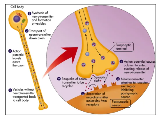
Communication between neurons happens when chemical signals, the neurotransmitters, are transmitted across a gap between the membranes of the pre- and post-synaptic cells, the synaptic cleft. This chemical transmission is known as the synaptic mechanism, and can be divided into four steps, in which two are presynaptic and the other two postsynaptic (Fig. 4). The dendrites act as small arms radiated from the cell body that received messages from other nerves. These messages either stimulate or destimulate the nerve. The cell body ‘collects’ the total sum of the received messages (neurotransmitters), and ‘stores’ them in vesicles.iv
iv Vesicle is a small, intracellular, membrane-enclosed pocket that stores and transports substances within a cell, as
Figure 4. Cartoon showing a synaptic mechanism between two neurons. The primary steps of
this process are: the synthesis of the neurotransmitter; the storage and release of the neurotransmitter; the transmitter’s interaction with a receptor in the postsynaptic membrane; and the removal of the neurotransmitter from the synaptic cleft.[6]
The synthesis of the neurotransmitters and the subsequent formation of the vesicles can be considered the starting point of the synaptic mechanism. So, assuming that the overall stimulation of the nerve by the received messages is great enough, an electrical signal is fired down the axon. The myelin sheaths insulate the signal as it passes down the axon. This transportation of the vesicles down the axon is achieved due to the action potential (Fig. 3). These vesicles conducting signals traveled down the axon to the presynaptic terminal, considered the synaptic button (knob-shaped swelling). The depolarization of the presynaptic terminal membrane, i.e. the opening of the calcium channels and consequent influx of Ca2+ ions, results on the stimulation of the nerve.
These generated action potential influences the vesicles “full to the limit” of neurotransmitters to fuse with the presynaptic terminal membrane. The neurotransmitters are then released from the vesicles into the synaptic cleft, and it targets its reciprocal receptor on the postsynaptic membrane of the next neuron. The effect of the neurotransmitter on the postsynaptic membrane will depend on the
nature of the neurotransmitter, the nature of the postsynaptic receptors, and whether the postsynaptic ion channels are voltage-gated or chemically-gated. Most neurotransmitters are removed from the synaptic cleft by neurotransmitter transporters in a process called reuptake (uptake). Without reuptake, neurotransmitters can continue to stimulate or inhibit the firing of the postsynaptic neurons.
1.1.2 Neurotransmitters – The Chemical Messengers
There is a large variety of messengers that interact with receptors and that differ quite significantly in structure and complexity. Some neurotransmitters are simple in structure, such as monoamines (e.g. acetylcholine, noradrenaline, dopamine and serotonin) or amino acids [e.g. γ-aminobutyric acid ― GABA (see also chapter 1.2.1), glutamic acid and glycine]. Other chemical messengers are more complex in structure and include lipids, purines, neuropeptides, peptide hormones, and even enzymes.[3]
Thus, different neurotransmitters operate at different parts of the nervous system and have different effects. Some neurotransmitters promote the transmission of impulses while others inhibit it.[7] In general, a nerve releases mainly one type of
neurotransmitter, and the receptor which awaits it on the target cell will be specific for that messenger. However, that does not mean that the target cell has only one type of receptor protein. Each target cell has a large number of nerves communicating with it and they do not all use the same neurotransmitter. Therefore, the target cell will have other types of receptors specific for those other neurotransmitters.
1.1.3 Receptors
A receptor (Rs) is a protein molecule, usually embedded within the cell membrane with part of its structure facing the outside of the cell (Fig. 5). The protein surface has a complicated shape containing hollows, ravines, and ridges, and somewhere within this complicated geography there is an area that has the correct shape to accept the incoming messenger. This area is known as the binding site and is analogous to the active site of an enzyme. The molecule which binds to this site is called a ligand and may be a peptide (such as a neurotransmitter), a hormone, a pharmaceutical drug, or a toxin. When the ligand fits into the binding site, it ‘switches on’ the receptor molecule and a message is received. In this process, ligand does not undergo a chemical reaction. It fits into the binding site of the receptor protein, passes on its message, and then leaves unchanged. This process depends highly on shape; i.e. the messenger binds to the receptor and induces it to change shape. This change subsequently affects other components of the cell membrane and leads to a biological effect.
Figure 5. Cartoon showing a schematic structure of a receptor. Cell surface receptors possess
an extracellular domain for binding ligands (e.g., growth factors, adhesion molecules), a transmembrane domain, and an intracellular or cytoplasmic domain. Receptors may be unbound, bound, or coupled with other membrane-associated molecules.[8]
1.1.3.1 Neurotransmitter Receptors
Most neurotransmitter receptors can be divided into two types, on the basis of their structure and function (Fig. 6):
Ion channel receptors or ligand-gated ion channelv ― ionotropic receptors [e.g. GABAA Rs (see also chapter 1.2.2)]: They are part of a five-protein ion channel structure, a glycofive-protein which cross the cell membrane. This receptor is part of the ion channel structure, so the binding of a chemical messenger leads to the rapid response that is crucial to the speed and efficiency of nerve transmission. When the receptor binds a ligand, it changes shape and this has a specific effect on the protein complex which leads to the channel opening – a process called gating. Ions channels can select between different ions. There are cationic ion channels for sodium (Na+), potassium
(K+), and calcium (Ca2+) ions. When these channels are open, they are generally
excitatory and lead to depolarization of the cell. There are also anionic ion channels for chloride ion (Cl-), controlled by the GABAA and glycine receptors.
These ion channels generally have an inhibitory effect when they are open, hyperpolarization of the cell. There are at least three families of receptors involved in the control of ion channels and these are classified by the number of transmembrane (TM) domains that they contain (TM, 3-TM, 2-TM). The 4-TM ion channels include the GABAA receptor.
G-protein-coupled receptor ― metabotropic receptors (or 7-TM Rs): They are receptors which do not directly affect ion channels or enzymes. Instead, they active a signalling protein called G-proteinvi which then initiates a
signalling cascade involving a variety of enzymes. G-protein coupled receptors are known as metabotropic or slow receptors, due to their indirect action. Examples include GABAB, glutamate and dopamine receptors.
v Ion channels controlled by chemical messengers are called ligand-gated ions channels. Ion channels in excitable
cells such as nerves are controlled by the membrane potential of the cell, and are called voltage-gated ion channels.
Figure 6. Cartoon showing a ligand-gated ion channel 4-TM receptor (GABAA R) and a
G-protein-coupled receptor (GABAB R).[6]
1.1.4 Drugs Action on the CNS
A communication system is clearly essential for a normal function of the human body and it is crucially dependent on a chemical messenger, the neurotransmitter. Hence, it should be possible for other chemicals, like drugs, to interfere or interact with this system. If this communication should become defective, it could lead to a variety of ills such as depression, heart problems, schizophrenia, muscle fatigue, and many other problems.
What sort of things could go wrong? One problem would be if too many messengers
were released. The target cell could (metaphorically) start to overheat. Alternatively, if too few messengers were sent out, the cell could become slow or inert. It is at this point that drugs can play a role by either acting as replacement messengers (if there is a lack of the body’s own messengers), or by blocking the receptors for the natural messengers (if there are too many host messengers).
Drugs that interact with receptors are amongst the most important in medicine. They can be characterized as:
Agonist: A drug that produces the same response at a receptor as the natural messenger. An agonist should have the following characteristics: i) the correct binding groups; ii) the binding groups properly positioned; iii) the right size for the binding site.
Antagonist: A drug that binds to the receptor without activating it, and which prevents an agonist or a natural messenger from binding. The antagonist may bind to a totally different part of the receptor. This process could alter the shape of the receptor protein leading to the distortion of the neurotransmitter receptor which is then unable to recognize the natural neurotransmitter. This form of antagonism is non-competitive, as the antagonist is not competing with the neurotransmitter for the same binding site.
Inverse agonist: An inverse agonist has the same effect as an antagonist; it binds to a receptor, fails to activate it, and prevents the normal chemical messenger from binding. In addition, it is capable of preventing the inherent activity of a receptor. Some receptors (e.g. the GABA and dihydropyridine receptors) are found to have an inherent activity even in the absence of the chemical messenger.
Uptake inhibitor: A drug that inhibits the transport of neurotransmitters into axon terminals or into storage vesicles within terminals. For many transmitters, uptake determines the time course of transmitter action so that inhibiting uptake prolongs the activity of the transmitter. Many clinically important drugs are uptake inhibitors although the indirect reactions of the brain, rather than the acute block of uptake itself, are often responsible for the therapeutic effects.
Partial agonist: A drug which acts like an antagonist by blocking an agonist, but which retains some agonist activity itself. There are several possible explanations for this behaviour: i) the partial agonist binds to the receptor in order to have an agonist effect. However, it may bind in such a way that the conformation change induced is not ideal and subsequent effects of receptor activation are decreased; ii) the partial agonist may be capable of binding to a receptor in two different ways by using different binding regions in the binding site. One of them activates the receptor (agonist effect), but the other does not (antagonist effect); iii) the partial agonist may be capable of distinguishing between different types of receptor. Receptors which bind a particular chemical messenger are not all the same, and there are various receptor types and subtypes. A partial agonist might distinguish between them, acting as an agonist at one subtype, but as an antagonist at another one.
1.2 GABA and GABA Receptors
γ-Aminobutyric acid or GABA (1, Fig. 7), is an amino acid neurotransmitter widely distributed throughout the neuraxis.vii
Figure 7. γ-Aminobutyric acid (GABA).
While GABA 1 is found in some peripheral tissues, and there is evidence that it may regulate neuronal activity in the intestines, lungs, and bladder, its predominant effects are in the CNS. GABA 1 activity is mediated by GABA receptors (GABA Rs). Because activation of neuronal GABA R generally results in hyperpolarization, this amino acid is considered an inhibitory neurotransmitter. Given the number of GABAergic
neuronsviii in the brain, and their widespread distribution, GABA 1 is the major
inhibitory neurotransmitter in the CNS. So, given its ubiquity, and relatively high concentrations in the CNS, GABA 1 plays a major role in mediating or modulating most, if not all, CNS functions. Evidence for this is provided by the fact that GABA R agonists and antagonists display a wide variety of pharmacological effects such as anxiolysis, hypnosis, muscle relaxation, amnesia, cognitive enhancement, stimulant, and anticonvulsant activities.[9] It is this ubiquity that has hindered drug development
because non selective GABA R agonists and antagonists have generalized effects on CNS function.
GABA Rs have been traditionally classified into two distinct types, the ionotropic GABAA R and the metabotropic GABAB R. These two classes of receptors are
functionally, molecularly and pharmacologically different. GABAA Rs are pentameric,
ligand-gated ion channels composed of different subunits which are highly permeable to chloride. GABAB Rs, in contrast, are heterodimers coupled to G-proteins. GABAC Rs
are the most recent and the least studied of the three major classes of GABA Rs. They are ligand gated ion channels, but their physiological roles are still being unravelled and the pharmacology of these receptors is also under development. The fast-inhibitory actions of GABA 1 are mediated by the activation of GABAA Rs in the
brain[10] and GABAC Rs in the retine,[11] whereas its slow and prolonged actions are
mediated by GABAB Rs.[12, 13]
1.2.1 GABA ― from Metabolite to Neurotransmitter
Gamma-aminobutyric acid 1 or zwitterion gamma-aminobutyric acid (at physiologic pH) was first identified in the mammalian brain in 1950’s by three research groups.[14-16] Nowadays, it is believed to be the primary endogenousix inhibitory
neurotransmitter in the mammalian CNS, being perhaps the most comprehensively studied inhibitory neurotransmitter in the mammalian CNS. It has been estimated that about 40% of synapses in the brain are GABAergic.[17]
Figure 8. Biosynthetic pathway and metabolism of GABA.
GABA 1 is synthesized in GABAergic neurons by alpha-decarboxylation of the L -glutamic acid 2 (or of glutamate, which is the carboxylate anions and salts of -glutamic acid) (Fig. 8). The rate-determining enzyme which catalyzes this step is the L-glutamic acid decarboxylase (GAD). GAD requires vitamin B6 (active form: pyridoxal phosphate)
as a cofactor, which can be used to regulate the levels of GABA 1. After being synthesized, GABA 1 is transported into vesicles by a vesicular neurotransmitter transporter (VGAT) (Fig. 9). GABA 1 can be released either vesicularly or non-vesicularly (by reverse transport). GABA 1 is degraded by a transamination reaction
that is catalysed by GABA transaminase (GABA-T). This enzyme also depends on pyridoxal phosphate as its cofactor. The resulting product, succinic semialdehyde 3 (SSA) is converted to succinic acid 4, which has negative-feedback inhibition on the enzyme GAD, thereby decreasing the conversion of L-glutamic acid to GABA 1. Therefore, if insufficient quantities of GABA 1 are present, GAD is activated due to the lack of end-product inhibition resulting from a lower concentration of the end-product, succinic acid. [14, 18, 19]
Figure 9. Cartoon showing the GABA-mediated inhibition.[20]
GABA-mediated action is an inhibitory ion channel synapse (Fig. 9). Glutamate (or L -glutamic acid, 2) is synthesized in the presynaptic terminal, and consequently transformed in GABA 1. GABA 1 is stored in synaptic vesicles and released into the synapse, upon presynaptic depolarization. Reuptake by GABA-T occurs by surrounding neurons in the axon or by glial cells from the extracellular space. GABA Rs are located at pre- and postsynaptic sites. Postsynaptically, GABA 1 activates the GABAA Rs, which
stimulate the influx of chloride ions through the GABA-gated chloride channel, and GABAB Rs (not in the picture). GABAB R cause presynaptic inhibition by suppressing
calcium influx and reducing transmitter release, and achieve postsynaptic inhibition by activating potassium currents that hyperpolarize the cell.[14, 18, 19, 21]
1.2.2 GABA
AReceptors
GABAA Rs are the most important inhibitory neurotransmitter receptors in the
mamalian CNS. They are allosteric proteinsx members of the ligand-gated ion channel
superfamily. GABAA Rs are heteromericxi chloride channels composed of five
homologous subunits that share a common structure: a large amino-terminal extracellular domain and four TM domains, with a large intracellular domain between TM3 and TM4 (Fig. 10). GABAA Rs have structural and functional homology with a
superfamily of cys-loop ligand-gated ion channel receptors.
GABAA Rs are activated by brief releases of GABA 1 into the synaptic cleft.[22] The
activation of GABAA R by GABA 1 opens the intrinsic ion channel, enabling flux of
chloride through the channel into the cell, leading to subsequent hyperpolarization of neurons. Thus, occurs an increase of the intraneuronal concentration of chloride ion, and thereby a reduction in neuronal activity. GABAA Rs besides GABA 1 activation are
also modulated by a variety of different drugs. GABAA Rs are clinically relevant drug
targets for anti-convulsant, anxiolytic and sedative-hypnotic agents. Moreover, deficits in the functional expression of GABAA Rs are critical in epilepsy, anxiety disorders,
cognitive deficits, schizophrenia, depression and substance abuse. Understandably, there has been considerable interest in determining the cellular mechanisms that regulate GABAA R accumulation on the neuronal plasma membrane.[23] This receptor
has an inherent activity; it does not have a ‘fixed’ inactive conformation, is continually changing shape such that there is equilibrium between the active conformation and
x Allosteric proteins: a protein whose shape is changed when it binds a particular molecule. In the new shape the
protein's ability to react to a second molecule is altered.
different inactive conformations. In that equilibrium, most of the receptor population is in an inactive conformation but a small proportion of the receptor is in the active conformation.[3]
Figure 10. Cartoon showing the structure of a GABAA R. (A) Structure of the 4-TM domains of
a single subunit of the GABAA R in a lipid bilayer (yellow lines connected to blue sphere): TM
domain 2 is shown as the shaded barrel that lines the pore of the ion channel. The disulfide bond in the C-terminal extracellular domain, characteristic of the family of cys-loop receptors is depicted as a yellow line. (B) Schematic plan view of a GABAA R containing five subunits
with the pore in the centre of the heteromeric molecule.[24]
1.2.2.1 GABA
AReceptor Binding Sites
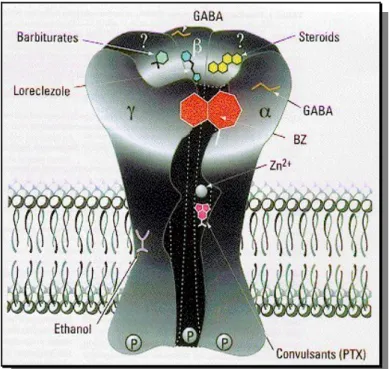
GABAA receptors have a rich pharmacology in that they have a number of separate
allosteric binding sitesxii for a variety of drugs that can modulate the activity of GABA
1 (Fig. 11). These includes: barbiturates, e.g. thiopental 5; non-barbiturate intravenous anesthetics, e.g. (R)-(+)-etomidate 6; neuroactive steroids, e.g. lanosterol 7; sedatives, e.g. loreclezole 8; benzodiazepines (BZ), (see also chapter 1.3), e.g. clonazepam 9; ethanol and other alcohols; volatile anesthetics, e.g. (S)-isoflurane 11; channel blockers or picrotoxin (PTX) site, e.g. picrotoxinin 10 and ionic zinc (Fig. 12). A distinct binding site that mediates the stimulatory actions of lactones (see also
xii Allosteric Site refers to a protein binding site other than the one used by the normal ligand, which affects the
activity of the protein. An allosteric inhibitor that binds to an allosteric binding site induces a change of shape in the protein which disguises the normal binding site from its ligand.
chapter 1.2.4.1) is presumed to exist, although no definitive evidence to support this claim exists.[25, 26] GABAA Rs are antagonized by bicuculline (BMC) 12 and insensitive
to (R)-(-)-baclofen (13, Fig. 13).[27]
Figure 11. Three-dimensional cartoon of the GABAA R on the plasma membrane with
ligand-binding sites indicated: GABA site, barbiturate site [e.g. barbiturates and loreclezole 8, benzodiazepine site (BZs and non-BZs), general anaesthetics site (e.g. steroids and ethanol) and convulsant site (PTX).[10]
Until now, only nineteen GABAA receptor subunits, molecularly distinct, have been
identified. Based on sequence homology, they are divided into eight subunit classes (designed α, β, γ, δ, ε, θ, π and ρ), some of which have multiple isoforms (designed numerically): α(1–6), β(1–3), γ(1–3), δ, ε, θ, π and ρ(1–3).[28] The subunit composition
influences fundamental features of the receptor, including sensitivity to GABA, channel kinetics and desensitizationxiii, neuronal location and pharmacological
properties. GABAA receptors with different subunit composition have different
physiological and pharmacological properties, are differentially expressed throughout
xiii Desensitization, in pharmacology, is the loss of responsiveness (i.e. the responsive to stimulation) to the
continuing or increasing dose of a drug. For medical purposes, is a method to reduce or eliminate an organism's negative reaction to a substance or stimulus.
the brain and are targeted to different subcellular regions. For instance, receptors composed of α1, α2, α3 or α5 subunits together with β and γ subunits are benzodiazepine-sensitive, which are largely synaptically located and mediate most phasic inhibition in the brain.[29] By contrast, those composed of α4 or α6 subunits
together with β and δ subunits formed a specialized population of predominantly extrasynaptic receptor subtypes that mediate tonic inhibition and are insensitive to benzodiazepine modulation.[30]
Figure 12. Drug binding sites and respective ligand drugs of the GABAA R.
Figure 13. Chemical structure of BMC 12 and (R)-(-)-baclofen 13.[27]

![Figure 9. Cartoon showing the GABA-mediated inhibition. [20]](https://thumb-eu.123doks.com/thumbv2/123dok_br/15447942.1026490/51.892.141.748.412.802/figure-cartoon-showing-gaba-mediated-inhibition.webp)


![Figure 20. Chemical structure of 1,4-benzodiazepine 57, 1,4-benzodiazepine-2,5-dione 58 and pyrrolo[2,1-c][1,4]benzodiazepine-5,11-dione 59](https://thumb-eu.123doks.com/thumbv2/123dok_br/15447942.1026490/65.892.184.711.802.996/figure-chemical-structure-benzodiazepine-benzodiazepine-dione-pyrrolo-benzodiazepine.webp)
![Figure 22. Proposed mechanism for the studied reactivity of isatoic anhydride. [87]](https://thumb-eu.123doks.com/thumbv2/123dok_br/15447942.1026490/67.892.192.697.261.486/figure-proposed-mechanism-studied-reactivity-isatoic-anhydride.webp)