UNIVERSIDADE TÉCNICA DE LISBOA
Faculdade de Medicina Veterinária
R
ISK ASSESSMENT CONCEPTUAL MODEL OF OCCURRENCE OF
M
YCOBACTERIUM AVIUM SUBSPECIES PARATUBERCULOSIS
IN UNPROCESSED BEEF AND BOVINE MILK
DIOGO FILIPE PEREIRA MARQUES
CONSTITUIÇÃO DO JURI ORIENTADOR
Professor Doutor Fernando Jorge Silvano Boinas Professor Doutor Søren Saxmose Nielsen Professora Doutora Yolanda Maria Vaz
Professor Doutor Virgílio da Silva Almeida CO-ORIENTADOR
Professor Doutor Søren Saxmose Nielsen Professor Doutor Virgílio da Silva Almeida
2009
UNIVERSIDADE TÉCNICA DE LISBOA
Faculdade de Medicina Veterinária
R
ISK ASSESSMENT CONCEPTUAL MODEL OF OCCURRENCE OF
M
YCOBACTERIUM AVIUM SUBSPECIES PARATUBERCULOSIS
IN UNPROCESSED BEEF AND BOVINE MILK
DIOGO FILIPE PEREIRA MARQUES
DISSERTAÇÃO DE MESTRADO INTEGRADO EM MEDICINA VETERINÁRIA
CONSTITUIÇÃO DO JURI ORIENTADOR
Professor Doutor Fernando Jorge Silvano Boinas Professor Doutor Søren Saxmose Nielsen Professora Doutora Yolanda Maria Vaz
Professor Doutor Virgílio da Silva Almeida CO-ORIENTADOR
Professor Doutor Søren Saxmose Nielsen Professor Doutor Virgílio da Silva Almeida
2009
i
T
RAINING DESCRIPTIONDuring a 8-month curricular training period the candidate was enrolled at the Faculty of Veterinary Medicine (FMV-UTL) – Technical University of Lisbon at the Department of Epidemiology & Economics and Veterinary Public Health (01/09/2008 – 28/01/2009), and at the Faculty of Life Sciences (LIFE-KU) – University of Copenhagen at the Epidemiology Group of the Department of Large Animal Sciences (02/02/2009 – 04/05/2009).
During all the training period Professor Virgílio da Silva Almeida was the Co-Supervisor.
In the first part of the training period, Dr. Telmo Nunes was assigned as supervisor and the training had as main objective the development of scientific skills in epidemiological & risk analysis and data management under the topic of Bluetongue Surveillance in Portugal. During the FMV-UTL training period several small projects were developed:
Project 1:
o Topic: Bluetongue prevalence estimation in Portugal in 2005 from pre-movement tests data;
o Learning objectives: bluetongue disease; bluetongue vectors; epidemiological indicators; data management and analysis; data geographical representation; Project 2:
o Topic: Types of confidence intervals for a proportion; o Learning objectives: confidence intervals;
Project 3:
o Topic: Geographical representation of the evolution of Bluetongue virus restriction zones between 2004 and 2008 in Portugal;
o Learning objectives: data geographical representation; data management; Project 4:
o Topic: Animal and herd densities in Portugal;
o Learning objectives: data geographical representation; data management; Kernel densities; sampling processes;
Project 5:
o Topic: Risk factors for the Culicoides occurrence at herd level in Portugal; o Learning objectives: bluetongue risk factors; bluetongue vectors; survey
design; data management and analysis; epidemiological measures of association;
Project 6:
o Topic: Portugal Culicoides occurrence modelling;
o Learning objectives: bluetongue risk factors; bluetongue vectors; data management and analysis; remote sensing; logistic regression models;
ii
In order to develop the above mentioned projects, the following software programmes were explored: MS Office Access, MS Office Excel, ArcGIS, GvSIG, JMP 7 and SAS.
During the training at FMV-UTL the candidate also participated in: Animal Health Department seminars:
o Presentation:
“Types of confidence intervals for a proportion” by Diogo Marques (FMV-UTL) – 21th October 2008, FMV-UTL;
o Purpose: Overview of the main types of confidence intervals for a proportion used in epidemiology;
o Participants: Telmo Nunes (FMV-UTL), Hugo Martins (FMV-UTL), Solange Pacheco (FMV-UTL), Ana Duarte (FMV-UTL);
Workshops:
o GIS Workshop: Presentation:
“Geographical Information Systems (GIS) applied to Epidemiology” by Hugo Martins (FMV-UTL), 13th - 14th October 2008;
Purpose: Introduction to GIS, GvSIG and ArcGIS training;
Participants: Solange Pacheco (FMV-UTL), Ana Duarte (FMV-UTL), Emanuel Garcia (FMV-UTL), Rui Cepeda (FMV-UTL), Vasco Martins (FMV-UTL), André Silva (FMV-UTL), Miguel Figueiredo (FMV-UTL).
o Statistics Workshop:
Presentation: “Data analysis and statistical tests” by Filipa Matos Baptista (LIFE–KU, FMV-UTL) – 08th - 09th January 2009, FMV-UTL; Purpose: Statistical methods applied to epidemiology, SAS training; Participants: Virgílio Almeida (FMV-UTL), Telmo Nunes (FMV-UTL),
Hugo Martins (FMV-UTL), Solange Pacheco (FMV-UTL), Ana Duarte (FMV-UTL), Emanuel Garcia (FMV-UTL).
Conferences and Congresses:
o “II Technical Journeys – Caçador Pecuária/Batallé/OPP” - Seminar on Swine Production – 07th November 2008, Leiria;
o “IV Congress of Veterinary Sciences Portuguese Society / I Iberian Congress of Epidemiology”, 27th – 29th November 2008, INRB-INIA/Fonte Boa.
iii
In the second part of the training period the candidate joined the Epidemiology Group working on Paratuberculosis at LIFE-KU – Denmark. Professor Søren Saxmose Nielsen was his supervisor and the main objective of the training was the assessment of MAP occurrence in unprocessed beef and milk.
During the training period two seminars were organized by Søren Saxmose Nielsen: Start-up Seminar:
o Presentations:
“Paratuberculosis” by Søren Saxmose Nielsen (LIFE-KU) – 05th February 2009, LIFE-KU;
“Short overview of Microbial Risk Assessment” by Sara Monteiro Pires (Food-DTU) - 05th February 2009, LIFE-KU;
o Purpose: Discuss the objectives, outline, boundaries and expected results of the candidate’s work;
o Participants: Hans Houe (LIFE-KU), Jens Frederik Agger (LIFE-KU), Liza Rosenbaum Nielsen (LIFE-KU), Filipa Matos Baptista (LIFE-KU, FMV-UTL), Lis Alban (Danish Meat Association), Heidi Mikkelsen (Vet-DTU) and Antonio Vieira (Food-DTU).
Follow-up Seminar: o Presentations:
“MAP occurrence in unprocessed beef and milk” by Diogo Marques (FMV-UTL) – 28th April 2009, LIFE-KU;
“Implication of MAP infection on cattle food chain” by Hisako Okura (LIFE-KU) – 28th April 2009, LIFE-KU;
o Purpose: Discuss the results of the candidate’s work; Provide Hisako Okura inputs to her PhD. study on MAP risk assessment.
o Participants: Søren Saxmose Nielsen (LIFE-KU), Jens Frederik Agger (LIFE-KU), Liza Rosenbaum Nielsen (LIFE-KU), Nils Toft (LIFE-KU), Erik Rattenborg (Danish Dairy Board), Heidi Mikkelsen (Vet-DTU), Sara Monteiro Pires (Food-DTU), Syed Sayeem Ahmed (LIFE-KU), Torben Dahl Nielsen (LIFE-KU).
v
A
CKNOWLEDGMENTSAt the end of this work, I want to express a word of thanks to all the people who have contributed directly or indirectly to its success:
To Virgílio Almeida for accepting to be my Co-Supervisor, for his invaluable help and motivation throughout this work and his exciting and always funny Epidemiology teaching; To Telmo Nunes for being my unofficial but exceptional Portuguese supervisor, for his
constant enthusiastic will of teaching, for encouraging to think on my own;
To Filipa Matos Baptista for her friendship, motivation, exceptional helping and strength she gave during all training, especially while I was in Denmark;
To my Portuguese department colleagues Hugo Martins, Ana Duarte and Solange Pacheco for all their pleasant and unforgettable moments spent during the training period; To Armando Louzã, Yolanda Vaz, Mário Melo, Maria Isabel Neto, José Pedro Lemos for
the funny and always agreeable lunches and “Nespresso Coffee-breaks”;
To Søren Saxmose Nielsen for being my Supervisor. His availability, support and guidance during my stay in Denmark were exceptional and a reference to my future;
To all my Danish department colleagues for allowing me to be part of the team and enjoy the relaxing and “delicious” moments at the “Monday’s Cake Party”, especially to Hans Houe for accepting me at the department, to Jeanne Oakman for her constant happiness and contagious laughs, and to Sayeem Ahmed for his company at the office;
To all the friends I made during my stay in Denmark in particular to Teresa Santos, Ida Engelund, Ana Ortega, Belén Pinilla, Bruno De Meo, Eros Ricci, Agustin, Tamara and Pilar for their friendship and for making my stay in Denmark a great and unforgettable life experience;
To Sara Pires for the encouragement and inspiration regarding the Epidemiology and Risk Analysis, and for the willingness to help and availability to be present at the seminars; To all my other friends Duarte, Anabela, Emanuel, Joana Afonso, Joana Marcelino,
Daniela, Tiago, Catarina Antão, Margarida Ramada, Claudia Alves and “BodyAttack-Colleagues” for being always present and for their friendship;
At last but not the least, I cannot forget to thank to all my family for their support, ongoing motivation and positivism during all good and bad moments. For them a Huge Thank You.
With all my thanks Diogo Marques
vii
R
ESUMOModelo conceptual da avaliação do risco de ocorrência do
Mycobacterium avium subspécie paratuberculosis em carne e leite de bovino não
processados
A etiologia da Doença de Crohn é actualmente desconhecida e o Mycobacterium avium subspécie paratuberculosis (MAP) tem sido proposto como um dos possíveis agentes etiológicos.
Apesar de não ter sido ainda estabelecida uma relação causal entre a Doença de Crohn e o MAP, este tem sido frequentemente isolado em humanos. O consumo de leite e carne é considerado um possível veículo do MAP, justificando-se por isso a avaliação do risco de ocorrência do MAP nos produtos mencionados.
Este trabalho teve como objectivo avaliar a probabilidade de ocorrência do MAP em carne e leite de bovino, não sujeitos a processamento tecnológico.
De forma a consultar e avaliar informação existente, foi feita uma pesquisa bibliográfica exaustiva sobre a disseminação da infecção por MAP e sobre o isolamento do MAP por cultura bacteriológica de tecidos. A principal lacuna identificada foi a ausência de informação detalhada sobre: i) o mecanismo de disseminação do MAP e a importância da contaminação fecal; ii) a relação entre a disseminação do MAP e outros indicadores (sinais clínicos, lesões macroscópicas, resposta imunitária); iii) a prevalência do MAP na carne e no leite nos diferentes estadios de infecção.
A ausência desta informação não permite a avaliação do risco e a consequente definição de medidas específicas com vista à sua mitigação. Neste trabalho são descritas as árvores de eventos, pressupostos, informação necessária e as lacunas no conhecimento.
Estudos futuros são necessários para disponibilizar a informação inexistente e para desenvolver e aperfeiçoar testes de diagnóstico para detecção directa do MAP na carne e no leite de bovino.
Palavras-chave: Mycobacterium avium subspécie paratuberculosis; Infecção disseminada do MAP; Leite; Carne; Bovinos; Árvore de eventos; Análise de risco.
ix
A
BSTRACTRisk assessment conceptual model of occurrence of
Mycobacterium avium subspecies paratuberculosis in unprocessed beef and bovine
milk
Crohn’s Disease aetiology is currently unknown and Mycobacterium avium subspecies
paratuberculosis (MAP) has been proposed as its etiologic agent.
Despite the absence of a causal relationship between MAP and Crohn’s Disease, MAP has been frequently isolated in humans. Milk and beef consumption is considered a possible MAP source. Thus the risk of MAP occurrence on these products should be assessed.
The main objective of this work was to assess the probability of MAP occurrence in unprocessed beef and bovine milk.
In order to assemble the available data, an exhaustive literature search was made on the dissemination of MAP infection and MAP isolation by bacteriologic tissue culture. The main knowledge gaps found were the lack of detailed information on: i) MAP dissemination mechanism and the importance of faecal contamination; ii) relation between MAP dissemination and other indicators (clinical signs, gross lesions, immune response); iii) MAP prevalence on beef and milk by stage of infection.
Due to the lack of this information, the risk assessment and the characterization of the risk mitigation measures could not be performed. The risk model pathways, its assumptions, data required and knowledge gaps are described in this work.
Further research is needed to make available the mentioned knowledge gaps and to develop and to improve diagnostic tests for direct MAP detection on beef and bovine milk.
Keywords: Mycobacterium avium subspecies paratuberculosis; Disseminated MAP infection; Milk; Beef; Bovine; Risk model pathway; Risk assessment.
xi
C
ONTENTS1 Introduction ... 1
1.1 Project objectives ... 1
1.2 Problem definition ... 2
1.2.1 Mycobacterium avium subspecies paratuberculosis and Crohn’s Disease ... 2
1.2.2 Human MAP sources - Water ... 2
1.2.3 Human MAP sources – Beef and Milk ... 3
1.2.4 Map occurrence on products entering the food chain and the support of the risk assessment ... 4
2 Paratuberculosis ... 6
2.1 Aetiology ... 6
2.2 Epidemiology ... 6
2.3 Pathogenesis and Clinical signs ... 7
2.4 Pathology ... 8
2.5 Danish Paratuberculosis Control Programme ... 10
2.5.1 Diagnostic testing ... 10
2.5.2 Risk levels ... 11
2.5.3 Recommendations ... 12
2.5.4 Herd classification ... 12
2.6 Animal status and Stages of Infection ... 13
2.6.1 Animal Status ... 16
2.6.2 Stages of infection ... 13
2.7 MAP diagnostic methods ... 18
2.7.1 Diagnostic test types ... 19
2.7.2 Agent detection tests ... 19
2.7.3 Immune response detection tests ... 22
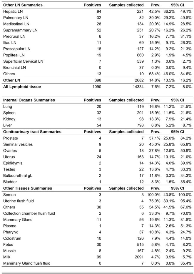
3 Distribution of MAP in tissues ... 25
3.1 Introduction ... 25
3.2 Materials and Methods ... 25
3.3 Results ... 26
3.3.1 Animal population characterization ... 32
3.3.2 Characterization of analyzed tissues ... 33
3.4 Discussion ... 37
3.5 Conclusions ... 44
3.5.1 Study results ... 45
3.5.2 Study limitations ... 46
3.5.3 Knowledge gaps of MAP distribution in tissues ... 46
xii
4 Risk assessment conceptual model ... 49
4.1 Hazard Identification ... 49
4.2 Release Assessment ... 49
4.3 Risk model pathway – Milk ... 49
4.3.1 Data availability and risk description ... 56
4.4 Risk model pathway – Muscle ... 58
4.4.1 Data availability and risk description ... 69
4.5 Conclusions ... 71
5 References ... 73
6 Annex ... 82
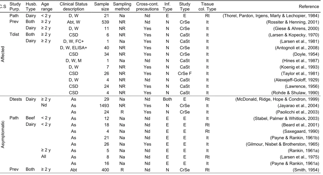
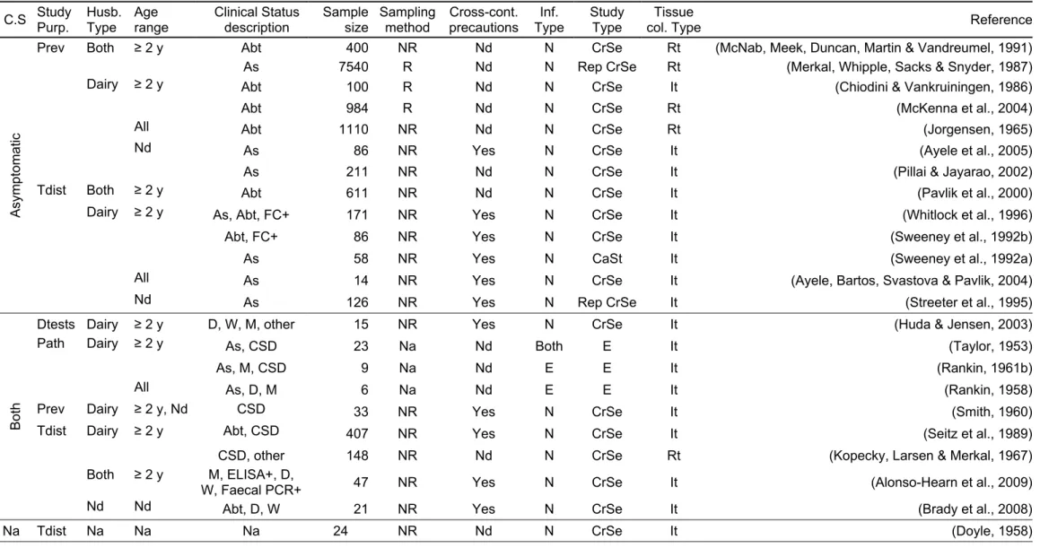
6.1 Annex 1 - Tissue culture results recorded from the reviewed studies. ... 82
T
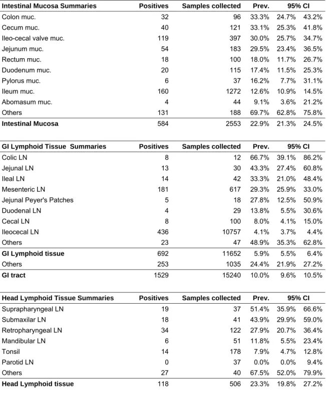
ABLE LIST Table 1 – Range of reported sensitivity (Se) and specificity (Sp) of serum antibody ELISA (SELISA) and milk antibody ELISA (MELISA) for three animal statuses: affected, infectious and infected. 23 Table 2 – Characteristics of the reviewed studies where MAP was isolated by tissue culture (part 1). 28 Table 3 – Characteristics of the reviewed studies where MAP was isolated by tissue culture (part 2). 29 Table 4 – Results from Annex 1 summarized by tissue (Part 1). Prevalence (Prev.) and 95% Confidence interval (CI) of MAP culture isolation by tissue and larger tissue sections. ... 34Table 5 – Results from Annex 1 summarized by tissue (Part 2). Prevalence (Prev.) and 95% Confidence interval (CI) of MAP culture isolation by tissue and larger tissue sections. ... 35
Table 6 - Milk risk model pathway legend ... 54
Table 7 - Milk risk model pathway description: list of variables and data availability. ... 55
Table 8 – Muscle risk model pathway legend. ... 66
xiii
F
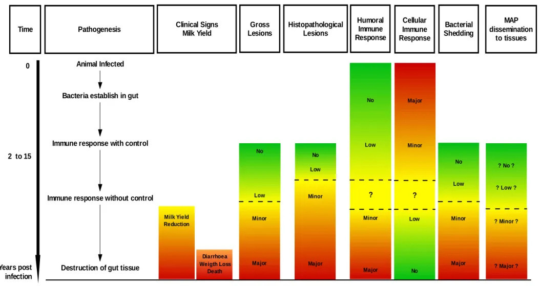
IGUREL
ISTFig. 1 – Schematic representation of the different risk levels and risk groups from the Danish ParaTB control Programme regarding the pathogenesis of MAP infection (adapted from Nielsen & Toft (2008)). ... 12 Fig. 2 – Schematic representation of “The Iceberg Effect” and the Stages of Infection in an infected
herd. Proportions adapted from Whitlock & Buergelt (1996) considering a population of 100 animals with 25 infected. ... 15 Fig. 3 – Schematic representation of the different animal status in an infected herd population.
Proportions for each status are unknown. ... 17 Fig. 4 – Summary of most commonly used MAP diagnostic tests. IFN-γ – Interferon Gamma; CF –
Complement Fixation; AGID - Agarose Gel Immunodiffusion; ELISA - Enzyme-Linked Immunosorbent Assay (adapted from Nielsen et al.(2001). ... 19 Fig. 5 – Schematic representation of pathogenesis of MAP infection and Clinical Signs, Gross Lesions, Histopathological Lesions, Humoral and Cellular Immune Responses, Bacterial Shedding and MAP dissemination to tissues. The point in time when the event pass from “Low” to “Minor” is not well known. Regarding the immune responses, the point in time when a cellular immune response shift to a humoral immune response is unknown. The relation between MAP dissemination to tissues and the previous columns need further investigation. ... 47 Fig. 6 - Milk risk model pathway part 1 – herd characteristics. ... 51 Fig. 7 – Milk risk model pathway part 2 – animal characteristics given that an infected herd gives test positive result. The dotted line represents the lack of data to support the relation between MAP dissemination to milk and the previous boxes in the figure. ... 52 Fig. 8 – Milk risk model pathway part 2 – animal characteristics given that an infected herd gives test negative result. The dotted line represents the lack of data to support the relation between MAP dissemination to milk and the previous boxes in the figure. ... 53 Fig. 9 - Muscle risk model pathway part 1 - herd characteristics. ... 60 Fig. 10 – Muscle risk model pathway part 2 – animal characteristics given that an infected dairy herd gives test positive result. ... 61 Fig. 11 – Muscle risk model pathway part 2 – animal characteristics given that an infected dairy herd gives test negative result. ... 61 Fig. 12 – Muscle risk model pathway part 2 – animal characteristics given that an infected non-dairy
herd gives test positive result. ... 62 Fig. 13 – Muscle risk model pathway part 2 – animal characteristics given that an infected non-dairy
herd gives test negative result. ... 62 Fig. 14 – Muscle risk model pathway part 3 – animal characteristics given that a Red or High risk
group animal (from dairy herd) - observable factors that could be investigated at slaughterhouse environment. It is assumed that an animal in the Red risk group or in the High risk group showing clinical signs at the ante mortem inspection will be rejected. The dotted line represents the lack of data to support the relation between MAP dissemination to muscle and the previous boxes in the figure. ... 63 Fig. 15 – Muscle risk model pathway part 3 – animal characteristics given that a Yellow/Green or
Medium/Low risk group animal (from dairy herd) - observable factors that could be investigated at slaughterhouse environment. The dotted line represents the lack of data to support the relation between MAP dissemination to muscle and the previous boxes in the figure. ... 64
xiv
Fig. 16 – Muscle risk model pathway part 3 – animal characteristics given that a High risk group animal (from non-dairy herd) - observable factors that could be investigated at slaughterhouse environment. It is assumed that an animal in the High risk group showing clinical signs at the ante
mortem inspection will be rejected. The dotted line represents the lack of data to support the
relation between MAP dissemination to muscle and the previous boxes in the figure. ... 65 Fig. 17 – Muscle risk model pathway part 3 – animal characteristics given that a Medium/Low risk
group animal (from non-dairy herd) - observable factors that could be investigated at slaughterhouse environment. The dotted line represents the lack of data to support the relation between MAP dissemination to muscle and the previous boxes in the figure. ... 66
E
QUATIONL
ISTEq. 1 – Wilson Score Interval equation. ... 26
A
BBREVIATIONSL
ISTAGID Agarose Gel Immunodiffusion
CD Crohn’s Disease
CF Complement Fixation
ELISA Enzyme-Linked Immunosorbent Assay IFN-γ Interferon gamma
LN Lymph Node
MAP Mycobacterium avium subspecies paratuberculosis
OIE Office International des Epizooties - The World Organisation for Animal Health ParaTB Paratuberculosis
PCR Polymerase Chain Reaction Se Sensitivity
Sp Specificity ZN Ziehl-Neelsen
1
1 Introduction
1.1 Project objectives
During 3 months, the candidate joined the Epidemiology Group of the Department of Large Animal Sciences at the Faculty of Life Sciences – University of Copenhagen. This was possible due to a student mobility grant of ERASMUS/SOCRATES programme.
In the framework of the Danish control programme of bovine paratuberculosis, a start-up seminar of MAP occurrence on beef and milk was organized and experts on relevant matters were present. During the seminar, the objectives, outline, boundaries and expected results of the present dissertation were discussed.
The main objective initially proposed was the assessment of MAP occurrence in unprocessed beef and bovine milk. In order to accomplish this objective, an exhaustive literature-based search was made to understand the relevant aspects of Paratuberculosis and MAP infection, to assess the current knowledge and to identify unavailable inputs required to develop the proposed risk assessment.
Due to considerable knowledge gaps it was not possible to accomplish the main objective but the available and non-available risk assessment inputs were described and proposals for MAP risk assessment model pathways were made.
This dissertation is composed by four parts:
1. Introduction (objectives and problem definition); 2. Overview of Paratuberculosis and MAP infection;
3. Literature revision concerning MAP isolation by tissue culture and MAP distribution on tissues;
4. A proposal of risk assessment model pathways to assess the risk of MAP occurrence in unprocessed beef and bovine milk.
2
1.2 Problem definition
1.2.1 Mycobacterium avium subspecies paratuberculosis and Crohn’s Disease
Crohn’s Disease (CD) is a chronic disease that affects the gastro-intestinal tract of humans. Usually, it occurs in young adults showing as major symptoms diarrhoea and chronic pain (Grant, 2005; Waddell et al., 2008).
Chiodini, Van Kruiningen, Merkal, Thayer & Coutu (1984) isolated Mycobacterium sp. organisms from several patients with CD. This finding revived the idea of a mycobacterial cause for this human disease (Chiodini, Van Kruiningen, Merkal, Thayer & Coutu, 1984). Since then, Mycobacterium avium subspecies paratuberculosis (MAP) has been isolated from several patients and a possible link between CD and MAP has been discussed in the last decades. Some of the findings that support this link are: the similarities of clinical signs between CD and bovine paratuberculosis (in which MAP is the etiological agent); detection of MAP on faeces, intestinal tissues and blood from CD patients; demonstration of a serological response to MAP antigens in Crohn’s patients; and anti-MAP antibiotic therapy resulting in remission or reduction of clinical signs. Several potential roles of MAP in CD patients have been suggested: MAP could be the primary infectious agent of CD; a secondary invader in a host with a compromised intestinal barrier; or colonize the intestinal tract without causing any harm (Grant, 2005). Because there are inconclusive and contradictory studies, difficulties on the isolation of MAP organisms and unexpected results, it is very difficult to achieve a consensus between authors (Waddell et al., 2008; Grant, 2005). In a recent systematic review, Waddell et al (2008) analyzed many studies and several MAP isolation methods in CD patients and concluded that the “evidence of association is not strong, but should not be ignored”. Despite the absence of an irrefutable causal relationship between MAP and CD, MAP has been consistently isolated in humans and its source should be investigated. Food and water-borne sources have been suggested (Grant, 2005).
1.2.2 Human MAP sources - Water
Water as a MAP source has not been properly investigated and its potential as a vehicle of transmission of MAP to humans is unknown (reviewed in: Grant, 2005)1. Water containing
MAP may be consumed directly, incorporated into food, used to wash food products or surfaces used to manipulate food. MAP survival characteristics in water environments were analyzed by Larsen, Merkal & Vardaman (1956), Lovell, Levi & Francis (1944) and Whittington, Marshall, Nicholls, Marsh & Reddacliff (2004). The presence of mycobacteria in
3
piped supplies suggests that water standard treatments have little effect on these organisms. The chlorination effect was studied in experimentally contaminated water and MAP was not completely eliminated by this process (Whan, Grant, Ball, Scott & Rowe, 2001). These findings make water a plausible MAP source that should be properly investigated.
1.2.3 Human MAP sources – Beef and Milk
Beef, milk and milk products are food products of bovine origin largely consumed by humans. There are no specific regulations about MAP presence on food products. Thus contaminated milk and beef may be consumed by humans.
MAP occurrence on milk and milk products was studied by several authors and through different methods (reviewed in: Slana, Paolicchi, Janstova, Navratilova & Pavlik, 2008). MAP was isolated by culture on milk samples aseptically collected from individual cows (Alexejeff-Goloff, 1929; Streeter, Hoffsis, Bechnielsen, Shulaw & Rings, 1995; Sweeney, Whitlock & Rosenberger, 1992a; Taylor, Wilks & McQueen, 1981; Paolicchi et al., 2003; Ayele, Svastova, Roubal, Bartos & Pavlik, 2005; Pillai & Jayarao, 2002; Giese & Ahrens, 2000; Jayarao et al., 2004). MAP was also identified on milk and milk products after industrial processing steps and their effect on MAP survival or mitigation was discussed (Slana et al., 2008; reviewed in: Grant, 2006). However, MAP resistance to milk industrial processing steps is not broadly accepted due to contradictory results and some study designs were criticized. Differences between the laboratory techniques used and industrial procedures are the reasons suggested for these differences, e.g. initial MAP concentrations; MAP origin - culture collection/field strains; absence/presence of turbulent flow during heating to assure that all milk particles get the same treatment; heating methods, volumes of milk tested; and absence/presence of homogenization step (reviewed in: Grant, 2006; Rademaker, Vissers & Giffel, 2007).
Presence of MAP after various processing procedures is not fully understood. It may be attributable to its high heat resistance (e.g. cell clumping, phagocytosis) (Rademaker et al., 2007) or to contamination during the process. It can be due to cross contamination during laboratory analysis; leaks on the heat exchanger plates of pasteurization plants enabling raw milk to contaminate pasteurized milk; and use of pasteurizers during a long time between cleaning processes (milk dry matter deposits leading to decrease in heat treatment efficacy) (Cerf, Griffiths & Aziza, 2007).
Map isolation and quantification constrains are also encountered in the evaluation of milk processing steps. The real number of MAP present in raw or pasteurized milk cannot be precisely estimated because milk culture requires a chemical decontamination treatment which has adverse effects on MAP viability (Dundee, Grant, Ball & Rowe, 2001; Grant &
4
Rowe, 2004) ending on an underestimation of MAP numbers (Grant, Williams, Rowe & Muir, 2005). The dilution on bulk milk tank also influences the detection of MAP on raw and processed milk. Despite the lack of consensus on the effect of milk processing steps on MAP, the evidence of MAP occurrence on retailed milk makes this product a key MAP source to be investigated.
Regarding beef, little information is available about this possible source of MAP to consumers. MAP was isolated by culture from muscle tissue (Antognoli et al., 2008; Alonso-Hearn et al., 2009), MAP DNA was obtained from surface swabs collected from beef carcasses at the slaughterhouse after skinning and dressing (Meadus, Gill, Duff, Badoni & Saucier, 2008), and two MAP detection methods in ground beef were compared (Jaravata, Smith, Rensen, Ruzante & Cullor, 2007).
MAP organisms may be reduced or eliminated by heating during cooking procedures. However, the effect of heating processes on MAP survival in the muscle matrix has not been evaluated (Alonso-Hearn et al., 2009). Raw and undercooked meat consumption should also be considered. Some products such as sausages and minced meat are based on low quality meat, resulting in a larger probability of coming from infected cattle with high MAP contamination rates (Alonso-Hearn et al., 2009). Beef as MAP food source to consumers should be subjected to further research.
1.2.4 Map occurrence on products entering the food chain and the support of the risk assessment
Considering these three MAP sources to humans (water, milk and beef), the reason behind MAP presence on these products should be assessed.
Water contamination has been mainly attributed to contamination with faeces of infected animals and to MAP capacity to survive for long periods of time on the environment (reviewed in: Grant, 2005).
MAP isolation from milk, muscle and other tissues, for example non gastro-intestinal lymph nodes and internal organs led some authors to suggest the possibility of MAP dissemination inside the infected host (Alonso-Hearn et al., 2009; Antognoli et al., 2008; Dennis et al., 2008; Hines, Buergelt, Wilson & Bliss, 1987; Pavlik et al., 2000). Yet there is also the chance of contamination by MAP of faecal origin during tissue collection and/or food processing. The view of MAP entry into the food chain, its potential to survive along food processing, the lack of MAP regulations, the increased claim of food safety by consumers, the continuous search for better and more competitive products by the dairy and meat industries, and the commitment to promote Public Health by the veterinary authorities, made this mycobacteria a subject of study. In order to properly address the MAP source to humans, a MAP risk
5
assessment on the food chain would be of great importance for risk managers to take better and more cost-effective decisions.
This work will therefore contribute to the development of such assessment in the most important products of animal origin, beef and milk. Water as a MAP source was excluded regarding the amount of knowledge gaps and the lack of published studies. Therefore, and due to time limitations, animal products consumed by humans were given priority.
The assessment of MAP occurrence in beef and milk will be addressed as a qualitative risk assessment and will be limited to a release assessment, i.e. the final objective will be to evaluate the probability and the quantity of MAP shed by infected animals into specific animal products.
The exposure assessment step was not incorporated in this study because:
o the causal relationship between MAP and Crohn’s disease has not been established;
o MAP doses that causes an undesirable effect on humans are unknown;
o there is a lack of consensus about the effect of food processing steps on MAP survival/reduction.
As this dissertation was elaborated in the framework of a curricular training period in Denmark, the Danish control programme of bovine paratuberculosis will be used as a model. The assessment will be restricted to cattle because of the larger amount of scientific publications and the special focus on cattle of the Danish bovine paratuberculosis control programme.
The end-points assumed will be the milk at herd level (individual milk) and the carcass at the end of the slaughter line. These end-points are in agreement with the purpose of the release assessment.
This work will focus on MAP dissemination inside the host, rather than on faecal contamination during harvesting and processing steps.
6
2 Paratuberculosis
2.1 Aetiology
Paratuberculosis (ParaTB) is a chronic, infectious disease caused by Mycobacterium avium subspecies paratuberculosis (MAP). It is a Gram-positive, intracellular facultative, acid-fast bacillus that is slow-growing, and has complex nutritional requirements in culture. It requires an organic source of iron and mycobactins to grow in vitro (reviewed in: Chiodini, Van Kruiningen & Merkal, 1984). Through genomic DNA analysis it was shown that it has an unique insertion sequence: IS900 (Green et al., 1989). The genus Mycobacterium consists of about 50 species that are grouped according some similarities. The most known are the
Mycobacterium tuberculosis complex (including M. tuberculosis and M. bovis),
Mycobacterium leprae and Mycobacterium avium-intracelullare complex (including
Mycobacterium avium and MAP) (Buergelt, Bastianello & Michel, 2004; Coetzer & Tustin,
2004). Cell wall elements of mycobacteria not only help in the survival and resistance of the agent against host defences but are also responsible for the host strong immunological response (reviewed in: Clarke, 1997). MAP has been shown to survive on the environment at different conditions. It can survive not only in dry conditions (Larsen, Merkal & Vardaman, 1956) but also in freezing environment (Larsen et al., 1956; Richards & Thoen, 1977) or water, soil and faeces (Jorgensen, 1977; Larsen et al., 1956; Lovell, Levi & Francis, 1944; Whittington, Marshall, Nicholls, Marsh & Reddacliff, 2004). It may stay viable in slurry, even when submitted to heat treatments (Olsen, Jorgensen & Nansen, 1985; Jorgensen, 1977) and in different water sources (Whittington, Marsh & Reddacliff, 2005). Recent studies suggest rather effective survival strategies namely an interaction between MAP, protozoa, nematodes and insects (Rowe & Grant, 2006) and the move into a dormancy state when MAP is submitted to unfavourable conditions (Whittington et al., 2004).
2.2 Epidemiology
Paratuberculosis occurs on several ruminant species but it is most studied on domestic ruminants. It is a widespread infection in Europe with cattle within-herd prevalence ≈ 20% and between-herd > 50% in cattle herds and > 20% in small ruminants flocks (Nielsen & Toft, 2009).
MAP can be transmitted by either direct or indirect contact between infected and susceptible animals. Transmission occurs mainly by faecal-oral route. Shedding animals play an important role contaminating the feed, water and pastures (reviewed in: Clarke, 1997). Transmission can also occur in utero (Doyle, 1958; Sweeney, Whitlock & Rosenberger, 1992b), by ingestion of infected colostrum and milk from infected cows or when the teats are
7
contaminated with faeces (Streeter et al., 1995; Sweeney et al., 1992a). Contaminated feeding utensils, herd worker’s shoes or clothing, wildlife ruminant reservoirs and certain veterinary procedures (e.g. rectal examination) can also transmit MAP (Sweeney, 1996). Between-herd transmission is thought to be mostly caused by purchases of healthy carriers. However, a break on the herd biosecurity or a direct contamination between herds, for example a common water source, may result on a cluster of paratuberculosis cases bearing in mind the resistance of MAP in environment (Sweeney, 1996; Whittington et al., 2005). The animals that are more vulnerable to infection are the neonates and some studies point to the existence of an age-dependent resistance (Bendixen, 1978; Buergelt, Hall, McEntee & Duncan, 1978; Larsen, Merkal & Cutlip, 1975; Rankin, 1958; Rankin, 1961b; Rankin, 1961a; Rankin, 1962; reviewed in: Windsor & Whittington, 2009). Hypothesis to explain calves’ susceptibility are: (i) an age-related difference on cellular immune response (Windsor & Whittington, 2009); (ii) effect (dilution or harmful effect) that the functional rumen of adult animals may have on MAP before they reach the gut (Windsor & Whittington, 2009); (iii) the existence of an “open gut” in calves that besides allowing some macromolecules such as immunoglobulins from colostrum to penetrate the mucosa to be absorbed, it may permit the entrance of MAP into the animal intestinal wall (Sweeney, 1996). Some animals could become asymptomatic carriers and shed the agent during their lives (reviewed in: Chiodini et al., 1984). Passive transfer or a pass-through of MAP is referred when a faecal culture positive exist together with a tissue culture negative (however the sample collection procedure may have neglected the infected sites) (Whitlock, Rosenberger, Sweeney & Spencer, 1996). Passive shedding has also been reported after oral inoculation and from animal living in a heavily contaminated environment (Hines II et al., 2007; Sweeney, Whitlock, Hamir, Rosenberger & Herr, 1992)
2.3 Pathogenesis and Clinical signs
The pathogenesis of Paratuberculosis can be divided into three main stages of infection: early infection, subclinical infection and clinical disease (Coussens, 2001).
MAP is ingested orally with contaminated material. Mycobacteria are transported in vacuoles across the M cells to macrophages in Gut Associated Lymphoid Tissue (mainly in Peyer’s patches) (Momotani, Whipple, Thiermann & Cheville, 1988). These patches reach their maximum development about the time of birth and progressively disappear afterwards (reviewed in: Clarke, 1997; Reynolds & Morris, 1983). After undergoing phagocytosis MAP organisms suffer degradation activities within the macrophage, are processed and presented to T lymphocytes or remain intact inside the phagocytic cell (Coussens, 2001; Stabel, 2000b). During the subclinical stage of infection there are no clinical symptoms. A granuloma develops that may allow the growth of the pathogen inside it by protecting infected cells from
8
cytotoxic immune effects (reviewed in: Coussens, 2001; Zurbrick & Czuprynski, 1987). Sporadic destruction of infected macrophages inside granulomas can lead to bacterial shedding observed in faecal cultures from subclinically infected animals. Intermittent shedding not only provides a way to MAP moving to new infection sites, leading to large damage portions of the intestinal tract but also to a continuous low-level stimulation of humoral immunity. This could convert a low-undetectable antibody response into detectable levels of antibodies in middle to late-stage of subclinical infections (reviewed in: Coussens, 2001). The initial cell-mediated response of the early and subclinical stages of infection is replaced by a non-protective humoral response at the clinical stage (Coussens, 2001; Stabel, 2000b; Stabel, 2000a) allowing for a rapid dissemination of the infection throughout the host (Bendixen, 1978; Stabel, 2000a) and the development of new lesions.
The balance between host (local and systemic immunity, age), MAP (infective dose, route of infection, virulence and survival capabilities) and environmental factors will determine the progression from the subclinical stage to the appearance of symptoms.
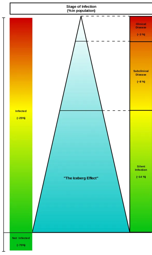
The symptoms are usually unspecific like a chronic progressive weight loss with chronic or intermittent diarrhoea. However, in advanced stages of infection, animals may show mandibular oedema, hypoproteinaemia and become cachetic or even die (reviewed in: Clarke, 1997). Correlation between disease progression and some measurable production indicators such as milk yield has also been investigated. Although is not an observable clinical sign, deviations in milk yield can be used together with other laboratory diagnosis techniques to characterize the stage of infection and the animal status, and take management measures (Nielsen, Krogh & Enevoldsen, 2009; Nielsen & Toft, 2008). Though young animals are the more susceptible to infection, clinical disease generally develops only after 3-5 years of age (Chiodini et al., 1984). Animals with clinical signs are considered the “tip of the iceberg” (Whitlock & Buergelt, 1996)2. However infected animals have higher
probability of being culled due to secondary infections, such as mastitis and metritis, than from severe clinical stages of late disease (Merkal, Larsen & Booth, 1975).
2.4 Pathology
Usually the concomitant existence of clinical symptoms, pathological signs and isolation of organism are representative of a later stage of the disease in contrast to early stages where this association is not a routine finding (Brady, O'Grady, O'Meara, Egan & Bassett, 2008; Clarke, 1997; Pavlik et al., 2000; Pérez, Marín & Badiola, 1996).
In a study of 32 infected cattle by Buergelt et al (1978) the most common pathologic gross findings were chronic enteritis, chronic intestinal lymphangitis and mesenteric
2 Whitlock & Buergelt (1996) suggested that for each 3 animals with clinical signs (stages III and IV) may exist 8 animals with subclinical disease (stage II) and 14 animals with silent infection (stage I) in an population with 25 infected animals.
9
lymphadenopathy with major lesions present at the distal ileum. Other pathological signs observed were corrugated and thickened intestinal mucosa, prominent and dilated subserosal lymphatics, cachexia, or atrophy of skeletal muscle and fat. Less frequent lesions were alopecia, and endocardial and aortic calcification. Gross lesions were detected rarely in colon, cecum or rectum (Buergelt et al., 1978).
Various histopathological lesions types can be observed both in sheep (Carrigan & Seaman, 1990; Clarke & Little, 1996; Pérez et al., 1996; Rajya & Singh, 1961; Stamp & Watt, 1954) and cattle (Buergelt et al., 1978; González et al., 2005). Some theories to explain these different lesions types are: (i) that the pathogenicity may vary between MAP strains (Stamp & Watt, 1954); (ii) that this variety of lesions is dependent upon the host immune defences (Bendixen, 1978; Buergelt et al., 1978; Shulaw, Bechnielsen, Rings, Getzy & Woodruff, 1993; Stamp & Watt, 1954); (iii) or that different lesions represent different stages of the disease (Pérez et al., 1996; Rajya & Singh, 1961). In cattle, the histopathologic lesions were described by Buerguelt et al (1978) and by González et al (2005). The latter examined 167 animals and in 116 (70%) they found MAP infection associated lesions which were categorized on focal, multifocal, diffuse multibacillary, diffuse lymphocytic and diffuse intermediate. Focal lesions were observed in distal ileum and lymph nodes and were described as “well-demarcated, small granulomas formed by macrophages with abundant, slightly foamy, pale cytoplasm and large nuclei with sparse chromatin”. On the other hand multifocal lesions (the correspondent to the “mild” and “moderate” forms described by Buerguelt et al (1978)) correspond to focal and well-demarcated granulomas in intestinal lamina propria in addition to lymphoid tissue. It was also observed that sometimes the same animal showed lesions in different regions of the intestine. In diffuse lesions (the correspondent to the “advanced” lesions described by Buerguelt et al (Buergelt et al., 1978)) an inflammatory infiltrate occurred in several areas of the intestine causing diffuse and severe granulomatous enteritis. Animals were divided into three different subtypes according to the main cell type present in the infiltrate and the amount of acid-fast bacteria (AFB). Diffuse multibacillary lesions were named when “macrophages, with foamy cytoplasm and also the appearance of epithelioid cells” appeared diffusely infiltrated in the intestinal wall and mycobacteria were present in large numbers in all sections of the intestine and lymph nodes. Another type of diffuse lesion is the lymphocytic or paucibacillary type where “lymphocytes were the main inflammatory cells infiltrating the lamina propria”. Although AFB was present, they were always in low numbers. González et al (2005) described also an intermediate diffuse category in which the “infiltrate contained abundant lymphocytes, plasma cells, giant cells and macrophages, either isolated or forming small granulomas”. However there was variation in the cellular content of the infiltrate between animals and between samples from the same animal. In this type of lesion mycobacteria were always detected (González et al., 2005).
10
Though it is uncertain whether different pathological forms, especially diffuse lesions, represent sequential or divergent stages of paratuberculosis, an association between histopathologic lesions and immunity responses was transposed from human leprosy to ruminant paratuberculosis (mainly in sheep) (González et al., 2005). According to that multibacillary lesions may be associated with marked humoral peripheral responses, paucibacillary lesions with a strong cellular immune response, and focal lesions with initial or latent forms of infection associated with high cellular immune responses (Clarke & Little, 1996; González et al., 2005; Pérez et al., 1996).
2.5 Danish Paratuberculosis Control Programme
(adapted from Nielsen (2007) and Nielsen, Jepsen & Aagaard (2007))
The Danish control programme for paratuberculosis started in February 2006. This voluntary programme aims to provide farmers with tools to control paratuberculosis and to reduce the prevalence of this disease. The programme follows a risk-based approach at farm level, where the cows are classified to different groups representing different stages of infection in order to manage the risk according to each risk group.
By June 2007 approximately 1140 (23%) dairy herds of the 4900 Danish dairy herds joined the programme. These herds are primarily large herds with an average herd size of 137 cow/herd compared to the national average of 110 cows/herd (Nielsen, 2007). The farmer’s main purposes of participation in the programme were the certification with 4-10 years, the control to avoid production losses, the control to increase animal health and control to increase food safety (S.S. Nielsen, personal communication, February 5th, 2009).
2.5.1 Diagnostic testing
The distribution of animals by risk groups is done according to the result of an individual antibody ELISA test performed on milk quality control samples without an obligatory confirmatory test (faecal culture). An ELISA positive test result is considered when corrected optical density values are > 0.3.
Each herd is tested 4 times a year. Each cow is tested 3 times year (not tested during the dry period). This testing scheme and the test used have as objective an early detection of the infectious animals being the most sensitive as possible. Due to the high test frequency an overall low specificity of the combined test is expected.
The test gives many positive results due to the focus on sensitivity rather than specificity. This characteristics needs to be taken into account in policy making. Every test-positive animal is regarded as a possible MAP disseminator, however, there are several types of status regarding the test positive results: animals could be infectious; could be infected but
11
still have to progress to disease; could acquire MAP sporadically from a contaminated environment; some could be false-positive.
It is recommended that cows aged 2 – 4 years should be tested more frequently and older cows may be tested less frequently (Nielsen & Ersboll, 2006). It was shown that ELISA test is able to detect almost all infected cattle that shed MAP (Nielsen & Ersboll, 2006), although the age-span of testing positive is from 2 to 11 years of age. With frequent testing most animals are detected at the time they are infectious (Nielsen & Toft, 2006).
2.5.2 Risk levels
Regarding the three colour system for risk groups, the test negative animals are referred as “Green cows”. These cows, on the day of the test, are considered non-infectious, possibly not infected and are considered low risk cows. Cows with the classification Yellow (intermittent positive and negative results) and Red (frequent positive results) are infected, infectious and considered high-risk cows. The Yellow cows could be in a stage of controlling the infection and they may be clinically affected. Red cows are in a stage incapable of controlling the infection and are all considered affected.
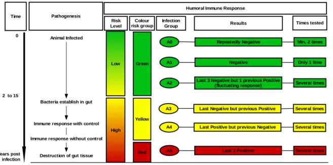
Another classification by “infection group” is used to infer differences on milk production losses. Based on repeated testing, cows are divided into 6 infection groups regarding their antibody profiles (Nielsen, 2007; Nielsen, 2008) (Fig. 1):
A0 – repeated negative (minimum 2 samples);
A1 – negative, but only 1 test result (usually at the beginning of the 1st lactation); A2 – positive on last sample and negative on previous test;
A3 – last 3 tests negative, but with 1 previous positive result;
A4 – last result negative, but more positive results have occurred previously (fluctuating response);
A5 – last 2 or more results positive;
Cows with positive reactions are considered to be high risk animals and potentially infectious but only cows from groups A4 and A5 have reduced milk production3. The allocation of these
animals in the animal status classification is: Infected – A2, A3, A4, A5;
Infectious – A2, A3, A4, A5 – but most important are A4 and A5 (milk yield decreased);
Affected –A4 and A5.
3 A decline in milk production is associated with progression of MAP infection and antibody profiles. It was assessed that milk production losses can start 300 days before the first positive antibody test (Nielsen et al., 2009). This information is important to characterize the animal status and to evaluate the stage of infection.
12
2.5.3 Recommendations
Selective culling strategies should be based on the following conditions: repeated high test-levels; affected animal (with decreased milk yield, diarrhoea, high SCC and other clinical signs), within-herd prevalence of Red and Yellow cows; other factors such as lameness, age and poor performance in general. Recommendations by the three colour risk groups are:
Green older cows are ideal for colostrum production. Green cows may calve other Green cows. The hygiene level can be lower than high risk cows;
Red cows are recommended not to calve again. Should be culled prior the next calving and not allowed near the calving area;
Yellow cows may calve again under special conditions. They should calve in an isolation of Green cows, should be kept in a single pen and thoroughly cleaned after each calving;
Yellow and Red cows should not provide milk or colostrum to feed calves.
2.5.4 Herd classification
The probability of freedom of MAP infection among cows in a herd is estimated based on annual ELISA testing without obligatory confirmatory testing. This probability is calculated based on test prevalence and its correction for test imperfection (True Prevalence).
Low
High
Green
Yellow
Red
A0 Repeatedly Negative Min. 2 times
A1 Negative Only 1 time
A2 Last 3 Negative but 1 previous Positive
(fluctuating response) Several times
A3 Last Negative but previous Positive Several times
A4 Last Positive but previous Negative Several times
A5 Last 2 Positive Several times
Results Humoral Immune Response
Times tested Infection Group Colour risk group Risk Level Pathogenesis Time 0 2 to 15 Years post infection Animal Infected
Bacteria establish in gut
Immune response with control
Immune response without control
Destruction of gut tissue
Fig. 1 – Schematic representation of the different risk levels and risk groups from the Danish ParaTB control Programme regarding the pathogenesis of MAP infection (adapted from Nielsen & Toft (2008)).
13
2.6 Animal status and Stages of Infection
In the context of this dissertation the stages of infection and the animal status will be described.
2.6.1 Stages of infection
General nomenclature
Inapparent (silent) infection4:
Is the absence of observable clinical signs and of changes in measurable production indicators (Thrusfield, 2005).
Subclinical disease:
Is defined when “infection occurs without overt clinical signs” but may include changed measurable production indicators (Thrusfield, 2005) as reduction in milk yield and chronic weight loss.
Clinical disease:
This denomination is used when cattle express typical clinical signs of the disease and changes in measurable production indicators.
Affected animals:
Animals that may have production indicators decreased, such as milk yield or animal weight, and/or observable clinical signs of MAP infection;
4 In the context of this dissertation these animals are also considered as “asymptomatic animals”. “Asymptomatic” nomenclature is used on the literature revision regarding MAP distribution in tissues, in opposition to “affected” animals because it is broadly used in published studies.
14
Stages of Infection description
Three stages of infection were proposed by Coussens (2001) to explain the relation between MAP infection, host immunity response and existence of clinical signs. Other authors proposed a four stage disease description (adapted from: Whitlock & Buergelt, 1996; Whitlock, Wells, Sweeney & Van Tiem, 2000) (Fig. 2):
Stage I – Silent / Inapparent Infection:
o Age group: Calves, heifers, young livestock up to 2 years; o Clinical signs: No;
o Measurable production indicators (weight and milk yield): Normal; o Detectable antibody response: No;
o Detectable MAP shed in faeces: No; o MAP demonstration in tissues: Yes; o Possible animal status: Infected.
Stage II – Subclinical disease: o Age group: Adults; o Clinical signs: No;
o Measurable production indicators (weight and milk yield): Decreased; o Detectable antibody response: Yes;
o Detectable MAP shed in faeces: Intermittently; o MAP demonstration in tissues: Yes;
o Possible animal status: Infected, probably infectious and affected.
Stage III and IV – Clinical disease and Advanced clinical disease: o Age group: Adults;
o Clinical signs: Yes;
o Measurable production indicators (weight and milk yield): Decreased; o Detectable antibody response: Yes;
o Detectable MAP shed in faeces: Yes; o MAP demonstration in tissues: Yes;
15
"The Iceberg Effect"
Subclinical Disease (~8 %) Clinical Disease (~3 %) Silent Infection (~14 %) Stage of Infection (%in population) Infected (~25%) Not Infected (~75%) In fe c te d H e rd P o p u la ti o n
Fig. 2 – Schematic representation of “The Iceberg Effect” and the Stages of Infection in an infected herd. Proportions adapted from Whitlock & Buergelt (1996) considering a population of 100 animals with 25 infected.
16
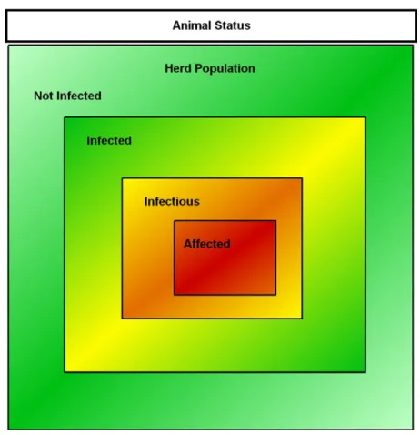
2.6.2 Animal Status
The following animal status classification was proposed on an evaluation study of the diagnostic tests accuracy and will be adopted in this dissertation. Animals were classified in Infected, Infectious and Affected (adapted from: Nielsen & Toft, 2008) (Fig. 3):
Infected animals:
o In these animals MAP persistence lasted long enough to stimulate an immune response, to produce clinical signs or to be detected by agent cultivation; o Cattle assigned to this status may become infectious and affected in later
stages of infection;
o It is assumed that once a cow has an established infection, the infection persists for life;
o These animals pose a risk of becoming infectious and cannot be declared MAP-free;
o They are an economic burden when there is decrease in milk/weight and transmission to other animals.
Infectious animals:
o These animals shed MAP at the time of testing according to the test used; o Their infectious status can also be decided on the basis of the detection of an
immune response (Nielsen, Grohn & Enevoldsen, 2002); o These animals are considered infected and may be affected;
o They may include non-infected animals which may be passive shedders of MAP (transient infectious status);
o MAP shedding is mainly observed in faeces, but it may also occur in milk and be transferred in utero.
o These animals are considered an economic burden because in short-term they experience decreased milk production and on long-term they transmit MAP to susceptible animals in the herd.
17 Affected animals:
o This status is assigned when cattle have decreased milk yield or weight loss (or any other production indicator) and/or clinical signs characteristic of MAP infection;
o These animals are considered infected and probably infectious;
o Their value is low because they may have reduced milk yield, decreased weight and probably low value at slaughter. There is a risk they will die from the infection.
Fig. 3 – Schematic representation of the different animal status in an infected herd population. Proportions for each status are unknown.
18
2.7 MAP diagnostic methods
None of the available diagnostic methods are 100% MAP specific and sensitive. Thus, detection and characterization of MAP infections are a hard task. MAP infection may be detected by clinical diagnosis or laboratory techniques. Due to the dynamics of MAP infection both options are dependent on the stage of infection and animal status.
MAP clinical signs were described previously. Usually they are associated with advanced stages of infection and must be confirmed by laboratory techniques. MAP suspicion by clinical diagnosis is influenced by veterinarian diagnostic skills, herd owner disease knowledge and capacity to detect affected animals, and the type of clinical signs. Clinical diagnosis sensitivity and specificity are unknown. In some countries, due to specific MAP legislation, the fear of being officially classified as MAP infected herd results in underreporting of suspicious signs. This fact decreases the sensitivity of the clinical diagnosis (Anon, 2004b).
A laboratory diagnostic test should be chosen based on the analyte to be detected (bacteria, bacterial constituents, immune response). Due to dynamics of the MAP infection the immune response, bacterial load, potential shedding or MAP dissemination through the host do not necessarily follow the same pattern or time course. Therefore, the major obstacle for obtaining a correct diagnosis is the time-dependent responses (Nielsen, 2002).
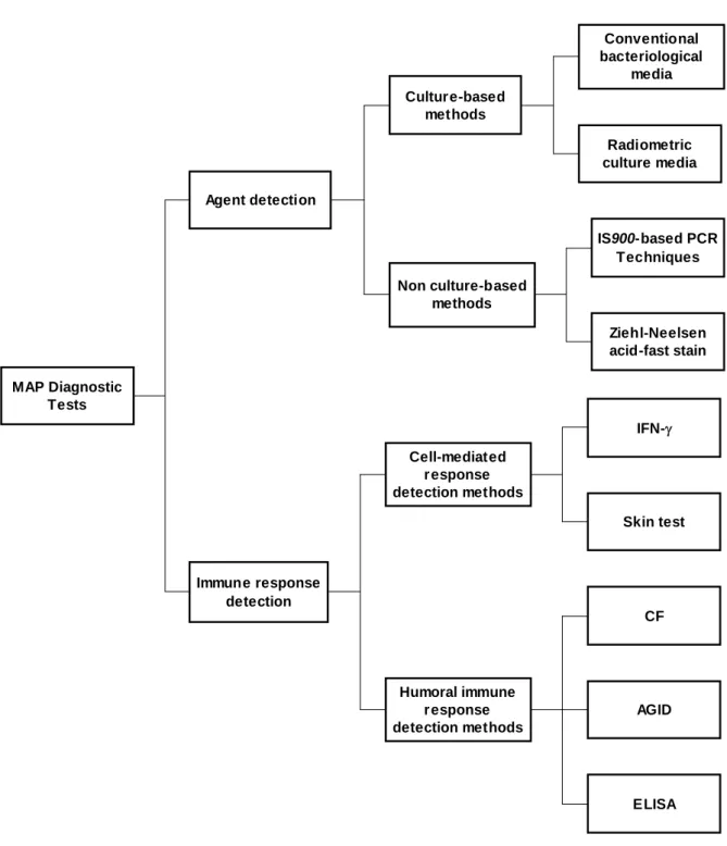
Two main diagnostic patterns can be followed: techniques that detect the agent and techniques that detect the immune response. MAP laboratory diagnosis methods were described elsewhere (Nielsen, Nielsen, Huda, Condron & Collins, 2001) (Fig. 4).
19
2.7.1 Diagnostic test types
MAP Diagnostic Tests Immune response detection Agent detection Non culture-based methods Culture-based methods Radiometric culture media Conventional bacteriological media Ziehl-Neelsen acid-fast stain IS900-based PCR Techniques Humoral immune response detection methods Cell-mediated response detection methods IFN- Skin test ELISA CF AGID
Fig. 4 – Summary of most commonly used MAP diagnostic tests. IFN-γ – Interferon Gamma; CF – Complement Fixation; AGID - Agarose Gel Immunodiffusion; ELISA - Enzyme-Linked Immunosorbent Assay (adapted from Nielsen et al.(2001).
2.7.2 Agent detection tests
Agent detection is one of the most used methods to diagnose MAP infection. Several matrices have been used to detect MAP such as faeces, milk and other tissues. Depending on the method used, the detected analyte may be the bacteria (bacterial stain – ZN, culture)
20
or bacteria and/or its constituents (PCR techniques). As a basic principle for agent detection, MAP must be present in the sample and available in detectable quantities. MAP isolation major problem is to ensure that MAP is present in the collected sample. About tissue culture, Whitlock et al (1996) mentioned that it may be necessary to collect samples from up to 100 sites per animal in order to obtain a positive culture of tissue specimen from an infected animal. Thus the sampling collection method, the sample processing steps and the agent detection method chosen are very important aspects to bear in mind.
2.7.2.1 Decontamination and concentration methods
In order to reduce fungal and bacteriologic contamination an appropriate decontamination procedure is necessary. MAP cells may be damaged and growth inhibited. The HPC (hexadecylpyridinium chloride) has been the most commonly used decontaminant, but others have also been reported e.g. benzalkonium chloride and oxalaic acid (reviewed in: Nielsen et al., 2001).
Pre-incubation methods and the use of antimicrobial agents have also been described. Some associations between chemical and antimicrobial agents for decontamination and culture methods are recommended (reviewed in: Nielsen et al., 2001).
To increase MAP recovery from samples concentration methods should be applied before cultivation process. Centrifugation and sedimentation methods are used but the former is described as having higher detection and lower contamination rates. Filtration is a technique used to concentrate MAP samples using the MAP cell clumping property. Immunomagnetic separation (IMS) is also used to extract MAP from heterogeneous samples. It has been used in milk samples and also prior to PCR (reviewed in: Nielsen et al., 2001).
2.7.2.2 Culture-based methods
Since its first isolation by bacteriologic culture, MAP isolation methods have evolved a lot but special growth requirements and some difficulties on the cultivation continue to be the main obstacles. In order to reduce the presence of fungi and other bacteria the decontamination step is particularly important. Isolates should be submitted to mycobactin dependency tests to differentiate it from other mycobacteria,5 (reviewed in: Manning & Collins, 2001).
Two types of culture methods are available: conventional and radiometric. MAP growth velocity is different between methods. The conventional method takes between 12 and 16 weeks, while radiometric method takes only 5 to 8 weeks. The time difference is due to the
5 MAP growth only on media supplemented with mycobactin J, an iron chelator required for in-vitro Map growth. The sample is streaked to media with mycobactin and to media without mycobactins, and the growth patterns are compared (reviewed in: Manning & Collins, 2001).
21
detection process. In the conventional method, slants of media are inspected until a colony is visible. In the radiometric method, a machine monitors sample-inoculated bottles of media for 14C-labelled products of bacterial metabolism. Metabolic products are detectable before the organism forms a colony sizeable enough to be observed on standard slants of media by naked eye. Both the analytical and the diagnostic sensitivity of the radiometric method are higher than the conventional culture. The automation of the process is another advantage (reviewed in: Manning & Collins, 2001).
Herrold’s Egg Yolk and Löwenstein-Jensen mediums complemented with mycobactins J are the most used mediums in conventional culture. Regarding the radiometric type, a commercial product known as the BACTEC system is used (reviewed in: Nielsen et al., 2001).
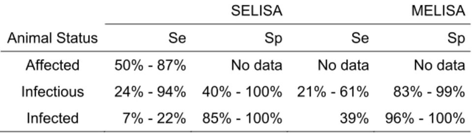
Culture methods sensitivity and specificity are dependent on several aspects. MAP tissue culture sensitivity and specificity have not been estimate thoroughly but they were reported elsewhere: sensitivity - 4% [0.2% - 15%] ; specificity – 97% [92% - 99%] (reviewed in: Anon, 2004b). Regarding faecal culture, the results were reported by animal status: Affected, Se – 70%, Sp – 100%; Infectious, Se – 74%, Sp – 100%; Infected, Se – 23% - 29%, Sp – 98% (Nielsen & Toft, 2008).
2.7.2.3 Non culture-based methods
IS900 has been used as the MAP genetic marker on PCR techniques since the discovery of the insertion sequence. IS900-based PCR can be applied directly on samples or as a confirmatory step after conventional culture, radiometric culture or immunomagnetic separation processes (reviewed in: Nielsen et al., 2001).
Due to its high sensitivity and fast response it is broadly used. However it also has some limitations. False-negative reactions may occur due to the presence of certain substances such as urea, haemoglobin or heparin that may inhibit enzymatic reactions; and MAP cell wall resistance. False-positive reactions are also described and may be caused by: laboratory contamination; IS900 sequence is not so MAP specific, as it was previously assumed – “IS900-like sequences” (Englund, Bolske & Johansson, 2002; Cousins et al., 1999); and amplification of DNA from dead bacteria and spheroblasts – these PCR positive results may be considered as a false-positive reaction when used together with a culture method (they will give a culture negative result) (reviewed in: Nielsen et al., 2001).
Although its drawbacks, IS900-based PCR is a fast technique and it is currently the method used to detect MAP infection in samples where MAP spheroblasts (viable and non-cultivable MAP forms) are present (Cousins et al., 1999; Chiodini, Vankruiningen, Thayer & Coutu, 1986).
22
The Se and Sp of direct PCR on faecal samples were reported: Se – from 2% [0% - 5%] to 39% [10% - 90%]; Sp – 98% [95% - 100%] (Anon, 2004b).
Another method of agent detection is by direct stain of samples followed by microscopic examination. Direct acid-fast staining of faecal samples has a low sensitivity and is very difficult to accurately distinguish MAP from non-pathogenic bacteria. Tissues direct staining is a commonly used process on samples collected at necropsy or by biopsy. Usually the Ziehl-Neelsen (ZN) acid-fast stain is the method used to highlight rod-shaped organisms (reviewed in: Manning & Collins, 2001).
MAP identification by PCR techniques and by ZN may be hampered in paucibacillary forms of disease (reviewed in: Manning & Collins, 2001).
2.7.3 Immune response detection tests
To measure an immune response, first it must have occurred at a detectable intensity regarding the test used and the timing of the testing. Measurable immunological reactions can be grouped into cell-mediated or humoral immune responses. In the former, the skin test (intradermal injection of a MAP purified protein derivate) and interferon-gamma (IFN-γ) can be used. In humoral immune responses, the agarose gel immunodiffusion (AGID) test, the complement-fixation (CF) test and the absorbed indirect enzyme-linked immunosorbent assay (ELISA) are the most commonly applied tests (reviewed in: Nielsen et al., 2001).
None of the mentioned tests is perfect (Se – 100%; Sp – 100%) and one of the major difficulties in their evaluation is the use of bacteria detection as the “gold standard” of infection. As bacteria detection methods are not perfect, the test results may be over or underestimated.
False-positive reactions in serological tests may be due to cross-reactions with other micro-organisms because the antigens used are crude and may contain antigens common to other bacteria of the order Actinomycetales. On the other hand, the animal may develop a protective immunologic response, recover after being exposed and be considered resistant. The problem of differentiating between resistant and infected animals is difficult to overcome by immunologic techniques since these methods give an indication of exposure, not infection (reviewed in: Chiodini et al., 1984).
False-negative responses may occur due to an insufficient antibody level (e.g. early infection) or to a state of anergy developed in clinical cases attributable to the protein-losing enteropathy. An animal that had responded to antigen but fail to respond in subsequent testing may be anergic to that specific antigen and infected or may have recovered from infection (Chiodini et al., 1984; reviewed in: Buergelt et al., 2004)