Cancer of the Head and Neck
Paper presented at the International Workshop on Cancer
of the Head and Neck, New York City, May 10-14, 1965
Washington BUlTERWORTHS
SURGICAL INDICATIONS FOR HEAD AND NECK TUMORS IN CHILDREN
The field of Pediatric Cancer is an ever growing one. Its importance is caused not only by the increased incidence, which to a certain extent runs parallel to that of congenital malformations, but by the significant rise in its percentage as a cause of death in the pediatric age group (0-14 years), now that infectious diseases are better controlled. It is given importance also by the increased awareness of doctors in its existence. Above all however, its importance is due to the fact that childhood tumors are no longer doomed to a hopeless prognosis.
It is extremely important that everyone realizes that childhood cancer can be cured in the same way as that in an adult.For this reason it is most important that pediatricians should become 'cancer minded.' Children's tumors are by no means a rarity: in the last 10 years 237 children with malignant tumors were seen at the Portuguese Cancer Institute. About one-third of these tumors were located in the head and neck (Table 1).
It is true that the type of tumors that are normally seen at a given Hospital sometimes does not reflect the true incidence of those tumors but rather the particular local interests and tendencies. At the beginning of 1960 a separate Department of Pediatric Cancer, one of the first of its kind in Europe, was opened at the Portuguese Cancer Institute in Lisbon, resulting in a marked increase in the number of pediatric neoplasms seen in that Hospital (1955/59
=
89; 1960/64=
147).Contrary to what happens in adults, in whom epithelial tumors are by far the most common (around 90 per cent of all tumors), they are extremely rare in children. If we exclude our patients suffering from xeroderma pigmentosum,
only 8 per cent of our patients with malignant tumors belong to that group. In children the various types of sarcomas, embryomas, and teratoid tumors predom-inate. An ulcerated skin tumor in this age group is more often originated in the deeper structures; in fact it is usually a sarcoma of soft somatic tissues rather than an epithelioma.
If certain tumors, such as retinoblastoma, are discovered mostly during the first four or fiveyears, lymphosarcoma, reticulum-cell sarcoma and thyroid tu-mors become more frequent thereafter. From a predominance of sarcomas we progressively change into an adult pattern at the end of puberty.
Topographic distribution by age (years) 0-1 1-4 5-9 10-14 Total
Eye ... 2 25 6 1 34 Bones ... 0••••••••••••••• - 3 8 11
Soft somatic tissues ... - 4 3 3 10
Oral cavity and pharynx ... 0••••••• 0•••• 0- 1 5 3 9
Lymphatic system ... 00000" 0•••••••••• 0- 1 1 4 6
Skin... o•• o•••• 0••••• - 1 I 1 3
Thyroid gland ... 0•00•0•0•••••••• - 2 2
Xeroderma (multiple tumors)... 0•000• 0000000000000- I 3 2 6
Total.............. 0••••••••••••••••••••••••••• 2 36 /9 24 81 Histologic types byage Retinoblastoma ... 00••••••• 0••••••••••••••••• J 24 6 32 Sympathicoblastoma ... 00000••••••••• 0.- J / Malignant neurinoma ... 00.0.0 •• 0•••••• 000000••••• - J / Reticulosarcoma ... 0••••••••••••••••••••• 0•• - 3 7 II Hodgkin's disease (localized)... 0.- J 1
Reticuloendothelioses ... 0••••••• 00••••••••• 0000 .- / Malignant hemangioendothelioma ... 00••••••••••• 0o- J 2 Embryonal rhabdomyosarcoma ... 0••• - 2 3 Fibromyxosarcoma ... 0•000••••• - I 2 Myxosarcoma ... 00000•••• - / Liposarcoma ... 00000••••• - / Osteosarcomas and condrosarcomas ... 000•00- 4 4 Undifferentiated sarcoma ... - /
Undifferentiated adenocarcinoma (of the thyroid
gland) ... - J /
Papillary adenocarcinoma (ofthe Thyroid gland) ... - 1 /
Malignant melanoma ... 0•••••••••••••••• - I 1
Squamous and basal cell carcinoma ... 0•••••• 0- 3 5 Squamous and basal cell carcinoma, melanoma,
(xeroderma) ... 0•••••••••• 0•• 00- 1 3 2 6
No histology available... 0•• 0••••••••••••••••••• 1 2 2 1 6 Total.................. 00•0••••••••••••••• 2 36 /9 24 81 PA THOGENESIS
Hormonal factors
Although not explainable in the light of our present knowledge, there is normally a decrease in incidence of malignant neoplasms during the prepubertal period. That this low incidence may be related to hormonal factors is a distinct possibility.
Heredity
Heredity seems to play an important role in certain head and neck tumors in children, considering that retinoblastoma and xeroderma pigmentosum are usu
As far as location is concerned many differences exist between children and adults. Central nervous system tumors (including retinoblastoma), soft tissue tumors, and tumors of the lymphatic and hematopoetic systems predominate in children, but on the other hand neoplasms of the skin,larynx, or esophagus are certainly a rarity.
Some tumors in children may present at birth or at least appear during the first months of life: it is therefore essential that the possibility of a malignant tumor be considered in the differential diagnosis of anyhead and neck lump. The high frequency of inflammatory tumors about the head and neck in children and particularly their altered evolution brought about by the use of antibiotics, together with the high frequency of tumorous congenital malformations about that region, make differential diagnosis sometimes exceedingly difficult.Cervical lymphadenopathy, particularly when unilateral and painless, must always arouse suspicion, for lymphosarcoma, reticulum-cell sarcoma, localized Hodgkin's d:s
-ease, metastatic thyroid carcinoma, and so on,may bepresent.
A short waiting period under close observation is normally indicated, although too much delay may often lead to disaster. Not more than a few weeks should elapse before a biopsy isperformed fora lump the origin of which has not become obvious during that period.
Aspiration biopsy
Aspiration biopsy is particularly valuable, especiallyas an office or out-patient procedure. It may not onlydetermine thepresence of pus,blood, or other liquid but may also obtain a small specimen for cytological or histological examina-tion.
Surgical biopsy shouldbe planned soasto bedirected towards themain lesion and performed in a place where its scar may easily be excised at the time of definitive surgery if this isrequired. The incision should fall into the natural skin creases, attempting as much as possible to followthe concept of the esthetic units of the face. Biopsy performed on a benign lesion should leave no more than a negligible scar. Cosmetic reasons alone, particularly in female patients, should not deter one from performing a timelybiopsy which, whenever possible, should be of the 'total excision'type.
Often, particularly in smaller children, biopsy is better performed under general anesthesia because, if the possibility of immediate pathologic study through quick frozen-sections is available, definitive surgery can then be carried out if necessary.
Considering that it is the only way of obtaining a correct histological diag-nosis, essential for instituting the right therapy, the role of biopsy cannot be
overemphasized. All tissues removed at biopsy or operation, no matter how benign they may seem or how convinced one is of the diagnosis,must always be sent for histological examination.
Histology
One aspect that needs to be emphasized is that the histological appearance of many benign childhood tumors closely resembles that of adult malignancies, as happens with juvenile melanoma. True malignant melanoma was only found once in our series, although ~any cases of juvenile melanoma were treated. The treatment of malignant melanoma is no different from that advised for adults, and consists mainly of the widest practicable excision. Elective radical lymph node dissection is only justified if the presence of metastatic nodes is suspected. Cellular non-differentiation of tumor tissues-which in the adult normally means high malignancy-is many times found in benign teratoid tumors. Frequent mitosis, invasion, and infiltration of surrounding tissues, usually a bad sign, are frequently found in benign childhood tumors, namely the hemangiomas. In our series of many thousand of these tumors, only two true malignant hemangioen-dotheliomas were found. These should be treated by using surgery whenever possible, associated with postoperative roentgentherapy.
Conversely, certain tumors which appear to be benign when using the usual histological criteria, as happens with certain fibrous tissue tumors, show definite clinical malignancy with persistent local recurrence after an apparently adequate excision and leading to death of the patient due to progressive invasion and destruction. Some of the so-called fibromas of the mandible are no more than low grade fibrosarcomas, normally non-metastasizing but requiring from the very beginning an aggressive approach. This aggressive approach means that bone has to be largely resected and not only conservative tumor excision performed. Another interesting feature of some childhood tumors is that they possess the possibility of undergoing spontaneous cure even in the presence of generalized soft tissue metastases, as happens with neuroblastomas. Some of these tumors, composed of not completely differentiated cells,may progressively differentiate and change into a less malignant or even benign type of tumor. This justifies the performance, in the presence of generalized metastases, of total or even partial excision of these tumors, complemented by roentgentherapy and/or chemother-apy.
In this age group early diagnosis is as important as in any other. The same holds true for benign tumors, which in certain cases may cause death by virtue of their location, sudden growth, hemorrhage, and so on. The proper timing for operation is usually as early as possible. Modern advances in pediatric surgery and anesthesiology make it safe to operate on any child at any age, provided careful preoperative, intraoperative, and postoperative management are used. The all-important aim of the surgeon must be the complete, wide, and safe surgical excision of the tumor, always bearing in mind that reconstructive prob.
lems and mutilation fears must be considered as secondary to the cure of the patient.
We must be conservative if possible and neveroperate without having planned
reconstruction carefully: if our patients are going to survive they will need
acceptable function and alsoacceptable cosmetic appearance. To be conservative
one must fully understand the nature and natural history of the tumor one is
dealing with.
At present surgery seems to be our most powerful tool in the treatment of
head and neck tumors in children. Nevertheless, surgery must be considered as
only part of the total care of the child. Often it has tobe associated with other
types of treatment such as roentgentherapy and chemotherapy, as isthe case in
Fig.1.-Eleven-year-old boy, suffering from Xeroderma Pigmentosum, had a squamous-cell carcinoma of the left ear invading the middle ear. He was treated by wide excision of the tumor, together with partial temporal bone resection preserving the facial nerve and followed by immediate recon
embryonal rhabdomyosarcoma, lymphosarcoma, sympathicoblastoma, localized Hodgkin's disease, and so on.
In retinoblastoma enucleation is often indicated, in unilateral cases,but orbital exenteration must always be contemplated if the tumor has extended beyond the eye itself. In these cases it is at times necessary to effect a total parotidectomy and radical neck dissection, with immediate facial nerve grafting if this nerve has to be sacrificed during removal of metastases. Roentgentherapy and chemo-therapy are normally associated with surgery. In Xeroderma Pigmentosum trea
t-ment is exclusively surgical (FIGS. 1 and 2), if we exclude prophylaxis, whichis mostly avoidance of exposure to sunlight and the use of protective cover
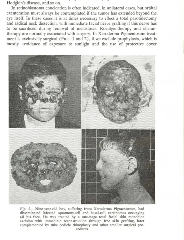
Fig. 2.-Nine-year-old boy, suffering from Xeroderma Pigmentosum, had disseminated infected squamous-celI and basal-celI carcinomas occupying alI his face. He was treated by a one-stage total facial skin monobloc excision with immediate reconstruction through free skin grafting, later complemented by tube pedicle rhinoplasty and other smalIer surgical
embryonal rhabdomyosarcoma, lymphosarcoma, sympathicoblastoma, localized Hodgkin's disease, andso on.
In retinoblastoma enucleation is often indicated, inunilateral cases, but orbital exenteration must always be contemplated if the tumor has extended beyond the eye itself. In these cases it is at times necessary to effect a total parotidectomy and radical neck dissection, with immediate facial nerve grafting if this nerve has to be sacrificed during removal of metastases. Roentgentherapy and chemo-therapy are normally associated with surgery. In Xeroderma Pigmentosum treat-ment is exclusivelysurg'ical(FIGS. 1 and 2), if we exclude prophylaxis, whichis mostly avoidance of exposure to sunlight and the use of protective cover
Fig. 2.-Nine-year-old boy, suffering from Xeroderma Pigmentosum, had
disseminated infected squamous-cell and basal-cell carcinomas occupying
all his face. He was treated by a one-stage total facial skin monobloc
excision with immediate reconstruction through free skin grafting, later
complemented by tube pedicle rhinoplasty and other smaller surgical pro-cedures.
creams. In this disease the appearance of malignant tumors is frequent, particu-larly in the face as the place more exposed to actinic radiation, and generally imposes diathermy excision of all small lesions until a time when a staged total facial skin replacement is justified. Roentgentherapy in these patients is to be strongly condemned. In other rare cutaneous neoplasms treatment is similar to that of adults, consisting of wide excision and immediate reconstruction;
how-ever, radical neck dissection is only justified if metastases are suspected (FIG.
3 ).
It is astonishing to see the adaptability of children to the large mutilations
which may be necessary. A child should never be lied to before operation. If he
is old enough to understand, a simple explanation of what is to come will often
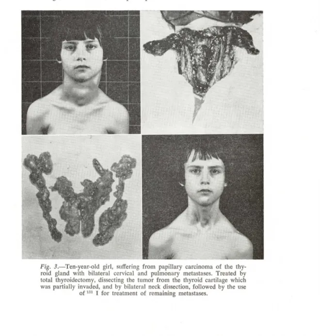
Fig. 3.-Ten-year-old girl, suffering from papillary carcinoma of the thy-roid gland with bilateral cervical and pulmonary metastases. Treated by total thyroidectomy, dissecting the tumor from the thyroid cartilage which was partially invaded, and by bilateral neck dissection, followed by the use
help him enormously in the postoperative and convalescent periods. The essen-tial features that are likelyto upset himshould be discussed: wemust make him understand why he is to be operated upon but not indulge in lengthy explana-tions, particularly about mutilations, which mayupset him more than help.The same holds true as regards parents. We must 'treat' not only the child but also his family: sooner or later the child will reflect the attitude of his parents. In many cases parents are aware that they may lose their child and it is the duty of doctors to minimize their suffering asmuch as possible. Above all they should never be given the impression that hope is completely lost or that everything possiblehasnot been done.
It is unquestionable that the outlook for children with cancer of the head and neck has changed and that nowadays many tumors do have a good chance of cure, provided adequate and timely treatment is given. Surgery is indicated in all localized lesions and even many times for those lesions that have extended beyond their original limits. Excision of the tumor is often associated with monobloc dissection of the regional nodes. Even embryonal rhabdomyosar-coma, probably one of the most refractory tumors to treatment in children, may many times permit if not a complete cure, at least a prolonged survival. The surgeon must never lose hope and stop fighting: only then may he give his little patients the best chances for a cure that should never be denied to them.