DEVELOPMENT OF METHODS BASED ON
BIOMIMETIC SYSTEMS FOR EVALUATION OF
ANTIOXIDANT PROPERTIES
Tese do 3⁰ Ciclo de Estudos Conducentes ao Grau de Doutoramento em Ciências Farmacêuticas na Especialidade de Química Analítica
Trabalho realizado sob a orientação de
Prof. Doutora Marcela Alves Segundo (Orientadora) Prof. Doutor José Luís Fontes da Costa Lima (Co-Orientador)
Doutora Marlene Susana Dionísio Lúcio (Co-Orientador)
iv
Acknowledgements
Though the following dissertation is an individual research work, I could never have reached the heights or explored the depths without the help, support, guidance and efforts of a lot of people. Therefore, here is a small tribute to all those people.
To Faculdade de Farmácia da Universidade do Porto (FFUP) for receiving me as a PhD student.
To Fundação para a Ciência e Tecnologia (FCT), for the PhD grant
SFRH/BD/41627/2008.
To Prof. Dr. José Luís Costa Lima for the challenge of the PhD in this department and for providing me all the necessary conditions to do my work.
To my supervisor Dr. Marcela Segundo, for her excellent guidance and for inspiring me the qualities of being a “good scientist”. Special thanks also for her friendship, enthusiasm, availability and effort made for the realization of this work.
To Dr. Marlene Lúcio for the co-supervision and guidance of my work.
To Prof. Dr. Salette Reis, my special thanks for accompanying the progress of my work and for being always available when I needed.
To all my colleagues and friends in the FFUP, for the help and friendship.
To Prof. Dr. Luis Camacho and Dr. Juan Casares for receiving me in the department of Física Química y Termodinámica, Universidad de Cordoba. The time spent there had an enormous contribution in my knowledge about how to do research. Thanks also for their friendship
To Luís Magalhães my special thanks for accompanying the progress of my work and for his friendship.
To my friend Mariana Arêde for the help in laboratory work and her friendship. Thank you.
v
To all my friends who were always present and available.
To all my family for the support and the encouragement.
To my lovely parents and my sisters, all my thanks. Without their help this work could never be done. They were always supporting and encouraging me along these years to pursue my interests.
vi
function and controlling systems towards homeostasis.
This high degree of complexity derives from the diversity of biological processes, namely the reactions involved in cellular respiration, which depend on the presence of oxygen, fostering an expectable increase in reactive species of oxygen, among others.
During the past few years, studies towards the causes and consequences about the production of free radicals in organisms were performed, along with studies aiming to unveil biological targets prone to attack and the possible consequences from these events. Actually, nowadays it is clearly known that the biological membranes are the main targets of free radical attacks, resulting in peroxidation conditions. In organisms, there are natural antioxidants, such as glutathione, uric acid and ascorbic acid, which are able to neutralize the action of these reactive species, avoiding the damage caused by them.
Aiming at studying how the generation of free radicals affect biological stability, several experiments were carried out along this thesis, focusing on understanding the role, the efficacy and possible interactions between biological membranes and free radicals. Moreover, some aspects related to the location of these antioxidants within the membrane (hydrophilic/hydrophobic media) and how the products of such reaction can interfere with membrane structure were focused. For this, different biomimetic models were assessed, namely micelles, liposomes and Langmuir monolayers, using lipid mixtures similar to those found in biological membranes. During these work, standards, reference compounds for evaluation of peroxidation were applied, including Trolox and vitamin E. Moreover, methodologies relying on application of fluorimetric probes were developed, aiming to achieve a simple method with optimized experimental conditions in order to evaluate peroxidation in different locations of membrane models (aqueous phase, interface and lipophilic media). Other biomimetic models were also assessed, with different features concerning their size, lamellar structure and lipidic composition, in order to enlighten how these variation and complex aspects interfere with the evaluation of oxidative or antioxidant capacity of different compounds.
vii
Os sistemas biológicos exibem uma elevada complexidade no que concerne a estrutura, constituição, função e sistemas de controlo da sua homeostase.
A sua elevada complexidade deve-se à diversidade de processos biológicos a eles inerente, nomeadamente as reações envolvidas na respiração celular, cuja dependência de oxigénio em muitos deles, acarreta um natural incremento de espécies reativas de oxigénio, entre outras.
Ao longo do tempo foi-se estudando e conhecendo algumas das causas e consequências da produção de radicais livres no organismo, assim como o estudo dos seus alvos de ataque e possíveis alterações daí resultantes, estando hoje bastante bem relatadas que as membranas biológicas pela sua constituição essencialmente lipídica, são um dos maiores alvos de ataque de radicais livres com consequente peroxidação. Existem no organismo antioxidantes naturais como a glutationa, o ácido úrico e o ácido ascórbico capazes de eliminar estas espécies reativas, evitando os danos potencialmente causados por estas. No sentido de estudar a forma como a geração destes radicais afetam a estabilidade biológica, foram realizados ao longo deste trabalho alguns ensaios com o intuito de perceber melhor o papel, a eficácia e as eventuais interações com as membranas biológicas dos radicais produzidos, a forma como estes antioxidantes biológicos protegem as membranas dependendo da sua localização (meio hidrofílico/meio hidrofóbico), assim como os produtos da reação radical/antioxidante poderão interferir com as características membranares. Para tal recorreu-se ao uso de diferentes modelos biológicos, nomeadamente com a preparação de micelas, lipossomas e monocamadas de Langmuir, utilizando misturas lipídicas de composição semelhante à das membranas biológicas. Ao longo de todo o trabalho realizado foram utilizados calibradores, compostos de referência para estudo de peroxidação lipídica, Trolox e vitamina E.
Foram ainda utilizadas metodologias envolvendo a utilização de sondas fluorescentes por forma a otimizar algumas condições experimentais baseada em metodologia simples, a serem utilizadas em estudos de avaliação da peroxidação lipídica, em diferentes localizações da membrana (meio aquoso, interface e meio lipofílico).
Foram estudados modelos biomiméticos distintos em tamanho, número de lamelas e composição como forma de incrementar o conhecimento sobre a forma como estas variações e complexidade interferem dificultando a avaliação da ação oxidante ou antioxidante de diferentes compostos.
viii
Abstract ... vi
Resumo ... vii
Table of contents ... viii
List of figures ... xiii
List of tables ... xix
List of abbreviations and symbols ... xxi
Chapter 1
1.General introduction ... 21.1 Biological membranes and oxidative stress ... 2
1.1.1 Structure and functions of biological membranes ... 2
1.1.2 Lipid peroxidation ... 4
1.2 Oxidative stress ... 7
1.3 Consequences to membrane and organisms exposed to oxidative stress .... 9
1.4 Biomimetic sytems ... 11
1.5 Methods for assessments of antioxidants ... 13
1.6 Scavenging capacity assays against specific ROS/RNS ...16
1.6.1 Peroxyl radical (ROO•) scavenging capacity assays ...16
1.6.2 Other ROS/RNS scavenging capacity assay ... 18
1.6.3 Scavenging capacity assays against stable, non-biological radicals and evaluation of total reduction capacity ... 20
1.7. Methods for antioxidants assessment with biomimetic structures ... 21
ix
1.7.4 Measurement of oxidation ... 45
1.7.5 Applications ... 46
1.8 References ... 47
Chapter 2
2.Material and methods
... 602.Material and Methods ...61
2.1. Introduction ...61
2.2. Reagents, solutions and samples ...61
2.3. Fluorimetric measurements ... 62
2.4. ORAC method – quantification ... 63
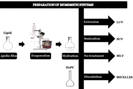
2.5. Preparation of biomimetic lipid systems ... 63
2.6. Surface pressure – area measurements in monolayers ... 65
2.7 Brewster angle microscopy (BAM) in monolayers ... 67
x phosphatidylcholine monolayers under peroxidation conditions
through Brewster Angle Microscopy ... 69
3.1 Introduction ... 70
3.2 Reagents and solutions ... 71
3.3 Results ... 71
3.3.1 π-A measurements ... 71
3.3.2 BAM observation ...77
3.4 Discussion and conclusions ... 81
3.5 References ... 89
Chapter 4
4.Interaction of soluble biological antioxidants with phosphatidylcholine monolayers ... 944.1 Introduction ... 95
4.2 Reagents and solutions ... 97
4.3 Results ... 97
4.3.1 π-A measurements ... 97
4.3.2 BAM observation ... 103
4.4 Discussion ... 106
xi
structures ... 111
5.1Introduction ... 112
5.2 Reagents and solutions ... 113
5.3 Preparation of lipid structures ... 114
5.4 Assessment of antioxidant capacity ... 114
5.5 Calculation of antioxidant capacity as area under curve ... 115
5.6 Results and discussion ... 115
5.6.1 Characterization of structures ... 115
5.6.2 Oxidation profile of fluorescein probe ... 116
5.6.3 Application to antioxidants ... 117
5.7 Conclusions ... 118
5.8 References ... 119
Chapter 6
6. High-throughput fluorimetric methodologies for assessment of antioxidant capacity of drugs at different locations of phospholipid bilayer ... 1216.1. Introduction ... 122
6.2. Materials and methods ... 124
xii
6.2.4 Calculation of antioxidant capacity values expressed as IC50 or IC25 126
6.3. Results and discussion ... 127
6.3.1 Influence of probe concentration ... 128
6.3.2 Influence of lipid and AAPH concentration ... 129
6.3.4 Ability of NSAIDs to counteract lipid peroxidation in different locations of phospholipid bilayers ... 133
6.4. Conclusions ... 139
6.5 References ... 139
Chapter 7
... 143xiii
Chapter 1
General introduction
Figure 1.1 Representation of the chain-reaction process and inhibition in the presence of antioxidants.
Figure 1.2 Classification of inducers of lipid peroxidation.
Figure 1.3 Schematic representation of equation and its components applied for calculation of peroxidation rate.
Figure 1.4 Schematic relationship oxidative stress and tissue injury.
Figure 1.5 Representation of the relationship between Food and/or drugs with Equilibrium, health or disease.
Figure 1.6 Schematic illustration of : (A) structure of the micelles; (B) liposomes structure (size and lamellarity); (C) Monolayer representation.
Figure 1.7. Schematic representation of competitive and non-competitive approaches, for in vitro determination of antioxidant capacity.
Figure 1.8 Schematic representation from the net integrated area under the fluorescence decay curves.
Figure 1.9 Physical and chemical factors that affect the peroxidation of liposomal lipids.
Figure 1.10 Molecular structure of thermolabile azo-compounds used for generation of peroxyl radicals.
xiv Figure 2.1 Schematic representation of techniques used in this work for preparation of biomimetic systems.
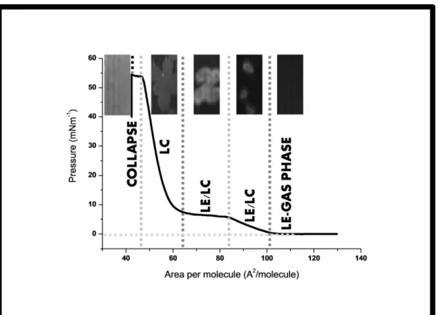
Figure 2.2 Isotherm representation, of DPPC monolayer obtained with a NIMA
601 (Nima Technology, Coventry, UK) Langmuir trough (total area = 600 cm2;
subphase volume c.a. 400 mL of the phosphate buffer) and BAM images at different pressures and lipid phases (LE-lipid expanded; LE/LC-lipid expanded/lipid condensed; LC - lipid condensed and monolayer collapse).
Chapter 3
Insights about α-tocopherol and Trolox interaction with
phosphatidylcholine monolayers under peroxidation conditions through Brewster Angle Microscopy
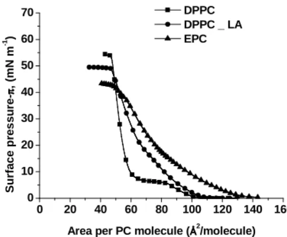
Figure 3.1 Surface pressure-area isotherms of monolayers formed by DPPC (■), DPPC + LA (●) and EPC (▲) upon a phosphate buffer (pH 7.4) subphase.
Figure 3.2 Surface pressure-area isotherms for different lipid systems, with monolayers formed and compressed above subphases containing phosphate buffer pH 7.4 (■), AAPH solution (●), Trolox solution (▲) and AAPH + Trolox solution (▼). A: DPPC ; B: DPPC + LA; C: EPC.
Figure 3.3 Surface pressure-area isotherms for lipid systems containing α-tocopherol, with monolayers formed and compressed above subphases containing phosphate buffer pH 7.4 (without α-tocopherol: ■; with α- tocopherol: ▲) or AAPH solution (without α-tocopherol: ●; with α-tocopherol: ▼). A: DPPC ; B: DPPC + LA; C: EPC.
Figure 3.4 Brewster angle micrographs of DPPC monolayer at different surface pressures upon monolayer compression.
xv Figure 3.6 Brewster angle micrographs showing representative features for monolayers of DPPC and DPPC + LA. A, DPPC monolayer with AAPH in the subphase; B, DPPC monolayer with Trolox in the subphase; C, D, DPPC monolayer with AAPH and Trolox in the subphase; E, DPPC monolayer with α-tocopherol; F, DPPC monolayer with α-tocopherol and AAPH in the subphase; G, DPPC + LA monolayer with AAPH in the subphase; H, DPPC + LA monolayer with AAPH and Trolox in the subphase.
Figure 3.7 Brewster angle micrographs showing representative features for monolayers of DPPC + LA and α-tocopherol in phosphate buffer subphase (A-D) or with AAPH in the subphase (E-H).
Figure 3.8 Schematic representations of interaction between monolayer and aqueous phase components.
Figure 3.9 Domain patterns formed in DPPC monolayers for surface pressures of
10 and 30 mN m-1 in absence of antioxidants and in presence of either
α-tocopherol or Trolox.
Chapter 4
Interaction of soluble biological antioxidants with phosphadylcholine monolayers
Figure 4.1 Surface pressure-area isotherms for different lipid systems, with monolayers formed and compressed above subphases containing phosphate buffer
pH 7.4 (▬), AAPH solution (▬), glutathione solution (▬), and AAPH +
glutathione solution (▬). The solid bars represent the area per DPPC molecule
xvi
acid solution (▬). The solid bars represent the area per DPPC molecule where LC
domains can be visualized by BAM.
Figure 4.3 Surface pressure-area isotherms for different lipid systems, with monolayers formed and compressed above subphases containing phosphate buffer
pH 7.4 (▬), AAPH solution (▬), uric acid solution (▬), and AAPH + uric acid
solution (▬). The solid bars represent the area per DPPC molecule where LC
domains can be visualized by BAM.
Figure 4.4 BAM images obtained for different monolayers (DPPC, DPPC + LA and EPC), with AAPH and/or antioxidants in the subphase (glutathione, ascorbic acid or uric acid).
Chapter 5
Evaluation of antioxidant capacity using lipidic biomimetic structures
Figure 5.1 Fluorescein oxidation profile in presence of different biomimetic structures
Figure 5.2 Fluorescein oxidation profile in presence of different biomimetic structures and antioxidant Trolox.
Chapter 6
High-throughput fluorimetric methodologies for assessment of antioxidant capacity of drugs at different locations of phospholipid bilayer
xvii The concentration ratio between large unilamellar vesicles of egg L-α-phosphatidylcholine (EPC) and fluorogenic probe was fixed at 300:1, respectively. Figure 6.2. Fluorescence intensity obtained for fluorescent hydrophilic probe (fluorescein), fluorescent interface probe (5-dodecanoylaminofluorescein, DDAF)
and for fluorogenic lipophilic probe C11-BODIPY581/591 in the presence of different
concentrations (µM) of large unilamellar vesicles of egg L-α-phosphatidylcholine (EPC). Fluorescein was added to LUV suspension immediately before the
measurements, while the DDAF and C11-BODIPY581/591 were incorporated during
vesicles preparation. For experiments using C11-BODIPY581/591, the concentration
of peroxyl radical generator (AAPH) was fixed at 10 mM. The circles show the EPC concentration selected for further studies.
Figure 6.3. Relative fluorescence intensity (%) of fluorescein (6 nM), DDAF (50
nM) and C11-BODIPY581/591 (50 nM) in the presence of large unilamellar vesicles of
egg L-α-phosphatidylcholine (EPC) (800 µM) and different concentrations (µM) of peroxyl radical generator AAPH: A) (1) 0, (2) 25, (3) 50, (4) 75, (5) 100, (6) 150, (7) 200; B) (1) 0, (2) 25, (3) 50, (4) 75, (5) 100, (6) 150; C) (1) 5; (2) 10, (3) 25, (4) 50, (5) 75. The arrows indicate the concentration of AAPH (50 mM) that was selected.
Figure 6.4. Relative fluorescence intensity (%) of fluorescein (A and B) and antioxidant capacity (C and D) obtained in the presence of different concentrations of indomethacin (0, 5, 10, 15, 20 µM) and tolmetin (0, 5, 10, 15, 20, 30, 40 µM) in large unilamellar vesicles of EPC (800 µM) and with the AAPH concentration (50 mM). Antioxidant capacity was determined using the following equation,
[(AUCTrolox/NSAIDs ‒ AUCblank) / (AUCblank)] x 100. For both drugs, AUC was
calculated after 240 min. LUV:FL, correspond to the fluorescent intensity obtained by liposomes in the presence of aqueous fluorescent probe (fluorescein) in the absence of peroxyl radical species.
xviii 16, 20 µM), etodolac (0, 5, 10, 15, 20, 30 µM) and indomethacin (0, 5, 10, 15, 20, 30 µM). The numbers (1 to 6) correspond to increasing concentrations of Trolox or NSAIDs. Relative fluorescence was determined by dividing the fluorescence intensity at a given time by the maximum of fluorescence intensity obtained for probe’s oxidation in the absence of antioxidant species (see section 2.4).
xix
Chapter 1
General introduction
Table 1. Principal reactive species (non radicals) free radicals presents in organism.
Table 2. Principal reactive species (non radicals) presents in organism. Adapted from (Halliwell and Gutteridge, 2007)
Table 3 Methods for assessment of antioxidant capacity using LUVs
Table 4 Methods for assessment of antioxidant capacity using MLVs
Table5 Methods for assessment of antioxidant capacity using SUVs
Table 6 Methods for assessment of antioxidant capacity using other biomimetic systems
Chapter 3
Insights about α-tocopherol and Trolox interaction with
phosphatidylcholine monolayers under peroxidation conditions through Brewster Angle Microscopy
Table 3.1. Characteristic parameters (elastic modulus, collapse pressure and collapse area) of the Langmuir monolayers tested on aqueous subphases containing the peroxidation inducer (AAPH) and antioxidants (Trolox and α-tocopherol)
xx monolayers
Table 4.1. Characteristic parameters (elastic modulus, collapse pressure and collapse area) of the Langmuir monolayers tested on aqueous subphases containing the peroxidation inducer (AAPH) and antioxidants (glutathione, ascorbic acid, uric acid)
Chapter 5
Evaluation of antioxidant capacity using lipidic biomimetic structures
Table 5.1. Sensitivitya (relative fluorescence × min × µM-1) for antioxidants in
presence of different biomimetic structures
Table 5.2. Ratioa between the sensitivities attained for a given antioxidant and that
attained by Trolox in presence of different biomimetic structures
Chapter 6
High-throughput fluorimetric methodologies for assessment of antioxidant capacity of drugs at different locations of phospholipid bilayer
Table 6.1. Peroxyl radical scavenging capacity of Trolox and nonsteroidal
anti-inflammatory drugs (NSAIDs) expressed as IC50 and IC25 values obtained with
CHAPTER 1
General introduction
2 1. INTRODUCTION
1.1 Biological membranes and oxidative stress
1.1.1 Structure and functions of biological membranes
The cells are the basic structural and functional unit of organisms and, at the same time, they are highly organized with many functional organelles constituted by one or more lipid membranes (Demel et al., 1967).
Each biological cell is enclosed by its outer plasma membrane which controls the interaction between the cell and its environment. This applies both to the relatively small cells of bacteria or prokaryotes, which have no cell nucleus and without defined compartments, and to the much larger cells of eucaryotes, which have such a nucleus and compartments delimited. The latter class of cells is present in all animals and plants as well as in single-celled microorganisms such as amoeba or yeast (Lipowsky and Sackmann, 1995). Furthermore, all eukaryotic cells contain internal membranes which represent the boundaries of the internal organelles such as the nucleus, mitochondrial, Golgi complex, chloroplasts, among other structures.
The fluid mosaic model introduced the idea that membranes were formed by a fluid bilayer in which proteins and lipids could move freely (Singer, 1993). Nowadays, the extended fluid mosaic model contemplates additional structural and functional restraints on membrane organization (Vereb et al., 2003).
One general construction principle which has emerged from molecular cell biology is the use of membranes of different composition, organization, complexity and diversity, in order to organize space into different compartments (Lipowsky and Sackmann, 1995). The basic function of biomembranes is to provide different spatial compartments and to act as a highly selective barriers for the exchange of molecules between the different compartments, sustaining the concentration gradients between theses compartments, fostering signal transduction and mechanical support (Escribá et al., 2008).
As indicated all membranes of eukaryotic cells separate functional compartments, but the cell surface membrane - the plasma membrane - is an extreme. It is the frontier, with different physical and functional properties. The plasma membrane is composed by a lipid bilayer (Edidin, 2003), ensuring the above mentioned functions because it is composed of specific mixtures of different lipids (mainly amphiphilic) and proteins.
3 of cellular process, thus requiring the maintenance of their proper structure and function (Brown and London, 1998; Kinnunen, 1991; Kinnunen et al., 1994), and this is correlated with their composition, essentially lipid bilayer to which proteins and carbohydrates may be associated or covalently linked.
The lipids are a heterogeneous group, with amphiphilic structure, with a hydrophilic head group and usually two lipophilic hydrocarbon chains. The essential characteristic of the lipids is its insolubility or very low solubility in water. Lipids have also been acknowledged as key elements in numerous processes (Escribá et al., 2008). Despite the matrix of lipids, whose structure and composition is far from simple, due to the number of different lipid molecules found in the plasma membrane (that can exceed 1000). The high number of structures formed by lipids in vitro indicates that the structural properties of membranes can greatly vary in vivo, since the interaction of lipid molecules to form membranes is not determined by covalent bonds, like it is with amino acids in proteins (Escribá et al., 2008). Membrane lipids can be classified into three main groups: i) Glycerol-based lipids, ii) cholesterol and iii) ceramide-based sphingolipids.
Furthermore, glycerophospholipids can be classified according to their headgroup,
originating: phosphatidylethanolamine, phosphatidylcholine, phosphatidylserine,
phosphatidylglycerol and phosphatidylinositol 4,5-bisphosphate.
In the membranes, the lipids aggregate and form bilayers, which are two-dimensional systems, with thermodynamic phases, e.g, fluid phase at high temperatures and one or several gel or solid-like phases at low temperatures. Membrane lipids participate in dynamic interactions that facilitate changes in their relative position in membranes, and that determine membrane thickness, surface packing, along with lateral and rotational mobility (Escribá et al., 2008). Hence, it is a difficult task to study the membrane structure because it presents polymorphisms, requiring a variable composition to act as mimetic to the biological membranes (Cullis et al., 1996).
One of the most important components of biomembranes is cholesterol that interacts with the phosphate headgroup of phospholipids, whereas the bulky steroid region interacts with phospholipid acyl chains, bearing an important role in regulating membrane fluity, membrane packing, non-lamellar phase propensity and the formation of microdomains. The proteins present on membrane, also have many functions, correlated with their position. Their role includes allowing the passage of substances into and out of the cell, acting as receptors of the membrane, receiving signals from substances that carry a message to the cell, promoting the adhesion of adjacent cells in a tissue, working as an anchor point for the cytoskeleton (Cullis et al., 1996). Therefore, most of the important
4 roles in membranes have been attributed to the membrane proteins. Hence, the proteins can inserted into the membrane or partially integrated into it, being designated as intrinsic or extrinsic proteins, respectively.
The transmembrane proteins have many interactions with lipids pertaining to membranes, because these last molecules interact with amino acids from the proteins in the hydrophobic environment of the membrane core while the interactions taking place at the interface are more less defined, and to a certain extent regulated by the features of the proteins (Escribá et al., 2008).
Furthermore, both lipids and proteins interact for determining the barrier function in the cells and they may be both subjected to regulatory processes in response to pathophysiological situations or nutritional/pharmacological interventions, which in turn may alter the activity and functions of the membrane.(Escribá et al., 2008) Lipids and proteins may respond to environmental situations and conditions, which can promote changes in the structure and function, consequently causing changes in cell functions, their homeostasis, communication and responses.
In conclusion, membranes constitute a meeting point for lipids and proteins, both fulfilling prominent roles in almost all cellular processes.
1.1.2 Lipid peroxidation
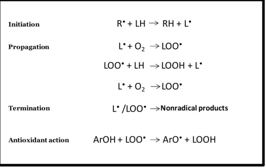
The lipids are the most studied components of the membrane, including aspects pertaining to its oxidation, because of they susceptibility, as one important target to attack by free radicals (the direct promoters of peroxidation). Hence, the lipid peroxidation products (Figure 1) are frequently used as to assess indices oxidative stress, usually coming from attack by free radicals to polyunsaturated fatty acids.
Lipid peroxidation plays a central role in many physiologic and pathological processes (Mattson, 2004; Poon et al., 2004a; b; Schnitzer et al., 2007b), and may be promoted by different inducers, as depicted in Figure 2.
5 Figure 1.1: Representation of the chain-reaction process and inhibition in the presence of antioxidants. Adapted from (Stojanovic et al., 2001)
Figure 1.2: Classification of inducers of lipid peroxidation. Adapted from (Schnitzer et al., 2007b)
Nonradical products
R
•+ LH RH + L
•ArOH + LOO
•ArO
•+ LOOH
Initiation Propagation
L
•+ O
2LOO
•LOO
•+ LH LOOH + L
•L
•+ O
2LOO
• Termination Antioxidant actionL
•/LOO
•LH
Chemically-induced peroxidation Physically-induced peroxidation Autooxidation (heat, air) Irradiation (γ, UV) Free radical generators Transition metal inducer Lipid soluble (e.g. AMVN) Metal dependent (e.g. Fe/ascorbate) Non-enzymatic (e.g. Cu2+) Enzymatic Metal independent (e.g. AAPH) Water soluble Free radicals Non-radicals ROS, RNS6 Therefore, much effort has been devoted to gain understanding about the mechanism responsible for peroxidation, especially in the context of oxidative stress-related diseases. The lipid compounds representing the end-products of these pathways must bestow significant evolutionary advantages to the cellular or multicellular systems in which they reside, implying particular functional roles for each component (Cullis et al., 1996).
Peroxidation may also be induced by free radicals, that produced within the system (e.g. upon metal-catalyzed decomposition of preformed hydroperoxides). The free radicals, involved in many biochemical and physiological processes, are formed due to the continuous use of oxygen by aerobic cells and their level are kept under control by the different endogenous antioxidants which remove oxidizing species at a similar rate at which they are formed (Gutierrez et al., 2003).
Figure 1.3: Schematic representation of equation and its components applied for calculation of peroxidation rate. Adapted from (Halliwell and Gutteridge, 1999)
The rate of peroxidation (V) is given by Figure3. When the rate of free radical production is constant and the membrane is homogeneous, the dependence of peroxidation on
membrane properties is governed by the two rate constants: Kp and Kt. The first is therate
constant of propagation of the free radical chain reaction and second is the rate constant of termination of the chain reaction.
Biomembranes may possess different susceptibility to lipid peroxidation since they contain proteins, antioxidants, cholesterol and different kinds of lipids (Udilova et al., 2003).
R
i2K
tV = K
p[LH]
√
√
R
i2K
tK
p[LH]
Kpis the rate constant of propagation of the free radical chain
reaction.
[LH] is the concentration of oxidizable lipids in the relevant compartments, namely in the bilayer or in the relevant compartments
Stead y- state concentration free radicals: square root of the ratio between the rate of free radical production (Ri) and the
rate constant of termination of the chain reaction (Kt).
a)
b)
7 1.2 Oxidative stress
All organisms are exposed to stressful conditions such as oxidative stress due to metabolic reactions (Costantini and Verhulst, 2009), but the term “oxidative stress” cannot be defined in universal terms, possibly because there are several types of “oxidative stress” due to the complexity of the biological systems and the mechanism of peroxidation (Dotan et al., 2004). Therefore, the term oxidative stress is defined intuitively and qualitatively, resulting in apparent inconsistencies between the results of different studies where this term is not well defined. The most common definition of oxidative stress is the imbalance between the concentrations of reactive species of oxygen and nitrogen (ROS and RNS, respectively) and the defense mechanisms of the body (Dotan et al., 2004; Pinchuk et al., 2012; Sies, 1986).
Nevertheless, some other definitions are also applicable, namely: 1) The rate at which oxidative damage is generated (Costantini and Verhulst, 2009); 2) Existence or development of oxidative damage attributed to species considered as markers of this type of damage. We proposed that the term oxidative stress of any given type (Dotan et al., 2004) is context-dependent and that oxidative stress evaluation should be based on the use of the most sensitive probe of oxidative damage (Dotan et al., 2004) ; 3) Is possible to quantify the oxidative stress with the ratio between the lag in presence and absence of the studied antioxidant (Pinchuk et al., 2012).
In some definitions for oxidative stress the idea that it is continuous variable and unlikely to ever be exactly zero is implicit since pro-oxidants are continually produced and some oxidative damage is always generated (Costantini and Verhulst, 2009).
The term or concept of the “oxidative stress” appears associated a to lipid peroxidation, because the lipids are labile components of membranes prone to oxidation, and therefore potentially affected causing changes in membrane properties and, consequently, structure and function. In this context, the biomedical literature is full of claims that free radicals and other reactive species (RS) are involved in human diseases (Halliwell and Gutteridge, 2007).
In the Table 1 and 2 presents a list of reactive species (RS) involved in biological oxidative stress.
8 Table 1.1. Principal reactive species (non radicals) free radicals presents in organism. Adapted from (Halliwell and Gutteridge, 2007).
Free radicals Nomenclature Types Formula
Reactive oxygen species ROS Superoxide O2•- Hydroxyl OH• Hydroperoxyl HO2• Carbonate CO3 •-Peroxyl RO2• Alkoxyl RO• Carbon dioxide CO2 •-Singlet O21∑g+ Reactive chlorine species RCS Atomic chloride Cl• Reactive Bromine species RBS Atomic bromine Br• Reactive nitrogen species RNS Nitric oxide NO•
Nitrogen dioxide NO2•
Nitrate NO3•
Table 1.2. Principal reactive species (non radicals) presents in organism. Adapted from (Halliwell and Gutteridge, 2007)
Non radicals Nomenclature Types Formula
Reactive oxygen species ROS Ozone O3
Singlet oxygen 1O2 Hydrogen peroxide H2O2 Hypochlorous acid HOCl Peroxynitrite anion ONOO -Peroxynitrite anion ONOO- Peroxynitrous acid ONOOH Alkyl peroxynitrite ROONO Nitroxyl anion NO -Nitrosyl cation NO+ Nitronium anion NO2+ Nitrous acid HNO2 Dinitrogen trioxide N2O3 Dinitrogen tetraoxide N2O4 Nitryl chloride NO2Cl
9 1.3 Consequences to membrane and organisms exposed to oxidative stress
Peroxidation of lipids, particularly polyunsaturated fatty acid residues (PUFA) of phospholipids and cholesterol esters, and consequently, oxidative stress in cells and organism, is a process of marked implications (Pinchuk et al., 2012), therefore it is generally accepted that such an imbalance plays a pivotal role in many pathologies (Dotan et al., 2004).
The consequences of oxidative stress can include any, or any combination of, the following, to an extent that depends on the cell type and the severity of the oxidative stress, including increased proliferation, adaptation of the cell or organism by upregulation of defence systems (completely protecting against damage or protecting against damage to some extent but not completely or over protect), cell injury, senescence, and ultimately cell death (Halliwell and Gutteridge, 2007).
For an organism exposed to oxidative stress, it is important to evaluate its “oxidative status” from the composition of its body fluids and tissues with respect to oxidation products, promoters and/or inhibitors of peroxidation.
Regarding membrane biophysics, it is possible to establish a causal relationship between chemical and biophysical consequences of lipid peroxidation (Schnitzer et al., 2007b). Essentially, the peroxidation of polyunsaturated fatty acid residues of membrane phospholipids result in changes of the membrane composition, which also alter the physical properties of the membrane (Schnitzer et al., 2007b).The peroxidation of lipids is usually accompanied by oxidation of membrane proteins (Mattson, 2004), thus, all oxidation processes affect the composition, packing, fluidity, structure and function of the biological membranes. Hence, the but the evaluation of the consequences to membrane is indeed difficult, because of the various and complexes process and consequently, in many cases, the results of different studies are apparently contradictory, namely concerning conclusions regarding “fluidization” or ”rigidization” of the biological membranes as consequence of the peroxidation. (De Guidi et al., 2005; Marathe and Mishra, 2002) Upon these changes in biological membranes, the organisms are affected. In fact, there are various studies in the literature about the effect of oxidative stress and lipid peroxidation
10 in cytotoxicity, cellular damage, pathogenesis of cancer, genetic alterations, diabetes mellitus, cardiovascular pathologies (atherosclerosis), different malignant diseases (rheumatoid arthritis, cystic fibrosis, intestinal ischaemia), virus infections (including AIDS), and neurodegenerative diseases (Halliwell and Gutteridge, 2007; Schnitzer et al., 2007b; Udilova et al., 2003) (including over 150 disorders)(Costantini and Verhulst, 2009).
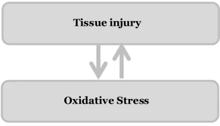
However, in most diseases, oxidative stress is a consequence and not a cause of the disease (Figure 1.4), for example, many of the biological consequences of excess radiation exposure are due to oxidative damage and dietary deficiencies (Halliwell and Gutteridge, 2007).
Although it has not been yet conclusively understood if reactive species are a cause or consequence, the answer probably differs for each pathological condition and may even differ from patient to patient depending on his/her antioxidant oxidative defence status.
Figure 1.4: Schematic relationship oxidative stress and tissue injury.
All these observations suggest that oxidative stress plays a role in human diseases taking to the proposal that health might be improved by increased dietary intake of antioxidants (Ames et al., 1993), because there exist a health balance between oxidants (such as free radicals) and antioxidants (such as vitamins E and C and protective enzymes) in vivo (Baublis et al., 2000). Beyond these, drugs (including both prescription only and over the counter medicines) with antioxidant indications may also have a functional relationship between health status and disease state (Aruoma, 1996; Pryor, 1982). This role that food and drugs might play in the management of health is shown schematically in Figure 1.5.
Tissue injury
11 Figure 1.5: Representation of the relationship between Food and/or drugs with Equilibrium, health or disease. Adapted from (Aruoma et al., 1998)
1.4 Biomimetic sytems
Over time, the need to study the structure and dynamic changes in biomembranes (and consequently in the organisms) couple to addressing the problem of applying methodologies that are not comparable to biological systems, biomimetic systems, such as liposomes, have been employed (Lúcio et al., 2007) in order to try to make conditions more closer to those found in vivo.
Several types of model structures have been employed so far, including here liposomes. Liposomes are in fact an important system in their own right in medical, cosmetic and industrial applications (Samuni et al., 2000), however, in many cases their instability upon storage is one of the major obstacles to their more spread use.
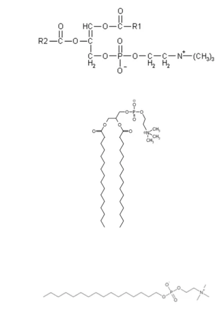
For investigating the basic characteristic of lipid membranes, simpler models, such as micelles, are also applicable. They are usually formed by a surfactant, for example miltefosine (hexadecylphosphocholine, HePC). HePC is an current used as alternative
12 drug to reduce and postpone the emergence of resistance of the protozoan parasite Leishmania donovani (usually fatal if untreated) upon conventional therapy for Visceral Leishmaniasis with pentavalent antimonials (Menez et al., 2007; Rangel-Yagui et al., 2005). Furthermore, HePC is a structural analogue of phospholipids, therefore a good and simple biomimetic system that, form a micellar system, which have been used to increase the aqueous solubility of hydrophobic compounds. However, in order to have formation of micelles, it is necessary to use concentrations higher than critical micellar concentration (CMC), which can pose problems for expensive lipids or can cause spectral interferences upon measurements.
Liposomes constitute a system used in investigation for commercial application (cosmetic area, among others), medical and pharmaceutical industry (as previously mentioned) with special attention to its physical properties, protocol for preparation, formation and fusion mechanism, pH gradients, membrane transport, capabilities, and interaction with biological structures (Santos, 2002). Indeed, liposomes are colloidal vesicles where a membrane is formed by one or more phospholipids (having a hydrophobic tail and a hydrophilic head to assemble the vesicular structures) bilayers. Liposomes have been classically applied as a drug carriers, as many kinds of molecules can be encapsulated in this inner phase while hydrophobic molecules can be trapped in the bilayer of the lipid membrane. In addition, liposomes can be modified with different molecules anchored at the surface, applied as biomimetic systems (Kojima et al., 2008), with special interest to areas, mostly due to their well characterized membrane properties.
The liposomes most frequently used are the large unilamellar vesicles (LUVs), with diameters larger than 100 nm and the small unilamellar vesicles (SUVs), with diameters between 20 to 50 nm. There structure is well bearing simple interpretation for interaction phenomena, comparatively to multilamellar vesicles. There are also oligolamellar vesicles (OVs), namely SOV, LOV and GOV, corresponding to small, large and giant variants, respectively (Figure 1.6).
Beyond the size and lamellar number, the chemical nature of the phospholipids that constitute of utmost importance because they determine other parameters of the vesicles structures, namely the charge, the stability, the membrane phase, the curvature of bilayer(s) and the formation of domains.
Other model frequently applied as a membrane surrogate is composed by monolayers, which have been known considerably longer before than micelles or liposomes. The amphiphilic nature of the substances dictates the orientation of the molecules at the interface (air/water). Lipid monolayers are very well defined, stable, homogeneous bidimensional systems with planar geometry. Although monolayers do not reflect the
13 complexity of membrane structure, they are considered useful models to learn about interactions at interfaces (Deleu et al., 2005) a 2D system.
Therefore, the various available models of biomembranes: micelles, liposomes and monolayers, can be select for the studies pertaining to interactions with biological membranes. In Figure 5 the structure of these models, concerning its size and lamellarity, are represented.
As mentioned previously, it is possible to study many properties/characteristics of the biological membranes with the use of liposomes, including their applications to studies of peroxidation of liposomal lipids.
1.5 Methods for assessments of antioxidants
In biological systems, the oxidation of membrane and lipoprotein lipids plays a central role in many pathology processes (Mattson, 2004; Poon et al., 2004a), especially in the context of oxidative stress-related pathologies referred to above. The aerobic organisms survive only in presence of oxygen, necessarily producing free radicals. Therefore, the antioxidant defences are extremely important in this mechanism as inhibitors of lipid peroxidation. The antioxidants can be synthetized in vivo or taken in from the diet. Similarly to the therms “oxidative damage” and “oxidative stress”, is the term “antioxidant” widely used but surprisingly difficult to define clearly (Halliwell and Gutteridge, 2007). In this context, there is also is an increasing interest in the efficacy of antioxidant activity of the inumerous naturally occurring molecules in food and biological systems, with potential to minimize oxidative damage in vivo and/or to retard the oxidation of easily oxidizable materials food (Lúcio et al., 2007).
Several methods have been applied to measure antioxidant capacity of tissues, biological fluids or single classes of antioxidants, usually with the aim to quantify oxidative stress (Halliwell and Gutteridge, 2007) or protection conferred by foreign compounds.
14
Biomimetic systems
Figure 1.6: Schematic illustration of : (A) structure of the micelles; (B) liposomes structure (size and lamellarity); (C) Monolayer representation.
A
B
MVL
MLV LUV SUV OLV
GOV LOV SOV
GUV C
Aqueous subphase
Air
Hydrophobic tail Hydrophilic head15 The evaluation of antioxidant capacity in matrixes such as plasma, beverages, vegetables, and fruits as well as that of pure compounds (i.e., phenols, peptides) has become a rather disputable issue during the past decade. Methods and data are questioned for providing meaningful information to interested parties (Nenadis et al., 2007). Regardless, current methods are still frequently applied, providing an enormous amount of information in the past few years.
Among the several available methods, they can be classified into two groups, depending on the reaction mechanism: methods based on hydrogen atom transfer (HAT) and methods based on electron transfer (ET) (Huang et al., 2005).
The majority of HAT-based assays apply a competitive scheme (depicted schematically in Figure 7), in which antioxidant compound and target (representing a biomolecule which may be attacked in vivo) compete for the reactive species (in this work, represented by thermally generated peroxyl radicals through the decomposition of azo-compounds). Hence, the assessment of antioxidant capacity can also be divided concerning their simple (non-competitive) or more complex (competitive) reaction schemes, as depicted schematically in Figure 1.7:
Reactive species Reduced reactive
species
Target Oxidized target
Antioxidant Oxidized antioxidant
16 Figure 1.7: Schematic representation of competitive and non-competitive approaches, for in vitro determination of antioxidant capacity. Adapted from (Magalhães et al., 2008)
In the competitive Scheme, the determination of antioxidant capacity relies on the quantification of an extra compound (probe) that facilitates the analytical measurement. In most of the competitive assay, the probe is the target species or its oxidized form. Moreover, the probe can also be a compound added after the above cited reaction that allows the quantification of the remaining reactive species or target molecules. Using the first type of strategy, the antioxidant capacity of a given compound is dependent on: (i) the concentration ratio of the target, probe, antioxidant and reactive species produced, (ii) the rate of the reaction between antioxidant compound and probe or target molecules, (iii) if the rate of oxidized probe is due only reactive species attack, (iv) rate of target and probe. On the other hand, in the non-competitive scheme, putative antioxidant compound reacts with the reactive species in the absence of any other competing agent. In this case, only two components are involved in the initial reaction mixture: the antioxidant compound and the reactive species. Therefore, this strategy deploys a reaction simpler than that applied in competitive schemes, suitable for screening assays (Magalhães et al., 2008).
1.6. Scavenging capacity assays against specific ROS/RNS
1.6.1 Peroxyl radical (ROO•) scavenging capacity assays
Several methods have been proposed to address these determinations, from which the oxygen radical absorbance capacity (ORAC) and total radical trapping antioxidant parameter (TRAP) are included. Indeed, the ORAC assay is one of the most common
Reactive species Reduced reactive
species
Antioxidant Oxidized antioxidant
17
methods for assessing scavenging capacity against ROO• as a HAT method (Frankel and
Meyer, 2000), developed initially by Cao, Alessio, and Cutler (1993).
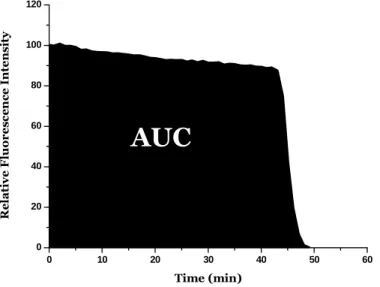
The principle of this assay is based on the intensity of fluorescence decrease of the target/probe along time under reproducible and constant flux of peroxyl radicals, generated from the thermal decomposition of AAPH in aqueous buffer (Magalhães et al., 2008). In the presence of a sample that contains chain-breaking antioxidants, the decay of fluorescence is inhibited. The method measures the ability of the antioxidant in the sample to protect. In ORAC assay, the reaction is monitored for extended periods (≥ 30 min) and the quantifications is based in the area under curve (AUC) that represents the oxidation of the probe along time. The protective effect of antioxidants is evaluated from
the net integrated area under the fluorescence decay curves (AUCsample – AUCblank) - Figure
1.8, and results are expressed as µM of Trolox equivalents. The advantage of the AUC approach is that it can be applied for antioxidants that exhibit distinct lag phases and also to those samples that have no lag phases. The principles of the ORAC assay can be adapted to determine the action against other reactive oxygen species (Ou et al., 2001). The assay is based on the intensity of fluorescence decrease of the target/probe along time under reproducible and constant flux of peroxyl radicals, generated from the thermal decomposition of AAPH in aqueous buffer. In the presence of a sample that contains chain-breaking antioxidants, the decay of fluorescence is inhibited (Glazer, 1990).
Figure 1.8: Schematic representation from the net integrated area under the fluorescence decay curves.
The TRAP assay was introduced by (Wayner et al., 1985), for the determination of the antioxidant status of human plasma. This method was based on the measurement of the
0 10 20 30 40 50 60 0 20 40 60 80 100 120 R e la ti v e F lu o r e s c e n c e I n te n s it y Time (min)
AUC
18
time period in which oxygen uptake was inhibited by plasma during a controlled ROO•
peroxidation reaction induced by the thermal decomposition of an azo-compound. In this assay, the target was the human plasma while the oxygen consumed in the oxidation of plasma material is the probe molecule used to follow the action of antioxidants. The measurement is based on the “lag time” that corresponds to the time period between the beginning of the assay and the beginning of the oxidation of the target molecules. One of the major problems with the original TRAP assay lies in the utilization of the oxygen electrode as detector, since it may not maintain its stability over the period of time required (Magalhães et al., 2008).
1.6.2 Other ROS/RNS scavenging capacity assay
Along with peroxyl radicals, other ROS/RNS subject biological organisms to oxidative stress. A summarized account of current merhods for evaluation of scavenging capacity
superoxide radical anion (O2•-), hydroxyl radical (HO•), hypochlorous acid (HOCl),
singlet oxygen (1O2) and nitric oxide radical (NO•) is given in the following
paragraphs. A more comprehensive account can be found elsewhere (Magalhães et al., 2008).
Superoxide radical anion (O2•-) is produced as a result of the donation of one electron to
oxygen. This radical arises either from several metabolic processes or following oxygen activation by irradiation (Halliwell, 2006). Generally, the analytical methods for
determination of O2•- scavenging capacity make use of the system XOD/hypoxanthine or
xanthine at pH 7.4 to generate superoxide anion radical.
Due to the high reactivity of hydroxyl radicals, almost anything in biological systems can
be regarded as an HO• scavenger. Hence, this task is not performed by any specific
molecule or enzyme. Thus, the evaluation of direct scavenging of HO• may be irrelevant for
evaluation of antioxidant action of a compound or matrix, simply because very high concentrations of scavenger are required to compete with adjacent molecules in vivo or in
the food matrix for any HO• generated. For these reason, it is more relevant and useful to
quantify the capacity of putative antioxidants to scavenge or block the formation of its
precursors (O2•-, H2O2, HOCl) and/or to sequester free metal ions related to HO•
formation. Scavenger compounds that act in in this way would behave as preventive antioxidants.
There are a several in vitro methodologies for determination of HO• scavenging capacity,
19 products from deoxyribose is inhibited by antioxidants; ii) formation of DMPO-OH adduct is measured by ESR and it is inhibited by antioxidant; iii) DHBA, formed due to oxidation of salicylic acid, measured using HPLC-ED and its formation is inhibited by antioxidants; iv) fluorescence decay along time due to oxidation of fluorescein is delayed/inhibited by antioxidants (ORAC method with different inducer); v) CL emission due to oxidation of luminal is inhibited by antioxidants (Magalhães et al., 2008).
HOCl is generally obtained from the enzymatic system myeloperoxidase/H2O2/Cl- or by
acidifying commercial sodium hypochloride to pH 6.2 with sulphuric acid(Aruoma, 1997). The former approach can be applied if the sample species do not interfere with HOCl
generation (e.g. inhibition of myeloperoxidase activity or direct reaction with H2O2). In the
second approach, the determination of the concentration of HOCl solution must be performed daily (Halliwell et al., 1995). For assessment of scavenging capacity against HOCl, direct spectrophotometric determination of this species along time is performed. Alternative methods are based on luminal-derived chemiluminescence.
Singlet oxygen (1O2) is an excited state of molecular oxygen that has no unpaired electrons
and it is known to be a powerful oxidizing agent, reacting directly with a wide range of
biomolecules (Davies, 2004). Due to its decay to the lower energy ground state, 1O2 emits
characteristic phosphorescence at 1270 nm. Therefore, the 1O2 scavenging ability of several
compounds was measured through the decay rates of the light intensity (Tournaire et al., 1993; Wilkinson et al., 1995).
NO has emerged as a fundamental signaling molecule regulating virtually every critical cellular function, as well as a potent mediator of cellular damage in a wide range of conditions. Recent evidence indicates that most of the cytotoxicity attributed to NO is
rather due to peroxynitrite -nitric oxide radical (NO•), for this reason it has indeed pivotal
role in the regulation of diverse physiological and pathophysiological processes (Pacher et al., 2007).
Vriesman et al. developed a relatively simple method for the quantification of NO•
scavenging capacity of sulfur-containing compounds in aqueous solution using an
amperometric NO• sensor. The assessment of NO• scavenging capacity has also been
20 1.6.3 Scavenging capacity assays against stable, non-biological radicals and evaluation of total reduction capacity
Several methods have been reported for evaluation of total antioxidant capacity, namely those relying on colored radicals, 2,2’-azinobis-(3-ethylbenzothiazoline-6-sulphonate)
radical cation (ABTS•+) and 2,2′-Azinobis-(3-ethylbenzothiazoline-6-sulphonate) radical
cation. Other methods, relying to electron transfer, have also been successfully employed, including here Ferric reducing antioxidant power (FRAP assay), Folin-Ciocalteu reducing capacity (FC assay) and Total reducing capacity estimated by electrochemical methods.
The TEAC assay, mediated by ABTS•+ species, involves the generation of the long-lived
radical cation chromophore ABTS•+ which has absorption maxima at 414, 645, 734, and
815 nm. The original TEAC assay, developed by Miller et al., was based on the activation of
metmyoglobin, acting as peroxidase, with H2O2 to generate ferrylmyoglobin radical, which
then reacted with ABTS to form the ABTS•+ radical cation (Miller et al., 1993).
In this strategy, the sample to be tested is added previously to the formation of the ABTS•+
radicals formed and the lag phase, which corresponds to the delay time in radical formation, is measured (competitive scheme-Figure 7). This method has been executed using commercial available Kits for standardized total antioxidant status measurement in an individual’s serum or plasma. The disadvantage is the price and the reagent cost per sample estimated in the Kit-TEAC assay is approximately nine times that in the ORAC assay (Cao and Prior, 1998).
In DPPH assay, the purple chromogen radical 2,2-diphenyl-1-picrylhydrazyl radical
(DPPH•) is reduced by antioxidant/reducing compounds to the corresponding pale yellow
hydrazine (Magalhães et al., 2008). The scavenging capacity is generally evaluated in organic media by monitoring the absorbance decrease at 515-528 nm until the absorbance remains constant (Magalhães et al., 2008) or by electron spin resonance (Calliste et al., 2001).
The FRAP assay measures the ability of antioxidants to reduce the ferric
2,4,6-tripyridyl-s-triazine complex [Fe(III)-(TPTZ)2]3+ to the intensely blue coloured ferrous complex
[Fe(II)-(TPTZ)2]2+ in acidic medium (Benzie and Strain, 1996; 1999). This method has also
been adapted to 96-well microplate reader, giving better reproducibility and higher sample throughput (Tsao et al., 2003).
The exact chemical nature of the Folin-Ciocalteu reagent is not known, but it is accepted that is contains phosphomolybdic/phosphotungstic acid complexes (Singleton and Rossi, 1965). The chemistry behind the FC assay relies on the transfer of electrons in alkaline medium from phenolic compounds and other reducing species to molybdenum, forming
21 blue complexes that can be detected spectrophotometrically at 750-765 nm (Singleton et al., 1999).
Finally, the electrochemical properties of pure compounds, foods, and biological samples may be used for the evaluation to their reducing/antioxidant capacity, since the electric oxidation potential has conceptually relation with the expected antioxidant capacity. This determination has been frequently automated using flow injection analysis schemes (Magalhães et al., 2009).
1.7 Methods for antioxidants assessment with biomimetic structures
A schematic representation of all factors, including physical and chemical features, are depicted in Figure 1.8. It is important to highlight that a major concern in these studies should be the effect of antioxidants depends on both the composition, physical properties and structure (LUV, MLV, SUV, etc) of the liposomal system chosen and the inducer.
Figure 1.9: Physical and chemical factors that affect the peroxidation of liposomal lipids. Adapted from (Schnitzer et al., 2007b)
Peroxidation of liposomal lipids, effects of liposomal composition and physico-chemical properties Chemical factors Physical factors Surface charge Lateral phase separation Headgroups Oxidizability of lipids Specific binding of inducer Fluidity, lateral difusion Oxidation of headgroup Number of Bisallylic hydrogens Number of Double bonds Position of double bonds Cis-trans isomerism Water permeability
22 In order to illustrate the different conditions used for oxidation/antioxidant evaluation with biomimetic structures, Tables 1.3 to 1.6 are presented, where a summary lipid, preparation method, oxidation initiator, principle of measurement of detection system, calibrator, data treatment, applications, references for each method given. Selected aspects of these methodologies are discussed in the following sections.
1.7.1 Type of biomimetic systems
In studies of lipid peroxidation the biomimetic structures of the biological systems, used were essencially micelles, MLVs, LUVs and SUVs, comprising structures with different lipid composition, size, lamellarity, stability and preparation protocol, whose nomenclature was defined in the 1980s.
As discussed before, liposomes are classified by their lamellarity and size range (Figure 5). Both these aspects are relevant towards lipid peroxidation for at least two reasons: i) they constitute a simple and convenient system where the oxidation process can be reproduced with different levels of complexity and ii) liposomes are currently being used for diagnostic and therapeutic purposes and lipid oxidation may impair their stability.
Concerning their complexity features, all structures being about advantages and disadvantages. For instance, the MLVs have a more simpler preparation comparatively for LUVs and SUVs, but the interpretation of results may not be so easy because the structure is not well defined, with several lipidic layers.
Therefore, when using MLVs in the presence of inducers of oxidation the outer monolayer is more to oxidative attack, but the layer inside are not accessible.
Hence, unilamellar liposomes seem to be a more suitable model for studding the free radical oxidation that take place in vivo, because biomembranes, in general, consist of unilamellar structures. The LUVs have a more resemblance with the biological membrane structure but studies as those performed by Barclay et al (1987) demonstrated that the oxidizability of PC in unilammelar liposomes was higher than that in multilamellar liposomes, because of the different packing of the lipid chain in LUV compared to that in MLV. Furthermore, despite the more complex LUV preparation its size is well defined.
23 T ab le 3 M et h o d s fo r as se ss m en t o f an ti o xi d an t ca p ac it y u si n g L U V s. L ip id P r e p a r a ti o n m e th o d O x id a ti o n In it ia to r P r in c ip le o f M e a s u r e m e n t o r D e te c ti o n S y s te m C a li b r a to r D a ta t r e a tm e n t A p p li c a ti o n s R e fe r e n c e D P P C + L A L ip id d is so lu ti o n o rg an ic so lv en t, e va p o ra ti on o f so lv en t, h yd ra ti o n w it h T ri s b u ff er p H 7. 4 , v o rt ex a gi ta ti o n , e xt ru si o n A A P H A b so rb an ce m ea su re m en t at 2 3 3 n m d u e to fo rm at io n o f co n ju ga te d ie n es α -t o co p h er o l % i n h ib it io n o f o xi d at io n n .a (C as te ll i et al ., 1 9 9 7) E g g o r S o y b e a n P C L ip id d is so lu ti o n o rg an ic so lv en t, e va p o ra ti on o f so lv en t, h yd ra ti o n w it h T ri s-H C l b u ff er p H 7 .4 , v o rt ex a gi ta ti o n , so n ic at io n , e xt ru si o n A A P H o r A M V N ( w as m ix ed w it h P C b ef o re p re p ar in g li p os o m es ) H P L C q u an ti fi ca ti o n o f fo rm at io n o f P C h yd ro p er o xi d es n .a R at e o f in it ia ti on an d r at e o f p er o xi d at io n n .a (K o ga e t al ., 19 9 7) P C L ip id d is so lu ti o n o rg an ic so lv en t, e va p o ra ti on o f so lv en t, h yd ra ti o n w it h T ri s-H C l b u ff er p H 7 .4 , v o rt ex a gi ta ti o n , so n ic at io n , e xt ru si o n A A P H H P L C q u an ti fi ca ti o n o f fo rm at io n o f P C h yd ro p er o xi d es B H T E va lu at io n o f h yd ro p er o xi d es co n ce n tr at io n v s ti m e p ro fi le E xt ra ct s of c er ea l gr ai n s (Z ie li n sk i an d K o zl o w sk a 2 0 0 0 ) P C o r D M P C L ip id d is so lu ti o n o rg an ic so lv en t, e va p o ra ti on o f so lv en t, h yd ra ti o n w it h H ep es b u ff er p H 7. 4 , e xt ru si o n C u (I I) M ea su re m en t o f T B A R S ( ab so rb an ce at 5 3 0 n m ) an d p er ox id es (S C N - m et h o d ) n .a E va lu at io n o f m al o n d ia ld eh yd e (M D A ) eq u iv al en ts v s ti m e p ro fi le X an th o p h yl ls (R en ge l et al ., 2 0 0 0 ) E g g P C L ip id d is so lu ti o n o rg an ic so lv en t, e va p o ra ti on o f so lv en t, h yd ra ti o n w it h H ep es b u ff er p H 7. 4 , e xt ru si o n n .a M ea su re m en t o f ch an ge s o n a cy l ch ai n c o m p o si ti o n (G C ) n .a E va lu at io n o f re si d u al f ra ct io n o f ac yl c h ai n s al o n g ti m e E st im at io n o f li p o so m e o xi d at io n u p o n st or ag e in p re se n ce o f te m p o l a n d α -to co p h er o l (S am u n i e t al ., 2 0 0 0 ) S o y b e a n P C n .g A A P H H P L C q u an ti fi ca ti o n o f co n ju ga te d d ie n es n .a E va lu at io n o f P C -O O H f or m at io n al o n g ti m e; r at e o f o xi d at io n N ew ly s yn th es iz ed co m p o u n d s (4 G B E 4 3 a n d 2 B B E 4 3 ) an d α -to co p h er o l (E zu re e t al ., 2 0 0 1)