DEVELOPMENT OF CHITOSAN-GRAFTED
POLYCAPROLACTONE COPOLYMER FOR
MUCOADHESIVE NANODELIVERY SYSTEMS
ANDREIA MARINA DE SOUSA ALMEIDA
DISSERTAÇÃO DE MESTRADO APRESENTADA
À FACULDADE DE ENGENHARIA DA UNIVERSIDADE DO PORTO EM ENGENHARIA BIOMÉDICA
Faculdade de Engenharia da Universidade do Porto
Development of chitosan-grafted
polycaprolactone copolymer for mucoadhesive
nanodelivery systems
Andreia Marina de Sousa Almeida
Dissertation for the Master Thesis in Biomedical Engineering
Supervisor: Bruno Sarmento, Ph.D.
vii
“Success is getting what you want.
Happiness is wanting what you get”
ix
Resumo
Durante a última década, uma vasta gama de formulações para a administração oral de fármacos anticancerígenos tem sido utilizada na prática clínica. No entanto, este desenvolvimento é dificultado pela baixa solubilidade nos fluidos gástricos e intestinais e consequente diminuta biodisponibilidade destes fármacos. Assim, o uso de veículos transportadores de fármacos, como as micelas poliméricas, está a ser amplamente investigado como estratégia de formulação devido à sua capacidade de encapsular fármacos de forma estável e com grande eficiência, aumentando a sua solubilidade e possibilitando ainda a sua penetração nos tecidos devido ao seu pequeno tamanho. Nesta dissertação, foram produzidas micelas poliméricas à base de quitosano (CS) e policaprolactona (PCL) com o objetivo de melhorar o perfil farmacocinético do paclitaxel (PTX), após a administração oral. Copolímeros de quitosano e policaprolactona (CS-g-PCL) foram sintetizados através de uma reação carbodiimida por amidação sendo caracterizados por transformada de Fourier de espectroscopia de infravermelho (FTIR), análise de ressonância magnética nuclear (RMN) e ângulo de contacto. Os resultados sugerem a formação do copolímero através do aparecimento de bandas amidas no espetro de FTIR, pelo aparecimento dos picos de ambos os polímeros no espectro do novo copolímero na análise de RMN e ainda pela maior hidrofobicidade quando em contacto com a água. Averiguou-se a concentração micelar crítica (CMC) do copolímero formado pelo método condutimétrico, obtendo-se a concentração de 3.7 × 10⁻³ mg/mL. A caracterização morfológica das micelas foi realizada por microscopia eletrónica de transmissão (TEM) e a determinação do diâmetro médio, índice de polidispersão e carga superficial foram determinados por espalhamento de luz dinâmico (DLS) e espalhamento de luz eletroforético (ELS), respetivamente, obtendo-se valores de 408 nm, 0,335 e 30,9 mV para a formulação otimizada, escolhida para os ensaios subsequentes. A eficiência de encapsulação (82%) foi determinada por cromatografia líquida de alta performance (HPLC). O perfil de libertação do PTX encapsulado nas micelas e do PTX livre foi estudado in vitro em condições intestinais simuladas. Verificou-se que as micelas controlaram a libertação por até 8 h. A citotoxicidade dos polímeros CS e PCL, do copolímero, das micelas com PTX encapsulado e o PTX livre foi testada contra células epiteliais intestinais (HT29-MTX e Caco-2). Verificou-se ausência de toxicidade para os polímeros isoladamente nas concentrações testadas. O copolímero apresentou toxicidade à maior concentração testada (1 mg/mL). O aumento da concentração de PTX livre e de micelas com PTX encapsulado levaram à diminuição da viabilidade celular, contudo, o sistema micelar apresentou sempre menor toxicidade em comparação com o fármaco livre. Por fim, foi realizado o ensaio de permeabilidade para o fármaco livre e para o sistema micelar em modelo de monocultura de células Caco-2 e em modelo de co-cultura de células Caco-2/HT29-MTX. Verificou-se que o sistema micelar desenvolvido teve uma permeação superior ao fármaco livre nos dois modelos intestinais testados. Assim, o sistema micelar desenvolvido mostrou ultrapassar as barreiras fisiológicas com desempenho superior ao anticancerígeno atualmente utilizado em terapia clínica. Pode concluir-se que o sistema micelar realizado no âmbito desta dissertação apresenta potencial para ser utilizado como sistema de libertação de fármacos hidrofóbicos, nomeadamente fármacos anticancerígen
xi
Abstract
During the last decade, a wide range of formulations for oral administration of anticancer drugs have been used in clinical practice. However, this development is sometimes hampered by low solubility in gastric and intestinal fluids and consequent decrease in bioavailability of these drugs. Thus, the use of drug carriers, such as polymeric micelles, is being widely investigated as strategy of formulation due to its ability to encapsulate drugs in a stable way with great efficiency, increasing its solubility and also allowing tissue penetration due to their small size. In this dissertation, polymeric micelles were produced based on chitosan (CS) and polycaprolactone (PCL) in order to improve the pharmacokinetic profile of paclitaxel (PTX) after oral administration. Chitosan-grafted-polycaprolactone (CS-g-PCL) copolymer was synthesized through a carbodiimide reaction by amidation being subsequently, characterized by Fourier Transform Infrared Spectroscopy (FTIR), Nuclear Magnetic Resonance analysis (NMR) and contact angle. The results suggest the formation of a new copolymer by the appearance of amide bands in the FTIR spectrum, appearance of both polymers peaks in the new copolymer spectrum in NMR analysis and, also higher hydrophobicity when in contact with water. The critical micelle concentration (CMC) of the copolymer, investigated by conductimetric method, was 3.7 × 10⁻³ mg/mL. The micelles morphology was characterized using transmission electron microscopy (TEM) and the determination of the average diameter, polydispersity index and surface charge were determined by dynamic light scattering (DLS) and electrophoretic light scattering (ELS), respectively, obtaining values of 408 nm, 0.335 and 30.9 mV for the optimized formulation chosen for subsequent tests. The encapsulation efficiency (82%) was determined by high performance liquid chromatography (HPLC). The release profile of free PTX and PTX loaded micelles was studied in vitro in simulated intestinal conditions. The micelles were able to control the drug release up to 8 hours. The cytotoxicity of CS and PCL polymers, copolymer, the PTX loaded micelles and free PTX was tested against intestinal epithelial cells (HT29-MTX and Caco-2). It was observed the absence of toxicity to polymers individually for all tested concentrations. The copolymer presented toxicity at the highest concentration tested (1 mg/mL). The increase of free PTX and PTX loaded micelles led to a decreased of cell viability, however, the micellar system showed lower toxicity when compared to the free PTX. Finally, it was performed the permeability assay for the free drug and the micellar system in the Ca2 monoculture model and Ca2/HT29-MTX co-culture model. It was observed that the micellar system developed had superior permeation of PTX when compared to the free drug in both intestinal models tested. Thus, the micellar system developed was able to overcome the physiological barriers with better performance than the anticancer drug currently used in clinical therapy. Therefore, it can be concluded that the PTX-CS-g-PCL micelles developed in this dissertation is a potential drug carrier for the controlled release of hydrophobic drugs, particularly, anticancer agents.
xiii
Acknowledgements
First of all, I would like to thank all my family, but particularly my parents, who always believed in me, for their unconditional love and support over all these academic years. My brother and my sister in law, by their wise advices and to my dear goddaughter, Joana, for all contagious joy, even after a hard day of work. A heartfelt thanks to João, my boyfriend, for all the help, support and care during these two years. Without you it would not be possible.
My sincere thanks to FEUP - Faculdade de Engenharia da Universidade do Porto, INEB – Instituto de Engenharia Biomédica/i3S- Instituto de Investigação e Inovação em Saúde and iinfacts – Instituto de Investigação e Formação Avançada em Ciências e Tecnologias da Saúde for having accepted me and cooperated in my dissertation.
My supervisor, Professor Bruno Sarmento, to whom I am sincerely grateful for have accepted me as a master student in his team, for all the support and guidance. Thanks to him I started in the world of research, and despite the early days were difficult, today I am grateful and happy for believing in me and for having created me a passion of doing research. It took months of professional exponential growth, but also personal. Thank you!
To all my teammates, but particularly those who worked closely with me - Rute Nunes, Ana Costa, Cassilda Reis, Flávia Sousa and Fabíola Prezotti - for all the fundamental teachings for my growth and success and, above all, for the friendship that we build. A special thanks to Daniella Souza for complicity, by working together, for the laughter and the good mood and high spirits that always brought to the lab. Thanks to all from the heart!
All of my other friends who have grown with me since childhood - Alice Ribeiro, Diana Coelho and Rita Loureiro - and the friends of the city that I bring in my heart - Ana João Nóbrega, Cláudia Montes, Beatriz Pereira, Cristiana Cabral, Sara Ferreira and Marlene Silva - by friendship, support in difficult days and the help that you have been offering me, in one way or another, over the last few years. Thank you!
And last, but far from least, Sofia Marques, friend and classmate, for all the problems, questions and concerns that we had together, for always heard me, and have a positive response to me and encouraging me. Thank you for having been part of this stage, although far but always close. Thanks for the patience, the complicity and for always being there for me.
xv
List of Contents
Resumo ... ix
Abstract ... xi
Acknowledgements ... xiii
List of Contents ... xv
List of Figures ... xvii
List of Tables ... xx
Abbreviations and Symbols ... xxii
Chapter 1 ... 1
Introduction ... 1
1.1 - Context and motivation ... 1
1.2 - Objectives ... 2
1.3 - Document structure ... 2
Chapter 2 ... 4
Literature Review ... 4
2.1 Colorectal cancer ... 4
2.2.1Anatomy and physiology of gastrointestinal system ... 5
2.2.2Pathways through the intestinal epithelial barrier ... 7
2.2.3Intestinal in vitro models ... 9
2.2.3.1 Caco-2 model... 10
2.2.3.2 Caco-2/HT29-MTX model ... 11
2.2.3.3 Caco-2/HT29-MTX/Raji B model ... 11
2.3 Anticancer drug model - Paclitaxel ... 11
2.4 Polymeric micelles as drug delivery vehicles ... 13
2.5 Chitosan and amphiphilic derivatives ... 14
2.5.1Biological properties of chitosan ... 16
2.5.2Chitosan amphiphilic derivatives ... 17
2.5.2.1 Alkylation ... 17
2.5.2.2 Acylation ... 18
2.5.2.2.1 N-acylation ... 18
2.5.2.2.2 O-acylation ... 20
2.5.3Chitosan derivatives in drug delivery of anticancer drugs... 21
2.6 Polycaprolactone ... 23
Chapter 3 ... 27
Material and Methods ... 27
3.1 Chemical material ... 27 3.2 Cell culturing ... 27 3.3 Methods ... 28 3.3.1Degree of acetylation ... 28 3.3.2Molecular weight ... 28 3.3.3Synthesis of chitosan-grafted-polycaprolactone ... 29 3.3.4Characterization of chitosan-grafted-polycaprolactone ... 30
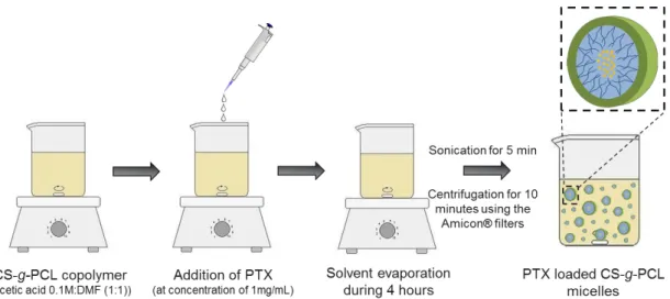
3.3.5Preparation of paclitaxel-loaded chitosan-grafted-polycaprolactone micelles ... 30
3.3.6Characterization of paclitaxel-loaded chitosan-grafted-polycaprolactone micelles .... 31
3.3.7In vitro paclitaxel release studies ... 31
3.3.8Cytotoxicity studies ... 32
3.3.9Permeability studies ... 33
3.3.10 Statistical analysis ... 33
Chapter 4 ... 35
Results and Discussion ... 35
4.1 Chitosan characterization ... 35
4.1.1Degree of acetylation ... 35
4.1.2Molecular weight ... 38
4.2 Synthesis and characterization of chitosan-grafted-polycaprolactone ... 39
4.2.1NMR analysis ... 39
4.2.2FTIR analysis ... 40
4.2.3Contact angle ... 41
4.2.4Critical micelle concentration ... 42
4.3 Characterization of paclitaxel loaded chitosan-grafted-polycaprolactone ... 43
4.3.1Particle size, size distribution and zeta potential ... 43
4.3.2Paclitaxel encapsulation efficiency ... 43
4.4 In vitro release studies... 45
4.5 Cytotoxicity studies ... 46 4.6 Permeability studies ... 48
Chapter 5 ... 53
Conclusion ... 53
5.1 Future perspectives ... 54References ... 56
xvii
List of Figures
Figure 2.1 - Organization of digestive tract by layers: mucosa, submucosa, muscularis propria
and serosa. 6
Figure 2.2 - Schematic representation of the architecture of the small intestine (A) and the colon (B) as well as all the cell types present in an intestinal villus. 7 Figure 2.3 - Schematic representation of the transport across the intestinal epithelium.
Molecules can across the intestinal epithelial barrier by paracellular transport (blue line) and by transcellular transport through passive diffusion (green line), transcytosis via normal
enterocytes (yellow line) or via M-cells (red line). 8
Figure 2.4 - Morphological aspect of Caco-2 cells (A) and HT29-MTX cells (B) at a resolution of 20 µm. The images were taken with Nikon TE2000-U microscope equipped with digital
camera and controlled via the Nikon ACT-1 program. 9
Figure 2.5 - Schematic representation of a Transwell® that consist in a well plate plus insert. The epithelial cells are seeded on the top of the membrane where the compounds are placed and the permeability occurs the apical to the basolateral compartment. 10 Figure 2.6 - Schematic representation of the Caco-2 monoculture model setup. 10 Figure 2.7 - Schematic representation of the Caco-2/HT29-MTX co-culture model setup. 11
Figure 2.8 - Chemical structure of paclitaxel. 12
Figure 2.9 - Schematic representation of cell cycle. 12 Figure 2.10 - Schematic representation of drug encapsulation from self-assembled nanosystems.
14 Figure 2.11 - Deacetylation of chitin to chitosan. 15 Figure 2.12 - The soluble–insoluble transition of chitosan occurs at about pH 6. 16 Figure 2.13 - Reaction scheme to obtain N-alkyl derivatives of chitosan. 18 Figure 2.14 - Acylation modification by carbodiimide reaction. 19 Figure 2.15 - Acylation synthesis from the use of EDC and NHS. 19 Figure 2.16 - N-acyl chitosan reaction by anhydrides. 20 Figure 2.17 - Reaction of protection through the phthaloyl group. 20
Figure 2.18 - Reaction of O-substitution through the protection of amine groups with CH3SO3H. 21
Figure 2.19 - Synthesis of polycaprolactone from ε-caprolactone by ring-opening polymerization. 24 Figure 3.1 - Synthesis scheme the reaction of chitosan with polycaprolactone through chitosan
modification by N-acylation. 29
Figure 3.2 - Schematic representation of the method used to prepare PTX loaded CS-g-PCL
micelles. 31
Figure 4.1 - First derivative UV-spectra for various concentrations of acetic acid and N-acetyl-D-glucosamine. The point where all the acetic acid spectrum cross is the zero crossing point.
36 Figure 4.2 - H values as a function of GlcNAc concentration. 36 Figure 4.3 - First derivative UV-spectra for a commercial chitosan sample and various
concentrations of acetic acid and N-acetyl-D-glucosamine. 37
Figure 4.4 - 1H NMR chitosan spectrum with group’s identification (A) and with calculated areas
(B). 37
Figure 4.5 - Plots of the ηsp/C and ln(ηrel)/C, from Huggins and Kraemer equations,
respectively, versus concentration of the chitosan samples. 38
Figure 4.6 - 1H NMR spectra of (A) CS-g-PCL in HCl/D
2O 1% (v/v), (B) PCL in CDCl3 and (C) CS in
HCl/D2O 1% (v/v). 40
Figure 4.7 - ATR-FTIR absorbance spectra for detection of surface functional groups. (A)
CS-g-PCL copolymer, (B) CS-g-PCL and (C) CS. 41
Figure 4.8 - Water contact angle of chitosan (A) and CS-g-PCL copolymer (B) after 15 seconds
of analysis. 42
Figure 4.9 - Conductivity curve as a function of the log concentration of the copolymer. 42 Figure 4.10 - Standard curve calibration for paclitaxel quantification using HPLC. 44 Figure 4.11 - Transmission electron microscopy images of CS-g-PCL free drug micelles (A) and
PTX-CS-g-PCL micelles (B) with a scale bar corresponding to 0.5 µm and 1 µm, respectively. 45 Figure 4.12 - Cumulative in vitro release of PTX-CS-g-PCL micelles () and free paclitaxel (●) in
0.1% Tween®-80 containing phosphate-buffered saline (pH 7.4, w/v) at 37 ºC (n=3, mean ±
SD). 46
Figure 4.13 - Cell viability of chitosan, PCL and CS-g-PCL against Caco-2 (A and C) and HT29-MTX (B and D) cell lines, respectively, at concentrations of 0.01-1000 µg/mL after incubation for 4 h (A and B) and for 24 h (C and D). Values were reported as mean ± SD (n = 8). (*), (**), and (***) denotes a significant difference (p<0.05, p<0.01 and p<0.001),
respectively. 47
Figure 4.14 - Cell viability of loaded micelles and free PTX against Caco-2 (A and C) and HT29-MTX (B and D) cell lines, respectively, at concentrations of 0.01-10 µg/mL after incubation for 4 h (A and B) and for 24 h (C and D). Values were reported as mean ± SD (n = 8). (*) and (***) denotes a significant difference (p<0.05 and p<0.001), respectively. 48
xix
Figure 4.15 - In vitro cumulative permeability profiles of paclitaxel loaded micelles (▲) and free paclitaxel (○) across Caco-2 monoculture model. All experiments were conducted from the apical to basolateral compartment in HBSS at 37 ºC. Error bars represent mean ± SD
(n = 3). 49
Figure 4.16 - In vitro cumulative permeability profiles of paclitaxel loaded micelles (▲) and free paclitaxel (○) across Caco-2/HT29-MTX co-culture model. All experiments were conducted from the apical to basolateral compartment in HBSS at 37 ºC. Error bars
represent mean ± SD (n = 3). 50
Figure 4.17 - In vitro cumulative permeability profiles of paclitaxel loaded micelles across Caco-2 monoculture model (●) and across Caco-Caco-2/HTCaco-29-MTX co-culture model (). All experiments were conducted from the apical to basolateral compartment in HBSS at 37 ºC.
List of Tables
Table 2.1 - Biological and structural properties of chitosan. 16 Table 2.2 - Overview of amphiphilic chitosan derivatives used to encapsulate anticancer
drugs. 23
Table 2.3 - Summary of the polycaprolactone features. 24 Table 4.1 - Degree of deacetylation (DD) and degree of acetylation (DA) of chitosan
calculated through the first derivative spectra and 1H NMR analysis. 38 Table 4.2 - The particle size, polydispersity index (PdI), ζ-potential and theoretical drug
loading of PTX-CS-g-PCL micelles and CS-g-PCL micelles (empty micelles). Each value represents the mean ± SD of three independent measurements (n=3). 43 Table 4.3 - The theoretical and practical drug loading of PTX-CS-g-PCL micelles. Each value
represents the mean ± SD of three independent measurements (n=3). 44 Table 4.4 - TEER values before the permeability experiment. Each value represents the
mean ± SD of three independent measurements (n=3). 49 Table 4.5 - Apical compartment paclitaxel quantification (%). Each value represents the
Abbreviations and Symbols
List of abbreviations
ALP Alkaline phosphatase
ATCC American Type Culture Collection ATP Adenosine triphosphatase
ATR Attenuated total reflection
ATR-FTIR Attenuated total reflection Fourier transforms infrared spectroscopy BCS Biopharmaceutic Classification System
CAC Critical aggregation concentration CDCL3 Deuterated chloroform
CL Caprolactone
CMC Critical micelle concentration
CS Chitosan
CS-g-PCL Chitosan-grafted-polycaprolactone D2O Deuterium oxide
Da Dalton DA Degree acetylation DCC N,N’-Dicyclohexylcarbodiimide DCM Dichloromethane DD Degree deacetylation DL Drug loading
DLS Dynamic light scattering
DMEM Dulbecco's Modified Eagle medium DMF Dimethylformamide
xxiii DOX Doxorubicin
DS Degree substitution
EDC 3-(3-dimethylaminopropyl) carbodiimide EE Encapsulation efficiency
ELS Electrophorectic light scattering EPR Enhanced permeability and retention FBS Fetal Bovine Serum
FDA Food and Drug Administration
FTIR Fourier transform infrared spectroscopy
Gal-OCMC-g-SA Galactosylated O-carboxymethyl chitosan-graft-stearic GI Gastrointestinal
GlcN D-glucosamine units
GlcNac N-acetyl-D-glucosamine units HBSS Hank's buffered salt solution HCl Hydrochloric acid
HPLC High performance liquid chromatography IC50 Inhibitory concentration
i.v. Intravenous administration
M Molar
M-cells Microfold cells
MTT 3-(4,5-dimethylthiazol-2-yl)-2,5-diphenyltetrazolium bromide MTX Methotrexate
Mw Molecular weight
NEAA Non-essential amino acids
NHS N-hydroxisuccimide
NMR Nuclear magnetic resonance NOCS N-octyl-O-sulfate chitosan
OCH Oleoyl-chitosan
OCMC O-carboxymethylchitosan
OTMCS N-octyl-N-trimethyl chitosan
Papp Apparent permeability PBS Phosphate-buffered saline PCL Polycaprolactone
PCL-NHS Polycaprolactone succinimide ester PdI Polydispersity index
P-gp P-glycoprotein PTX Paclitaxel
PXT-CS-g-PCL Paclitaxel loaded chitosan-grafted-polycaprolactone micelles ROP Ring-opening polymerization
rpm Rotations per minute RT Room temperature SD Standard deviation
SEM Scanning electron microscopy SOC N-succinyl-N-octyl chitosan
Td Thermal decomposition temperature TEER Transepithelial electrical resistance TEM Transmission electron microscopy Tg Glass transition temperature TJ Tight junction Tm Melting Point 5-Fu 5-fluorouracil List of Symbols ε Epsilon η Eta µ Micro Ω Omega ® Registered ζ Zeta
Chapter 1
Introduction
The field of biomedical science is in constant work trying to develop new molecules or drug products capable of performing therapeutic functions. A special regard is given to cancer therapy, as the cure for tumour diseases is far from being achieved, even with appropriate therapeutic conditions. However, much work has been devoted to this area and, gradually, the progress are beginning to be notorious.
1.1 - Context and motivation
The colorectal cancer is a leading cause of death not only in Portugal, but in the world. Although the diversity of treatment methods such as surgery, chemotherapy and radiotherapy, 50% of patients
are recidivist in the disease [1]. The efficacy of well-established drugs can be improved with the
support of a biomaterial that confers, for example, adequate solubility and further bioavailability. This is the case of many anti-cancer drugs, such as paclitaxel (PTX), with limited efficiency due to low solubility.
PTX is an anticancer drug used in various types of cancer, including colorectal cancer. Its mechanism of action affects the cell division, leading to hyper stabilization of microtubules that stops mitosis in G2 and M phases, leading to cell death [2]. Besides being a hydrophobic drug and, therefore, with low solubility in aqueous environment, its commercial used form, Taxol®, entails drastic side effects to patients due to the employment of organic excipients to provide suitable solubilisation. Thus, currently, the demand of a biomaterial able to encapsulate such drugs is the major focus of research.
Polymeric micelles can be spontaneously formed in aqueous solution using amphiphilic polymers, as result of hydrophobic interactions between the segments of two polymers [3]. These systems have the ability to encapsulate in their hydrophobic inner lipophilic drugs and demonstrated better response in the treatment of disease due to the capacity of the drug to remain in the systemic circulation and the reduction of side effects when compared with the free drug [4]. Moreover, the drug can be protected from adverse conditions of the body such as enzymes or biological fluids, preserving their stability [5].
2 Introduction
Chitosan, a natural polymer, has already been described and their properties are primarily based on the fact that is biocompatible, biodegradable, non-toxic and have mucoadhesive properties, which ensures its permanence on the epithelium [6]. Besides, it is proposed here as the ideal candidate to be the hydrophilic component of polymeric micelles due to the presence of reactive groups (NH2, OH)
on its backbone. Thus, CS can be chemically modified by the addition of hydrophobic groups as moieties of polycaprolactone (PCL) allowing the preparation of CS amphiphilic derivatives that are able to self-assemble and form stable micelles [7].
Currently, it is possible to test this type of drug carriers through the intestinal in vitro models that mimic very closely the human intestine. These models allow the evaluation of permeability and drug absorption of these systems in order to develop a new one that will overcome all those barriers hitherto, leading to a therapeutically effective effect.
1.2 - Objectives
The main goal of this dissertation was to design a novel polymeric micelles as effective polymer-based drug delivery systems, achieving a controlled release of the encapsulated drug, PTX, in colorectal cancer cells after oral administration. This includes synthetize and characterize a new amphiphilic graft copolymer of CS and PCL with self-assembling properties in aqueous environment; produce polymeric micelles to encapsulate and enhance the solubility and stability of PTX; characterize the physical-chemical properties of micelles and drug kinetics in simulated biological media by an in vitro release study; evaluate their toxicity in intestinal cancer cells through the in vitro cytotoxicity studies; evaluate the PTX intestinal permeability formulated into polymeric micelles and free PTX in cell-based intestinal models developed by our group as an approach to improve the pharmacokinetic profile and bioavailability of PTX after oral administration.
The rationale for the overall concept of the project arises from the idea of developing a new biomaterial able to form nanosystems for the incorporation of anticancer drugs and, thus find a system capable of operating in the desired target, leading to a therapeutically effective treatment against cancer.
1.3 - Document structure
This dissertation is organised in five chapters. The first chapter is the Introduction, where the context and motivation for this work and the main goals inherent is evaluated. The second chapter is the Literature Review, which is divided into seven sections where is addressed the main problem proposed for this dissertation, the human intestine architecture as well as the models used to evaluate new formulations, a brief description of the anticancer drug used and an approach to polymeric micelles as drug carriers as well as a description the polymers that constitute them. Finally, a short review is made of the methods used to produce this type of copolymer.
Material and Methods of the all work performed is presented in chapter 3. The chapter 4 presents all the results and discussion of the work. Finally, in the last chapter is presented the main findings of this dissertation and referred to future prospects and possible improvements of this work.
4
Chapter 2
Literature Review
The use of drug carriers is of upmost importance for drug delivery, being extensively investigated in pharmaceutical research. Polymer-based drug delivery systems are promising as they enable enhancing therapeutic efficiency, thus, reducing the side effects common to most drugs. This chapter will address the issues related to biomaterials used to produce nanosystems for the encapsulation of hydrophobic drugs, with special attention to anti-cancer drugs with interest on colorectal tumors.
2.1 Colorectal cancer
Cancer is a leading cause of death worldwide and the number of deaths is predictable to continue to increase in the next decades. In Portugal, deaths from cancer increased 0.6% compared to the previous year, being the second cause of death in 2013 [8]. In total, 25.920 people died of cancer in 2013 in Portugal, which corresponds to 24.3% of mortality in the country, over 162 deaths in the previous year. The colorectal cancer is the second most common type of cancer in both men and women in Portugal [8] and the third higher incidence and fourth higher mortality worldwide in both genders [9].
The change in bowel habits (diarrhea or constipation), blood in the stool, abdominal pain, loss of appetite or weight loss are some of the most common symptoms that lead to diagnosis. Carcinoma of the colon and rectum develops from mucosal epithelial cells of these organs, often from precursor or pre-malignant lesions such as polyps. The lower gastrointestinal endoscopy is the method of choice in the diagnosis of these tumors because they allow to view the lining of the rectum and colon and collect small fragments for analysis for histological confirmation.
The treatment of colorectal cancer depends on the location, size, and extent of spread of cancer, as well as the health of the patient [10]. However, the treatment is based on surgery in combination with radiotherapy and/or chemotherapeutic agents such as antimetabolites (5-fluorouracil, 5-FU), topoisomerase inhibitors (doxorubicin, DOX), platinum salts (cisplatin) or taxanes (paclitaxel, PTX) [11], which is given after surgery.
Although tumor resection by surgery and conventional chemotherapy, nearly 50% of patients with colorectal carcinoma return to develop the disease [1]. This method of treatment becomes less effective to the extent that cannot eliminate cancer cell subpopulations with highly aggressive
5 Literature Review
chemotherapy resistance, the ability to increase the heterogeneity of the tumor and metastases development [12]. Thus, there is an urgent need to develop new approaches that could improve the existing chemotherapeutic treatment.
The advances in nanotechnology have enabled the delivery of anticancer drugs or chemotherapeutic agent through nanocapsules [13], micelles [14] or nanoparticles [15]. These transport vehicles have a great potential since they can overcome some barriers, such as the reduction of systemic toxicity, improving the solubility of biomolecules, particularly hydrophobic molecules and poorly soluble in water [16]. Additionally, these carriers have the ability to prolong the half-life of the drug in systemic circulation because of the lack of functional lymphatic drainage and due to the large abnormal tumor vasculature, which makes them unable to eliminate the biomaterials that were extravagated [17]. Thus, these vehicles tend to accumulate in the tumor over time through a mechanism called enhanced permeability and retention (EPR) effect [18, 19]. Particularly, mucoadhesive nanoparticles can adhere to the mucus slowing the particle transit time in the gut, thereby increasing the time available for the entrapped drug to be released and absorbed. Therefore, is expected a larger drug doses to be directly released to the intestine epithelial cells.
2.2 Intestinal architecture and cell models for permeability
studies
Currently, the pharmaceutical industry has a significant interest in the development of in vitro human small intestine models in order to evaluate strategies for the improvement the absorption of drugs. Thus, the determination of oral absorption is an important part in the development of new drug carriers. One approach to a simple and reproducible absorption model is to cultivate intestinal cells on permeable membranes, denominated Transwell®.
2.2.1 Anatomy and physiology of gastrointestinal system
The principal function of gastrointestinal (GI) tract is digestion and absorption of nutrients and vitamins that are ingested through food. The GI tract is arranged in order to facilitate this and others functions, being designed to prevent the entry of pathogens or undigested macromolecules. Actually, about 90% of the GI tract absorption occurs in the small intestine [20].
The intestinal tract begins after the stomach with the small intestine (duodenum, jejunum and ileum) and the large intestine (colon). Each part of the GI tract have anatomical, biochemical and physiological properties which command the parameters of digestion and absorption. Intestine wall is composed of layers that are not fully separated from each other, but are joined together by connective tissue and vascular and neuronal elements [21]. As can be seen in Figure 2.1, the GI tract is divided into four layers: 1) the mucosa (which is subdivided into epithelium, lamina propria, and muscularis mucosae), 2) the submucosa, 3) the muscularis propria and 4) the serosa.
Mucosa, the innermost layer, is the structural layer and functionally more complex and important because is where absorption occurs. Thus, the mucosa consists of three layers: a first coating layer comprises epithelial cells that are bathed in nutrients, commensal bacteria and mucus [22]. The epithelium is connected to the basal membrane covering the second layer, lamina propria. This layer consists of subepithelial connective tissue, blood capillaries and lymph nodes [21]. The third and deepest layer called the muscularis mucosae, which is constituted by a thin layer of smooth muscle.
Literature Review 6
and circular muscle is present with the goal of boost food through initiation of contractile peristaltic waves regulated by neuronal and hormonal form [23]. The outermost layer is referred to as the serosa which is a protective layer against spread of inflammatory and malignant processes [24].
Figure 2.1 – Organization of digestive tract by layers: mucosa, submucosa, muscularis propria and serosa.
Adapted from [25].
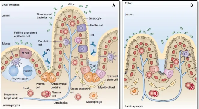
The epithelium of the small intestine consists of four different cell lines: cells that take up the villi (enterocytes, goblet cells and enteroendocrine) and differentiated cells that reside at the bottom of the crypts and secrete antimicrobial agents, Paneth cells and microfold cells (M-cells) [21, 26] (see Figure 2.2A). Crypts are tubular invagination of the intestinal epithelium being characterized by presenting in its base Paneth cells and stem cells, which continuously divide and are the source of all intestinal epithelial cells [26]. Already the villi are tiny finger-like projections, which increase the surface area by approximately 30-fold, being characterized by the presence of mature enterocytes, goblet cells and mucus secreting [27].
The human colon is very similar in its cellular organization and morphology to the small intestine as depicted in Figure 2.2B. The large intestine or colon begins in the right iliac region of the small intestine. Unlike the small intestine, the mucosa of the large intestine is not covered with villous projections but contains deep tubular pits, which increase in depth as they approach the rectum, as well as the muscularis propria and further increase the surface area for digestion and absorption by approximately 600-fold [21].
The epithelial cells of the colonic mucosa include enterocytes (absorptive cells), enteroendocrine cells, epithelial stem cells, goblet cell and microfold cells (M-cells) [21], nearly identical to cells in the small intestine.
All types of cells mentioned above have distinct functions:
a) Enteroendocrine cells: specialized endocrine cells of the gastrointestinal tract that differentiate from pluripotent stem cells with ability to coordinate the functioning of the intestine by specific hormone secretion;
b) Paneth cells: specialized cells that reside in the crypts and has a role in innate immunity through the secretion of proteins;
c) Goblet cells: produce and secrete mucins, which constitutes the mucus need for chemical and mechanical protection of the intestine;
7 Literature Review
d) Enterocytes: are columnar and highly polarized cells, and characterized by the presence of an apical brush border, which perform the around 80% of the absorption of nutrients through the epithelium [22, 28, 29];
e) M cells are specialized in transepithelial transport of macromolecules, particles and microorganisms.
Figure 2.2 – Schematic representation of the architecture of the small intestine (A) and the colon (B) as well as
all the cell types present in an intestinal villus. Adapted from [26].
2.2.2 Pathways through the intestinal epithelial barrier
Oral administration of therapeutic agents is the most common and preferred route of drug delivery for patients, in view of their ease of administration. However, drugs administered orally, as compared to those administered parenterally, are not directly available in the systemic circulation to make its therapeutic effect effective. Following oral administration of a drug, there are many barriers which the drug must pass to be absorbed by the intestine. Starting with the drastic pH changes at slightly acidic (pH 1.2-3.0) in the stomach to slightly alkaline (pH 6.5-8.0) in the end of the ileum proximal, lead to drastic changes of stability and solubility of the drug [27]. In the small intestine, which is the main site of absorption, the enzyme activity collects a number of proteolytic enzymes which may degrade the drug [27, 30]. In addition to the above parameters, molecular size, lipophilicity/hydrophilicity, site of action, efflux pathways and stability/solubility of the administered drug are others factors to be considered when considering administration of therapeutic oral agents.
Literature Review 8
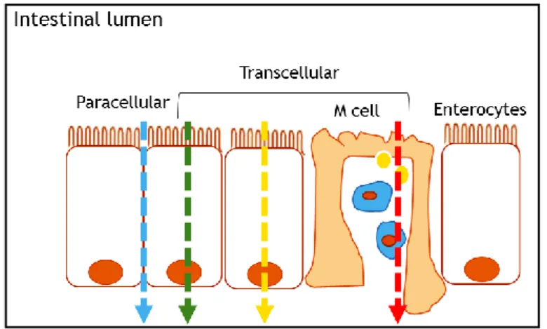
There are several absorption pathways in the intestinal epithelium such as by passive diffusion, which occurs through nonspecific permeability pathways (for small amphipathic molecules), by the paracellular route (to hydrophilic molecules of low molecular weight), specialized carriers to effect the facilitated absorption of molecules (sugars and amino acids) [27], among others (see Figure 2.3). Since larger molecules such as peptides, proteins and anticancer drugs can be absorbed by endocytosis [31].
Figure 2.3 – Schematic representation of the transport across the intestinal epithelium. Molecules can across the
intestinal epithelial barrier by paracellular transport (blue line) and by transcellular transport through passive diffusion (green line), transcytosis via normal enterocytes (yellow line) or via M-cells (red line).
Some reports demonstrated that particles with about 100-200 nm could be internalized by the receptor-mediated endocytosis, whereas large particles have to be phagocytized [32]. On the other hand, larger molecules must be transported by paracellular route, which corresponds to the passage of molecules across epithelial cells. Consequently, tight junctions (TJs) should open to occur this passage with biomaterials such as chitosan (CS) that have this ability [33]. Actually, the movement through the epithelium can occur the apical to basolateral side, which corresponds to the movement from the intestinal lumen to underlying tissue and circulation, and on the contrary, to the basolateral to apical side. The epithelium, reinforced by TJs and efflux transporters, exclude molecules larger than 200 Da via paracellular transport [34].
The TJs are an adhesion region between adjacent epithelium (endothelial cells) that regulates the paracellular flow. There are several proteins in this adherence area that are responsible for opening and closing it [26]. The combined role of P-glycoprotein (P-gp) which is an efflux pump family of adenosine triphosphate (ATP) binding cassette transporter and acts contrary to the absorption of drugs [35], with cytochrome P450 3A, an enzyme that metabolizes drugs has been recognized as one of the main determinants of intestinal wall absorption [36]. Still, even when administered a significant amount of drug and is carried to the intestinal epithelium, the first pass metabolism by the liver will reduce substantially the initial dose to a dose fraction which will pass, later, to the systemic circulation [37].
For these reason, the use of polymeric micelles as drug carriers is a promise to overcome these and other barriers and reach the target with less degradation and maximum quantities. The opening of the intestinal cell barrier, through a breakdown of TJs, can increase the absorption of drugs delivered orally.
9 Literature Review
2.2.3 Intestinal in vitro models
Since in vivo studies are complex and time consuming, gastrointestinal digestion and intestinal absorption in vitro models have been developed [22, 38, 39].
Caco-2 cells (Figure 2.4A), derived from human colon adenocarcinoma, are very used as model to mimic human intestinal epithelium and study intestinal absorption of drugs, as these cells suffer spontaneous enterocytic differentiation in culture and are polarized with well-established TJs [20]. Also, Caco-2 cells present apical brush border with abundant microvilli and enzymatic activity of digestive enzymes such as alkaline phosphatase (ALP) [22]. Since the permeation of drugs across monolayers of Caco-2 cells are close to the characteristics of permeation of human intestinal mucosa, this model has been suggested for predicting oral drug absorption in humans [20]. However, our group, started to use C2BBe1, a clone of Caco-2, as is described in even better mimic the enterocyte because the parental Caco-2 cells have an heterogeneous population not displaying always the brush border typical of the enterocytes [40].
Similarly, HT29 cell lines has its origin in human colorectal adenocarcinoma, and, after 21 culture can be differentiated into goblet cells, mimicking them [22]. Because of HT29 cells acquire a heterogeneous phenotype when cultured in modified medium [41], Lesuffleur established HT29-MTX cell line in a medium containing 10-6 M methotrexate (MTX) (see Figure 2.4B). This cell line grows in
a monolayer of polarized goblet cells that produce mucus, it has a discrete apical brush border with intestinal hydrolases and, contrasting with Caco-2 cells, do not express P-gp and have less TJs [41]. The main physiological function of mucus includes protecting the GI epithelium itself to be degraded by stomach acid and digested by gastric and intestinal enzymes.
Figure 2.4 – Morphological aspect of Caco-2 cells (A) and HT29-MTX cells (B) at a resolution of 20 µm. The images
were taken with Nikon TE2000-U microscope equipped with digital camera and controlled via the Nikon ACT-1 program.
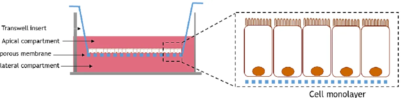
The Transwell® system comprises a well of a plate with an insert inside and represents a potential in vitro tool for investigating intestinal permeability lumen-to-blood drug, i.e., the permeability from apical compartment to the basolateral compartment (Figure 2.5). It is necessary to use human cell lines, preferably several lines of cells, with the goal of reproducing the intestinal heterogeneous population, mimicking better the human intestine [22]. The insert of this system includes a 10 mm thick membrane made of polyester or polycarbonate and is available with different pore sizes.
A B
Literature Review 10
Figure 2.5 – Schematic representation of a Transwell® that consist in a well plate plus insert. The epithelial cells
are seeded on the top of the membrane where the compounds are placed and the permeability occurs the apical to the basolateral compartment.
The integrity of each model can be easily monitored by measuring the transepithelial electrical resistance (TEER) which returns the absolute values of resistance per unit area [22]. The higher the value of TEER more TJs exist to modulate the transport of molecules by paracellular route.
2.2.3.1 Caco-2 model
Due to all the features described about the Caco-2 cell lines, the monoculture of these cells is the standard for the intestinal absorption in in vitro models.
As already mentioned, Caco-2 cells undergo spontaneous differentiation in the enterocyte, however, in the early stages of the culture (3-4 days), Caco-2 cells remain undifferentiated, showing only some apical microvilli [22]. Thus it is necessary to culture the cells as a monolayer for around 21 days to produce the differentiation into enterocytes, which are the main important cells in human intestine, able to form junctions between cells. Although they are derived from colon, they express most of the morphological and functional characteristics of the intestinal absorption cells, including enzymes typically expressed in enterocytes [22].
Some disadvantages are evident in this model, for example, no mucus production and no differentiation between cellular transport and intestinal metabolism [42], there is a high and variable P-gp expression and a low expression of metabolic enzymes [43], and the permeability of hydrophilic compounds is made by paracellular route, which makes this model less efficient because of TJs as compared with the human or animal intestine [42]. In this case, the Caco-2 model is used to investigate their utility in the study of passive absorption of drugs (Figure 2.6).
11 Literature Review
2.2.3.2 Caco-2/HT29-MTX model
To overcome some barriers of the model mentioned above, a new model was imposed. This model consists in a mixture of Caco-2 and HT29-MTX (mucus-producing) cell lines with the aim to generate more predictable results (see Figure 2.7).
Once HT29-MTX cells do not form TJs as tight as Caco-2 cells when cultured in monolayer, is given an increase in paracellular transport routes, making the model closer to the human intestine [44]. Also, the mucus production is important because it protects the organism against the entry of pathogens agents, however, acts as a barrier to absorption of drugs, affecting the retention time of the compound in the intestinal wall [45].
This co-culture model was studied by our group and it was found that the optimum ratio to obtain a closer mimic reality is 90% Caco-2 cells and 10% of HT29-MTX cells [38].
Figure 2.7 – Schematic representation of the Caco-2/HT29-MTX co-culture model setup.
2.2.3.3 Caco-2/HT29-MTX/Raji B model
A more realistic and complexed model is based on Caco-2 cells, HT29-MTX and Raji B, originally developed by our group [38, 44].
Raji B cells are derived from a human Burkitt's lymphoma, and when cultured with Caco-2 cells, these are induced to cell phenotype, resembling, functionally and morphologically to intestinal M-cells characterized by scarce and irregular microvilli [29] [38]. Since the presence of lymphocytes leads to the formation of cells that resemble intestinal M-cells in function and morphology, contributing to the transport of nanoparticles, and combined with the advantages of the co-culture model showed above, this model mimics even more by human intestinal epithelium.
Once M-cells are specialized in transepithelial transport of macromolecules, particles and microorganisms, this model is more complete compared to the models described above as will mimic more adequately the human intestine for development of new drugs administered orally also more suitable and closer to the in vivo model.
2.3 Anticancer drug model - Paclitaxel
Paclitaxel (PTX), a large antineoplastic agent isolated from the bark of Pacific Yew (Taxus brevifolia from family Taxaceae), is characterized for its potent anti-tumor capabilities against a broad spectrum of cancers, such as metastatic breast cancer, colon cancer, ovarian cancer, among others [46]. PTX is a white to off white crystalline powder, highly hydrophobic and insoluble in water
Literature Review 12
and has the molecular formula C47H51NO14 (Figure 2.8), which corresponds to a molecular weight (Mw)
of 853 Da.
Figure 2.8 – Chemical structure of paclitaxel. Adapted from [2].
Its mechanism of action is promoted by the polymerization of tubulin and hyper stabilization of microtubules in their polymerized form, leading to cell death [47, 48]. Microtubules are an integral component of eukaryotic cells and are involved in various cellular functions including mitosis, maintaining the form and movement of the cell, organelle transport between the cell interior and spindle formation during cell division. On the other hand, tubulin is a protein that form the microtubules. PTX alter the normal balance, forcing the polymerization of tubulin dimers leading to the formation of microtubules, making them stable by preventing depolymerisation and, therefore, making them dysfunctional [48]. Consequently, interrupts cell division, since the stabilization of microtubules interferes with the G2 and M phases of the cell cycle (and these cellular activities involves microtubules), causing cell death by disrupting the normal dynamics of the tubule needed for cell division and vital interphase process [2, 48] (see Figure 2.9).
Figure 2.9 – Schematic representation of cell cycle. Adapted from [49].
PTX is poorly soluble in aqueous medium, but can be dissolved in organic solvents such as dimethyl sulfoxide (DMSO) (50 mg/mL) and methanol (50 mg/mL) [50], and acetonitrile (15 mg/mL) and ethanol (40 mg/mL) [2]. This drug is rapidly destroyed in weakly alkaline medium, methanolic solutions and in strongly acidic methanolic solutions. For instance, a sample with 0.1% acetic acid added to methanol showed no signs of degradation for 7 days at room temperature (RT) or during 3 months at
13 Literature Review
4 ºC [51]. Due to the low solubility and poor permeability of PTX, it is classified under class IV of Biopharmaceutic Classification System (BCS) [52], which means being slightly permeable by the intestinal mucosa in addition to the poor solubility, as already discussed.
The commercial form of PTX, Taxol®, is designed in a vehicle consisting of Cremophor EL (polyethoxylated castor oil) and dehydrated ethanol (50:50, v/v) [53]. This form is clinically used after dilution in isotonic saline solution before intravenous administration (i.v.). However, a drastic number of side effects are reported as hypersensitivity, peripheral neuropathy, hypotension, neurotoxicity, nephrotoxicity [54], among others. Furthermore, i.v. may cause other problems such as infections and thrombosis. Therefore, several alternatives to the use of Cremophor EL-based vehicle have been explored to reduce these and other side effects and improve the therapeutic efficacy of PTX. One such alternative is polymeric micelles [55] to be administered orally as it is the most cost-effective treatment method due to less need for visits and remained in the hospital staff and for the preparation and administration of the treatment.
The half maximal inhibitory concentration (IC50) is a measure of the effectiveness of a substance in inhibiting a specific biological or biochemical function by half. The IC50 of PTX is in the range of 2.5-7.5 nM [56]. It is important to have knowledge of this value because it indicates the amount of PTX required to inhibit a particular biological process by half, in this case, to inhibit cell division. Clinically, the dose generally accepted is 200-250 mg/m2 [2].
Studies conducted in HeLa cells (from human cervical cancer origin) and fibroblasts using PTX (0.25-10 mM) showed that PTX blocked cells in the G2 and M phase of the cell cycle [57]. Already in human colorectal cell lines (HCT116) at PTX concentration of 3-10 nM, there was inhibition of mitosis leading to cell death [58].
In general, the PTX in vitro activity is greater than the activity of cisplatin, DOX, and 5-Fu against human tumors and equal to or less than docetaxel [46].
2.4 Polymeric micelles as drug delivery vehicles
Polymeric micelles are amphiphilic polymers that could be self-assembled into a core-shell nanostructure based on hydrophobic and hydrophilic segments with a range of diameters of 10-200 nm [59, 60]. This micellar system has the advantage of improve stability in the bloodstream, large capacity encapsulation, despite the reduced size, are not toxic and yet have a controlled release of the drug [61]. The time of drug circulation in the blood is higher [18, 19], providing better permeability to anticancer drugs by improving its delivery from the deep blood vessels to tumors [17]. Additionally, these carriers are typically accumulated in extensive vasculature sites, such as solid tumors and sites of inflammation, due to EPR effect [18, 19], as already mention above.
The fact that these systems are organized in core-shell type structures, make them selected for drug delivery applications. The stability of these processes is given by the decrease of the Gibbs free energy, which makes these systems thermodynamically more stable, this process is called self-assembly [62, 63]. For being an amphiphilic system, the hydrophobic moiety is protected from the aqueous medium, and therefore the hydrophilic part is in contact therewith (see Figure 2.10). The hydrophobic core provides space for the encapsulation of hydrophobic drugs such as PTX or DOX, and the hydrophilic shell allows the inclusion of hydrophobic drugs and it also provides stability, protecting from biological environment [64]. Taking in account these properties, polymeric micelles have become a promising carriers of hydrophobic drugs. They are not easily absorbed in the GI tract,
Literature Review 14
providing the reduction of the side effects and they are protected from environmental stimuli such as enzymes and gastric pH [16, 65].
Figure 2.10 – Schematic representation of drug encapsulation from self-assembled nanosystems.
Chemical modifications of polymers are the most efficient and well-known method to create self-assembled systems. These changes lead to the formation of amphiphilic polymer derivatives by the addition of hydrophobic groups, such as alkyl and acyl groups, to the hydrophilic polymer backbone. These target drug delivery systems are developed to prevent drug degradation, as well as their loss, to avoid the harmful side effects and to increase the bioavailability of the drug in the required site of interest [16].
If the external components of micelles are usually comprised by hydrophilic polymers as polyethyleneglicol (PEG) or chitosan (CS), to form the micellar core, it has been used various polyesters such as poly (L-lactide), poly(glycolic acid-lactide) and PCL [65]. The latter have advantages over others polyesters by always keeping elastic at RT, which makes it highly permeable [66, 67], and decomposes more slowly, being optimal for controlled release drugs [3].
There are two concepts that have to be explained about micelles: the first is the critical micelle concentration (CMC), which is the minimum concentration required for a given polymer to form micelles by self-assembly. The second is the critical aggregation concentration (CAC), which is the minimum concentration required for a given polymer to aggregate. Typically, the CMC values are higher than the CAC value [68]. The CMC of the polymer-based micelles depends on the hydrophobic character of the molecule [69] and a Mw of the blocks [70]. There are several methods to calculate the CMC, such as by conductivity measurements, interfacial tension or fluorescence spectroscopy [64]. However, in the case of polymer-based micelles, the CMC is very low, being the fluorescence spectroscopy the best choice because of its high sensitivity [64].
The hydrodynamic diameter of polymeric micelles is possible to be determined by dynamic light scattering (DLS) method [71] and the morphology of the micelles can be visualized by transmission electron microscope (TEM) and by scanning electron microscope (SEM) methods [60]. Differing the composition and size of the micelles of hydrophobic segments and hydrophilic of the materials that compose them, it is possible to regulate the properties of the micelles as the size and loading capacity [60].
2.5 Chitosan and amphiphilic derivatives
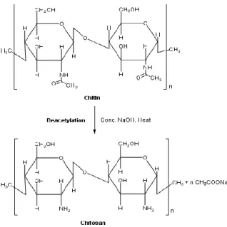
Polysaccharides are widely distributed in nature and, among them, CS is receiving increasing attention. CS is a linear polysaccharide prepared from chitin, which is the second most abundant natural polymer in the world, after cellulose [72, 73]. Chitin can be found in the exoskeleton of
Hydrophilic polymer Hydrophobic group (graft) Drug
15 Literature Review
insects, crustaceans and some cell wall of fungi. CS, derivatived from chitin, can be obtained by alkaline deacetylation or enzymatic hydrolysis [73]. The reaction product is CS only when the average degree of acetylation (DA), is equal to or less than 50%. The DA is used to characterize the average content of N-acetyl-D-glucosamine units (acetylated unit) while degree of deacetylation (DD) is the number of D-glucosamine units (deacetylated unit) [7, 72]. Thus, CS is constituted by N-acetyl-D -glucosamine (GlcNAc units) and of D-glucosamine units (GlcN units), linked by glycosidic linkages β-(1,4) (Figure 2.11) [6, 72-74].
This polymer shows solubility in moderately acidic medium. Its solubility in acidic medium occurs due the protonation of the NH2 functional group on the C-2 position of the D-glucosamine repeating
unit [72, 74, 75].
CS has one amino group and two hydroxyl groups in the repeating glycosidic residue, providing reactive sites for a variety of reactions, which is an advantage over other polymers [76]. Several types of CS can be obtained and with different physicochemical properties such solubility, pKa and viscosity,
which depend on parameters like DD and Mw [73, 77, 78].
Figure 2.11 – Deacetylation of chitin to chitosan.
The DD and the Mw are two of the most important physicochemical properties of this polymer. An increase on the DD, leads to an increase of its viscosity, due to high and low deacetylated CSs having different conformations in aqueous solutions [79]. Additionally, the CS solution viscosity can be also affected by factors as temperature and concentration [79]. For example, when CS concentration increases or the temperature of the solution decreases, the viscosity increases.
CS is a polycation whose charge density depends on DA and pH. Due to the progressive amine protonation of pendant groups, the solubility is pH dependent. Therefore, CS is only soluble at pH values of approximately 6 or less [6, 75, 80]. This feature turns CS into biopharmaceutical of interest, along with its properties of mucoadhesiveness and ability to open epithelial TJs [33]. In fact, at low pH (<6), CS amine groups are protonated and become positively charged, leading to polycationic behavior [7, 81]. By contrast, at a higher pH value (above around 6), CS amines are deprontonated and reactive (Figure 2.12).
Literature Review 16
Figure 2.12 - The soluble–insoluble transition of chitosan occurs at about pH 6.
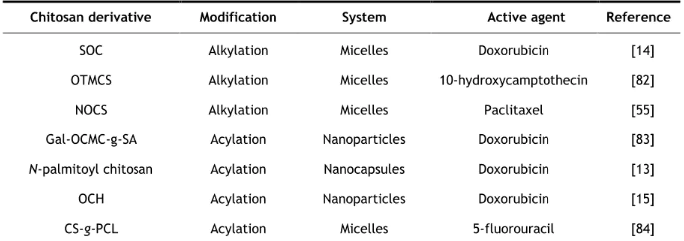
To improve CS solubility in physiological media, its biological properties and expand its potential applications, CS derivatives have been synthesized [13-15, 55, 82-84]. Indeed, macromolecules with amphiphilic character have become a subject of great interest over the last years.
2.5.1 Biological properties of chitosan
CS is a polymer of great interest in pharmacy, medicine and tissue engineering due to its biological activities such as non-toxicity [85-88], antimicrobial activity [72, 73, 89-92], biocompatibility [7, 93, 94], biodegradability [7, 95-97], mucoadhesiveness and ability to open, temporarily, the tight junctions of the epithelium [94, 98, 99], thereby facilitating the permeation of drugs [6, 100-103]. These properties may double the therapeutic effects making CS a polymer with pronounced potential for biomedical and biopharmaceutical applications, especially for drug delivery.
The presence of NH2 groups in CS is the reason why it exhibits much higher potential as compared
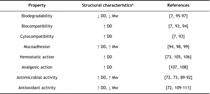
with chitin [7] and what makes it an excellent polymer for pharmaceutical applications. Also, as shown in Table 2.1, it is possible understand the CS biological properties and the relationship between its structural characteristics (DD and Mw). For instance, the lower the DD and Mw, the higher the CS biodegradability [7, 95-97]. Moreover, CS plays a role in hemostasis, but independently of the coagulation cascade [96]. Okamoto and colleagues showed that CS reduces clotting time in a dose-dependently manner, with the ability to aggregate platelets [104].
Table 2.1 — Biological and structural properties of chitosan.
Property Structural characteristicsa References
Biodegradability ↓ DD, ↓ Mw [7, 95-97] Biocompatibility ↑ DD [7, 93, 94] Cytocompatibility ↑ DD [7, 93] Mucoadhesion ↑ DD, ↑ Mw [94, 98, 99] Hemostatic action ↑ DD [73, 105, 106] Analgesic action ↑ DD [107, 108] Antimicrobial activity ↑ DD, ↑ Mw [72, 73, 89-92] Antioxidant activity ↑ DD, ↓ Mw [72, 109-111]
17 Literature Review
The properties of CS mentioned above make the CS an exciting and promising excipient for the pharmaceutical industry, with a large margin for development. However, CS amphiphilic derivatives have, indeed, attracted the particular attention among researchers working in biomedical engineering, and particularly, in drug delivery, given the fact that they improve physical and chemical properties of CS when used alone [77].
2.5.2 Chitosan amphiphilic derivatives
A large number of CS amphiphilic derivatives have been reported in the literature over the past years. CS amphiphilic derivatives result from the attachment of hydrophobic structures to the hydrophilic CS backbone due to the presence of hydroxyl and amine groups, and can be achieved by using different methodologies such as alkylation [112-115] and acylation [62, 116-123] . Generally, the chemical derivatization of CS can be regioselective due to the presence of reactive sites C2-amine, C3-hydroxyl and C6-hydroxyl. The properties of products of the regioselective reactions are strongly influenced by the distribution of substituents groups along the polymer chain and, therefore, it is possible to obtain N-substituted, O-substituted or N,O-substituted CS derivatives. In general, the reaction products of an amphiphilic CS are, among others, N-alkyl chitosan, acyl-chitosan and graft derivatives. Interestingly, the introduction of a hydrophobic molecule on CS backbone modifies its rheological behavior, being therefore used as rheological modifiers in pharmaceutical industries [124]. However, such substitutions may be controlled over the reaction conditions applied [68, 113], as is shown here.
2.5.2.1 Alkylation
The alkylation is one of the most frequent modifications of CS. Alkylation reactions occur through the grafting of alkyl chains in the structure of the CS. In other words, alkyl CS is obtained by the introduction of alkyl groups on the amine groups of CS by reductive amination of CS [125]. Generally, this substitution reaction is carried out in heterogeneous conditions and owing to the semi-crystalline character of CS, amorphous regions will be more accessible than ordered ones, giving rise to substituted products displaying block distribution patterns.
The hydrophobic character of alkylated CS is dependent on the length of the chains, i.e., the longer the alkyl chains, the higher the polymer hydrophobicity [126]. Additionally, the higher the average degree of substitution (DS), the more hydrophobic will be the modified CS.
Desbrieres et al., [125], developed a procedure to obtain alkylated derivatives of CS from its swollen structure and modified after precipitation by neutralization, which improves the accessibility to the reactive sites. Generally, aldehydes and ketones are employed as alkylating agents and the Schiff bases resulting from reaction with CS are converted to N-alkyl CS derivatives by reduction with sodium borohydride (NaBH4) or sodium cyanoborohydride (NaBH3CN) (Figure 2.13), the reaction
efficiency depending on the choice of the reducing agent [68]. Indeed, NaBH3CN is commonly used
Literature Review 18
Figure 2.13 - Reaction scheme to obtain N-alkyl derivatives of chitosan.
CS modifications can dramatically alter its properties, therefore, it is necessary to characterize the properties and medical safety of these new derivatives [127]. Indeed, the presence of hydrophobic interactions between the alkyl chains improves the properties of modified CSs: the C12 is the minimum length of the alkyl chain for the hydrophobic behavior be effective; the solutions of such CS derivatives are usually non-Newtonian, mainly when the length of alkyl chains increases; also de C12 alkyl chain length proved to be more efficient to improve film formation and mechanical properties; the efficiency of this modification increases with increasing DS [126].
Apart from their properties, alkyl chitosan derivatives have been widely used in drug delivery. In Section 2.5.3, examples are given of this modification.
2.5.2.2 Acylation
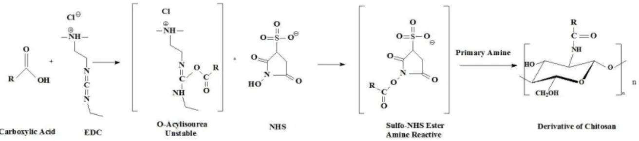
The addition of hydrophobic groups is also carried out by acylation reactions. Acylated CS was proposed as a drug carrier for drug delivery, essentially due to its hydrophobic association that promotes self-assembly of nanoparticles or micelles. The advantage of the acylation of CS over its alkylation occurs because the acylation allows the introduction of new groups in sites C2-amine, C3-hydroxyl and C6-C3-hydroxyl [68]. In the case of O-acylation, the presence of the ester bond in the structure of the resulting CS derivative permits its degradation by the action of lipase enzymes, making it a biodegradable polymer [68].
The acylation of CS makes possible its solubilization in organic solvents due to the introduction of hydrophobic groups in the polymer. However, the solubility of acylated CS in water depends on the acyl chain length and on DS [68, 128]. This means that short acyl chains and relatively low DS result in water soluble CS derivatives but increasing the DS will dramatically decrease the water solubility.
2.5.2.2.1 N-acylation
The N-acylation of CS may be achieved by activation of carboxylic acids through reaction with carbodiimides as well as by using reactive carboxylic acid derivatives, such as anhydrides and acyl chlorides.