Trabalho final de mestrado:
Treatment of Myeloma Cast Nephropathy
associated Acute Kidney Injury
Terapêutica da Lesão Renal Aguda associada
à Nefropatia por Cilindros em Mieloma
Múltiplo
Clínica Universitária Medicina II
Aluno: Tomás Laia McGuire, nº 12877
Orientador: Dr. João Madeira Lopes
CONTENTS
Abstract/Resumo ... 4
Introduction ... 5
Epidemiology ... 5
Etiology ... 5
Molecular basis of disease ... 6
Pathophysiology of Cast Nephropathy ... 7
The M component ... 7
Light chain metabolism ... 8
Diagnosis ... 9
Prophylaxis ... 10
Treatment strategy ... 10
General measures ... 11
Myeloma chemotherapy – decreasing FLC production ... 12
Extracorporeal FLC removal ... 13
Discussion ... 17
Summary ... 20
4
ABSTRACT/RESUMO
Cast nephropathy, also known as Myeloma Kidney, is a frequent complication of Multiple Myeloma. It results from the interaction of excess free immunoglobulin light chains (FLC) with Tamm-Horsfall protein in the distal tubule and thick ascending limb of the loop of Henle, forming dense, mucous, casts, which cause tubular obstruction and rupture, resulting in local inflammation and fibrosis. The kidney injury is irreversible if appropriate therapy is not rapidly instituted.
A prompt, sustained reduction of serum FLC concentration leads to improved renal recovery rates. However, the optimal therapeutic strategy to achieve this reduction is yet to be determined. While the role for chemotherapy and correction of precipitating factors is clear, the extracorporeal removal of FLCs is being investigated, with the use of high cut-off hemodialysis membranes particularly promising.
A Nefropatia de Cilindros, também conhecida por Rim de Mieloma, é uma complicação frequente em doentes com Mieloma Múltiplo. Resulta da interação de cadeias leves de imunoglobulinas livres (FLCs) com a proteína de Tamm-Horsfall no túbulo contornado distal e ramo ascendente da ansa de Henle, formando cilindros densos e mucosos que causam obstrução e rutura tubular, que resulta em inflamação local e fibrose. A lesão renal é irreversível se a terapêutica apropriada não for rapidamente instituída.
Uma diminuição mantida da produção de FLCs traduz-se em melhores taxas de recuperação da função renal. Contudo, a estratégia terapêutica óptima para conseguir aquela redução ainda não foi estabelecida. Enquanto que o papel da quimioterapia e da correção dos factores precipitantes é claro, a remoção mecânica das FLCs está a ser investigada, sendo particularmente promissor o uso de membranas de hemodiálise de elevado “cut-off”.
INTRODUCTION
Multiple Myeloma (MM) is a disease defined by the proliferation of malignant plasmatocytes in the bone marrow, tumor-produced monoclonal protein in the blood or urine and associated organ dysfunction.1 The clinical presentation is typically dominated by the CRAB signs and symptoms, standing for hyperCalcemia, Renal failure, Anemia and Bone lesions (pain or fractures), but may also include susceptibility to infections, and, more rarely, neurological symptoms, clotting abnormalities and features of hyperviscosity.
EPIDEMIOLOGY
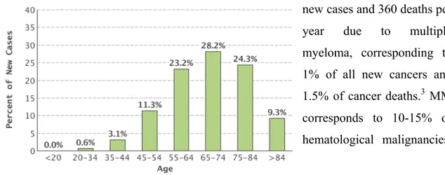
A rare cancer, MM is responsible for slightly less than 1% of all new cancer cases worldwide. Incidence rates are highest in developed countries in North America, Europe and Australia/New Zealand, where its incidence can be almost 5 per 100 000 persons. Incidence is low in Asia, except western Asia.2 In Portugal, there are approximately 520 new cases and 360 deaths per year due to multiple myeloma, corresponding to 1% of all new cancers and 1.5% of cancer deaths.3 MM corresponds to 10-15% of hematological malignancies.
It is slightly more common in males than females, with a worldwide incidence ratio of 1.4/1. In the USA, it affects people of African-American descent approximately twice as much as whites, while people of Asian origin have a much lower incidence.4
The median age at diagnosis is approximately 70 years. Despite differences in incidence, response to therapy and prognosis are similar across different ethnicities.
ETIOLOGY
The etiology of MM is poorly understood, in part due to its low incidence. The presence of a first degree relative (but not second, or third-degree relatives) has shown an increased risk of developing multiple myeloma. Obesity and acquired immune
6
deficiency syndrome (AIDS) are possible risk factors, as well as chronic exposure to ionizing radiation. Alcohol and tobacco consumption have shown inconsistent associations with risk of MM. Fish and seafood consumption appear to be protective factors. Additionally, a hormonal component is presumed, due to the higher incidence in males.5
MOLECULAR BASIS OF DISEASE
Multiple myeloma results from the abnormal malignant proliferation of plasma cells derived from a single post-germinal-centre B-cell clone. While it is often diagnosed de
novo, most studies point to an evolution from the premalignant condition Monoclonal
Gammopathy of Undetermined Significance, or MGUS.6 This condition is present in 3-4% of the general population over 50 years of age, increases in prevalence with age, and carries a risk of progression to MM of approximately 1% per year.7
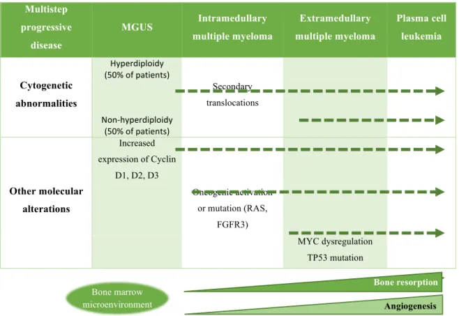
Multistep progressive disease MGUS Intramedullary multiple myeloma Extramedullary multiple myeloma Plasma cell leukemia Cytogenetic abnormalities Hyperdiploidy (50% of patients) translocations Secondary Non-hyperdiploidy (50% of patients) Other molecular alterations Increased expression of Cyclin D1, D2, D3 Oncogenic activation or mutation (RAS, FGFR3) MYC dysregulation TP53 mutation Bone resorption Angiogenesis Bone marrow microenvironment
Figure 2 - Multistep pathogenesis of Multiple Myeloma. Both MGUS (Monoclonal gammopathy of undetermined significance) and Multiple Myeloma share early chromosomal abnormalities, typically involving immunoglobulin heavy chain translocations or trisomies. Secondary translocations are quite rare in MGUS; activation of oncogenes such as RAS and FGFR3 and dysregulation of MYC and TP53 determine drug resistance and tumor progression. Besides the molecular changes in neoplastic plasmatocytes, changes of bone microenvironment, specifically resorption and aberrant angiogenesis are features of disease progression. 86
PATHOPHYSIOLOGY OF CAST NEPHROPATHY
The M component
The presence of a monoclonal protein is a defining feature of MM, presenting in almost all patients. While only 82% of patients will have a corresponding peak or band in serum protein electrophoresis (SPEP), 97% of patients will have a detectable M-component if immunofixation of serum or urine is performed in addition to SPEP.8
However, up to 3% of patients will not have any detectable monoclonal protein in the blood or urine,8 traditionally defined as non-secretory MM.9 Approximately 60-70% of these patients will, however, have an altered kappa/lambda ratio in a serum Free Light Chain (sFLC) assay.10–12
The remainder will have true non-secretory myeloma, with 85% having evidence of cytoplasmic immunoglobulin on bone marrow immunofixation of plasmatocytes, but a defect in its secretion, and 15% will have non-producer myeloma.13 As immunoglobulins and free light chains are nephrotoxic, patients with non-secretory myeloma have no risk of Ig-mediated renal lesion.
A review of 1027 patients revealed the following M-component distribution: 52% IgG, 21% IgA, 2% IgD, 0,5% IgM, 2% are
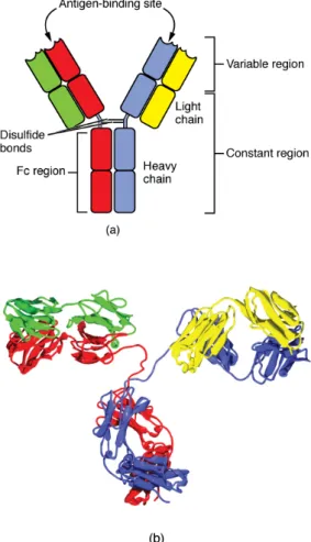
Figure 4 – The typical four chain structure of a generic antibody (a) and the corresponding three-dimensional structure of the antibody IgG2 (b). The basic unit of an immunoglobulin is built of two identical heavy and two identical light chains, arranged in a symmetrical Y-shape. Human Igs can belong to one of 5 classes (isotypes), IgA, IgD, IgE, IgG and IgM, which correspond respectively to alpha, delta, epsilon, gamma or mu class heavy chains. The light chains can be kappa or lambda. Both light and heavy chains have a variable region, corresponding to the antigen-binding site and a constant, structural region. Light chains have a molecular weight of approximately 25 kDa. 88
Figure 3 – Typical tracing of serum protein electrophoresis of a multiple myeloma patient, with M-component present in the gamma region. Less often, it is in the beta or alpha-2. even more rarely, a biclonal peak can be seen.87
8
Bi-clonal and 16% contains only light chain, with kappa being twice as common as the lambda isotype.
Light chain metabolism
During normal Ig synthesis, light chains (LCs) are produced in slight excess to allow the correct assembly of the immunoglobulin.14 Up to 500-1000 mg of polyclonal Free Light Chains (FLC) produced each day by the immune system, released into circulation, freely filtered through the glomerulus and efficiently endocytosed by the epithelial cells of the proximal tubule, with less than 10 mg of polyclonal FLCs excreted in the urine each day.15
In MM, there is an overproduction of FLCs, such that they exceed the proximal tubule’s catabolic capacity and are excreted in large quantities, classically described as Bence-Jones proteinuria, after Henry Bence Bence-Jones, the physician who first described the protein in 1847.16 In the distal tubule, the FLCs interact with Tamm-Horsfall protein (THP), an 80 kDa glycoprotein of uncertain function secreted by cells in the thick ascending limb of the loop of Henle. A 9 amino acid sequence in THP binds to the complementarity determining region 3 (CDR3) of the FLC.17 As this is a variable region, different FLCs have varying nephrotoxic potential.
The binding of FLCs to THP creates dense mucous casts, leading to tubular obstruction and rupture, resulting in subsequent interstitial inflammation, fibrosis, which define Cast Nephropathy (CN). Additionally, increased FLC concentration, decreased flow through the nephron (as in hypovolemia), acidic pH, loop diuretics promote FLC aggregation and lead to cast formation in animal models.18,19 Radiocontrast media, hypercalcemia and nephrotoxic drugs (such as
non-steroidal
anti-inflammatory drugs and
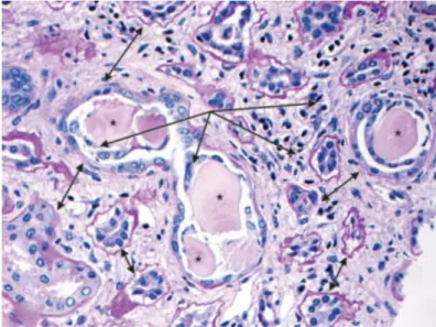
Figure 5- light microscopy of a kidney with cast nephropathy stained with PAS, with characteristic features of CN marked. * - casts, PAS negative. Some are fractured. The single-headed arrows mark inflammatory infiltrate in the kidney interstitium and surrounding the casts. The double headed arros mark an increase in distance between the tubules, due to edema (acute) or tubular atrophy (chronic).89
angiotensin-converting enzyme inhibitors) have also been implicated.18,20,21 In addition to cast formation, CN can present with proximal tubule damage. Some light chains are resistant to lysosomal degradation and accumulate in the proximal tubule cells.18,22
If severe enough, the histological damage resulting from these processes will translate clinically to overt acute kidney injury (AKI).
DIAGNOSIS
Up to 25-50% of patients with MM will develop mild renal impairment during the
course of the disease. AKI is frequently the inaugural manifestation, with up to 10% of patients dialysis-dependent at presentation.8,23 Renal insufficiency in MM has been defined by the International Myeloma Working Group as a creatinine clearance < 40 mL/min as calculated by the CKD-EPI or MDRD equations or serum creatinine > 2 mg/dL resulting from the disease.24 However, these equations are only validated in patients with stable creatinine levels, and the use of the AKIN or RIFLE criteria is recommended, with the caveat that they have not been studied in detail in MM patients.25
Regarding the etiology of renal impairment, cast nephropathy is the most common etiology, responsible for 33 to over 60% of renal impairment in some series.26 Monoclonal immunoglobulin deposition disease and AL amyloidosis are the other major causes. However, a significant minority of patients may have renal impairment not related to MM, such as diabetic nephropathy or hypertensive arteriosclerosis.27 Definitive diagnosis can be made by renal biopsy. However, it can often be distinguished from other
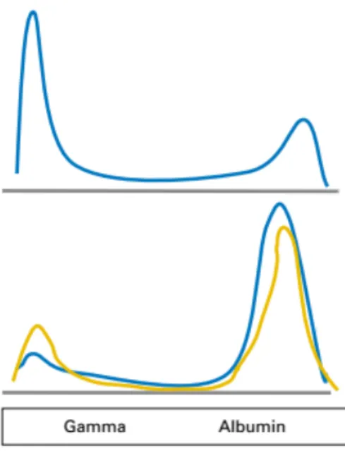
Figure 6 – diagram of urine electrophoresis. The top figure corresponds to a selective proteinuria typical in cast nephropathy, while the bottom depicts the non-selective or predominantly albumin proteinuria of amyloidosis, monoclonal immunoglobulin deposition disease, and other unrelated entities.
List 1 – Kidney diseases in multiple myeloma classified by site of injury 90,91
Glomerular
• Primary amyloidosis (AL or AH) • Monoclonal immunoglobulin deposition disease (light chain, heavy chain or both) • Miscellaneous (cryoglobulinemia, proliferative glomerulonephritis)
Tubular
• Cast nephropathy • Distal tubular dysfunction • Acquired Fanconi syndrome
Interstitial
• Plasma cell infiltration • Interstitial nephritis
Other causes
• Hyperuricemia • Hypercalcemia
10
causes noninvasively. The quantification and electrophoresis of a 24-hour urine sample can determine whether the proteinuria is selective for light chains, non-selective or albumin-rich. Monoclonal Immunoglobulin Deposition Disease (MIDD) and amyloidosis typically present with nephrotic syndrome (albumin preponderance). Therefore, in patients with high sFLCs (> 500 – 1500 mg/L) and significant Bence-Jones proteinuria, a renal biopsy can often be discarded if there are no confounding factors. If the patient presents with nonselective proteinuria, significant albuminuria and low sFLC (< 500 mg/L) a renal biopsy will most likely be necessary to distinguish between the multiple entities.25 A biopsy of subcutaneous fat positive for Congo-red may diagnose amyloidosis.
PROPHYLAXIS
Various strategies have been employed to reduce the risk of developing CN in high-risk patients: vigorous hydration, avoidance of loop diuretics and alkalinization of urine. The latter has its physiologic basis in the manipulation of electric charges on FLCs to modulate their affinity for THP. The isoelectric point (iP) of FLCs is 5.1. By increasing the pH in the distal tubule above the iP, the FLCs would lose positive charges and have a reduced binding with the anionic THP.18,19 However, there is no evidence as to its effectiveness either in preventing CN in humans or aiding in its reversal in overt CN.18,28
In order to specifically target pathological interaction of FLCs with THP, a cyclized peptide was synthesized which inhibited the binding of FLCs to THP in vitro and prevented the development of CN in an animal model.29 By simulating the CDR3 region of the FLCs and binding to the Light-Chain Binding Domain on THP,30 it was effective in preventing CN when co-administered with nephrotoxic light chains in mice, even when given 4 hours after the nephrotoxic light chains. While an innovative proof-of-concept, only one of each type of light chain, kappa and lambda, was evaluated and similar trials are yet to be attempted in humans.
TREATMENT STRATEGY
As the development of acute kidney injury in CN is multifactorial, management should contain both general measures to correct precipitating and aggravating factors such as hypercalcemia and volume depletion as well as therapy targeted at reducing serum FLC
concentration. This includes chemotherapy to decrease production, as well as extracorporeal removal of the FLCs.
General measures
The principal objective of therapy is to decrease the concentration of light chains in the distal tubules and avoid their precipitation, to avoid further kidney injury. This can be achieved by correction of volume depletion, hypercalcemia, hyperuricemia and avoidance of nephrotoxic drugs (nonsteroidal inflammatory drugs, radiocontrast dye, angiotensin-converting enzyme inhibitors, angiotensin-II receptor antagonists).25
Typically, isotonic IV fluids are used for rehydration, with a bolus of 500-1000 mL and further administration at a rate of 100-200 mL/hour until the patient is euvolemic, with particular care to avoid volume overload in patients with congestive heart failure (CHF). Once the patient is euvolemic, administration should be titrated to a urine output of 100 mL/hour.31 Furosemide was considered a mainstay of treatment by forcing diuresis, but this approach is not supported by evidence and enhances cast formation, so should only be considered in patients with CHF and clear signs of fluid overload.32
Hypercalcemia leads to renal vasoconstriction and enhances cast formation.18 It normally courses with volume depletion, and should be treated accordingly.33 IV Bisphosphonates are a mainstay of severe hypercalcemia management. Both zoledronate (3-4 mg over 15-30 min) and pamidronate (60-90 mg over 2-6 hours) have been used in hypercalcemia combined with AKI.34–36 Dosage should be adjusted for renal function. Zoledronate is the more effective agent but has been associated with greater worsening of kidney function than pamidronate, so the latter may be preferred. Importantly, both may cause collapsing focal segmental glomerulosclerosis and acute tubular necrosis.37,38
Bisphosphonates can take up to 48-72 hours to have effect on calcium levels. Therefore, for severe hypercalcemia, calcitonin can be used. The subcutaneous or intramuscular administration of 4-8 units/kg every 6-12 hours can reduce serum calcium levels by 2 mg/dL for up to 72 hours, limited by the development of tachyphylaxis, with no adjustment for renal function required.39
The alkalinization of urine is often attempted in many treatment centers. This treatment is not supported by evidence.28
12
Myeloma chemotherapy
The key to effectively treating cast nephropathy is the prompt and significant decrease in FLC concentration. The current treatment paradigm is a bortezomib-based regimen, in conjunction with high dose corticosteroids in the first
month and immunomodulatory drugs such as thalidomide and lenalidomide. While the specifics of myeloma chemotherapy go beyond the scope of this thesis, an overview of the current therapeutic options will be described.
Bortezomib is effective at rapidly lowering FLC concentration and may be renoprotective by its effects on the NF-κB pathway in proximal tubule cells. It is not metabolized by the kidney and therefore does not require dose adjustment for renal impairment. Multiple studies have confirmed its benefits in renal response and time to renal response, dialysis independence and mortality. As such, it is considered a cornerstone of CN and MM therapy, comparing favorably to cytotoxic chemotherapy, lenalidomide and thalidomide based regimens.25,40 The subcutaneous administration appears to have similar results as intravenous, and leads to less adverse effects.41,42 Other proteasome inhibitors are undergoing investigation, and have not proven to be superior to bortezomib. Both carfilzomib and ixazomib (the first oral proteasome inhibitor) have been studied in relapsed multiple myeloma and can be administered in patients with a creatinine clearance ≥ 30 mL/min.43,44
High dose steroids (typically administered as 3 pulses of 40 mg dexamethasone given 4 days on, 4 days off for a 28-day cycle)45,46 given during the first month of therapy have been associated with a more rapid renal response, in comparison with low dose steroids, (1.6 to 46 months) even in treatment regimens that included bortezomib and lenalidomide or thalidomide.47
The immunomodulatory drugs currently in use, lenalidomide and thalidomide, have been shown to be safe and effective in MM associated renal impairment. While thalidomide does not require dose adjustment, lenalidomide has renal excretion and requires dosing modifications. Pomalidomide is ongoing study for use in moderate to
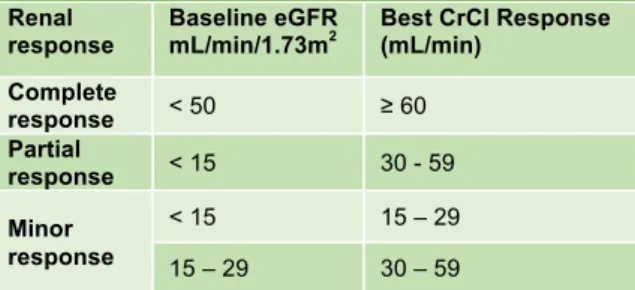
Table 3. Criteria for the definition of renal response to antimyeloma therapy
Renal
response Baseline eGFR mL/min/1.73m2 Best CrCl Response (mL/min)
Complete response < 50 ≥ 60 Partial response < 15 30 - 59 Minor response < 15 15 – 29 15 – 29 30 – 59
Abbreviations: CrCl, creatinine clearance; eGFR,
estimated glomerular filtration rate based on the CKD-EPI or MDRD formula
severe renal impairment, having shown to be safe and effective in patients with GFR ≥ 45 mL/min.25
Renal impairment does not preclude autologous stem cell transplantation (ASCT), and is the treatment of choice in newly diagnosed MM patients eligible. An adjustment of the melphalan dose is required but appears to be equally effective.48 ASCT in patients with renal impairment at the time of the procedure is associated with a greater mortality.48–50
Extracorporeal FLC removal
The FLCs have a molecular weight of approximately 25 kDa, and therefore their removal depends on either plasma exchange, or renal replacement therapy using membranes with pores of sufficient caliber: Hemofiltration or High Cut-Off hemodialysis.
Importantly, these therapies depend on concomitant chemotherapy in order to cause a decrease in production of the FLCs.
Plasma exchange
The therapeutic exchange of plasma has been used experimentally in acute kidney injury associated with multiple myeloma since at least 1978 with conflicting results.51 However, only 4 randomized clinical trials (RCT) have been done to address the issue of whether plasmapheresis adds any benefit to chemotherapy in MM-associated kidney injury.
The oldest one included 29 patients, 16 of which had biopsy proven cast nephropathy. The control group (n=14, 11 dialysis-dependent) was offered chemotherapy with methylprednisolone and cyclophosphamide, as well as peritoneal dialysis if needed. The study group (n=15, 13 dialysis-dependent) was offered plasma exchange and hemodialysis as necessary in addition to identical chemotherapy. With 5-7 daily sessions, 13/15 (87%) patients in the trial group recovered renal function with serum creatinine levels decreasing to below 2.5 mg/dL, compared to only 2 patients (14%) becoming dialysis-independent in the control group. Survival at 1 year was improved in the plasmapheresis group (66 vs 28%).52
A study by Johnson (n=21) forced diuresis with furosemide and chemotherapy with or without plasma exchange (n=11 and n=10, respectively) and found no significant
14
difference in overall survival or recovery of renal function; however, the plasmapheresis group had a much faster decrease of serum M-protein.53
The largest trial with published results included 97 patients with acute renal impairment due to MM. 58 received 5-7 plasma exchanges and chemotherapy with either melphalan-prednisolone or vincristine-adriamycin-dexamethasone and 39 received only chemotherapy. After 6 months, there was no statistical difference between groups on the composite outcome of death, dialysis dependence or GFR < 30 mL/min (58% study vs 69% control).54
Finally, the MERIT trial, having enrolled only 77 of the planned 286 patients, closed without publishing results.55 However, results presented at a meeting failed to show any benefit of plasmapheresis over conventional chemotherapy.56
High cut-off hemodialysis
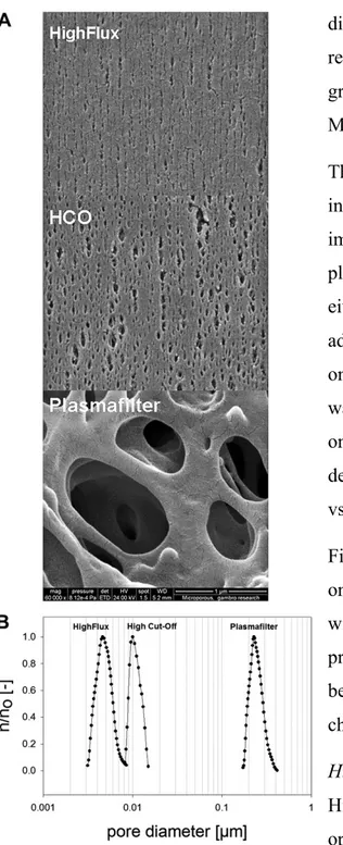
High cut-off hemodialysis (HCO-HD) operates on the same principles as conventional dialysis, but uses a membrane with larger pores (typically around 0.008 to 0.01 µm, compared to 0.003-0.006 µm for high-flux dialysis membranes). Molecules up to 50-60 kDa are filtered effectively, but due to lack of uniformity in pore size, the membranes will allow some loss of slightly larger molecules, such as albumin (66.5 kDa)57. Kappa FLC monomers and lambda FLCs dimers, with molecular weights of 22.5 and 45 kDa respectively, are effectively filtered by HCO-hemodialysis in vitro and
in vivo.58
Figure 7 – A: scanning electron microscopy of high-flux, high cut-off and plasmafilter membrane surfaces. B: respective membrane pore size distribution.57
Several case reports and case series have been published, adding HCO-HD to chemotherapy and overall showing its effectiveness to quickly reduce FLCs in addition to chemotherapy. The largest published study to date enrolled 67 patients (biopsy-confirmed CN in 37 of the 38 biopsied patients), and offered HCO-HD combined with chemotherapy (bortezomib-based in 58%). 63% of patients became dialysis independent. Interestingly, a significant association was found between dialysis independence and both initiation of dialysis within 7 days of renal failure and sustained FLC reduction by days 12 and 21.59 Another study prospectively evaluated 21 patients with CN-associated kidney injury (15/21 biopsy-confirmed CN, with highly probable CN in the remainder),60 which were treated with HCO-HD and either bortezomib-dexamethasone-thalidomide and hematopoietic stem-cell transplantation (HSCT) if eligible or bortezomib-melphalan-prednisone in not eligible for HSCT. Of the 21 patients, 16 became dialysis independent, with sFLC decreases of > 90%. The 3 year overall survival was 67%, with no early deaths observed.60 Many other case series report similar effectiveness for HCO-HD in renal response.61–64
Two RCTs have been registered to study the efficacy of HCO-HD in combination with bortezomib-based chemotherapy: Studies in Patients With Multiple Myeloma and Renal Failure Due to Myeloma Cast Nephropathy (MYRE)65 and the European Trial of Free Light Chain Removal by Extended Haemodialysis in Cast Nephropathy (EuLITE).66 The first, MYRE, recruited 284 patients and aims to compare bortezomib-dexamethasone with cyclophosphamide + bortezomib-bortezomib-dexamethasone in biopsy proven CN patients not requiring HD. In dialysis-dependent patients, the trial will add either HCO-HD or conventional high-flux HD to chemotherapy BD. The trial is expected to end in September 2017. The second, EuLITE, ended in but its results have yet to be published. In the study, 84 patients were randomized to either high-flux HD or HCO-HD and offered a bortezomib-doxorubicin-dexamethasone chemotherapy regimen. The outcomes of both studies include renal response as well as overall survival.
Other techniques
Dialysis is a mainstay of acute kidney injury therapy and should be used for the usual indications of kidney failure: such as uremia and refractive hyperkalemia. Due to the small pore size, FLCs do not diffuse in meaningful quantity across conventional dialysis membranes and are therefore not effective in FLC removal. There are reports of FLC removal by adsorption onto synthetic polymethylmethacrylate dialysis membranes both
16
in vitro58 and in vivo67,68. These techniques are limited by membrane saturation and therefore require either replacing the membrane during renal replacement therapy or adding a second one in series.
Most research regarding high cut-off membranes has been applied to hemodialysis. However, the same membranes can be used for hemodiafiltration (HDF).
A single center prospective cohort study compared hemodiafiltration using a heat sterilized high-flux propylene membrane dialyzer with hemodialysis and hemodiafiltration using a HCO membrane, in addition to chemotherapy (bortezomib-based in 75% of patients) in a group of 16 patients with confirmed or probable CN. The population included new-onset and relapsed MM. The tracked outcomes were FLC reduction ≥ 65%, albumin loss and renal recovery. Of the 10 patients that survived, 9 were successfully weaned of dialysis. While there was a statistically significant difference in lambda FLC reduction between hemodiafiltration with HCO and conventional HF-dialyzer – 78 to 61% respectively – both HDF and HCO-HDF were effective in reducing kappa FLC in approximately 70% of patients. Albumin losses were also much larger with usage of HCO dialyzers, with 21 g/session if HD was performed and 63 g/session with HDF. RRT with the conventional dialyzer resulted in a loss of only 7 g/session. Importantly, there was no significant difference in renal outcome.69
Another novel approach is hemodiafiltration with endogenous reinfusion of ultrafiltrate (SUPRA-HFR) which combines the convection and diffusion of HDF with adsorption. A dual chamber dialyzer is used with a first convective stage with a cut-off of 42 kDa and a second, diffusive, stage. The ultrafiltrate from the convective stage is circulated through a resin cartridge with high affinity for FLCs and reinfused before the second stage, thus allowing the adsorption of FLCs and other high protein-bound toxins. This technique has been applied to CN-associated kidney injury with two published case series, one with 3 and another with 4 patients. In the first,70 one patient became dialysis independent, and two did not recover kidney-function. In the second study,71 3 out of 4 patients achieved reductions of sFLC concentration over 65% and became dialysis independent. Of note, none of the patients studied had any significant albumin loss.
DISCUSSION
Myeloma cast nephropathy is the most frequent cause of irreversible renal failure in multiple myeloma and associated with a markedly worse prognosis if renal impairment persists.23 Studies have shown that a quick decrease in sFLC concentration was associated with recovery of renal function72,73, with one study showing that a 60% or greater reduction by 21 days of therapy was associated with renal recovery in 80% of patients.74
The role of effective chemotherapy decreasing production of FLCs and therefore reducing their concentration is undisputed. However, in renal impairment the FLCs are excreted at a reduced rate, leading to their accumulation and perpetuating the end-organ damage75. This suggests that extracorporeal removal by mechanical means could be an important adjuvant therapy to allow for kidney recovery. The co-precipitating factors of cast nephropathy, such as hypercalcemia, dehydration, nephrotoxic drugs and contrast agents can lead to overt renal failure in patients with underlying disease. In these cases, symptomatic treatment is warranted.
The affinity of each FLC’s CDR3 for THP is the major determinant in their co-precipitation and cast formation. Increased FLC concentration in the distal tubule, as can occur in dehydration and hypercalcemia, favors this interaction, as do a low pH and increased sodium concentration due to loop diuretics. Supportive care aims to create an environment less favorable to cast formation. Volume resuscitation, targeting a urine output of 3 L/day, and correction of severe hypercalcemia both with bisphosphonates is recommended25. Special care should be taken with pamidronate and zoledronate, as these are contraindicated in patients with eGFR < 30 mL/min. Denosumab is not metabolized by the kidney and can be used in persistent hypercalcemia, but can lead to dangerous hypocalcemia. Dialysis is not effective at removing FLCs, but should be offered for the usual indications. Urine alkalinization does not appear to improve outcomes despite being commonly used.
Bortezomib-based chemotherapy, particularly with dexamethasone and thalidomide (VDT), is the current standard-of-care in patients with new-onset MM and CN76. Specifically, the proteasome inhibitor has been shown to rapidly reduce tumor burden and FLC production, and in combination with dexamethasone in the first month77 improves the likelihood of some recovery of renal function. The further addition of
18
immunomodulatory drugs, such as thalidomide or lenalidomide, appears to be beneficial. Other proteasome inhibitors may be of benefit in relapsed or refractory myeloma.
Renal impairment does not exclude ASCT or the therapy required49 and patients stand to recover renal function, particularly when offered bortezomib.78 However, patients with renal impairment have significantly increased transplant-related complications and mortality (mortality of 4%, compared to < 1%).48–50
Because of the delay between institution of chemotherapy and the decrease of sFLC concentration, there is a physiological basis for the mechanical removal of FLCs as they can potentially reduce the FLC burden immediately.
Plasmapheresis has been used experimentally for decades with mixed reports of effectiveness. Of the 4 RCTs performed, only in one did the study group perform significantly better than control.52 The limitations may be due to limited number of patients, disparate outcomes or simply outdated methodologies: the trials were performed before the widespread availability of bortezomib-based chemotherapy and sFLC concentration assays. A case series combining novel agents and plasmapheresis in patients with high-probability or biopsy-proven CN showed a significant decrease in sFLC concentration and partial or better renal response in 12 of 14 patients.79 However, the limitations may instead lie with the technique: plasma exchange sessions are typically short (2 hours or less), resulting in limited clearance of the extravascular compartment. As much as 85% of molecules of similar size are retained in the extravascular56,58,80, and any benefits might be the result of a good response to chemotherapy. Another issue with the technique is the removal of essential proteins such as albumin, intact immunoglobulins and clotting factors. As such, the only undisputed indication for plasma exchange in MM is hyperviscosity syndrome.81
High cut-off hemodialysis overcomes many of the limitations of plasmapheresis: the membranes used have a cut-off of 50-60 kDa, which is similar to the glomerulus. However, due to some lack of pore uniformity, molecules such as albumin are also removed, particularly if convection is applied, albeit at a lower rate than the FLCs, inflammatory cytokines and myoglobin they were designed to remove. While the multiple case series and reports published attest to the effectiveness of HCO-HD in combination with chemotherapy – particularly using novel agents – the results from the
two ongoing clinical trials should clarify whether there is a benefit to HCO-HD beyond that of the chemotherapy. As of now, the data is encouraging: partial renal responses or better were achieved in over 60% of patients treated,82,60,62,59,61 and FLC reduction of 60-70%.83 There are cases described in which HCO-HD was not effective in clearing FLCs due to their aggregation in polymers larger than the membrane cut-off, a limitation which had not been previously considered.84 Effectiveness notwithstanding, the use of HCO dialyzers is significantly more expensive than other renal replacement therapies. A prospective cost-effectiveness study has been published claiming it would result in cost savings due to greater life expectancy and savings by avoiding chronic hemodialysis, with the caveat of the authors’ significant ties to the dialysis equipment manufacturer.85
While most of recent research has focused on HCO-HD, when hemodiafiltration is used, conventional HD membranes can be permeable to FLCs. They may be more effective clearing kappa chains, which more frequently present as monomers.69 The addition of adsorptive resins to a HDF system allows the reinfusion of the ultrafiltrate cleared from FLCs, resulting in an albumin sparing alternative.70,71 These techniques hold promise but require further investigation to clarify a potential role and benefits compared to HCO-HD.
20
SUMMARY
• Multiple myeloma is a rare hematological cancer caused by a malignant proliferation of plasmatocytes.
• Cast nephropathy is a frequent complication of multiple myeloma and can often be the inaugural presentation of the disease. It is caused by the precipitation of immunoglobulin free light chains with Tamm-Horsfall glycoprotein in the distal tubule and ascending limb of the loop of Henle.
• The risk of developing CN is related to the FLC burden, their affinity to THP, volume depletion, hypercalcemia, tubular pH, loop diuretics and nephrotoxic drugs.
• Supportive therapy for CN consists in correction of the precipitating factors: administration of fluids, bisphosphonates for severe hypercalcemia.
• Bortezomib based-chemotherapy is the current standard of care for MM treatment in patients with CN. Adding dexamethasone to initial therapy, but not maintenance therapy, increases the likelihood of renal recovery. Whichever the treatment regimen chosen, it should be instituted as quickly as possible to increase the likelihood of recovery of renal function.
• The extracorporeal removal of FLCs is a promising area of investigation but conclusive benefits have yet to be proven:
o Plasmapheresis has been studied the longest, and has so far not shown to increase survival or renal recovery. However, most of the studies published were performed before the availability of FLC quantification and novel agents such as bortezomib so some experts advocate its usage. o Hemodialysis using high cut-off membranes appears to be extremely
effective in rapidly decreasing FLC burden in small case series, and two multicentric RCTs are ongoing to evaluate its role. It can be prohibitively costly and causes significant albumin loss.
o Hemodiafiltration may be an effective alternative to HCO-HD, and FLC adsorption may have a role to play. Its usage has been very limited up to now, but may increase as it is significantly less expensive and causes less albumin loss than HCO-HD.
o Importantly, the mechanical removal of FLCs alone is not sufficient; chemotherapy to decrease tumor burden and FLC production is required
to improve outcomes. Patients not responding to chemotherapy probably do not stand to gain from continued FLC removal.
• ASCT can be performed in patients with kidney failure, and is associated with increased overall survival and renal recovery. However, patients with renal impairment at transplantation have increased transplant-associated mortality.
22
REFERENCES
1. Singhal, S. & Mehta, J. Multiple Myeloma. October 351, 802–810 (2006).
2. Ferlay, J. et al. Cancer incidence and mortality worldwide: Sources, methods and major patterns in GLOBOCAN 2012. Int. J. Cancer 136, E359–E386 (2015). 3. Ferlay, J. et al. Cancer incidence and mortality patterns in Europe: Estimates for
40 countries in 2012. Eur. J. Cancer 49, 1374–1403 (2013).
4. Howlader, N. et al. SEER Cancer Statistics Review, 1975-2011. National Cancer
Institute (National Cancer Institute, 2014).
5. Caini, S. et al. Food of animal origin and risk of non-Hodgkin lymphoma and multiple myeloma: A review of the literature and meta-analysis. Crit. Rev. Oncol.
Hematol. 100, 16–24 (2016).
6. Kyle, R. A. et al. A long-term study of prognosis in monoclonal gammopathy of undetermined significance. N. Engl. J. Med. 346, 564–569 (2002).
7. Wadhera, R. K. & Rajkumar, S. V. Prevalence of monoclonal gammopathy of undetermined significance: a systematic review. Mayo Clin. Proc. 85, 933–42 (2010).
8. Kyle, R. a et al. Review of 1027 patients with newly diagnosed multiple myeloma. Mayo Clin. Proc. 78, 21–33 (2003).
9. Swerdlow, S. H. et al. WHO Classification of Tumours of Haematopoietic and
Lymphoid Tissues. World Health Organization Calssification of Tumours of Haematopoietic and Lymphoid Tissue 4th, (International Agency for Research on
Cancer, 2008).
10. Drayson, M. et al. Serum free light-chain measurements for identifying and monitoring patients with nonsecretory multiple myeloma. Blood 97, 2900–2902 (2001).
11. Katzmann, J. a et al. Elimination of the need for urine studies in the screening algorithm for monoclonal gammopathies by using serum immunofixation and free light chain assays. Mayo Clin. Proc. 81, 1575–8 (2006).
Diagnostic performance of quantitative ?? and ?? free light chain assays in clinical practice. Clin. Chem. 51, 878–881 (2005).
13. Smith, D. B., Harris, M., Gowland, E., Chang, J. & Scarffe, J. H. Non-secretory multiple myeloma: a report of 13 cases with a review of the literature. Hematol.
Oncol. 4, 307–13 (1986).
14. Maack, T. Renal handling of low molecular weight proteins. Am. J. Med. 58, 57– 64 (1975).
15. Berggård, I. & Peterson, P. A. Polymeric Forms of Free Normal κ and λ Chains of Human Immunoglobulin. J. Biol. Chem. 244, 4299–4307 (1969).
16. Jones, H. B. On A New Substance Ocurring In The Urine Of A Patient With Mollities Ossium. Philos. Trans. R. Soc. London 95, 55–62 (1848).
17. Huang, Z. Q. & Sanders, P. W. Localization of a single binding site for immunoglobulin light chains on human Tamm-Horsfall glycoprotein. J. Clin.
Invest. 99, 732–736 (1997).
18. Sanders, P. W. & Booker, B. B. Pathobiology of cast nephropathy from human Bence Jones proteins. J. Clin. Invest. 89, 630–639 (1992).
19. Holland, M. D., Galla, J. H., Sanders, P. W. & Luke, R. G. Effect of urinary pH and diatrizoate on Bence Jones protein nephrotoxicity in the rat. Kidney Int. 27, 46–50 (1985).
20. Mussap, M. & Merlini, G. Pathogenesis of renal failure in multiple myeloma: Any role of contrast media? Biomed Res. Int. 2014, (2014).
21. Smolens, P., Barnes, J. L. & Kreisberg, R. Hypercalcemia can potentiate the nephrotoxicity of Bence Jones proteins. J. Lab. Clin. Med. 110, 460–5 (1987). 22. Leboulleux, M. et al. Protease resistance and binding of Ig light chains in
myeloma-associated tubulopathies. Kidney Int. 48, 72–79 (1995).
23. Bladé, J. et al. Renal failure in multiple myeloma: presenting features and predictors of outcome in 94 patients from a single institution. Arch. Intern. Med.
158, 1889–1893 (1998).
24
the diagnosis of multiple myeloma. Lancet Oncol. 15, e538–e548 (2014).
25. Dimopoulos, M. A. et al. International Myeloma Working Group Recommendations for the Diagnosis and Management of Myeloma-Related Renal Impairment. J. Clin. Oncol. 34, 1544–1557 (2016).
26. Ivanyi, B. Frequency of light chain deposition nephropathy relative to renal amyloidosis and Bence Jones cast nephropathy in a necropsy study of patients with myeloma. Arch. Pathol. Lab. Med. 114, 986–987 (1990).
27. Nasr, S. H. et al. Clinicopathologic correlations in multiple myeloma: A case series of 190 patients with kidney biopsies. Am. J. Kidney Dis. 59, 786–794 (2012).
28. Med, B., Ed, C. R. & Permissions, T. R. Analysis and management of renal failure in fourth MRC myelomatosis trial. MRC working party on leukaemia in adults. Br. Med. J. (Clin. Res. Ed). 288, 1411–1416 (1984).
29. Ying, W. Z., Allen, C. E., Curtis, L. M., Aaron, K. J. & Sanders, P. W. Mechanism and prevention of acute kidney injury from cast nephropathy in a rodent model. J. Clin. Invest. 122, 1777–1785 (2012).
30. Ying, W. Z. & Sanders, P. W. Mapping the binding domain of immunoglobulin light chains for Tamm-Horsfall protein. Am. J. Pathol. 158, 1859–1866 (2001). 31. Pi, J. et al. A review in the treatment of oncologic emergencies. J. Oncol. Pharm.
Pract. (2015). doi:10.1177/1078155215605661
32. Practice, C., Legrand, S. B., Leskuski, D. & Zama, I. Annals of Internal Medicine Review Narrative Review : Furosemide for Hypercalcemia : An Unproven yet.
Ann. Intern. Med. 149, 259–264 (2008).
33. Glezerman, I. G. & Sternlicht, H. Hypercalcemia of malignancy and new treatment options. Ther. Clin. Risk Manag. 1779 (2015). doi:10.2147/TCRM.S83681
34. Major, P. et al. Zoledronic acid is superior to pamidronate in the treatment of hypercalcemia of malignancy: A pooled analysis of two randomized, controlled clinical trials. J. Clin. Oncol. 19, 558–567 (2001).
35. Pecherstorfer, M., Steinhauer, E. U., Rizzoli, R., Wetterwald, M. & Bergstr??m, B. Efficacy and safety of ibandronate in the treatment of hypercalcemia of malignancy: A randomized multicentric comparison to pamidronate. Support.
Care Cancer 11, 539–547 (2003).
36. Machado, C. E. & Flombaum, C. D. Safety of pamidronate in patients with renal failure and hypercalcemia. Clin Nephrol 45, 175–179 (1996).
37. Munier, A. et al. Zoledronic acid and renal toxicity: Data from French adverse effect reporting database. Ann. Pharmacother. 39, 1194–1197 (2005).
38. Weide, R. et al. Renal toxicity in patients with multiple myeloma receiving zoledronic acid vs. ibandronate: a retrospective medical records review. J Cancer
Res Ther 6, 31–35 (2010).
39. Vaughn, C. B. & Vaitkevicius, V. K. The effects of calcitonin in hypercalcemia in patients with malignancy. Cancer 34, 1268–1271 (1974).
40. Palumbo, A. et al. International Myeloma Working Group consensus statement for the management, treatment, and supportive care of patients with myeloma not eligible for standard autologous stem-cell transplantation. J. Clin. Oncol. 32, 587–600 (2014).
41. Moreau, P. et al. Subcutaneous versus intravenous administration of bortezomib in patients with relapsed multiple myeloma: A randomised, phase 3, non-inferiority study. Lancet Oncol. 12, 431–440 (2011).
42. Merz, M. et al. Subcutaneous versus intravenous bortezomib in two different induction therapies for newly diagnosed multiple myeloma: An interim analysis from the prospective GMMG-MM5 trial. Haematologica 100, 964–969 (2015). 43. Badros, a Z. et al. Carfilzomib in multiple myeloma patients with renal
impairment: pharmacokinetics and safety. Leukemia 27, 1707–14 (2013).
44. Philippe Moreau, MD et al. Paper: Ixazomib, an Investigational Oral Proteasome Inhibitor (PI), in Combination with Lenalidomide and Dexamethasone (IRd), Significantly Extends Progression-Free Survival (PFS) for Patients (Pts) with Relapsed and/or Refractory Multiple Myeloma (RRMM): The Phase 3 Tourmaline-MM1 Study (NCT01564537). Available at:
26
https://ash.confex.com/ash/2015/webprogram/Paper79829.html. (Accessed: 2nd May 2016)
45. Kastritis, E. et al. Reversibility of renal failure in newly diagnosed multiple myeloma patients treated with high dose dexamethasone-containing regimens and the impact of novel agents. Haematologica 92, 546–549 (2007).
46. Darda Bayraktar, U., Warsch, S. & Pereira, D. High-dose glucocorticoids improve renal failure reversibility in patients with newly diagnosed multiple myeloma. American Journal of Hematology 86, 224–227 (2011).
47. Dimopoulos, M. a et al. The role of novel agents on the reversibility of renal impairment in newly diagnosed symptomatic patients with multiple myeloma.
Leukemia 27, 423–429 (2013).
48. Lee, C.-K. et al. Dialysis-dependent renal failure in patients with myeloma can be reversed by high-dose myeloablative therapy and autotransplant. Bone
Marrow Transplant. 33, 823–8 (2004).
49. San Miguel, J. F. et al. Are myeloma patients with renal failure candidates for autologous stem cell transplantation? Hematol. J. 1, 28–36 (2000).
50. Parikh, G. C. et al. Autologous hematopoietic stem cell transplantation may reverse renal failure in patients with multiple myeloma. Biol. Blood Marrow
Transplant. 15, 812–6 (2009).
51. Isbister, J. P., Biggs, J. C. & Penny, R. Experience with large volume plasmapheresis in malignant paraproteinaemia and immune disorders. Aust. N. Z.
J. Med. 8, 154–164 (1978).
52. Zucchelli, P., Pasquali, S., Cagnoli, L. & Ferrari, G. Controlled plasma exchange trial in acute renal failure due to multiple myeloma. Kidney Int. 33, 1175–1180 (1988).
53. Johnson, W. J., Kyle, R. A., Pineda, A. A., O’Brien, P. C. & Holley, K. E. Treatment of renal failure associated with multiple myeloma. Plasmapheresis, hemodialysis, and chemotherapy. Arch Intern Med 150, 863–869 (1990).
54. Clark, W. F. Plasma Exchange When Myeloma Presents as Acute Renal Failure.
55. Gaskin, G. MyEloma Renal Impairment Trial: adjunctive plasma exchange in patients with newly diagnosed multiple myeloma and acute renal failure (MERIT): NCT00416897; ISRCTN37161699. Available at: https://clinicaltrials.gov/ct2/show/NCT00416897. (Accessed: 21st April 2016) 56. Cockwell, P. & Cook, M. The Rationale and Evidence Base for the Direct
Removal of Serum-Free Light Chains in the Management of Myeloma Kidney.
Adv. Chronic Kidney Dis. 19, 324–332 (2012).
57. Gondouin, B. & Hutchison, C. A. High Cut-off Dialysis Membranes: Current Uses and Future Potential. Adv. Chronic Kidney Dis. 18, 180–187 (2011).
58. Hutchison, C. a et al. Efficient removal of immunoglobulin free light chains by hemodialysis for multiple myeloma: in vitro and in vivo studies. J. Am. Soc.
Nephrol. 18, 886–895 (2007).
59. Hutchison, C. A. et al. Immunoglobulin free light chain levels and recovery from myeloma kidney on treatment with chemotherapy and high cut-off haemodialysis. Nephrol. Dial. Transplant. 27, 3823–3828 (2012).
60. Zannetti, B. A. et al. Bortezomib-based therapy combined with high cut-off hemodialysis is highly effective in newly diagnosed multiple myeloma patients with severe renal impairment. Am. J. Hematol. 90, 647–652 (2015).
61. Borrego-Hinojosa, J. et al. Treatment by long haemodialysis sessions with high cut-off filters in myeloma cast nephropathy: our experience. Nefrología 33, 515– 23 (2013).
62. Sinisalo, M., Silvennoinen, R. & Wirta, O. High cut-off hemodialysis and bortezomib-based therapy to rescue kidneys in myeloma-dependent cast nephropathy. Am. J. Hematol. 87, 640 (2012).
63. Bachmann, U. et al. Combination of bortezomib-based chemotherapy and extracorporeal free light chain removal for treating cast nephropathy in multiple myeloma. Clin. Kidney J. 1, 106–108 (2008).
64. Khalafallah, A. A. et al. Early application of high cut-off haemodialysis for de-novo myeloma nephropathy is associated with long-term dialysis-independency and renal recovery. Mediterr. J. Hematol. Infect. Dis. 5, 2013007 (2013).
28
65. Fermand, J.-P. & Bridoux, F. Treatment of Renal Failure Due to Myeloma Cast Nephropathy: Comparison of Two Different Chemotherapy Regimens and Evaluation of Optimized Removal of Monoclonal Immunoglobulin Light Chains Using a High Permeability Hemodialysis Membrane: NCT01208818; P081226. Available at: https://clinicaltrials.gov/ct2/show/NCT01208818. (Accessed: 25th April 2016)
66. Hutchison, C. a et al. European trial of free light chain removal by extended haemodialysis in cast nephropathy (EuLITE): A randomised control trial. Trials
9, 55 (2008).
67. Santoro, A., Grazia, M. & Mancini, E. The double polymethylmethacrylate filter (DELETE system) in the removal of light chains in chronic dialysis patients with multiple myeloma. Blood Purif. 35 Suppl 2, 5–13 (2013).
68. Fabbrini, P. et al. Polymethylmethacrylate Membrane and Serum Free Light Chain Removal: Enhancing Adsorption Properties. Blood Purif. 35, 52–58 (2013).
69. Rousseau-Gagnon, M., Agharazii, M., De Serres, S. A. & Desmeules, S. Effectiveness of haemodiafiltration with heat sterilized high-flux polyphenylene HF dialyzer in reducing free light chains in patients with myeloma cast nephropathy. PLoS One 10, e0140463 (2015).
70. Pendón-Ruiz de Mier, M. V. et al. Effectiveness of haemodiafiltration with ultrafiltrate regeneration in the reduction of light chains in multiple myeloma with renal failure. Nefrología 33, 788–96 (2013).
71. Pasquali, S. et al. A novel option for reducing free light chains in myeloma kidney: supra-hemodiafiltration with endogenous reinfusion (HFR). Journal of
Nephrology 28, 251–254 (2015).
72. Leung, N. et al. Improvement of cast nephropathy with plasma exchange depends on the diagnosis and on reduction of serum free light chains. Kidney Int. 73, 1282–1288 (2008).
73. Haynes, R. J., Read, S., Collins, G. P., Darby, S. C. & Winearls, C. G. Presentation and survival of patients with severe acute kidney injury and multiple myeloma: a 20-year experience from a single centre. Nephrol. Dial. Transplant
25, 419–26 (2010).
74. Hutchison, C. A. et al. Early Reduction of Serum-Free Light Chains Associates with Renal Recovery in Myeloma Kidney. J. Am. Soc. Nephrol. 22, 1129–1136 (2011).
75. Davids, M. S., Murali, M. R. & Kuter, D. J. Serum free light chain analysis. Am.
J. Hematol. 85, 787–790 (2010).
76. Dimopoulos, M. A. et al. Renal impairment in patients with multiple myeloma: A consensus statement on behalf of the International Myeloma Working Group. J.
Clin. Oncol. 28, 4976–4984 (2010).
77. Harrison, S. J. et al. The addition of dexamethasone to bortezomib for patients with relapsed multiple myeloma improves outcome but ongoing maintenance therapy has minimal benefit. Am. J. Hematol. 90, E86–E91 (2015).
78. Breitkreutz, I. et al. Bortezomib improves outcome after SCT in multiple myeloma patients with end-stage renal failure. Bone Marrow Transpl. 49, 1371– 1375 (2014).
79. Burnette, B. L., Leung, N. & Rajkumar, S. V. Renal improvement in myeloma with bortezomib plus plasma exchange. N. Engl. J. Med. 364, 2365–2366 (2011). 80. Cserti, C., Haspel, R., Stowell, C. & Dzik, W. Light-chain removal by
plasmapheresis in myeloma-associated renal failure. Transfusion 47, 511–514 (2007).
81. Chapdelaine, I. & Madore, F. Plasmapheresis in myeloma cast nephropathy. Clin.
Nephrol. 79, 72–77 (2013).
82. Heyne, N. et al. Extracorporeal light chain elimination: High cut-off (HCO) hemodialysis parallel to chemotherapy allows for a high proportion of renal recovery in multiple myeloma patients with dialysis-dependent acute kidney injury. Ann. Hematol. 91, 729–735 (2012).
83. Hutchison, C. A. et al. Serum free-light chain removal by high cutoff hemodialysis: Optimizing removal and supportive care. Artif. Organs 32, 910– 917 (2008).
30
84. Harding, S. et al. Aggregated serum free light chains may prevent adequate removal by high cut-off haemodialysis. Nephrol. Dial. Transplant. 26, 1438– 1440 (2011).
85. Grima, D. T., Airia, P., Attard, C. & Hutchison, C. a. Modelled cost-effectiveness of high cut-off haemodialysis compared to standard haemodialysis in the management of myeloma kidney. Curr. Med. Res. Opin. 27, 383–391 (2011). 86. Tuddenham;, H. A. V. D. C. E. Gda. R. G. Multiple Myeloma. N. Engl. J. Med.
364, 577–598 (2011).
87. Peter, B. Myeloma Protein. 60 (1982).
88. OpenStax. The Adaptive Immune Response: B-lymphocytes and Antibodies.
OpenStax CNX. (2013). Available at:
http://cnx.org/contents/AY5_7yUs@4/The-Adaptive-Immune-Response-B. (Accessed: 4th April 2016)
89. Leung, N. Treating myeloma cast nephropathy without treating myeloma. J. Clin.
Invest. 122, 1605–1608 (2012).
90. Sakhuja, V. et al. Renal involvement in multiple myeloma: a 10-year study. Ren.
Fail. 22, 465–477 (2000).
91. Heher, E. C., Rennke, H. G., Laubach, J. P. & Richardson, P. G. Kidney disease and multiple myeloma. Cjasn 8, 2007–17 (2013).