Effects of Voluntary Physical Activity and Endurance
Training on Cardiac Mitochondrial Function of Rats
Sub-Chronically Treated with Doxorubicin
Dissertation submitted to the Faculty of Sports, University of Porto to obtain the 2nd cycle in Physical Activity for Elderly, under the decree-law no. 74/2006 of 24 March
Supervisors. Professor Doutor António Ascensão Professor Doutor José Magalhães
Diogo Nuno Mariani Felix
Mariani, D. (2013). Effects of Voluntary Physical Activity and Endurance Training on Cardiac Mitochondrial Function of Rats Sub-Chronically Treated with Doxorubicin. Porto: D. Mariani. Master thesis presented to the Faculty of Sport, University of Porto.
KEY-WORDS: EXERCISE; HEART; BIOENERGETICS; MITOCHONDRIAL
F
UNDINGThe present work was supported by a research grant from the FCT (PTDC/DTP/DES/1071/2012 – FCOMP-01-0124-FEDER-028617) and from IJUP (PP_IJUP2011 253) to António Ascensão; from Research Centre In Physical Activity, Health And Leisure (CIAFEL) I&D UNIT (PEST-OE/SAU/UI0617/2011).
D
EDICATÓRIAA
GRADECIMENTOSAgora que finalizo o mestrado, gostaria de expressar os meus profundos e sinceros agradecimentos:
Aos Professores José Magalhães e António Ascensão, pela disponibilidade sempre demonstrada na orientação deste estudo, pelos sábios ensinamentos que me transmitiram, pelo incentivo, por estarem sempre presentes, pela paciência, amabilidade e pelos momentos de carinho e amizade sempre demonstrados.
Ao Centro de Investigação em Atividade Física, Saúde e Lazer (CIAFEL) por todo o apoio e colaboração prestados na realização deste estudo.
A todo o grupo de trabalho em especial à Inês, à Estela e à Mané, que me acompanharam em todos os momentos do protocolo experimental deste estudo, pela disponibilidade, pelo incentivo e pela amizade. Inês, obrigada por todas as explicações, pela paciência e por estares sempre disponível!
E a todas as pessoas e amigos que direta ou indiretamente contribuíram para a concretização deste trabalho.
ix
Table of Contents
1. Introduction ... 1
2. State of art ... 3
2.1- Age effects on cardiac function ... 3
2.1.1 Aging effects on cardiac mitochondria function ... 8
2.2. Exercise and cardioprotection ... 16
2.2.1 Exercise and cardiac mitochondrial adaptations ... 18
2.2.1.1. Morphological and biochemical adaptations ... 18
2.2.1.2. Mitochondrial biogenesis ... 20
2.2.1.3. Oxidative stress and antioxidant capacity ... 21
2.2.1.4. Cell death pathways ... 24
2.3. Doxorubicin: therapeutic agent vs. cardiotoxicity ... 25
2.3.1 Cardiac mitochondrial toxicity induced by DOX ... 27
2.3.1.1 Morphological evidences ... 28
2.3.1.2 Increased oxidative stress ... 29
2.3.1.3 Increased susceptibility to apoptosis ... 30
2.4. Exercise as a therapeutic and preventive strategy against DOX-induced cardiotoxicity. ... 31
2.4.1 Acute exercise ... 31
2.4.2 Chronic exercise ... 32
3. Aim ... 37
4. Materials and methods ... 39
4.1 Reagents ... 39
4.2 Animals ... 39
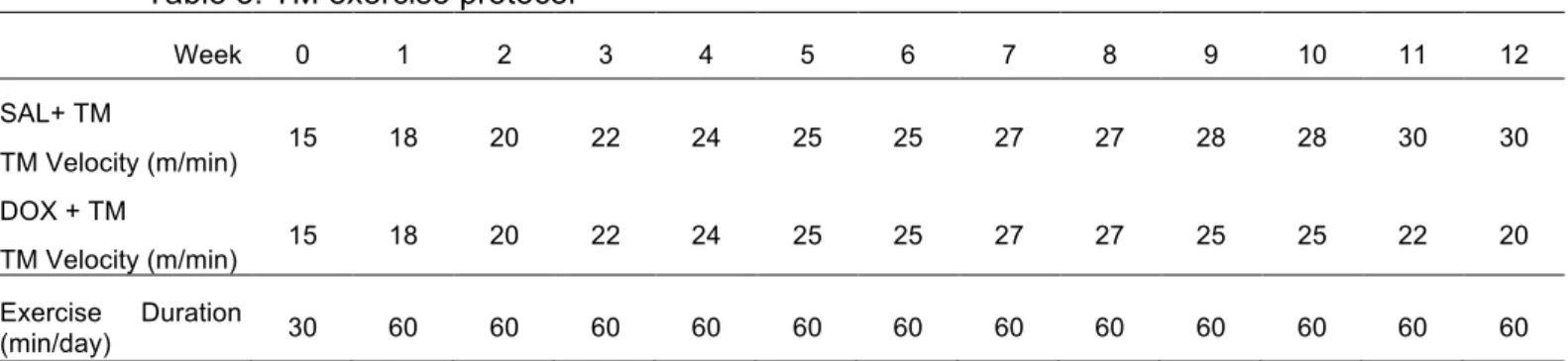
4.3 Exercise protocols ... 40
x
4.3.2 Voluntary physical activity ... 41
4.4 Doxorubicin treatment ... 41
4.5 Animal sacrifice, heart and soleus extraction ... 41
4.6 Isolation of heart mitochondria ... 42
4.7 Mitochondrial respiratory activity ... 43
4.8 Mitochondrial transmembrane electric potential ... 43
4.9 Mitochondrial osmotic swelling during MPTP induction ... 44
4.10 Mitochondrial oxidative damage ... 44
4.11 Soleus citrate synthase activity ... 45
4.12 Statistical analysis ... 45
5. Results ... 47
5.1. Characterization of animals and exercise protocols ... 47
5.2 Heart mitochondrial oxygen consumption ... 50
5.3 Heart mitochondrial transmembrane electric potential ... 51
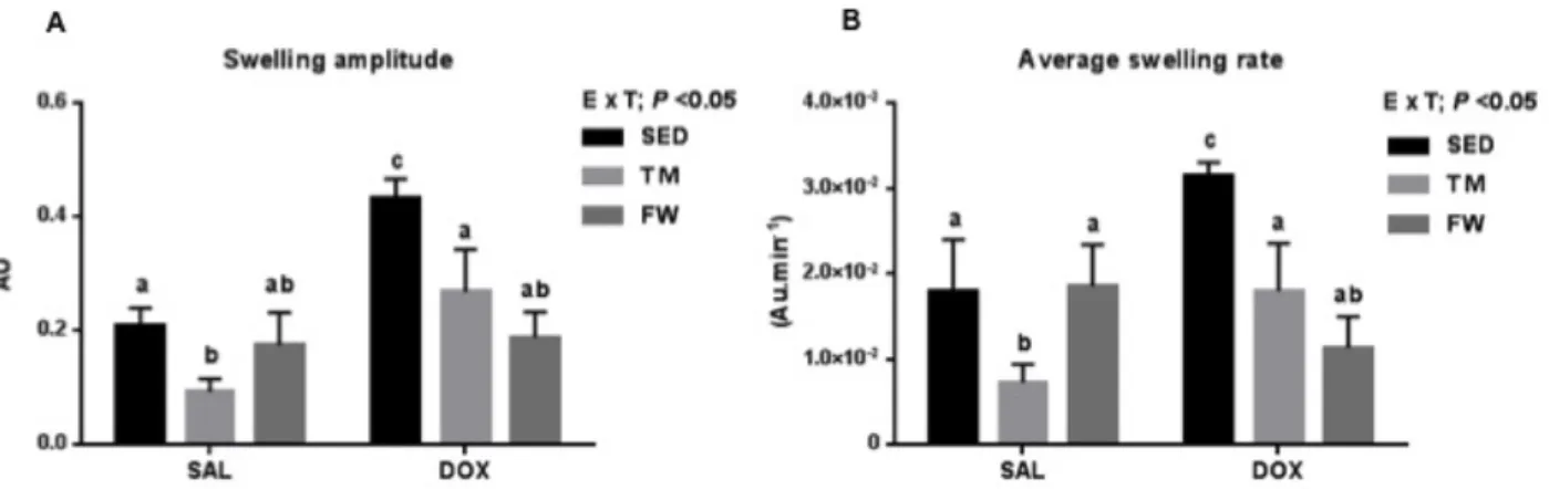
5.4 Mitochondrial osmotic swelling during MPTP induction ... 52
5.5 Oxidative stress markers ... 54
6. Discussion ... 55
6.1 Heart mitochondrial oxygen consumption and transmembrane electric potential .... 56
6.2. Mitochondrial osmotic swelling during MPTP induction ... 58
6.3 Oxidative stress markers ... 60
6.4 Meaning for exercise-induced cardioprotection in aging ... 61
7. Conclusion ... 63
xi
Figures
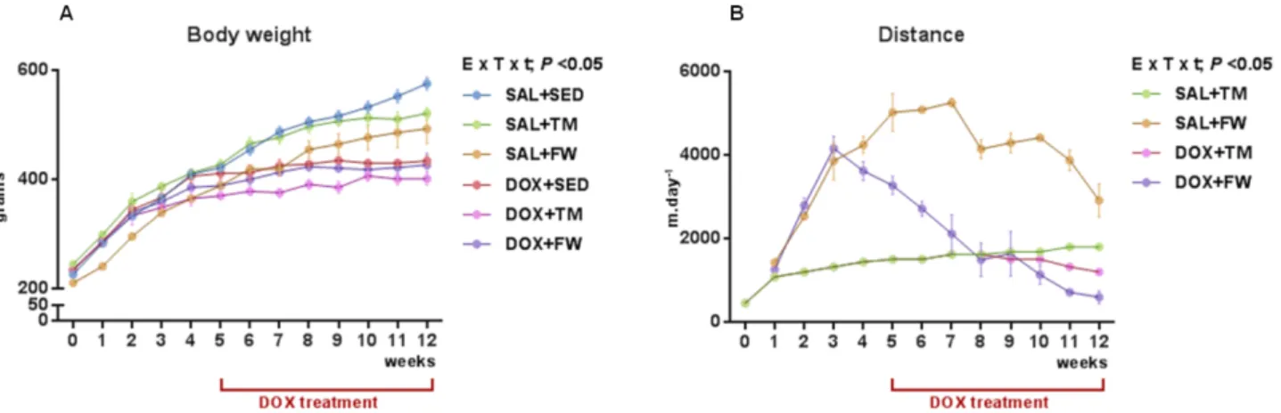
Figure 1. Representative scheme of reduced cardiorespiratory fitness (VO2max) in old adults. (adapted from Oxenham and Sharpe (2003)). ... 6 Figure 2. Mechanisms of DOX action and toxicity (adapted from Carvalho, Santos et al. 2009) ... 26 Figure 3. Effect of exercise and DOX treatment on (A) body mass over time and (B) distance covered per day by TM and FW groups during the 12 wks of protocol. ... 47 Figure 4. Effect of exercise and DOX treatment on (A) state 3 of heart mitochondrial respiration, (B) state 4 of heart mitochondria respiration, (C) RCR and (D) ADP/O.. ... 51 Figure 5. Effect of exercise and DOX treatment on heart mitochondria of heart ∆ψ fluctuations (A) maximal energization, (B) ADP-induced depolarization, (C) repolarization and (D) ADP phosphorylation lag phase. ... 52 Figure 6. Effect of exercise and DOX treatment on heart mitochondria to Ca2+-induced MPTP (A) Swelling amplitude; (B) Average swelling rate. ... 53 Figure 7. Heart mitochondrial (A) MDA and (B) reduced sulfhydryl contents.. ... 54
Tables
Table 1. Effects of aging on cardiovascular system ... 4 Table 2. Summary of some described mitochondrial-related alterations associated with DOX-induced cardiotoxicity and the modulation effect afforded by physical exercise against DOX (adapted from Ascensao, Oliveira et al. 2012) ... 34 Table 3. TM exercise protocol ... 40 Table 4. Animal data and yield of mitochondrial protein isolation ... 49
Equations
xiii
Resumo
O presente estudo teve como objetivo analisar o efeito de dois protocolos de exercício crónico distintos (treino em tapete rolante - TM e atividade física voluntária em roda livre - FW) na disfunção mitocondrial induzida pelo tratamento sub-crónico de doxorrubicina (DOX), uma potente droga antineoplásica bastante eficaz cuja principal limitação é a toxicidade cardíaca. Foram utilizados 32 ratos Sprague-Dawley macho jovens divididos em seis grupos (n = 6 por grupo): salino sedentário (SAL + SED), salino treinado (SAL + TM, 12 semanas de treino em tapete rolante), salino roda-livre (SAL + FW 12 semanas de atividade física em roda livre), tratado com DOX sedentário (DOX + SED [7 semanas de tratamento sub-crónico de DOX (2mg.kg-1.wk-1)], DOX + TM e DOX + FW. Foi analisada a funcionalidade mitocondrial cardíaca in vitro [consumo de oxigênio, potencial transmembranar (ΔΨ) e swelling osmótico], assim como os níveis de MDA e grupos sulfidril.
O tratamento com DOX afetou a funcionalidade mitocondrial cardíaca, alterando o consumo de oxigénio, o potencial transmembranar assim como o swelling osmótico durante a indução do poro de permeabilidade transitória mitocondrial (DOX + SED vs SAL + SED). A disfunção induzida pela administração de DOX no estado 3, no índice de controlo respiratório, ADP/O, no ΔΨ máximo, na repolarização, na lag-phase, amplitude e taxa de swelling, assim como no nível de MDA e no conteúdo grupos sulfidril foram revertidos pelos dois tipos de exercício crónico.
Ambos os protocolos de exercício estudados atenuaram a disfunção bioenergética das mitocôndrias cardíacas associada ao tratamento sub-crónico com DOX. Os nossos resultados são mais um contributo para o estudo dos efeitos cardioprotetores do exercício físico realizado antes, durante e após o tratamento com DOX, não só na população adulta mas também idosa.
PALAVRAS-CHAVE: EXERCÍCIO, CORAÇÃO, BIOENERGÉTICA,
xv
Abstract
The effects of two distinct chronic exercise models (endurance treadmill training – TM and voluntary free-wheel activity - FW) against mitochondrial dysfunction induced by sub-chronic treatment of doxorubicin (DOX), a potent antineoplastic drug known to induce a dose-related cardiac and mitochondrial toxicity, were analyzed.
Male young Sprague-Dawley rats were divided in six groups (n=6 per group): saline sedentary (SAL+SED), saline exercised (SAL+TM; 12-wks treadmill), saline freewheel (SAL+FW, 12-wks voluntary free-wheel), DOX+SED [7-wks sub-chronic DOX treatment (2mg.kg-1.wk-1)], DOX+TM and DOX+FW. In vitro
endpoints of heart mitochondrial function [oxygen consumption, membrane potential (ΔΨ) and osmotic swelling], MDA level and sulfhydryl groups content were evaluated.
DOX affected mitochondrial function as seen by oxygen consumption, ΔΨ endpoints and osmotic swelling during MPTP induction (DOX+SED vs. SAL+SED). DOX-induced impairments in state 3, respiratory control ratio, ADP/O, maximal ΔΨ, repolarization, ADP lag-phase, swelling amplitude and average swelling rate as well MDA level and sulfhydryl groups content were reverted by both TM and FW.
Both studied chronic models of physical exercise reestablished heart mitochondrial bioenergetic defects induced by sub-chronic DOX treatment. Our results contribute to the analysis of the cardioprotective effects of exercise performed before, during and after DOX treatment no only on adult but also in older population.
KEY-WORDS: EXERCISE; HEART; BIOENERGETIC; MITOCHONDRIAL
xvii
Abbreviations and Symbols
VO2max Maximal Oxygen Uptake
A.M. Before Midday
AIF Apoptosis Inducing Factor
AMPK Adenosine Monophosphate-Activated Protein Kinase ANT Adenosine Nucleotide Translocase
ATP Adenosine Triphosphate Ca2+ Calcium Ion
CAT Catalase
CHF Congestive Heart Failure CS Citrate Synthase
Cyc D Cyclophilin D
DNA Deoxyribonucleic Acid
DOX Doxorubicin DTNB Beta Dystrobrevin E Exercise ER Endoplasmic Reticulum FW Free Wheel G Glutamate GPX Glutathione Peroxidase GSH Glutathione h Hours H2O2 Hydrogen Peroxide
HSP Heat Shock Proteins
IFM Intermyofibrillar Mitochondria IR Ischemia Reperfusion KCl Potassium Chloride Kg Kilogram KH2PO4 Monopotassium Phosphate m Metro M Malate
xviii MDA Malondialdehyde MFN Mitofusin mg Milligram min Minute ml Milliliter mM Millimolar
MnSOD Manganese Superoxide Dismutase
MPTP Mitocohndrial Permeability Transition Pore mtDNA Mitochondrial Deoxyribonucleic Acid
MΩ Megaohm
NaCl Sodium Chloride
NADH Reduced Nicotinamide Adenine Dinucleotide
NADPH Reduced Nicotinamide Adenine Dinucleotide Phosphate
nmol Nanomol NO Nitric Oxide NS Non-Significant O2 Oxygen O2- Superoxide Radical ºC Degree Celsius OH. Hydroxyl Radical
PGC Proliferator-Activated Receptor Gamma RCR Respiratory Control Ratio
RNA Ribonucleic Acid
ROS Reactive Oxygen Species
SAL Saline
SEM Standard Error Of The Mean -SH Sulfhydryl groups
SOD Superoxide Dismutase
SSM Subsarcolemmal Mitochondria
T Treatment
t Time
TM Treadmill
xix TPP+ Tetraphenylphosphonium
VDAC Voltage Dependent Anion Channel (A-VO2)diff Arteriovenous oxygen difference
1
1. Introduction
Doxorubicin (DOX, or adriamycin) is a highly effective antibiotic used to treat several types of malignancies. Unfortunately, the clinical use of DOX is limited by the occurrence of a dose-related cardiac toxicity that results in life-threatening cardiomyopathy. DOX-induced cardiomyocyte dysfunction is associated with increased levels of oxidative damage involving mitochondrial bioenergetics collapse in the process (Wallace 2007). Actually, sub-chronic DOX treated rats reveal defects on heart mitochondrial function, which are accompanied by compromised mitochondrial electron transport chain activity and increased oxidative stress and damage (Berthiaume, Oliveira et al. 2005, Santos, Moreno et al. 2002).
Among the strategies advised to counteract the cardiac side effects associated with DOX treatment, physical exercise has been studied and recommended as a non-pharmacological tool against myocardial injury (Ascensao, Ferreira et al. 2007, Ascensao, Lumini-Oliveira et al. 2011, Ascensao, Oliveira et al. 2012). Previous work has suggested that the advantage of both acute (Ascensao, Lumini-Oliveira et al. 2010, Wonders, Hydock et al. 2008) and chronic exercise models (Ascensao, Ferreira et al. 2006, Ascensao, Magalhaes et al. 2005, Ascensao, Magalhaes et al. 2005, Chicco, Hydock et al. 2006, Chicco, Schneider et al. 2005, 2006) on the preconditioning of DOX-treated rats include the protection of cardiac tissue and mitochondria against induced impairments. Moreover, recent studies investigating the effects of exercise performed during and following models of late-onset cardiotoxicity caused by DOX provide evidence of exercise-induced cardioprotection in both adult and juvenile rat models (Hayward, Lien et al. 2012, Hydock, Lien et al. 2012). However, the cellular and molecular mechanisms underlying this protective phenotype induced by exercise are still elusive. In particular, whether perturbations in heart mitochondrial oxidative phosphorylation capacity and pro-oxidant redox modifications associated with cumulative DOX administration are modulated by long-term physical exercise performed during and after treatments is yet unknown.
2 We therefore aimed to analyze the effects of two types of long-term exercise with distinct characteristics, performed before and during the overall DOX treatment, on cardiac mitochondrial bioenergetics. Heart mitochondrial respiratory parameters associated with oxygen consumption, transmembrane electrical potential and osmotic swelling during mitochondrial permeability transition pore (MPTP) induction as well as markers of oxidative stress (sulfhydryl groups (-SH) and malondialdehyde (MDA) contents) were determined.
3
2. State of art
2.1- Age effects on cardiac function
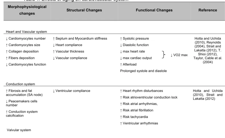
Aging can be characterized as a time dependent decline of maximal functionality that affects tissues and organs of the whole body (Figueiredo, Mota et al. 2008). The average human life span has markedly increased in modern society, a fact largely attributed to advances in medical and therapeutic sciences that have successfully reduced the severity of several diseased conditions (Chaudhary, El-Sikhry et al. 2011). However, elderly individuals continue to suffer the greatest burden from cardiovascular disease, including coronary heart disease that remains the leading cause of death in industrialized countries (Wei 2004). That can be understood as the result of several morphophysiological, structural and functional alterations, which we will further address in detail (Table 1).
The aging-induced increase on vascular stiffness, septum and myocardium thickness and fibrosis, is associated, among others, with: i) decreased myocyte number; ii) increasing cardiomyocyte size with alterations in calcium (Ca2+) homeostasis and iii) increased collagen fibers deposition, leading to cardiac diastolic dysfunction, increased afterload, and loss of arterial and heart compliance (Strait and Lakatta 2012). Importantly, these physiological-related impairments along with unchanged cavity size and increased left ventricle hypertrophy are the major characteristic of aging heart (Chaudhary, El-Sikhry et al. 2011). In fact, Dai et al (2009) reported that aging left ventricle mass index increased by around 75% compared to a young adult group, indicating the increase prevalence of left ventricular pathological hypertrophy with age. It was also reported reduction in diastolic function, as well as worsening of myocardial performance index (Dai, Santana et al. 2009).
Furthermore, the conduction system also undergo some structural alterations leading to heart dysfunction, including fat accumulation around sinoatrial node, which creates a partial or complete separation of the node from atrial tissue (Strait and Lakatta 2012), a marked decreased in peacemakers number cells
4 (Wei 2004) and an increased calcification in conduction system leading ultimately to atrioventricular conduction lock. Slow and prolonged systole and diastole are other aging-related heart features usually related to an aberrant Ca2+ handling (Strait and Lakatta 2012). The decreased diastolic function, which falls linearly with aging at a rate of about 6–7% per decade (Gates, Tanaka et al. 2003), is associated to a decrease in diastolic filling reported at rest and under stress conditions, as some daily tasks that promote an acute increase of physical demands. In addition, the degeneration of cardiac valvular apparatus is commonly reported, being valvular annular dilation found in the majority of older persons and is also associated with concomitant coronary artery calcification (Wei 2004).
Table 1. Effects of aging on cardiovascular system Morphophysiological
changes Structural Changes Functional Changes Reference
Heart and Vascular system ↓ Cardiomyocytes number ↑ Cardiomyocytes size ↑ Collagen deposition ↑ Fibers deposition ↓ Cardiomyocytes function
↑ Septum and Myocardium stiffness ↓ Heart compliance
↑ Vascular thickness ↓ Vascular compliance
↑ Systolic pressure ↓ Diastolic function ↓ max heart rate ↓ max cardiac output ↑ Afterload
Prolonged systole and diastole
Hotta and Uchida (2010), Reynolds (2004), Strait and Lakatta (2012), T.
Shioi (2012), Taylor, Cable et al.
(2004)
Conduction system ↑ Fibrosis and fat accumulation (SA node) ↓ Peacemakers cells number ↑ Conduction system calcification ↓ Ventricular compliance
↑ Heart rhythm disturbances
↑ Risk atrioventricular conduction lock ↑ Risk atrial arrhythmias,
↑ Risk atrial fibrillation ↑ Risk tachycardia ↑ Ventricular arrhythmias
Hotta and Uchida (2010), Strait and Lakatta (2012)
Valvular system
↑ Valvular annular dilatation Coronary artery calcification Reynolds (2004)
(↑) – increase; (↓) – decrease
5 Overall, these aging related heart morphophysiological, structural and functional alterations result in increased systolic pressure, one major risk factors for development of atherosclerosis, hypertension and stroke, and arterial fibrillation (North and Sinclair 2012).
As a consequence of all those age-associated alterations, cardiac function declines maximal functionality, being that, under basal conditions this decline may not be detected (Wessells and Bodmer 2007).
The most used standard index of cardiorespiratory fitness is the maximal oxygen consumption (VO2max), which declines approximately 10% per decade
starting at 20 to 30 years old (Souza 2012). VO2maxcan be estimated by the
Fick’s equation:
Equation 1: VO2max=Q×(A-VO2)diff
Q is cardiac output, and is the product of max heart rate and stroke volume, (A-VO2)diff is the arteriovenous difference of oxygen. As it is observed in figure 1,
based on Fick´s equation, aerobic exercise capacity depends on cardiac output and arteriovenous difference (Seeley, Stephens et al. 2005).
Reductions in peak heart rate, peripheral oxygen utilization and stroke volume appear to mediate the age-associated decline in VO2max. Actually, impairments
in cardiac filling and increased afterload leads to a decrease in heart rate, in a stress situation (Taylor, Cable et al. 2004) being the major responsible for much of the age-associated decrease in maximal cardiac output. Reductions in muscle oxygen delivery and therefore (A-VO2)diff are mainly due to reduced
and maldistribution of cardiac output. Also, a decline in skeletal muscle oxidative capacity with aging is reported, due in part to mitochondrial dysfunction, which appears to play a particularly important role in old age, where skeletal muscle VO2max is observed to decline by approximately 50%
even under conditions of similar oxygen delivery as young adult muscle (Betik and Hepple 2008).
6 Although controversial, some reports suggested that left ventricular systolic function remains relatively preserved and without significant alterations in left ventricular stroke volume and cardiac output at rest (for refs see Kappagoda and Amsterdam 2012, Lakatta 2002, Morley and Reese 1989). However, at peak exercise, stroke volume index is reduced in older individuals and is thought to be the consequence of age-related reductions in β-adrenergic stimulation, increases in vascular stiffness, aortic impedance and impaired left ventricular diastolic function (Spina, Turner et al. 1998).
Generally, the morphophysiological and functional alterations related to VO2max decline with aging are depicted in figure 1.
Figure 1. Representative scheme of reduced cardiorespiratory fitness (VO2max) in old
adults. (adapted from Oxenham and Sharpe (2003)).
Studies have demonstrated that advancing age is also associated with decreased heart rate variability (O'Brien, O'Hare et al. 1986, Yeragani,
7 Sobolewski et al. 1997). Heart rate variability is a reliable reflection of the many physiological factors modulating the normal rhythm of the heart. In fact, it provides a powerful mean of observing the interplay between the sympathetic and parasympathetic nervous systems and it is thought to reflect the heart’s ability to adapt to changing circumstances by detecting and quickly responding to unpredictable stimuli (Rajendra Acharya, Paul Joseph et al. 2006). It is particularly relevant on daily tasks, as it is important to maintain a normal lifestyle without manifestation of heart injury. Also, it has been suggested that a decreased responsiveness to β-adrenergic receptor in aged hearts might be responsible for decreased heart rate variability in aged people during exercise or stress (Chaudhary, El-Sikhry et al. 2011).
Ischemia-reperfusion (IR) injury is the primary pathological manifestation of coronary artery disease (Lennon, Quindry et al. 2004, Powers, Demirel et al. 1998). The level of IR-induced myocardial injury can range from a small insult, resulting in limited damage, to a large insult, culminating in major cardiac electrical and mechanical dysfunction and ultimately in death (Powers, Quindry et al. 2004). Nowadays, it is well established that numerous age-related cellular and functional changes occur in the heart that could lead to ischemia-reperfusion injury as i) alterations in cardiac gene expression; ii) increased oxidative stress and iii) reduced ability of the heart to response to stress (Powers, Quindry et al. 2004). In this regard, Starnes et al. (1997) reported that immature hearts tolerate and recover from hypoxia better than adult hearts, and that the sarcolemmal membranes of immature rat hearts seem to be less susceptible to damage from hypoxic stress than those of older group.
It is notable that throughout life, there are many impairments originating cardiac injuries and ultimately death. So, it is important to implement countermeasures to attenuate and minimize those consequences, which will enhance old people lifestyle and lifespan. Recently, several approaches have been investigated and physical activity has been shown to be an important countermeasure to protect against myocardial injuries (Ascensao, Ferreira et al. 2007, Kavazis 2009,
8 Kavazis, Alvarez et al. 2009, Powers, Quindry et al. 2004, Starnes and Taylor 2007).
Furthermore, among the several mechanisms or causes for cardiac dysfunction, mitochondrial abnormalities have a central role (for refs see Braunwald and Bristow 2000). In fact, due to the key mechanisms to which these organelles are associated such as energy production, ion regulation, pH control, Ca2+ homeostasis, redox reactions, control of cell signaling and apoptosis mitochondria assume a pivotal role in cellular functioning. For these reasons, mitochondria have been suggested as reliable sensors of cellular functionality and (dys)functional mitochondria correlates with (dys)functional heart tissue submitted to a variety of stimuli, including age (Wallace 2010).
Because heart is primarily a postmitotic tissue that exhibits a highly aerobic metabolism due to the abundance of large mitochondria, a dependence on healthy mitochondria for normal organ function is implicated (Judge and Leeuwenburgh 2007).
The following section will address the effects of aging process on the modulation of mitochondrial function and related mechanisms.
2.1.1 Aging effects on cardiac mitochondria function
In metabolically active and thus energy-demanding tissues such as the heart, mitochondria, as producers of most of the ATP necessary for metabolism via oxidative phosphorylation, have a very important role in the supply of energy for continuously contracting myocytes (Ascensão 2011). Additionally, mitochondria play other important roles as cellular redox and ion homeostasis, oxygen sensing, signaling and regulation of programmed cell death (for refs see Rabinovitch 2012, Sheu 2009). Therefore, mitochondrial integrity is vital for cellular homeostasis and cardiac performance (Chaudhary, El-Sikhry et al. 2011) being age-related heart mitochondrial dysfunction closely associated to
9 life limiting impairments, including congestive heart failure (CHF) (Chaudhary, El-Sikhry et al. 2011, Judge and Leeuwenburgh 2007, Pashkow 2011).
In 1956, Harman et. al, proposed the free radical theory of aging, postulating that the production of reactive oxygen species (ROS) is a major determinant of lifespan acting as important mediators responsible for the cellular damage seen in aged cells (Harman 1956). Later, it was defined that mitochondria are the main source of ROS and the main target of their injury being ROS produced within mitochondria almost 90% of the total ROS produced in the cell (Hearman 1972); so it was postulated as the “key organelles” initiating cellular processes leading to death (Chaudhary, El-Sikhry et al. 2011). Indeed, evidences suggest that with advanced age, mitochondrial production of ROS significantly increases in heart tissue (Judge, Jang et al. 2005), which leads to development of degenerative diseases (for refs see Rabinovitch 2012). Within cells, ROS are produced in multiple compartments and by multiple enzymes including reduced nicotinamide adenine dinucleotide phosphate (NADPH) oxidase at the plasma membrane, respiratory chain within mitochondria, lipid oxidation within peroxisomes and by clyclo-oxygenases and xanthine oxidase in the cytoplasm (Dai and Rabinovitch 2009, Dai, Rabinovitch et al. 2012). Although all of these source contribute to the overall oxidative damage, mitochondria are one of the most contributors for ROS generation as a byproduct of electron transfer during oxidative phosphorylation (Dai and Rabinovitch 2009). Most specifically, excess electrons from complex I and III can be transferred directly to oxygen (O2) to
generate superoxide anion (O2-), which is then converted to hydrogen peroxide
(H2O2) and ultimately into a hydroxyl radical (OH.), the most reactive ROS
species (Dai and Rabinovitch 2009). The unbalance between ROS production and the capacity of antioxidant machinery, favoring ROS production is usually called oxidative stress. Mitochondria have antioxidant enzymes that play a vital role in the protection against oxidative stress (Meng 2007), being manganese superoxide dismutase (MnSOD), catalase (CAT) and glutathione peroxidase (GPX) the most commonly cited (Ascensão 2003). In addition, there are non-enzymatic antioxidant compounds, both endogenous and exogenous, such as glutathione (GSH), vitamins C and E, and lipoic acid that play important roles in
10 ROS neutralization or in the attenuation of the effects caused by increased ROS production (Ji, Leeuwenburgh et al. 1998). Heat shock proteins (HSPs) are another system of cellular defense against oxidative stress (Ascensao, Ferreira et al. 2007, Hamilton, Staib et al. 2003, Powers, Locke et al. 2001). These “stress-induced proteins” are ubiquitous and highly conserved chaperones, important in the folding of new synthesized, damaged or transported proteins. Moreover, HSPs mediate mitochondrial protection against oxidative stress, namely HSP70 have been associated with myocardial protection (Gunduz, Senturk et al. 2004).
To understand the impact of oxidative stress, which is known to damage proteins, lipids, and deoxyribonucleic acid (DNA) (Shioi and Inuzuka 2012), some oxidative markers are analyzed in mitochondria as carbonil groups (protein oxidation), malondialdehyde (MDA) (lipid peroxidation marker) or thiobarbituric acid reactive substances (TBARS) (nonspecific marker for lipid peroxidation), among others. The accumulation of oxidant-induced damage in mitochondria may be a major contributing factor to the age-related alterations in myocardial function (Chen and Knowlton 2011, Ljubicic, Menzies et al. 2010, Pohjoismaki, Boettger et al. 2012). Nonetheless, some authors have suggested that throughout the aging process, the antioxidant capacity can be adjusted in response to prooxidant exposure (Ji, Leeuwenburgh et al. 1998). Localized oxidative stress in specific organs, tissues, and organelles may stimulate cellular uptake and synthesis of certain antioxidants under complicated genetic, hormonal, and nutritional regulation (Harris 1992). One possibility is that aged mitochondria produce more ROS stimulating antioxidant enzyme gene expression. This scenario is consistent with the finding that mitochondrial antioxidant enzyme activity showed a greater increase in the senescent myocardium (Ji 1993, Ji, Dillon et al. 1991, Rao, Xia et al. 1990, Vertechy, Cooper et al. 1989). Nevertheless, data seems to be controversial as no alteration (Bejma, Ramires et al. 2000, Tian, Cai et al. 1998) or decrease in antioxidant activity (Bagchi, Bagchi et al. 1996, Pritsos and Ma 2000) have also been reported. So, increased antioxidant activity may be interpreted as being beneficial because it provides better protection against oxidant-induced
11 damage, or it may be viewed as negative because it could indicate a need for enhanced antioxidant defenses due to increased oxidant production (Beckman and Ames 1998).
However, characterization of age-related changes in cardiac mitochondria has been challenged due to the fact there are two distinct populations of mitochondria in the myocardium (Fannin, Lesnefsky et al. 1999). Subsarcolemmal mitochondria (SSM) are located beneath the plasma membrane, whereas interfibrillar mitochondria (IFM) are found in parallel rows between the myofibrils. There are important biochemical and functional differences between SSM and IFM and it has been revealed that IFM are more adversely affected with age (Fannin, Lesnefsky et al. 1999, Judge, Jang et al. 2005), although Judge et al. (2007) suggests that further studies are required to determine the mechanisms contributing to these changes, and to further characterize differential effects of age upon the SSM and IFM populations in the heart.
Despite this fact, some authors report increased H2O2 production from SSM, but
not IFM, with age (Judge, Jang et al. 2005, Judge and Leeuwenburgh 2007). This can be a consequence of the lower antioxidant capacity on this subpopulation. Actually, whereas IFM MnSOD, GPX and CAT levels seem to increase with age, in SSM only MnSOD and GPX, but not with CAT, seems to be positively regulated. However, as MnSOD converts O2- radicals to H2O2,
increased MnSOD activity might also represent higher levels of H2O2. Also,
lower glutathione levels and increased oxidative damage have been suggested, supporting that oxidant production within the matrix of “old” IFM is greater than that in “young” IFM (Judge, Jang et al. 2005). Similarly, state 3 respiratory rates were also lower in “old” IFM. This has a particular concern because IFM are likely the major source of ATP production for myosin ATPase (Judge, Jang et al. 2005). Because high levels of ATP are required for both systolic contraction and diastolic relaxation, reduced availability of ATP as a result of IFM dysfunction could contribute to the alterations in cardiac contractility observed with age (Judge, Jang et al. 2005).
12 Besides nucleus, mitochondria are the only organelles in animal cells that possess their own DNA, the mtDNA, as well as related transcriptional and translational synthesis machinery. mtDNA is localized in matrix with physical proximity to the mitochondrial respiratory chain (Bratic and Trifunovic 2010). However, mitochondria cannot be synthesized de novo, instead they replicate in the cytosolic compartment through a process of division (for refs see Chaudhary, El-Sikhry et al. 2011). Importantly, the proteins that are encoded by mtDNA are vital for normal mitochondrial function and mtDNA does not have the protein protection as nuclear DNA and has less effective repair mechanisms (Desler, Marcker et al. 2011). Therefore, mutations in mtDNA through oxidative stress affect the expression and integrity of oxidative phosphorylation complexes and can cause mitochondrial dysfunction and increased ROS production (Wallace 2010). So, it is notable the interrelationship between oxidative phosphorylation complexes, ROS levels, mtDNA mutations and ultimately cell death. Furthermore, mutations in mtDNA and the resultant decline in mitochondrial activity observed in aged tissues are responsible for the increased generation of ROS, which in turn, will further negatively impact mitochondria causing further mtDNA damage. This “vicious cycle” concept postulated that accumulation of mtDNA mutations is exponential and associated with massive increase in ROS production (Lenaz 1998).
Being mitochondria strategic organelles essential for cell function and homeostasis, providing energy to the cell (Chaudhary, El-Sikhry et al. 2011), they must undergo some dynamic mechanisms. Mitochondrial turnover and death can occur via several processes that are suggested to be interrelated namely apoptosis, necrosis, autophagy and related dynamics of organelles. In this dynamic network biogenesis, fusion and fission are closely associated mechanisms (Chen and Knowlton 2011).
In fact, apoptosis is mediated by two pathways: the extrinsic and the intrinsic pathways, and both have been described in cardiac myocytes (Whelan, Kaplinskiy et al. 2010). The extrinsic apoptotic pathway can be triggered by Fas ligand or tumor necrosis factor (TNF)-α, which are expressed in cardiac
13 myocytes and have been implicated in cardiovascular pathology (Whelan, Kaplinskiy et al. 2010).
Intrinsic pathways involve the participation of endoplasmic reticulum (ER) and/or mitochondria (Kroemer, Galluzzi et al. 2007). Impairment in mitochondrial integrity, dynamics or metabolic activity may result in a range of deleterious effects to the cell, such as reduced ATP production, elevated cytosolic Ca2+, increased ROS release, release of proapoptotic factors as cytocrome c or caspases activation and Bax translocation, triggering cell death (Chen and Knowlton 2011). In mammalian cells, apoptosis is regulated by a variety of factors that are essentially either pro-life or pro-death (Goldspink, Burniston et al. 2003). Quantitation of the expression of genes involved in the apoptotic pathway might represent a good index of the probability for a cell to undergo apoptosis. As the number of Bax-expressing cells dramatically increases in left ventricular hypertrophy and left ventricular dysfunction (Condorelli, Morisco et al. 1999, Green and Reed 1998), it is suggested that mitochondrion is the primary organelle mediating the intrinsic apoptotic pathway in these conditions (Chiong, Wang et al. 2011)
Also, if excessive Ca2+ enters to mitochondria and enhanced oxidative stress conditions are present, a phenomenon known as permeability transition may occur (Ascensao, Lumini-Oliveira et al. 2011). The mitochondrial permeability transition is characterized by the loss of the impermeability of the mitochondrial membranes and it is suggested that this condition is mediated by the formation and opening of protein complex-like pores in the inner mitochondrial membrane, the mitochondrial permeability transition pore (MPTP) (for refs see Ascensao, Lumini-Oliveira et al. 2011). Increased pro-oxidant generation causing oxidative stress is one condition that augmented the susceptibility for the opening of these pores and the release of pro-apoptotic proteins within mitochondria as cytochrome c, SMAC/DIABLO and the apoptosis inducing factor (AIF), which will activate the caspase-related apoptotic pathways. It is suggested that the release of these proteins is dependent on the formation and opening of MPTP that cross the inner and the outer membranes leading to the loss of
14 mitochondrial membrane potential (Δᴪ), increased mitochondrial osmotic swelling and rupture of the outer mitochondrial membrane, which leads to death (Ascensao, Lumini-Oliveira et al. 2011). It is believed that the structure and regulation of this multi-protein complex comprises the outer membrane voltage-dependent anion channel (VDAC) as well as the inner membrane adenine nucleotide translocator (ANT) and cyclophilin D (Cyc D). Myocyte loss has been shown to occur in the aged rat heart and to precede the occurrence of ventricular dysfunction (Anversa, Hiler et al. 1986), being apoptotic cardiomyocyte death present under different conditions in humans (Haunstetter and Izumo 1998, Narula, Haider et al. 1996, Olivetti, Abbi et al. 1997).
Among the excess of biological phenomena affected by aging, the malfunction and decrease of biogenesis of mitochondrial biogenesis seems to exert some of the most potent effects on the organism (Lopez-Lluch, Irusta et al. 2008). If biogenesis is affected, it is reasonable to expect that mitochondrial turnover must be slower and the accumulation of modified lipids, proteins and DNA must also increase, further aggravating the conditions resulting on deficient activity of aged mitochondria (Lopez-Lluch, Irusta et al. 2008). The precise reason for the decrease in the rate of mitochondrial biogenesis during aging is currently unknown. However, it seems that both, extra- and intra-cellular regulatory factors of mitochondrial biogenesis are implicated. Specifically, peroxisome proliferator-activated receptor gamma coactivator (PGC1-α) has been shown to act as a common intracellular mediator during mitochondrial biogenesis induced by hormonal factors (Weitzel, Iwen et al. 2003), and adenosine monophosphate-activated protein kinase (AMPK) an intracellular regulator of mitochondrial biogenesis, which activity appears to be one of the main factors associated with deficient mitochondrial biogenesis (Reznick, Zong et al. 2007). PGC family members have gained particular interest because of their ability to drive virtually all mechanisms of mitochondrial biogenesis in the heart, including mitochondrial number, mitochondrial respiration, expression of oxidative phosphorylation and fatty acids oxidation genes, and ROS levels (Lehman, Barger et al. 2000). Decreased PGC-1α expression has been linked to the development of heart failure in mouse models (for refs see Moslehi, DePinho et
15 al. 2012), and decline in mitochondrial biogenesis and mitochondrial protein quality control in cardiac muscle was found in aging (Koltai, Hart et al. 2012) In addition, its well known that mitochondria are dynamic organelles that constantly undergo fission and fusion and it has been found to be vibrant organelles that continuously divide and fuse within the cell and have functions extending beyond energy production, including cell signaling (Liesa, Palacin et al. 2009). Disruption of fission and/or fusion can also lead to cellular dysfunction and to apoptosis. This dynamic mechanism is regulated by proteins controlling fission, such as hFis1 and Drp1, and fusion, such as mitofusin 1 and 2 (MFN1 and MFN2) and OPA1. The correct function of these proteins seems to be critical for normal mitochondrial activity, and their deregulation is associated with several pathologic conditions (Lopez-Lluch, Irusta et al. 2008). Indeed, the impairments on fission-related protein hFis1 has been associated with the process of senescence in mammalian cell cultures (Lee, Jeong et al. 2007). Moreover, depletion of hFis1 by RNA interference (RNAi) induces dramatic changes in mitochondrial structure, including the enlargement and flattening of the organelle. Futrthermore, elimination of any of the mitochondrial fusion proteins as MFN1, MFN2 or OPA1, induces mitochondrial fragmentation, as expected, being that down-regulation of Opa1 expression in cells by RNAi results in spontaneous apoptosis (for refs see Chen and Knowlton 2011). Overall, defects in the mitochondrial fission/fusion machinery and so loss of the symmetry between fusion and fission (Hoppins, Edlich et al. 2011) may contribute to the decline in mitochondrial function during aging. However, several fundamental questions remain to be answered (Bossy-Wetzel, Barsoum et al. 2003).
In the next section, the roles of physical exercise as a strategy to improve cardiac function in adult and old subjects as well as the mitochondrial-mediated mechanisms associated with exercise-induced cardioprotection will be addressed.
16
2.2. Exercise and cardioprotection
Cardiac damage is a major contributor to morbidity and mortality in industrialized countries; so it becomes important to develop strategies that result in cardioprotective phenotype. In this regard, several approaches have been investigated and physical activity has been shown to be an important countermeasure to protect against myocardial injuries (Bowles, Farrar et al. 1992, Harris and Starnes 2001, Powers, Demirel et al. 1998, Powers, Quindry et al. 2004). In fact, cardiorespiratory fitness is inversely related to cardiovascular and all-cause mortality and it has crucial role preventing heart injury (Kokkinos, Myers et al. 2010). Some reports had postulated exercise-induced benefits based on decreases of some risk factors to develop cardiac and myocardium impairments such as bodymass index, body weight, waist circumference, abdominal and visceral fat and consequently insulin resistance, triglyceride levels, blood pressure and, in general, metabolic syndrome-related parameters (for refs see Golbidi and Laher 2012). Therefore, chronic aerobic exercise is able, not only, to improve cardiovascular function in young healthy subjects, but also, and most importantly, in older people and those with cardiovascular risk factors (Hambrecht, Fiehn et al. 1998).
The study of the mechanisms responsible for exercise-induced cardioprotection has been ongoing for over decades and morphological and biochemical/molecular alterations have been considered as putative mechanisms of exercise-induced cardioprotection. Those include morphological adaptations of heart and coronary arteries, induction of myocardial HSPs, increased myocardial cyclooxygenase-2 activity, elevated ER stress proteins, nitric oxide production, improved function of sarcolemmal and/or mitochondrial adenosine triphosphate (ATP)-sensitive potassium channels and increased myocardial antioxidant capacity (for refs see Kavazis 2009).
For instance, exercise induces vascular remodeling and so, morphological alterations in coronary arteries through angiogenesis and arteriogenesis (Leung, Yung et al. 2008). Here, nitric oxide (NO) assumes important roles due its anti-inflammatory, vasodilator and platelet inhibitory effects (Landmesser and
17 Drexler 2005). Also, NO protects against inschemia-reprefusion (IR) injury in such a way that the heart responds to ischemia using nitric oxide species in a harmonized manner and these mechanisms could be based on inhibition of Ca2+ influx into myocytes, antagonism of β-adrenegic stimulation, reduction in cardiac oxygen consumption and ability to increase the expression of HSP70 (for refs see Golbidi and Laher 2011). HSPs protect cell against oxidative injury and apoptosis (Polla, Kantengwa et al. 1996) and furthermore, enhance recovery from acute myocardial cellular injury protecting heart for subsequent injury (for refs see Powers, Locke et al. 2001) by promoting restoration of dysfunctional enzymes and preventing aggregation of severely denatured proteins. The majority of evidences indicate that members of the 70-kDa family are the cytoprotective proteins most responsible for cell protection (for refs see Powers, Locke et al. 2001). The expression of HSP70 in cardiomyocytes is associated with increased cell survival and protection against ischemic damage and its now well established that acute and chronic exercise are able to induce increases in the expression of HSP70 (Kregel and Moseley 1996, Powers, Demirel et al. 1998), although in a temperature-dependent “fashion”.
The improved function of sarcolemmal ATP sensitive potassium channels (Powers, Quindry et al. 2008) and elevated ER stress proteins are other important exercise-induced cardioprotection-related alterations and both have special relevance during a cardiac insult (Golbidi and Laher 2012). The ER stress proteins help cellular homeostasis by maintaining intracellular Ca2+ regulation and protein folding during IR injury (Logue, Gustafsson et al. 2005). Considering the importance of mitochondrial machinery in the maintenance of cardiac function, mitochondrial-mediated mechanisms have also been associated with exercise induced cardioprotection phenomenon. This topic will be further discussed in the next sections.
18
2.2.1 Exercise and cardiac mitochondrial adaptations
As previously mentioned, mitochondrial adaptations may play a critical role in exercise-induced protection against cardiovascular impairments. The mechanisms behind this phenomenon remain unclear, however it may be related to morphological and biochemical adaptations including biogenesis, antioxidant production or resistance to cell death pathways (Ascensao, Ferreira et al. 2007, Kavazis, Alvarez et al. 2009, Kavazis, McClung et al. 2008). Those will be briefly addressed in the following section.
2.2.1.1. Morphological and biochemical adaptations
Heart is a highly oxidative organ, with a low rate of cell growth and slow turnover of proteins; so, it would be expected that heart might have a limited ability to adapt to acute and/or chronic conditions.
Although there are scarcely addressed outcomes about exercise-induced cardiac mitochondrial morphological adaptations in healthy hearts, the same cannot be stated regarding hearts under deleterious conditions. In fact, under deleterious conditions as DOX treatment, diabetes, or aging, morphological impairments on heart mitochondria have been reported (Ascensao, Oliveira et al. 2012, Searls, Smirnova et al. 2004). Despite this fact, in a study where trained animals were submitted to an endurance swimming training program, while the non-trained were not engaged in any exercise program Ascensao et al. (2006) reported that endurance swimming training per se caused notable changes in myocardial structure seen as an apparent increased glycogen content, intercalated discs showing a notorious scalloped appearance and evident signs of mitochondria biogenesis with elevated number of encroached mitochondria per fiber area, probably resulting in an increased volume/density of mitochondria. Also, mitochondria division, mild and focal loss of cristae density and organization as minimal degradation by-products, were also present in non-treated trained hearts. Another study developed by Searls et al. (2004)
19 with type I diabetes rats found in type I diabetes rats that 9 weeks of moderate exercise is able to reverse some of the phenotype of diabetic cardiomyopathy. Specifically, the mitochondrial quality, cytoplasmic area, and collagen cross-sectional area returned toward non-diabetic values with exercise.
Mitochondrial biochemical adaptations to exercise were also reported and several approaches have been trying to prove the exercise-induced modifications of some parameters involved in mitochondrial bioenergetics. Judge et al. (2005) reported that wheel running had no effect on mitochondrial protein yield, rates of oxygen consumption (states 4 and 3) or RCR. This was not entirely surprising given that other studies have shown that, unlike skeletal muscle, oxidative capacity of cardiac muscle is not increased in response to treadmill training, an exercise protocol that is typically much more intense than voluntary wheel running (for refs see Judge, Jang et al. 2005). Also, Starnes et al. (2007) reported that endurance training has no impact on mitochondrial oxidative phosphorylation, being that all oxidative phosphorylation parameters were similar in endurance trained and sedentary rats. Because the heart is highly oxidative, it is not expected to be as responsive to exercise-induced increases in oxidative capacity as skeletal muscle. Although, several reports suggested that exercise per se is able to improve mitochondrial protein yield, rates of oxygen consumption as well as cardiac function (for refs see Ascensao, Ferreira et al. 2007, Ascensao, Magalhaes et al. 2006, Chicco, Schneider et al. 2006, Powers, Lennon et al. 2002). Moreover, Kavasis et al. (2009) reported that the abundance of several proteins involved in bioenergetics was altered following endurance exercise in both SS and IMF mitochondria. Most specifically, the protein levels of several proteins involved in β-oxidation of fatty acids were increased following repeated bouts of endurance exercise. It is of especial concern as during the development of heart disease or genetic defects in mitochondrial fatty acid β-oxidation the myocardial energy source switches from fatty acid β-oxidation to glycolysis. Also, there are some controversial reports about endurance exercise benefits against Ca2+-induced mitochondrial
dysfunction. In fact, while Starnes et al. (2007) found that endurance exercise training had no influence when mitochondria was challenged with identical Ca2+
20 concentrations, being that mitochondria from trained and sedentary animals displayed similar declines in ATP production, French et al. (2008) found that exercise-induced protection against IR injury, in part, by attenuating IR-induced oxidative modification of important Ca2+-handling proteins and preventing subsequent calpain activation. In this study, MnSOD may had an important role as, the results support that exercise-induced increases in myocardial MnSOD activity attenuate the oxidation and degradation of Ca2+-handling proteins,
preventing calpain activation during IR. It has special concern because calpain activation within the myocardium has been directly linked with IR-induced necrotic and apoptotic cardiac myocyte death (French, Hamilton et al. 2008). Furthermore, several reports suggest that exercise per se is able to enhance cardiac mitochondria Ca2+ uptake capacity without MPTP induction (Ascensao,
Lumini-Oliveira et al. 2011, Kavazis, McClung et al. 2008, Marcil, Bourduas et al. 2006). Therefore, exercise may induce beneficial morphological and biochemical adaptations in mitochondria, which can be translated in a cardiac phenotype more functional and protected against deleterious stimuli.
2.2.1.2. Mitochondrial biogenesis
Due to the high-energy demand of the heart, a decline in mitochondrial function can result in a deterioration of cardiac performance (Li, Muhlfeld et al. 2011) and can lead to a wide variety of pathophysiological conditions. According to some reports, the malfunction of mitochondria and the decrease of mitochondrial biogenesis, together with increased oxidative damage, seem to exert some of the most deleterious effects on the organism (Guarente 2008, Lopez-Lluch, Irusta et al. 2008). Fortunately, although controversial, heart seems to have the ability to alter some gene expression profile and phenotype to produce new and more functional mitochondria (Lee and Wei 2007). Regular exercise or increased energy demand are important stimuli that lead to increased mitochondria biogenesis (for refs see Lopez-Lluch, Irusta et al. 2008). In fact, increases in cytosolic Ca2+ levels induced by exercise stimulates
21 calmodulin kinase which then promotes PGC1-α expression (Wu, Kanatous et al. 2002) that has been shown to be a major regulator of mitochondrial biogenesis (Rodgers, Lerin et al. 2005). Also, Geng et al. (2010) showed that in skeletal muscle-specific PGC-1α knockout mice, exercise-induced mitochondrial biogenesis and angiogenesis were significantly attenuated. However, exercise-induced mitochondrial biogenesis on cardiac muscle is still not fully illusive. In fact, although a significant amount of experimental data on mechanisms involved in exercise-induced mitochondrial biogenesis has been obtained in skeletal muscle, less information is available about the heart. In this regard, Li et al. (2011) reported no changes in mitochondrial biogenesis on left ventricle after 3 months of endurance exercise. Actually, in contrast to skeletal muscle in which adaptive changes in volume fraction of mitochondria can readily occur, this is not so frequent in cardiac muscle (Hood, Balaban et al. 1994). So, the exercise-induced benefits on heart mitochondria biogenesis are still controversial.
2.2.1.3. Oxidative stress and antioxidant capacity
With exercise performance, increased O2 consumption creates favorable
conditions for increased generation of ROS, an inevitable consequence that may increase oxidative stress at the organelle, cell, and tissue (Ji, Leeuwenburgh et al. 1998). However, if these stimuli are repeated over time, it may have a strong modulating effect on various defense systems in cardiac cells (Powers, Lennon et al. 2002). Also, in some deleterious conditions that lead to ROS production, as IR-induced myocardial injury, DOX administration or aging, physical exercise as been reported to induce benefic countermeasures. In fact, IR-induced myocardial injury is manifested due to the complex interaction of numerous factors but ROS generated by mitochondria during IR injury are believed to play key role in this process (for refs see Honda, Korge et al. 2005). During IR, mitochondrial ROS generation can lead to increased general oxidative stress and consequently Ca2+ overload, which could be of
22 special concern due its close relation with MPTP opening (Ascensao, Lumini-Oliveira et al. 2011), resulting in the release of proapoptotic proteins and subsequent activation of programmed cell death (Adhihetty, Ljubicic et al. 2007). Also, an additional production of ROS has been related with aging process and it has been also reported that ROS expression is upregulated during cardiac hypertrophy and heart failure (Gustafsson and Gottlieb 2009), being that, increased mitochondrial oxidant production is accepted as a cause of myocardial cell loss via apoptosis and necrosis (Judge, Jang et al. 2005). In this regard, exercise has been shown to provide intrinsic protection (Bowles, Farrar, & Starnes, 1992; M. B. Harris & Starnes, 2001; Powers et al., 1998; Powers et al., 2004) and mitochondria play an important role on this cardioprotective phenotype (Bejma, Ramires et al. 2000, Zhu, Zuo et al. 2007). In fact, the decreased mitochondrial oxidant damage (Judge, Jang et al. 2005) is an important adaptive strategy within mitochondria that contribute to cardioprotection.
Decreased oxidant damage can be understood as a result of both reduction of oxidant production or increased of antioxidant enzymes. Several reports had postulate that chronic endurance exercise results in a reduction of mitochondrial oxidant production (Judge, Jang et al. 2005, Starnes, Barnes et al. 2007) and enhanced mitochondrial antioxidant enzyme activity (Judge, Jang et al. 2005, Kavazis, McClung et al. 2008, Starnes, Barnes et al. 2007) which could reduce oxidative stress induced by different stress stimulators (Ascensao, Magalhaes et al. 2006, Lennon, Quindry et al. 2004, Powers, Locke et al. 2001). In general, most studies designed to investigate the influence of regular exercise on cardiac antioxidant activity focus on endurance training programs, and there are little available reports on the effect of sprint training in the modulation of antioxidant defenses (for refs see Ascensao, Ferreira et al. 2007). However, it seems clear that cardiac muscle tissue, when stimulated by acute exercise, reveals increased signs of cell damage due to oxidative stress. Despite this fact, Ascensao et al. (2011) reveal that a single bout of exercise increased SOD activity, and so, decreased oxidative stress.
23 As previous described, GPX, SOD and CAT are included on primary enzymatic antioxidant defenses and the relation of those with exercise has been hardly studied. Interestingly, current data have reported increases (Lennon, Quindry et al. 2004, Powers, Demirel et al. 1998), no changes (Ascensao, Magalhaes et al. 2005), or even decreases (Chicco, Schneider et al. 2005, Hong and Johnson 1995) in those antioxidant enzyme activities following endurance exercise training. These differences can be a consequence of some methodological differences, as characteristics of the animals and/or training protocols; biochemical procedures or type of markers and enzymes considered (Ascensao, Ferreira et al. 2007). However, the general approaches indicate that chronic exercise is responsible for either improved or unchanged levels of antioxidant enzymes activity.
Also, lipid peroxidation and protein oxidation following endurance training had been hardly studied and results are still controversial. In fact, both heart lipid peroxidation and protein oxidation have demonstrated increases (Aydin, Ince et al. 2007), no changes (Ascensao, Magalhaes et al. 2005, Chicco, Schneider et al. 2005) or decreases (Ascensao, Magalhaes et al. 2005) in response to endurance training. However, major tendencies may be indicative that, in general, endurance exercise training has some positive modulator effects on some enzymatic and non-enzymatic antioxidant systems (for refs see Ascensao, Ferreira et al. 2007). In addition, the rate of H2O2 production during
exercise performance has been studied, in order to analyze oxidative stress. Judge et al. (2005) showed that exercise could decrease H2O2 production in
myocardial mitochondria of male rats. More recently, Starnes at al. (2007) also reported a H2O2 decrease following an endurance exercise protocol.
Importantly, they found similar decreases in both IFM and SSM mitochondrial populations. However, different results are under apparently conflict since Marcil et al. (2006) did not find differences on H2O2 levels following an
endurance exercise protocol. It is possible that differences in sex or rat strain are responsible for the different results (Starnes, Barnes et al. 2007). However, it is generally accepted that endurance training induces adaptations within myocardial mitochondria, resulting in a decreased oxidative stress.
24 Furthermore, Navarro et al. (2004) reported that moderate treadmill exercise significantly decreased the aging-associated development of heart oxidative stress preventing the increase in protein carbonyls and TBARS contents of submitochondrial membranes and the decrease in antioxidant enzyme activities.
In general, it is accepted that endurance exercise induces cardioprotection against oxidative stress possible through an increased antioxidant enzyme activity and/or decreased oxidant production.
2.2.1.4. Cell death pathways
As reviewed in previous sections, apoptosis is one of the pathways of cell death and it is also the most studied phenomenon in which exercise induces protection. Kavazis et al. (2008) reported that exercise induces a cardiac mitochondrial phenotype that resists to apoptotic stimuli in both SSM and IMF. In this study, cytochrome c was described with primary role in oxidative adaptation within mitochondria and following an exercise training protocol cytochrome c release susceptibility after ROS challenge was decreased. Also, Cyc D and ANT levels decreased following exercise training, being that, ANT decreased in both SSM and IMF. Furthermore, exercise induces increased antioxidant (Ascensao, Ferreira et al. 2007, Powers, Locke et al. 2001) and exercise is related to attenuation of the oxidation and degradation of Ca2+ -handling proteins, which can lead to caspase-3 and -9 activation, and prevention of calpain activation (French, Hamilton et al. 2008). These reports suggest that both subfractions of mitochondria undergo biochemical adaptations in response to endurance exercise leading to decreased apoptotic susceptibility. In addition to IR, the cardiomyopathy-related to and STZ-induced type I diabetes and aging, the chronic endurance exercise also provide protection against tissue and mitochondrial deleterious effects of DOX treatment, a topic of special interest in the present dissertation. In the following section, the
25 mechanisms and consequences of the selective toxicity of this anti-cancer drug for the heart will be addressed.
2.3. Doxorubicin: therapeutic agent vs. cardiotoxicity
One of the mostly used chemotherapeutic drugs is the highly effective anthracycline DOX. However, its clinical use is limited by the dose-related and cumulative cardiotoxicity and consequent dysfunction that can lead to apoptosis (Ascensao, Magalhaes et al. 2005, Carvalho, Santos et al. 2009, Wallace 2003). In fact, DOX is an anthracycline prescribed alone or in combination with other chemotherapeutics for the treatment of various neoplasms such as leukemias, lymphomas, thyroid and lung carcinomas, several sarcomas, stomach, breast, bone and ovarian cancers (Wallace 2003). It has been shown that DOX antineoplastic activity is attributed to its ability to intercalate into the DNA double helix and/or to bind covalently to proteins involved in DNA replication and transcription, leading ultimately to cellular death through the inhibition of DNA, ribonucleic acid (RNA) and protein synthesis (Carvalho, Santos et al. 2009). More specifically, DOX can be classified as a topoisomerase II poison (Fortune and Osheroff 2000). Topoisomerase II is an essential enzyme that plays a role in virtually every cellular DNA process and as a result of this action DOX generate high levels of enzyme-mediated breaks in the genetic material of treated cells and ultimately trigger cell death pathways (Fortune and Osheroff 2000). It has been shown that DOX, by simple diffusion, enters cancer cells and binds with high affinity to a proteasome in cytoplasm forming a DOX proteasome complex that translocates into the nucleus in an ATP-dependent process facilitated by nuclear localization signals. Finally, DOX dissociates from the proteasome and binds to DNA due to its higher affinity for DNA than for the proteasome (Kiyomiya, Matsuo et al. 2001). This process is pictured on figure 2.
26 Figure 2. Mechanisms of DOX action and toxicity (adapted from Carvalho, Santos et al. 2009).
Unfortunately, the clinical use of DOX is limited due the occurrence of a multidirectional cytotoxic effects, being cardiotoxicity the most known (Berthiaume and Wallace 2007, Sardao, Pereira et al. 2008). Actually, the clinical use of DOX soon proved to be hampered by serious problems such as the development of resistance in tumor cells or toxicity in healthy tissues and heart seems to be a particularly susceptible organ to DOX-induced toxicity (Carvalho, Santos et al. 2009). Several mechanisms, in which mitochondria are involved, are proposed to be responsible for the increased heart sensitivity to DOX-induced toxicity including: i) a special affinity of DOX by cardiolipin, which is a major phospholipid component of the inner mitochondrial membrane in the heart; ii) a higher mitochondrial density per unit volume in cardiomyocytes when compared to other tissues; iii) the possible but controversial existence of a specific and external reduced nicotinamide adenine dinucleotide (NADH) dehydrogenase able to initiate DOX redox cycling and, consequently, increased formation of ROS and iv) the fact that the heart has low levels of antioxidant defenses when compared with other tissues (Lebrecht, Setzer et al. 2003, Sardao, Pereira et al. 2008, Wallace 2003).
27 However, despite the clinical manifestations of DOX-induced cardiomyopathy and the knowledge that DOX induces several cardiac ultrastructural changes, the mechanisms responsible for DOX-induced cardiac toxicity remain elusive. Nevertheless, clinical manifestations of DOX-induced cardiotoxicity can be acute and chronic but there is a wide variation in the frequency of clinical DOX-induced cardiotoxicity (Carvalho, Santos et al. 2009). Following some reports, acute effects in the heart, including arrhythmias, hypotension and several electrocardiographic alterations can be clinically controlled and frequently reversible, disappearing once the treatment ceases (van Acker, Kramer et al. 1996). On the other hand, chronic cardiotoxic effects induced by DOX are dose-dependent and culminate in CHF (Swain, Whaley et al. 2003). In both cases, mitochondria play an important role being classified as a primary target organelle of DOX-induced cardiotoxicity, evidenced by morphological and biochemical alterations. Interestingly, Hayward et al. (2012) suggested that all patients treated with anthracyclines are at greater risk of cardiotoxicity, particularly those individuals treated with these drugs at an early age. While it is still unclear why children are at a greater risk, it have been proposed that exposure to anthracyclines at a young age leads to chronically elevated hemodynamic stress and an eventual stunting of normal cardiac growth. Also, Huang et al. (2010) suggested that DOX exposure in juvenile rats decreased the ability of the heart to adapt to increases in workload as adults or olds.
2.3.1 Cardiac mitochondrial toxicity induced by DOX
Mitochondria are the producers of most of the ATP necessary for metabolism via oxidative phosphorylation. Also, they play other important roles as cellular redox and ion homeostasis, oxygen sensing, signaling and regulation of programmed cell death (for refs see Rabinovitch 2012, Sheu 2009). Therefore, mitochondrial integrity is vital for cellular homeostasis and cardiac performance (Chaudhary, El-Sikhry et al. 2011).