Original Article
Vascular function long term after Kawasaki disease: another
piece of the puzzle?
Fátima F. Pinto,1Inês Gomes,1Petra Loureiro,1Sérgio Laranjo,1 Ana T. Timóteo,2Miguel M. Carmo2 1
Pediatric Cardiology Department; Hospital Santa Marta, Centro Hospitalar de Lisboa Central (CHLC);2Cardiology Department; Hospital de Santa Marta, (CHLC), Lisbon, Portugal
Abstract Background: Kawasaki disease is an acute systemic vasculitis. Cardiac complications are frequent and include endothelial dysfunction in patients with coronary anomalies. Thus far, endothelial dysfunction in patients with no coronary lesions is poorly understood. Our aim was to access the vascular function in adolescents and young adults long term after Kawasaki disease, but without coronary aneurysms or any other cardiac risk factors. Methods: We carried out a single-centre prospective study in a Portuguese population. We evaluated two groups of subjects: (1) Kawasaki disease patients over 11 years of age, diagnosed>5 years ago, with no coronary lesions or any other risk factors for cardiovascular disease; (2) control group of individuals without cardiovascular risk factors. Patients and controls were clinically assessed. Endo-PAT and carotid intima-media thickness assessment were performed to determine vascular function. Results: In total, 43 Kawasaki disease patients were assessed and compared with 43 controls. Kawasaki disease patients presented a decreased reactive hyperaemia index compared with controls (1.59± 0.45 versus 1.98 ± 0.41; p < 0.001). Augmentation index was similar in both groups (−4.5 ± 7 versus −5 ± 9%; p 0.6). The mean carotid intima-media thickness was not significantly increased in the Kawasaki disease group. There were no statistically significant changes with regard to laboratory data. Conclusions: Children with Kawasaki disease may have long-term sequelae, even when there is no discernible coronary artery involvement in the acute stage of the disease. Further research is needed to assess whether known strategies to improve endothelial function would bring potential benefits to Kawasaki disease patients. Keywords: Kawasaki disease; vascular function; endothelium
Received: 28 November 2015; Accepted: 21 April 2016
K
AWASAKI DISEASE IS AN ACUTE SYSTEMIC VASCULITISof medium- and small-sized arteries that occurs predominantly in children under 5 years of age. It is now known to occur worldwide in children of all ethnicities, with different incidence rates.1 In Portugal, the incidence rate was 6.5 per 100,000 children aged 0–4 years in 2000–2011.2
A multifactorial aetiology is accepted, where an infectious trigger, in a favourable ambient environment, can act on genetically susceptible individuals causing Kawasaki disease.3–5 It is
typically self-limited, and its major complication is the occurrence of coronary artery aneurysms, which can develop in nearly 25% of untreated patients and 5–10% of treated patients. Hence, the treatment in the acute phase is focussed on preventing coronary ectasias and potential thrombotic occlusions.6
A good prognosis is generally expected for patients with Kawasaki disease, and no cardiac involvement or only mild and transient dilatation of the coronary arteries, without aneurysm formation, are observed in those who recover without sequelae. Although some studies performed in children and adolescents have hypothesised that previous Kawasaki disease is a potential risk factor for early onset of atherosclerosis,7–12 others have shown that the vascular changes represent a specific form of Correspondence to: F. F. Pinto, MD, Pediatric Cardiology Department, Hospital
de Santa Marta– CHLC, Rua de Santa Marta 50, 1169-024 Lisbon, Portugal. Tel:+ 35 121 359 4332; Fax: + 35 121 359 4326; E-mail: Fatima.Pinto@ hsmarta.min-saude.pt
vasculopathy. Burns et al reported theirfindings on autopsy studies of adults who died of complications related to undiagnosed Kawasaki disease in childhood. No classical histological changes in atherosclerosis were found. Instead, long-term histopathological abnormalities such as subacute and chronic vasculitis or luminal myofibroblastic proliferation were observed, causing increased thickness of the arterial wall and decreased lumen of the coronary arteries with replacement of normal endothelium in the affected areas, by dysfunctional endothelial cells, perpetuating the inflammatory process, expressed as abnormal inflammatory parameters.9,10,13,14
Patients with coronary artery aneurysms and those with regressed aneurysms may also have endothelial dysfunction and increased intima-media thickness of the carotid arteries.11–19There is conflicting evidence that an abnormal endothelial function can also be shown in patients without coronary involvement, long term after the Kawasaki disease episode.8,10,11,15,18–22
Understanding vascular function long term after Kawasaki disease can enlighten our knowledge of this pathological process and the need for changes in the long-term follow-up of these patients.
Endothelial function can be assessed using reactive hyperaemia in the peripheral arteries, a physiological response based on dilatation of the target artery in response to increased bloodflow after shear stress.23–25 Endothelial peripheral arterial tonometry (EndoPat) is a technique that measures changes in digital pulse volume, assessed at the finger tips, before and after reactive hyperaemia.26–29The augmentation index, a measure of arterial stiffness, is also obtained during EndoPat examination, and several studies have demonstrated its association with cardiovascular risk factors,30,31although it is still an exploratory method. Carotid intima-media thickness has been used as a non-invasive method to test early or subclinical ather-osclerotic changes, both in children and adults.32It has also been used as a surrogate marker for both coronary and peripheral artery atherosclerosis. Several studies were performed to studied carotid intima-media thickness changes in children with Kawasaki disease, showing conflicting results.7,18,19,33,34
The aim of this study was to access vascular function in adolescents and young adults long term after Kawa-saki disease, but without coronary aneurysms or any other cardiac risk factors; the present study was a single-centre, prospective study in a Portuguese population.
Materials and methods Overall
This present study was a single-institution, prospective study, and the investigators were blinded
to the study protocol. The study was approved by the Institutional Ethics Committee, and an informed consent was obtained from all subjects or their parents. Clinical evaluation, questionnaire administration, electrocardiography, and echocardiography were performed by the same investigator – a senior paediatric cardiologist. EndoPat and assessment of carotid artery intima-media thickness were per-formed always by the same senior operators, with extensive experience, for each technique, and they were unaware of patient or control status.
Study subjects
This study included 43 adolescents and young adults with previous history of Kawasaki disease without coronary aneurysms, followed-up at our centre. The Kawasaki disease group included patients diagnosed with the disease at least 5 years earlier (mean± SD: 15.8± 6.8 years) and aged more than 12 years (mean± SD: 19.1 ± 6.8 years); 43 age- and sex-matched healthy controls (mean± SD: 18.9 ± 5.4 years) with no previous history of cardiac disease comprised the control group.
All the study participants were Portuguese, caucasians, non-smokers, defined as individuals who had never smoked, were not born prematurely, and had no medical history of hypertension, hypergly-caemia, hypercholesterolaemia, or obesity, defined as body mass index>30 for adults or BMI >30 kg/m2 for adolescents under 20 years of age – ⩾ 95th percentile.35In addition, they did not have a family history of hereditary diseases such as homocystinuria or familial hypercholesterolaemia.
All subjects in the Kawasaki group had the diag-nosis of Kawasaki disease made in conformity with international guidelines,36 and cardiovascular exam-ination was performed by a paediatric cardiologist. The presence of coronary aneurysms was excluded in all patients; they were all discontinued from follow-up, at least 1 year after Kawasaki disease diagnosis, without symptoms or any cardiovascular sequelae.
The control group included healthy volunteers and normal subjects referred to the paediatric cardiology outpatient clinic because of asymptomatic heart murmur, chest pain, or other ill-defined symptoms, whose investigation excluded cardiac disease; all of them willingly took part in this study.
Investigation was deferred for at least 4 weeks in subjects with acute illnesses such as febrile illness or viral syndromes.
Study protocol
All subjects were advised against consuming food and drinks, except for water, overnight or for at least
12 hours, and against performing heavy exercise and consuming caffeine on the day before the examination.
We obtained the past history for Kawasaki disease in all subjects; we measured weight, height, and blood pressure automatically on the dominant arm in the sitting position using the oscillometric method after at least 15 minutes of resting. In total, three blood pressure measurements were obtained and the mean value was used. All subjects were coded before performing other techniques, so that the investigators would remain blinded to their condition.
Endothelial function was then assessed using Endo-PAT2000 (Itamar Medical Ltd, Cesarea, Israel) in a quiet examination room at 23–25°C before blood sampling for analysis. Venous blood samples were subsequently collected. EndoPat was repeated 3–4 weeks latter to assess reproducibility of this test. After having breakfast, all subjects underwent echocardiography, electrocardiogram, and bilateral carotid ultrasound examination.
Assessment of endothelial function
The Endothelial Pulse Amplitude Test (EndoPAT) is a CE (Conformité Européenne) mark-approved device that analyses reactive hyperaemia after compression of the non-dominant arm. This technique has been validated in adults and in different paediatric groups. The cut-off value of 1.67 indicates dysfunction for lower values. It has been demonstrated that lower Endo-PAT index is associated with increased cardio-vascular risk.37–40
The participants rested in the supine position with both hands at the same level. Specific finger probes for recording pulse amplitude volume curves were placed on the indexfinger of each hand, and a blood pressure cuff was placed on the non-dominant upper arm (study arm), whereas the other arm served as a the control (control arm). After continuous recording of the pulse amplitude volume curves signal from both hands for 10 minutes, the blood pressure cuff on the study arm was inflated to whichever pressure would be higher, 200 or 60 mmHg plus systolic blood pressure, for 5 minutes. The blood pressure cuff was then rapidly deflated to induce hyperaemia in the study arm while the pulse amplitude volume curves signal continued to be recorded for 10 minutes. Data were analysed automatically by the software of the device. In brief, the reactive hyperaemia index was calculated as the ratio of the average amplitude of pulse amplitude volume curves signal over 1-minute time interval, starting 90 seconds after cuff deflation, over the average amplitude of the PAT signal of the 210-second period before cuff inflation (baseline), which was then multiplied by a baseline correction
software factor. Reactive hyperaemia index values from the study arm were then normalised to the control arm, in order to compensate for potential systemic changes. A reactive hyperaemia index value⩽1.67 was used as the cut-off to diagnose endothelial dysfunction. We have demonstrated previously the feasibility, safety, and reproducibility of this test in adolescents with Kawasaki disease and normal controls.40
Assessment of arterial stiffness
Arterial stiffness was measured using the augmentation
index obtained during EndoPat examination.
Augmentation index is defined as an increment in pressure after thefirst systolic shoulder to the peak of aortic pressure and is expressed as a percentage of aortic pulse pressure. Pressure sensors within this particular finger probe capture the beat-by-beat finger pulse wave amplitude, which is then filtered, amplified, and graphically displayed on a computer screen. A com-puterised algorithm was generated to automatically identify peak pressures and the inflection point. The augmentation index is calculated by averaging the pulse wave amplitude data over a period of 210 seconds, and is defined as the difference between the second and the first systolic peaks expressed as a percentage of the pulse pressure. The augmentation index adjusted to 75 beats/minute was calculated by adjusting the augmentation index to a 75 beats/minute variable, as this measurement is influenced by heart rate. Both variables are still considered exploratory, and the results are analysed according to this view.
Blood sampling and analysis
Fasting total cholesterol, HDL cholesterol, LDL cholesterol and triglyceride levels as well as glucose and C-reactive protein levels were collected on the day of the visit after the vascular tests were completed.
Carotid ultrasound
Carotid ultrasound examination of both carotid arteries was performed to assess the thickness of the intima-media portion of the carotid artery as a marker of structural changes of the arterial wall. An increased carotid arterial intima-media thickness has been reported as a surrogate marker of early-onset atherosclerosis.4
The study was performed with a high-resolution, B-mode system equipped with a high-frequency, linear-phased array transducer (Acusson Sequoia echocardiography system C512, probe: 15 MHz–15 L8; Siemens). Measurements were performed by two senior operators, blinded to subjects diagnosis.
Subjects were allowed to rest in the supine position with slight contralateral hyperextension and lateral rotation of the neck. A clear three-lead electrocardiographic signal was simultaneously obtained. The probe was positioned laterally to obtain the best resolution image of the lumen–intima interface. Common carotid bifurcation and the proximal segment of the artery until the carotid sinus were identified in a longitudinal B-mode plane. The carotid arterial intima-media thickness measure-ments were obtained at the far wall of the common carotid at least in 1 cm segment at the distal end of the common carotid artery, according to the
recommendations of the American Society of
Echocardiography.41,42
The right common carotid artery and the left common carotid artery were examined, and the frames were digitally recorded. Measurements were performed offline with a semi-automatic software (SingoDynamics, Siemens, Munich, Germany); they included carotid arterial intima-media thickness means, maximums, and standard deviation. When-ever necessary, the automatic detected contour of the arterial wall was corrected manually. We also eval-uated the presence of atherosclerotic plaques, defined as a focal thickness of⩾ 50% of the adjacent wall or ⩾1.5 mm, or abnormalities of flow.
Statistical analysis
Patient characteristics including lipid profile and vascular variables were compared with the control group. Primary end points were EndoPat indices and carotid arterial intima-media thickness. Data were analysed using SPSS 20.0 software (SPSS, Chicago, Illinois, Unites States of America). Discrete variables are presented as frequency (percentage) or means± standard deviation. The normal distribution of variables was examined using the Shapiro–Wilk’s test. To compare them, between the Kawasaki disease group and the control group, Student’s t-test was used when normality was confirmed or the Mann–Whitney test when they did not follow a normal distribution. The analysis of categorical vari-ables was performed using theχ2-test. If the expected frequency was lower than 5, Fisher’s exact test was utilised. A multivariate regression analysis was used to determine the relationship between reactive hyperaemia index and carotid arterial intima-media thickness, indicative of disease and the different factors including the variables that were found to be statistically significant in the bivariate analysis or the clinical impact of which had been verified in previous studies, such as age, weight, body mass index, lipid profile, and elevated C-reactive protein. A p value of<0.05 was considered
statistically significant, for a confidence interval of 95%; the p values presented were not adjusted for multiple comparisons.
Results
Clinical characteristics
Kawasaki disease and control group patients did not differ significantly with regard to gender, age, weight, height, body mass index, and blood pressure. Both groups also had similar medical history, cardiovascular risk factors, and laboratory profile, including lipids, proteins, and C-reactive protein levels (Table 1), with no statistical significance in the univariate or multivariate analysis. There was a trend towards increased lipid profile levels in the Kawasaki disease group, with higher triglycerides (73.93 mg/dl [with a range from 143 to 24 mg/dl] versus 74.48 mg/dl [with a range from 140 to 32 mg/dl], p 0.73), total cholesterol (159.38 mg/dl[with a range from 187 to 111 mg/dl] versus 152.12 mg/dl [with a range from 178 to 110 mg/dl], p 0.14), HDL cholesterol (54.93 mg/dl [with a range from 74 to 36 mg/dl] versus 49.93 mg/dl [with a range from 69 to 30 mg/dl], p 0.04), and LDL cholesterol (96.93 mg/dl [with a range from 126 to 52 mg/dl] versus 93.61 mg/dl [with a range from 126 to 58 mg/dl], p 0.43). The only value with weak statistical significance was HDL cholesterol (p = 0.04).
All subjects had normal electrocardiograms and echocardiographic evaluation.
Kawasaki disease history
Patients in the Kawasaki disease group had the disease at 3.90± 2.03 years of age (with a range from 7.4 to 0.4 years). The elapsed time between the acute episode and the present study was 15.8± 6.8 years (with a range from 28 to 6 years), free of disease, and there were no relapses.
The vast majority of the patients had typical Kawasaki disease (95%), only two patients had the atypical form, all patients had the diagnosis accord-ing to established diagnostic criteria, and all had cardiovascular screening on the 1st day of diagnosis and on follow-up. On cardiac evaluation, 35 patients had normal coronary arteries and eight children (18.6%) had transient dilatation (Z-score<3), which returned to normal in about 1 month. Intravenous immunoglobulin was administered to 38 patients (88.4%) at an average of 7 days (with a range from 2 to 15 days) from fever onset. All patients were medicated with aspirin for 6 months, and cardiac
follow-up was maintained for an average of
Endothelial function
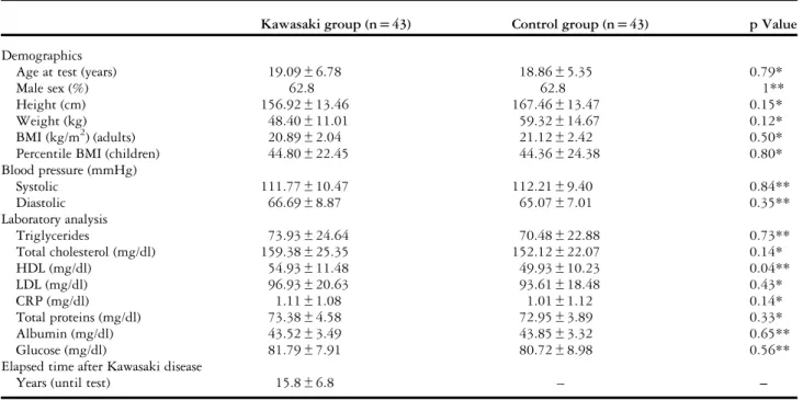
The Endothelial Pulse Amplitude Test (EndoPAT) was performed and repeated; the results demonstrated their reproducibility without statistically significant differ-ences between them in both groups: Kawasaki disease patients had a mean reactive hyperaemia index of 1.59± 0.45 and 1.62 ± 0.41 (first and second assess-ment, respectively), whereas control group had mean values of 1.98± 0.41 and 2.01 ± 0.43, (Table 2). There was a good correlation between both studies (Pearson’s correlation 0.958, p value<0.001).
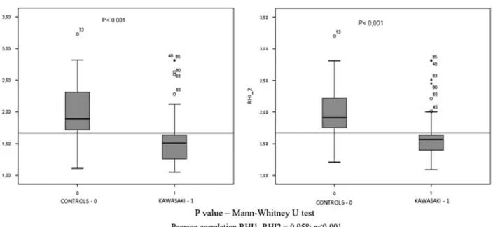
Endothelial dysfunction was found in the majority (n= 34; 79.06%) of Kawasaki disease patients, when compared with a significantly lower percentage of con-trol subjects (n= 8; 18.60%). There were significant group differences in pulse wave amplitude-derived reactive hyperaemia index, with Kawasaki disease patients having a significantly lower reactive hyper-aemia index when compared with the control group (1.59 and 1.62 versus 1.98 and 2.01; p< 0.001) (Fig 1). Pulse wave amplitude-derived augmentation index in both measurements were within the normal range and similar for both groups (−4.5 ± 7 and −4 ± 8 versus −5 ± 9 and −4.5 ± 9% p 0.61 and p 0.63 for Kawasaki and controls group, respectively) with no statistical significance; the same results were obtained for the augmentation index adjusted to 75 beats/minute (Table 2).
Carotid intima-media thickness
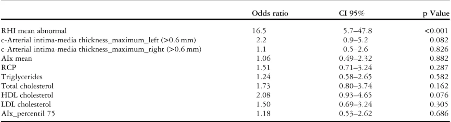
The maximum carotid arterial intima-media thickness results were slightly higher for the Kawasaki disease patients, but without significance (left common carotid artery maximum: 0.587± 0.100 versus 0.575 ± 0.065; p 0.22; right common carotid artery maximum: 0.598± 0.082 versus 0.574 ± 0.074; p 0.34), whereas the carotid arterial intima-media thickness mean values were similar for both groups, (left common carotid artery mean: 0.447± 0.088 versus 0.443 ± 0.059; p 0.93; right common carotid artery mean: 0.441± 0.073 ver-sus 0.438± 0.058; p 0.89) (Table 3). There were some outliers for these values in both groups; nevertheless, when we excluded the outliers, there was no change in the significance. We also found that 97.7 and 95.3% of subjects in the Kawasaki and control group, respectively, had mean carotid arterial intima-media thickness values under 6 mm. No plaques or flow abnormalities were identified in either group by carotid ultrasound imaging (Figs 2 and 3). In the multivariate analysis, the main factor associated with Kawasaki disease was an abnormal reactive hyperaemia index (odds ratio= 16.5; 95% confidence interval, 5.7-47.8; p < 0.001) (Table 4).
Discussion
Kawasaki disease occurs worldwide as both endemic and community-based epidemic forms in children of Table 1. Descriptive variables.
Kawasaki group (n= 43) Control group (n= 43) p Value
Demographics
Age at test (years) 19.09± 6.78 18.86± 5.35 0.79*
Male sex (%) 62.8 62.8 1**
Height (cm) 156.92± 13.46 167.46± 13.47 0.15*
Weight (kg) 48.40± 11.01 59.32± 14.67 0.12*
BMI (kg/m2) (adults) 20.89± 2.04 21.12± 2.42 0.50*
Percentile BMI (children) 44.80± 22.45 44.36± 24.38 0.80*
Blood pressure (mmHg) Systolic 111.77± 10.47 112.21± 9.40 0.84** Diastolic 66.69± 8.87 65.07± 7.01 0.35** Laboratory analysis Triglycerides 73.93± 24.64 70.48± 22.88 0.73** Total cholesterol (mg/dl) 159.38± 25.35 152.12± 22.07 0.14* HDL (mg/dl) 54.93± 11.48 49.93± 10.23 0.04** LDL (mg/dl) 96.93± 20.63 93.61± 18.48 0.43* CRP (mg/dl) 1.11± 1.08 1.01± 1.12 0.14* Total proteins (mg/dl) 73.38± 4.58 72.95± 3.89 0.33* Albumin (mg/dl) 43.52± 3.49 43.85± 3.32 0.65** Glucose (mg/dl) 81.79± 7.91 80.72± 8.98 0.56**
Elapsed time after Kawasaki disease
Years (until test) 15.8± 6.8 – –
BMI= body mass index; CRP = C-reactive protein Values are means± standard deviation
*Mann–Whitney U-test **Student’s t-test
all races. It is a medium-vessel vasculitis with predominant involvement of the coronary arteries. The search for an aetiological agent has been wide ranging. Kawasaki disease is accompanied by significant derangements in the immunoregulatory system3–5that lead to coronary inflammation and disruption with dilatation and aneurysm formation. Endothelial cell damage appears to occur as a result of this increased immune activity.
The long-term prognosis of Kawasaki disease, par-ticularly in patients with normal coronary arteries, mild dilatation, or regressed coronary aneurysms, remains uncertain at present. Several authors have demonstrated that 50% of aneurysms regress within several years with a normal angiographic lumen;6,10 however, histological studies and intra-coronary ultra-sound examinations have demonstrated persistent
abnormal arterial walls with marked thickening of intima, which may be caused by the proliferation of smooth muscle cells.12,15
Besides these morphological abnormalities, it has also been demonstrated that Kawasaki disease has a deleterious effect on coronary artery function, years after the acute presentation, particularly in patients with persistent coronary artery aneurysms when compared with control subjects.6,12,14,15 Abnormal coronary flow reserve has been demonstrated in Kawasaki disease patients with transiently dilated coronary arteries17 and in those without antecedent coronary artery dilation or ischaemia.43,44
Abnormalities in endothelial function,10,11,15,18,19,40 arterial stiffness,45–47and carotid intima-media thick-ness have also been observed.33,34,48 Nevertheless, controversy persists, and other studies have failed to
Figure 1.
Box plots of reactive hyperaemia index (RHI) testing in Kawasaki disease patients and controls using Mann–Whitney U-test. The red line represents the cut-off value of 1.67 for RHI (normal endothelial function RHI> 1.67).
Table 2. EndoPat results.
Kawasaki group (n= 43) Control group (n= 43) p Value
RHI (1) 1.59± 0.45 1.98± 0.45 <0.001* RHI (2) 1.62± 0.41 2.01± 0.43 <0.001* RHI mean (1_2) 1.60± 0.42 1.99± 0.43 <0.001* AIx (1) (%) −4.5 ± 7 −5 ± 9 0.61** AIx (2) (%) −4 ± 8 −4.5 ± 9 0.63** AIx mean (1_2) (%) −4.4 ± 7.5 −4.7 ± 9 0.62** AIx_percentil_75 (1) (%) −6 ± 7 −9 ± 7 0.78** AIx_percentil_75 (2) (%) −7 ± 8 −9 ± 9 0.87** AIx_percentil_75 mean (1_2) (%) −6.5 ± 7.5 −9 ± 8 0.09**
AIx= Augmentation Index; AIx@75 = Augmentation Index referred to 75 beats/minute; RHI = Reactive Hyperaemia Index Values are means± standard deviation
*Mann–Whitney U-test **Student’s t-test
demonstrate thosefindings.8,49This uncertainty about long-term vascular function of Kawasaki disease patients without history of coronary abnormalities is also reflected on different guidelines approach on life-long follow-up of these patients.50,51
Understanding the long-term vascular function in Kawasaki disease patients is an important issue, because its presence might represent ongoing inflammatory reaction and can preclude increased cardiovascular risk and the need for serial long-term cardiac follow-up.14,16–22,34,48,52,53 In addition, con-sidering that endothelium dysfunction is involved in the atherosclerotic process, this raises questions about similar pathological pathways.20–22,27,54
In this study, we found significant late endothelial dysfunction but absence of increased stiffness or increased carotid intima-media thickness in the
majority of Kawasaki disease patients when
compared with matched controls. The endothelial-dependent microvascular relaxation abnormality may be a consequence of ongoing systemic inflammatory process with sequelar damaged endothelium. The
finding of normal c-arterial intima-media thickness on both carotid arteries and the absence of differences between the two groups are consistent with the absence of subclinical atherosclerotic changes, but we can speculate that the observed histological changes in the arterial walls of Kawasaki disease patients might take longer time to evolve to atherosclerotic changes.33,34,48 Investigation of vascular function in Kawasaki disease patients has recently been revived with a number of similar studies published in the litera-ture. Most of these studies were conducted in chil-dren with Kawasaki disease having coronary artery aneurysms. Noto et al20found an increase in carotid arterial intima-media thickness, in both children and adults, late after the acute disease in patients with coronary artery lesions. Cheung et al45 have specu-lated that the increased carotid arterial intima-media thickness in Kawasaki disease patients without cor-onary involvement was associated with increased systemic arterial stiffness; our study demonstrates the opposite, as we did notfind a significantly increased augmentation index, a measure of arterial stiffness, or Table 3. Right and left common carotid intima-media thickness (c-arterial intima-media thickness) measurements.
Kawasaki group (n= 43) Control group (n= 43) p Value
c-Arterial intima-media thickness left maximum 0.587± 0.100 0.575± 0.065 0.22**
c-Arterial intima-media thickness left mean 0.447± 0.088 0.443± 0.059 0.93*
c-Arterial intima-media thickness left SD 0.063± 0.022 0.062± 0.016 0.89*
c-Arterial intima-media thickness right maximum 0.598± 0.082 0.574± 0.074 0.34*
c-Arterial intima-media thickness right mean 0.441± 0.073 0.438± 0.058 0.89*
c-Arterial intima-media thickness right SD 0.062± 0.015 0.076± 0.084 0.37*
Values are means± standard deviation *Mann–Whitney U-test
**Student’s t-test
Figure 2.
Box plots of the mean of two measurements of augmentation indices (AI and AI_Percentil_75) testing in Kawasaki disease patients and controls.
significantly increased carotid intima-media thick-ness, and we speculate this can represent different genetic polymorphisms.
The consistency of endothelial dysfunction, in this study, is in line with the one from Dhillon et al,11 which demonstrated abnormalflow-mediated dilation
Figure 3.
Box plots of left and right maximum (a and b) and mean (c and d) carotid intima-media thickness (c-arterial intima-media thickness) values in Kawasaki disease patients and controls.
Table 4. Logistic regression: calculated odds ratio value.
Odds ratio CI 95% p Value
RHI mean abnormal 16.5 5.7–47.8 <0.001
c-Arterial intima-media thickness_maximum_left (>0.6 mm) 2.2 0.9–5.2 0.082
c-Arterial intima-media thickness_maximum_right (>0.6 mm) 1.1 0.5–2.6 0.826
AIx mean 1.06 0.49–2.32 0.882 RCP 1.51 0.71–3.24 0.287 Triglycerides 1.24 0.58–2.65 0.582 Total cholesterol 1.73 0.80–3.74 0.162 HDL cholesterol 2.08 0.93–4.65 0.076 LDL cholesterol 1.50 0.69–3.24 0.305 AIx_percentil 75 1.18 0.53–2.62 0.686
of the brachial artery in 20 male subjects, who were studied 5–17 years after the onset of Kawasaki disease, irrespective of whether they had developed aneurysms during the acute phase. Endothelial dysfunction was also reported by Deng et al and Dalla Pozza et al48in some children with Kawasaki disease, but without coronary artery aneurysms. Our study adds evidence to these findings and suggests that the occurrence of Kawasaki disease may have lifelong consequences even when there is no coronary artery involvement. This issue, however, is rather controversial. Studies performed in Japanese subjects, of identical characteristics, reported abnormal flow-mediated dilation only in patients who had devel-oped coronary artery aneurysms during the acute stage of Kawasaki.19,26 A Canadian study of 52 Kawasaki disease patients concluded that there was no evidence of long-term endothelial cell dysfunction late after Kawasaki disease onset.8A recent American study demonstrated that a mixed population of Kawasaki disease patients with and without coronary abnormalities had normal vascular health 1 year after the Kawasaki disease episode.49It is unclear whether these conflicting results reflect methodological issues or are best explained by genetic differences of the studied populations.
Children with Kawasaki disease, with or without coronary artery lesions, seem to have a more adverse cardiovascular risk profile, which requires lifelong cardiac assessment and counselling, either sporadic or serial. Whether this is a cause or a consequence of an abnormal endothelial function has not yet been assessed. We also failed to demonstrate premature or subclinical atherosclerotic changes with the used methodology. What is noteworthy is the fact that, in our sample, almost 79% of Kawasaki disease patients had endothelial dysfunction, when compared with only 17% of the control subjects.
These findings suggest that children with
Kawasaki disease may have long-term sequelae, even when there is no discernible coronary artery involve-ment in the acute stage of the disease. Affected chil-dren should therefore be kept on long-term follow-up and counselled to reduce cardiovascular risk factors such as sedentary lifestyle, overweight, high-lipid and high-caloric meals, and alcohol or tobacco use. Further research is needed to assess whether known strategies to improve endothelial function would bring potential benefits to Kawasaki disease patients.
Acknowledgements
The authors thank Lurdes Medroa for her contribu-tion in performing the EndoPat studies and Helena Aidos for the statistical analysis.
Financial Support
This research received no specific grant from any fund-ing agency or from commercial or not-for-profit sectors. Conflicts of Interest
None.
Ethical Standards
The authors assert that all procedures contributing to this work comply with the ethical standards of the relevant national guidelines on human experimenta-tion and with the Helsinki Declaraexperimenta-tion of 1975, as revised in 2008, and has been approved by the institutional committees.
References
1. Kawasaki T. Acute febrile mucocutaneous syndrome with lymphoid involvement with specific desquamation of the fingers and toes in children. Arerugi 1967; 16: 178–222.
2. Laranjo S, Aidos H, Lourenço A, Rodrigues V, Pinto FF. Ten years of Kawasaki disease in Portugal: national database results. Rev Port Cardiol 2015; 34: 171.
3. Rowley AH. The etiology of Kawasaki disease: superantigen or conventional antigen? Pediatr Infect Dis J 1999; 18: 69–70. 4. Wang CL, Wu YT, Liu CA, et al. Kawasaki disease: infection,
immunity and genetics. Pediatr Infect Dis J 2005; 24: 998–1004. 5. Duong TT, Silverman ED, Bissessar MV, et al. Superantigenic activity is responsible for induction of coronary arteritis in mice: an animal model of Kawasaki disease. Int Immunol 2003; 15: 79–89. 6. Kato H, Sugimura T, Akagi T, et al. Long-term consequences of Kawasaki disease. A 0-to 21-year follow-up study of 594 patients. Circulation 1996; 94: 1379–1385.
7. Gupta-Malhotra M, Gruber D, Abraham SS, et al. Atherosclerosis in survivors of Kawasaki disease. J Pediatr 2009; 155: 572–577. 8. McCrindle BW, McIntyre S, Kim C, et al. Are patients after
Kawasaki disease at increased risk for accelerated atherosclerosis? J Pediatr 2007; 151: 244–248.
9. Newburger JW, Burns JC, Beiser AS, et al. Altered lipid profile after Kawasaki syndrome. Circulation 1991; 84: 625–631. 10. Mitani Y, Sawada H, Hayakawa H, et al. Elevated levels of
high-sensitivity C-reactive protein and serum amyloid-A late after Kawasaki disease: association between inflammation and late coronary sequelae in Kawasaki disease. Circulation 2005; 111: 38–43. 11. Dhillon R, Clarkson P, Donald AE, et al. Endothelial dysfunction
late after Kawasaki disease. Circulation 1996; 94: 2103–2106. 12. Iemura M, Ishii M, Sugimura T, et al. Long term consequences of
regressed coronary aneurysms after Kawasaki disease: vascular wall morphology and function. Heart 2000; 83: 307–311.
13. Orenstein JM, Shulman ST, Fox LM, et al. Three linked vasculo-pathic processes characterize Kawasaki disease: a light and trans-mission electron microscopic study. PLoS One 2012; 7: e38998. 14. Burns JC, Shike H, Gordon JB, et al. Sequelae of Kawasaki disease
in adolescents and young adults. J Am Coll Cardiol 1996; 28: 253–257.
15. Yamakawa R, Ishii M, Sugimura T, et al. Coronary endothelial dysfunction after Kawasaki disease: evaluation by intracoronary injection of acetylcholine. J Am Coll Cardiol 1998; 31: 1074–1080.
16. Silva AA, Maeno Y, Hashmi A, et al. Cardiovascular risk factors after Kawasaki disease: a case-control study. J Pediatr 2001; 138: 400–405.
17. Furuyama H, Odagawa Y, Katoh C, et al. Altered myocardialflow reserve and endothelial function late after Kawasaki disease. J Pediatr 2003; 142: 149–154.
18. Ikemoto Y, Ogino H, Terguchi M, et al. Evaluation of preclinical atherosclerosis byflow-mediated dilation of the brachial artery and carotid artery analysis in patients with a history of Kawasaki disease. Pediatr Cardiol 2005; 26: 782–786.
19. Noto N, Okada T, Yamasuge M, et al. Noninvasive assessment of the early progression of atherosclerosis in adolescents with Kawasaki dis-ease and coronary artery lesions. Pediatrics 2001; 107: 1095–1099. 20. Ross R. Atherosclerosis an inflammatory disease. N Engl J Med
1999; 340: 115–126.
21. Zeiher AM, Drexler H, Wollschlager H, et al. Modulation of coronary vasomotor tone in humans. Progressive endothelial dysfunction with different early stages of coronary atherosclerosis. Circulation 1991; 83: 391–401.
22. Vita JA, Keaney JF Jr. Endothelial function: a barometer for cardiovascular risk. Circulation 2002; 106: 640–642.
23. Furchgott RF, Zawadzki JV. The obligatory role of endothelial cells in the relaxation of arterial smooth muscle by acetylcholine. Nature 1980; 288: 373–376.
24. Forstermann U, Closs EI, Pollock JS, et al. Nitric oxide synthase isozymes. Characterization, purification, molecular cloning, and functions. Hypertension 1994; 23: 1121–1131.
25. Cooke JP, Rossitch E Jr, Andon NA, et al. Flow activates an endothelial potassium channel to release an endogenous nitrovaso-dilator. J Clin Invest 1991; 88: 1663–1671.
26. Kadono T, Sugiyama H, Hoshiai M, et al. Endothelial function evaluated byflow-mediated dilatation in pediatric vascular disease. Pediatr Cardiol 2005; 26: 385–390.
27. Deanfield JE, Halcox JP, Rabelink TJ. Endothelial function and dysfunction: testing and clinical relevance. Circulation 2007; 115: 1285–1295.
28. Hamburg NM, Palmisano J, Larson MG, et al. Relation of brachial and digital measures of vascular function in the community: the Framingham Heart Study. Hypertension 2011; 57: 390–396. 29. Kuvin JT, Patel AR, Sliney KA, et al. Assessment of peripheral
vascular endothelial function with finger arterial pulse wave amplitude. Am Heart J 2003; 146: 168–174.
30. Laurent S, Cockcroft J, Van Bortel L, et al. Expert consensus document on arterial stiffness: methodological issues and clinical applications. Eur Heart J 2006; 27: 2588–2605.
31. Newburger J, Keflioglu-Scheiber A, Opazo Saez AM, et al. Augmentation index is associated with cardiovascular risk. J Hypertens 2002; 20: 2407–2414.
32. Urbina EM, Williams RV, Alpert BS, et al. Noninvasive assess-ment of subclinical atherosclerosis in children and adolescents: recommendations for standard assessment for clinical research: a scientific statement from the American Heart Association. Hypertension 2009; 54: 919–950.
33. Meena RS, Rohit M, Gupta A, et al. Carotid intima-media thickness in children with Kawasaki disease. Rheumatol Int 2014; 34: 1117–1121. 34. Lee SJ, Ahn HM, You JH, et al. Carotid intima media thickness and pulse wave velocity after recovery from Kawasaki disease. Korean Circ J 2009; 39: 264–269.
35. Cole TJ, Bellizi MC, Flegal KM, et al. Establishing a standard definition for child overweight and obesity worldwide: international survey. BMJ 2000; 320: 1240–1243.
36. Ayusawa M, Sonobe T, Uemura S, et al. Revision of diagnostic guidelines for Kawasaki disease (the 5th revised edition). Pediatr Int 2005; 47: 232–234.
37. Kuvin JT, Karas RH. Clinical utility of endothelial function testing: ready for prime time? Circulation 2003; 107: 3243–3247. 38. Kuvin JT, Patel AR, Sliney KA, et al. Assessment of peripheral vascular endothelial function with finger arterial pulse wave amplitude. Am Heart J 2003; 146: 168–174.
39. Selamet Tierney ES, Newburger JW, Gauvreau K, et al. Endothe-lial pulse amplitude testing: feasibility and reproducibility in adolescents. J Pediatr 2009; 154: 901–905.
40. Pinto FF, Laranjo S, Paramés F, et al. Long-term evaluation of endothelial function in Kawasaki disease patients. Cardiol Young 2013; 23: 517–522.
41. Roman MJ, Naqvi TZ, Gardin JM, et al. Clinical application of non-invasive vascular ultrasound in cardiovascular risk strati fica-tion: a report from the American Society of Echocardiography and the Society for Vascular Medicine and Biology. J Am Soc Echocardiogr 2006; 11: 201–211.
42. Touboul PJ, Hennerici MG, Meairs S, et al. Mannheim carotid intima-media thickness and plaque consensus (2004-2006-2011). An update on behalf of the advisory board of the 3rd, 4th and 5th watching the risk symposia, at the 13th, 15th and 20th European Stroke Conferences, Mannheim, Germany, 2004, Brussels, Belgium, 2006, and Hamburg, Germany, 2011. Cerebrovasc Dis 2012; 34: 290–296.
43. Hamaoka K, Onouchi Z, Ohmochi Y. Coronaryflow reserve in children with Kawasaki disease without angiographic evidence of coronary stenosis. Am J Cardiol 1992; 69: 691–692.
44. Hauser M, Bengel F, Kuehn A, et al. Myocardial bloodflow and coronaryflow reserve in children with ‘normal’ epicardial coronary arteries after the onset of Kawasaki disease assessed by positron emission tomography. Pediatr Cardiol 2004; 25: 108–112. 45. Cheung YF, Wong SJ, Ho MHK. Relationship between carotid
intima-media thickness and arterial stiffness in children after Kawasaki disease. Arch Dis Child. 2007; 92: 43–47.
46. Ooyanagi R, Fuse S, Tomita H, et al. Pulse wave velocity and ankle brachial index in patients with Kawasaki disease. Pediatr Int 2004; 46: 398–402.
47. Mitani Y, Okuda Y, Shimpo H, et al. Impaired endothelial function in epicardial coronary arteries after Kawasaki disease. Circulation 1997; 96: 454–461.
48. Dalla Pozza R, Bechtold S, Urschel S, et al. Subclinical athero-sclerosis, but normal autonomic function after Kawasaki syndrome. J Pediatr 2007; 151: 239–243.
49. Tierney ESS, Gal D, Gauvreau K, et al. Vascular health in Kawasaki Disease. J Am Coll Cardiol 2013; 62: 1114–1121.
50. Newburger JW, Takahashi M, Gerber MA, et al. Diagnosis, treatment, and long-term management of Kawasaki disease: a statement for health professionals from the Committee on Rheumatic Fever, Endocarditis, and Kawasaki Disease, Council on Cardiovascular Disease in the Young, American Heart Association. Pediatrics 2004; 114: 1708–1733.
51. Newburger JW, Takahashi M, Gerber MA, et al. Guidelines for diagnosis and management of cardiovascular sequelae in Kawasaki disease (JCS 2008)– digest version. Circ J 2010; 74: 1989–2020.
52. Gersony WM. The adult after Kawasaki disease the risks for late coronary events. J Am Coll Cardiol 2009; 54: 1921–1923. 53. Gordon JB, Kahn AM, Burns JC. When children with Kawasaki
disease grow up; myocardial and vascular complications in adult-hood. J Am Coll Cardiol 2009; 54: 1911–1912.
54. Libby P, Ridker PM, Maseri A. Inflammation and atherosclerosis. Circulation 2002; 105: 1135–1143.