UNIVERSIDADE DE LISBOA
FACULDADE DE CIÊNCIAS
DEPARTAMENTO DE BIOLOGIA VEGETAL
Iron Man(ipulation): unravelling the iron responsive genes’
network in malaria parasites
Miguel Dinis Monteiro dos Santos
Mestrado em Bioinformática e Biologia Computacional
Especialização em Biologia Computacional
Dissertação orientada por:
Dr. Ksenija Slavic
Dr. Octávio Paulo
Acknowledgments
It would not be possible to finish this thesis without the help and support from several people. I will only mention the ones that were closer to me during this process, but I am truly thankful to all of you that contributed directly and indirectly to the outcome of this work.
I am truly thankful to Dr. Maria Mota for accepting me in her lab and allowing me to perform my Master thesis in her group. I want to thank to my internal supervisor, Dr. Octávio Paulo for all the support and confidence. To Ksenija, for being the best supervisor I could ever ask. Since the first day I felt your trust in my work and that you were always listening to my ideas and suggestions. At the same time, you were always frontal with me, teaching me in the best way you can, by correcting my mistakes and congratulating me when things were properly done. You will be truly important for everything I will do in my life after this, and I won’t never forget your advices (both personal and scientific).
To Torcato Martins and Filipa Domingos, for providing me with one of the best summer I had and being a major part of my scientific and personal growth.
To everyone in MMota lab, for the constant help and support, as well as the friendship and funny moments. Here I have to highlight some names: thank you Ângelo, Vanessa, Iset, Inês B. and João V., for sharing their knowledge, advices and for the challenging conversations, Liliana for all the help and enthusiastic teachings, to Jon for making every night The Night and to Maria Inês Marreiros for the unconditional friendship and support. If our lab is a family, for sure you are my sister.
To everyone in LFigueiredo and MPrudencio labs, for the help and friendship. I want to give a special thanks to Daniel Neves for the advices, the productive brainstorming and constant availability for help and to Fábio for the friendship. I am also thankful to António Mendes for the interesting discussions. Thank you to Ana Rita Grosso for the advices and help provided. I want to give a special thanks to the STAB VIDA staff for the good work and support.
To all my friends, for being the best I have and for being always there with all the patience and friendship: to Nina, one of my oldest friends, to Pedro for the nights and conversations and to Camila for the infinite patience and constant care; you are one of the people I wish more the best. To Chico and Carolina, two of my most recent friends without whom this year wouldn’t be the same: Chico for the deep, fun and honest friendship, you became like an older brother to me; Carolina for becoming one of my best friends and one of the most important people during this process, watching you overtake this year was truly inspirational. To my great friend João Simão: despite the distance I felt that you were always close. You are one of the people I respect and I am more proud of. To Maria Beatriz, my best and oldest friend. Every day you keep surprising me and make me feel lucky for met you. To Malhadas, my best friend: I know you would give an arm and a leg for me and I would do the same for you. I miss you and I am proud of you every day, I know things with you will never change.
To Marco, one of the most brilliant people I ever met and one of my best friends. For all the conversations, nights and honest advices (if some were difficult to hear, I cannot imagine how difficult they were to tell). I trust you with all my heart and you are one of the few people I feel I can be completely myself. Your unconditional friendship and support during the last years were indescribable and completely fundamental. Thank you for challenging me and make me break my limits. Thank you for being always frontal with me. You are an amazing person and I know you will be incredible in everything you do.
To Filipe, Bia, Inês (both) and Patrick for more than living with me, for being my home: you were part of everything during the last 5 years and I could not have been luckier for meeting you. You make me leave and enter at home with a smile every day and calling you family is not enough. We already came through so many, and there is nothing that I didn’t tell you already. I love you with all my heart. To everyone that passed through our house: James, Afonso, Dani and Alice. To James (the cat) and Luke for being partners in crime and friends for life.
To my Family, to whom I owe the person I am and everything I got and achieved. I could not be happier with you. To my lovely Mother, the woman of my life and the one that was always there. I do not have words to explain how much I love you and feel blessed for being your son. I wouldn’t change anything about you. To my lovely Father, one of the two best man I know. I hope one day I will be half the man you are. Thank you for teaching me what is truly important and for making me feel the best. To Tiago, my big brother: you are the other best man I know, my role model and a friend for life. I would not have achieved anything I did without your help, advices and unconditional and tireless support, especially during this year. Part of this work I owe to you, as well as most of what I am. Thank you for having your home always available for me, and for loving me as you do. You are the best brother I could ever ask. To Maria, my little sister and the joy of my eyes. You are such an inspiration! I hope I can be in every step of your life and you know that you can count with me for all your life. I love you since the day you were born. To my little babies Matilde and Simão for making me feel so loved, and to Maria Helena for loving me as a son. To my grandparents which would give everything for me, and all my uncles, aunts and cousins. I am the luckiest person ever.
To the most important person ever, to whom I already told everything I could and that were part of every step. Diana, every day I try to find a way to show you how much I love you and how much I am thankful to you. You are the person I respect and are more proud of and I keep learning with you every day. I have never met someone like you. I know with all my heart that you are the only person I will ever love. You never failed to me, and were by my side in every moment. I hope you will continue being part of my life as you are until the last day. I will always love you and I know we don’t need to tell anything to each other in order to know what we are feeling. Thank you my love, for being as you are. Thank you!
Resumo
A Malária é uma doença infeciosa causada por várias espécies de parasitas protozoários do género
Plasmodium. Entre eles, P. falciparum é o que provoca a maior mortalidade em humanos. O parasita
apresenta um ciclo de vida complexo, alternando entre o inseto vetor que transmite a forma infeciosa do parasita e o hospedeiro mamífero, no qual após a infeção o parasita passa por uma enorme replicação causando os sintomas da doença. Após a picada de um mosquito infetado, formas móveis denominadas esporozoítos são injetadas dentro do hospedeiro mamífero e viajam até ao fígado onde invadem hepatócitos. Após replicar dentro dos hepatócitos, milhares de novos parasitas são libertados para a corrente sanguínea onde invadem os glóbulos vermelhos. Durante a fase sanguínea da infeção o parasita passa por diversas formas de desenvolvimento, utilizando um programa de expressão génica altamente coordenado onde genes codificantes de proteínas para funções específicas de cada forma são ativados apenas quando são necessários. Ocasionalmente, algumas formas da fase sanguínea diferenciam-se em gametócitos, a forma sexual transmissível para os mosquitos. Durante este complexo ciclo de vida, para assegurar a sua sobrevivência e replicação, o parasita necessita de sequestrar nutrientes e micronutrientes de várias fontes dos tecidos do mosquito e do mamífero. Portanto, foi posta a hipótese de que os parasitas causadores de malária possuem mecanismos adequados de adaptação de maneira a possibilitar a utilização desta enorme variedade de recursos. O ferro é um dos micronutrientes mais importantes, estando envolvido em diversos processos celulares essenciais para a sobrevivência, como a replicação do ADN e a produção de ATP. Por outro lado, o ferro é altamente tóxico quando se encontra em excesso, o que implica que a sua concentração dentro das células tenha que ser mantida num intervalo restrito. Isto é tipicamente alcançado através da regulação da expressão de proteínas envolvidas na aquisição de ferro, armazenamento e metabolismo. Apesar do papel fundamental do ferro e da sua regulação para a sobrevivência celular, uma grande quantidade de informação básica encontra-se em falta no que diz respeito à forma como o parasita lida com este valioso recurso. De maneira a abordar esta questão, realizámos uma análise do transcriptoma total de P. falciparum durante a fase sanguínea em condições a curto prazo de privação de ferro e exposição a excesso de ferro. Ao identificar os genes diferencialmente expressos entre as diferentes condições e os respetivos controlos, pretendemos encontrar genes envolvidos na deteção e regulação de ferro. Os genes identificados como diferencialmente expressos possuem uma grande variedade de funções, desde fatores de transcrição e proteínas de ligação a ARN mensageiro até componentes do sistema ubiquitina-proteossoma, ARNs não codificantes, transferases de grupos metilo de histonas, vários transportadores e proteínas envolvidas no metabolismo. Estes resultados sugerem a existência de diversos níveis de regulação dos genes relacionados com o ferro, tanto ao nível da transcrição e da pós-transcrição, bem como ao nível da degradação de proteínas e mecanismos epigenéticos, indicando uma rede altamente complexa envolvida na manutenção da homeostasia do ferro em Plasmodium.
Abstract
Malaria is an infectious disease caused by several species of protozoan parasites of the Plasmodium genus. Among them, P. falciparum is the one causing the greatest mortality in humans. The parasite has a complex life-cycle, switching between the insect vector which transmits the infective form of the parasite and the mammalian host, in which upon infection parasite goes through enormous replication causing the symptoms of the disease. Upon the bite of an infected mosquito, motile forms called sporozoites are injected into the mammalian host which travel to the liver and invade hepatocytes. After replicating inside hepatocytes, thousands of new parasites are released into the bloodstream where they invade red blood cells. During the blood stage of the infection the parasite goes through several developmental stages, using a tightly coordinated gene expression program where genes encoding proteins for stage-specific functions are activated only when they are required. Occasionally, some blood stage forms differentiate into gametocytes, the sexual stage transmissible to the mosquitoes. During this complex life cycle, to support its survival and replication, parasite needs to scavenge nutrients and micronutrients from various sources of the mammalian and insect tissues. Thus, it is hypothesized that malaria parasites must have adequate mechanisms of adaptation to be able to use this wide range of resources. Iron is one of the most important micronutrients, as it is involved in several cellular processes essential for survival, such as DNA replication and ATP production. On the other hand, iron is highly toxic when found in excess, therefore its intracellular concentrations need to be kept in a very narrow range. This is typically achieved through regulation of expression of proteins involved in iron acquisition, storage and metabolic usage. Despite the critical role of iron and its regulation for cell survival, a great deal of basic information is missing regarding the way that malaria parasites deal with this valuable resource. In order to address this question, we performed genome-wide transcriptome analysis of P. falciparum blood-stage by RNA-seq in conditions of short-term iron deprivation and exposure to iron excess. By identifying the differentially expressed genes between the different conditions and the respective control, we aimed to find genes involved in iron sensing and regulation. The genes identified as differentially expressed had a great variety of functions, from transcription factors and mRNA-binding proteins to components of the ubiquitin-proteasomal system, ncRNAs, histone methyl-transferases, several transporters and metabolism-related proteins. These results suggest the existence of several layers of regulation of the iron-responsive genes, both at the levels of transcription and post-transcription, as well as protein degradation and epigenetic mechanisms, pointing to a highly complex network involved in the maintenance of iron homeostasis in Plasmodium.
Resumo alargado
A Malária é uma doença infeciosa causada por várias espécies de parasitas protozoários do género
Plasmodium. Apesar dos esforços para a prevenção, da redução no número de casos e do investimento
na investigação de novas terapias e vacinas anti-maláricas, esta doença continua a representar um grande fardo económico nas áreas endémicas. Entre as espécies existentes, P. falciparum é aquela que causa maior número de casos e maior mortalidade em humanos. Estes parasitas apresentam um complexo ciclo de vida, que se inicia quando uma fêmea de mosquito do género Anopheles (o hospedeiro invertebrado) injecta esporozoítos, uma forma infeciosa altamente móvel, na derme do hospedeiro mamífero (humanos no caso de P. falciparum) durante a sua refeição de sangue. Os esporozoítos viajam para a corrente sanguínea e dirigem-se ao fígado, onde invadem hepatócitos dentro dos quais se multiplicam em milhares de merozoítos. Estes viajam para a corrente sanguínea e invadem os glóbulos vermelhos, iniciando-se a fase sanguínea (assexuada) onde os parasitas apresentam um elevado grau de regulação da expressão génica, onde genes com funções específicas para cada forma são ativos apenas no exato momento em que são necessários. Durante a fase sanguínea o parasita assume diversas formas morfologicamente distintas: uma forma denominada anel devido à sua morfologia característica, os trofozoítos, altamente ativos metabolicamente, e os esquizontes. Dentro dos esquizontes formam-se novos merozoítos que irão invadir novos glóbulos vermelhos. Ocasionalmente, alguns merozoítos diferenciam-se em gametócitos feminino ou masculino, a forma sexuada infeciosa para os mosquitos. Quando os mosquitos picam um hospedeiro mamífero infetado, os gametócitos são transportados para o sistema digestivo destes onde vão dar origem aos respetivos gâmetas. O zigoto diploide móvel resultante da fecundação denomina-se oocineto, que invade as paredes do sistema digestivo do mosquito e diferencia-se num oocisto. Dentro deste ocorrem várias divisões mitóticas que darão origem a novos esporozoítos, que viajarão para as glândulas salivares dos mosquitos e serão transmitidos para um novo hospedeiro mamífero durante a próxima refeição de sangue.
Durante este complexo ciclo de vida, os parasitas lidam com diversos recursos do hospedeiro com uma larga gama de concentrações, o que exige que estes possuam um sofisticado sistema de deteção e regulação dos mesmos. Entre estes micronutrientes, o ferro possui uma elevada importância devido ao seu papel central em diversos processos celulares, desde a replicação do ADN e produção de ATP até à participação em diversas reações enzimáticas como cofactor. Este micronutriente possui características de oxidação-redução únicas que lhe conferem o seu papel central em diversas reações bioquímicas. O facto de alternar entre dois estados de oxidação (2+ e 3+) permite-lhe associar-se com proteínas, ligar-se
ao oxigénio e transportar eletrões, entre outras funções. Devido ao seu papel fulcral, este é altamente regulado na grande maioria dos organismos, desde as leveduras aos mamíferos onde diversos mecanismos de regulação ao nível da transcrição e da pós-transcrição atuam para manter a sua homeostasia, desde fatores de transcrição a proteínas que se ligam a ARN mensageiro. Apesar da sua elevada importância na relação parasita-hospedeiro, os mecanismos de regulação deste recurso em
Plasmodium são ainda altamente desconhecidos. Exceções são a identificação de dois transportadores
de ferro, o facto de o parasita regular a expressão da enzima hepática hepcidina, de extrema importância na regulação do ferro em mamíferos e do envolvimento do heme (grupo prostético da hemoglobina) no modo de ação da artemisinina, um dos medicamentos mais usados para combater a doença. Tendo estas evidências em conta, postulámos que estes mesmos mecanismos de regulação da expressão de genes envolvidos no metabolismo do ferro se encontram conservados no parasita, mantendo os seus níveis altos o suficiente, possibilitando a sua utilização sem que estes se tornem tóxicos.
Para a abordagem destas questões, as técnicas de sequenciação de larga escala de nova geração tornaram-se estratégias cada vez mais comuns e foram já aplicadas a diversos organismos incluindo parasitas do género Plasmodium. O facto de o seu genoma já se encontrar completamente sequenciado representa também uma vantagem neste tipo de estudos. Uma das técnicas mais utilizadas é a sequenciação do transcriptoma completo, ou seja, a determinação da expressão génica global de um dado organismo num dado momento denominada RNA-seq. Esta consiste na extração do ARN total de uma amostra biológica, construção de uma biblioteca de sequenciação através da conversão para ADN complementar, ligação de adaptadores que permitem a imobilização num suporte sólido e sequenciação utilizando nucleótidos artificiais que possuem um fluoróforo específico ligado. O sinal emitido será lido pelo sequenciador, permitindo a identificação das bases em cada posição. Esta técnica possibilita a abordagem de diversas questões, sendo a mais utilizada a identificação de genes diferencialmente expressos entre condições (organismo selvagem vs. organismo mutante ou situação controlo vs. tratamentos). Utilizando esta técnica, caracterizou-se o transcriptoma ao longo da fase sanguínea do parasita, revelando resultados muito interessantes. Ao longo desta fase do desenvolvimento ocorre uma onda de ativação e inibição dos genes específicos para cada forma morfológica e função específica, com cada gene tendo um único pico de expressão durante esta fase apenas quando é estritamente necessário. Pelos motivos descritos acima, propusemo-nos a identificar novos genes envolvidos na rede de regulação do ferro no parasita e, para tal, levámos a cabo uma experiência de sequenciação de ARN (RNA-seq) em diferentes condições de ferro de forma a identificar os genes diferencialmente expressos, que estarão potencialmente envolvidos neste processo. Para tal desenvolvemos um sistema artificial que introduz alterações na concentração de ferro do ambiente do parasita, nomeadamente uma condição de excesso e de deficiência de ferro. Após confirmarmos que o sistema era funcional e inofensivo para o desenvolvimento e viabilidade dos parasitas, identificámos diversos homólogos de genes envolvidos no metabolismo do ferro em organismos-modelo e testámos se a sua expressão era alterada pelos nossos tratamentos através de PCR quantitativo. Estes resultados validaram o nosso sistema, e permitiram-nos avançar para a experiência de RNA-seq, de maneira a caracterizar o transcriptoma de parasitas submetidos às diferentes condições de ferro. Para tal, utilizámos um protocolo no qual os fragmentos obtidos são alinhados contra o genoma de referência utilizando o software TopHat. De seguida procedemos à assemblagem dos transcritos utilizando o software Cufflinks e, por fim, realizámos a análise de expressão diferencial utilizando o software Cuffdiff. Devido ao facto de os parasitas dos nossos replicados biológicos, realizados em dias distintos, não se encontrarem exatamente na mesma forma morfológica, deparámo-nos com alguns problemas aquando do agrupamento dos nossos resultados. Ao compararmos os nossos dados de transcriptoma com os dados publicados ao longo da fase sanguínea, chegámos à conclusão que aquando da extração de ARN, os parasitas encontravam-se numa forma um pouco mais avançada no segundo dia quando comparando com o primeiro. A melhor solução foi a comparação das diferentes condições dentro de cada dia de experiência, tentando identificar genes com comportamentos semelhantes entre os tratamentos em cada dia.
Os genes identificados como diferencialmente expressos entre as condições de ferro e os respetivos controlos possuem uma grande variedade de funções, desde fatores de transcrição, proteínas que se ligam a ARN mensageiro, transferases de grupos metilo de histonas, enzimas envolvidas na degradação de proteínas pelo complexo ubiquitina-proteossoma, diversos transportadores e até ARNs não codificantes. Estes resultados demonstram que o parasita não mantém a homeostasia do ferro usando apenas uma estratégia, mas através de uma rede de genes e mecanismos altamente complexa e regulada a diversos níveis, o que permite a continuação deste projeto em diversas direções altamente interessantes.
Table of Contents
Acknowledgments ... I
Resumo ... IV
Abstract ... V
Resumo alargado ... VI
Table of Contents ... VIII
List of tables and figures ... X
List of abbreviations and symbols ... X
1 - Introduction ... 1
1.1 – The disease and the parasite ... 1
1.2 – Pathogen-host battle for iron ... 2
1.3 – Yeast iron sensing and regulation ... 4
1.4 – Iron sensing and regulation in mammalian cells ... 5
1.5 – Iron in Plasmodium: what do we know?... 6
1.6 – Welcome to the omics’ era! ... 7
1.6.1 – Our approach: RNA-seq! ... 8
1.7 – Plasmodium falciparum blood stage transcriptome: a unique gene regulation cascade mechanism among eukaryotes ... 9
2 – Thesis aims ... 13
3 – Methods ... 13
3.1 BLAST (Basic Local Alignment Search Tool) and Multiple Alignment ... 13
3.2 – Preparation of biological samples ... 13
3.2.1 – P. falciparum in vitro culture ... 13
3.2.2 – Giemsa staining of smears ... 14
3.2.3 – MCM composition ... 14
3.2.4 – Albumax II solution composition ... 14
3.2.5 – Washing of Human Blood ... 14
3.2.6 – Thawing of glycerolyte-frozen parasites ... 14
3.2.7 – Sorbitol synchronization ... 15 3.2.8 – Iron Treatments ... 15 3.2.9 – PhenGreen assay ... 15 3.2.10 – Viability assay ... 15 3.2.11 – RNA extraction ... 16 3.2.12 – cDNA synthesis ... 16 3.2.13 – Quantitative PCR (qPCR)... 17 3.3 – Sequencing ... 17
3.4 – RNA-seq bioinformatic analysis ... 18
3.4.1 – Quality control ... 18
3.4.2 – Mapping to reference genome ... 18
3.4.4 – Differential expression ... 20
3.5.5 – Downstream analysis ... 21
4 – Results ... 22
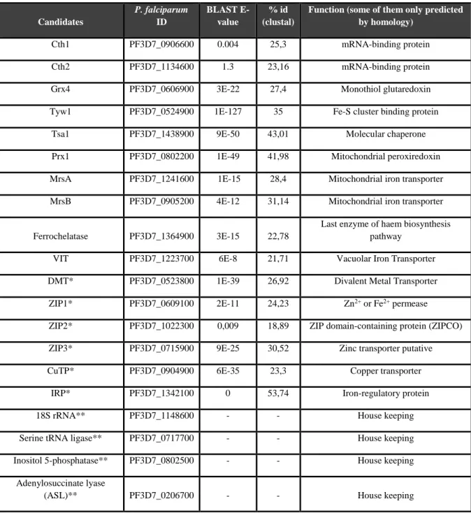
4.1 – BLASTing: in search for putative iron-responsive genes ... 22
4.1.1 – Iron treatments are changing iron levels in the iRBCs ... 24
4.1.2 – Iron treatments are not affecting parasites’ viability ... 26
4.1.3 – Iron treatments are affecting the expression levels of some candidate genes ... 26
4.2 – RNA-seq: genome-wide quest for iron-responsive mechanisms ... 28
4.2.1 – A replication issue: when the highly regulated P. falciparum blood stage transcriptome becomes a problem ... 29
4.2.2 – The clock-like pattern of gene expression ... 31
4.2.3 – Pairwise comparison: finding common patterns between treatments and experiments .... 32
5 – Discussion ... 37
References ... 41
List of tables and figures
Figure 1.1 - Plasmodium life cycle 1
Figure 1.2 - Transcriptional iron regulation in yeast 4
Figure 1.3 - Post-transcriptional iron regulation in yeast 5
Figure 1.4 - Post-transcriptional regulation by IRPs/IREs in mammals 6 Figure 1.5. – Solid-phase amplification 8
Figure 1.6. – Cyclic reversible termination 8
Figure 1.7. – Paired-End Reads 9
Figure 1.8. – P. falciparum IDC transcriptome 10
Table 4.1 – Candidate genes for which expression levels were tested after the iron treatments 24 Figure 4.1 – PhenGreen fluorescence of iRBCs after 6h (left) and after 24h (right) treatments 25 Figure 4.2 – Blood smears of infected RBCs in the different conditions 26 Figure 4.3 – Relative gene expression in P. falciparum after 6 hours with FeSO4 and DFO
compared to their corresponding control parasites 27
Table 4.2 – RNA concentration and ratios measured using Nanodrop and amount of RNA for each
sample 28
Figure 4.4 – Experimental workflow 28 Figure 4.5 - Agarose gels with RNA samples sent for sequencing 29 Figure 4.6 –Comparison of genes differentially expressed between the 2 experiments with the data published by Otto et. al confirms the shift in parasite development between experimental
days 30
Figure 4.7 – Multidimensional scaling (MDS) confirms the P. falciparum transcriptome pattern
across the blood stage 31
Figure 4.8 - Clustered heatmap shows the visual representation of the samples’ comparison based on the expression values of each transcript as log2(FPKM+1) 32
Table 4.3 – Summary of the number of genes differentially expressed in the experimental conditions (up and down-regulated) as well as the number of genes annotated as “unknown function” within the differentially expressed genes 34
Table 4.4 – Genes differentially expressed in the different iron conditions 35
Figure 5.1 – Plasmodium falciparum putative iron-responsive network 39
Table S1 – Raw sequence data statistics of each sample. 46
Figure S1 – Plot containing the average quality at each cycle 47
List of abbreviations and symbols
RBCs
Red Blood Cells
IDC
Intraerythrocytic Developmental Cycle
DNA
DeoxyRibonucleic Acid
Fe-S
Iron-Sulfur
ROS
Reactive Oxygen Species
GSH
Glutathione
ER
Endoplasmic Reticulum
mRNA
messenger Ribonucleic Acid
ARE
AU-rich Element
UTR
Untranslated Region
TCA
tricarboxylic acid
DMT1
Divalent Metal Transporter 1
TfR1/2
Transferrin Receptor 1/2
IRP1/2
Iron Regulatory Protein 1/2
IRE
Iron Responsive Element
ZIPCO
ZIP domain-containing protein
VIT
Vacuolar Iron Transporter
CRT
Cyclic Reversible Termination
micro
M
Molar
BLAST
Basic Local Alignment Search Tool
MCM
Malaria Complete Medium
DFO
Deferoxamine
qPCR
quantitative PCR
RIN
RNA integrity number
GTF
Gene Transfer Format
FPKM
Fragments Per Kilobase of transcript per Million mapped fragments
MDS
MultiDimensional Scaling
FDR
False Discovery Rate
SERA
Serine Repeat Antigen
lncRNA
long non-coding RNA
GO
Gene Ontology
ATP
Adenosine Triphosphate
HIF
Hypoxia Inducible Factor
1 - Introduction
1.1 – The disease and the parasite
Malaria is an infectious disease responsible for approximately 500,000 deaths and 200 millions of infections worldwide only in 2015 (Anon 2015). Despite the progress in research and the decrease in the number of cases worldwide, malaria is still a major health and economic concern in endemic areas. The disease is caused by several species of the protozoan parasite
Plasmodium, which are transmitted to the hosts
through the bite of infected Anopheles mosquitoes. Five Plasmodium species are infectious to humans: P. falciparum, P. vivax, P.
ovale, P. malariae and P. knowlesi. Among
these, P. falciparum is causing the greatest mortality (Anon 2015). Since for obvious reasons, in vivo studies in humans are complicated, species infectious to rodents like P.
berghei, P. yoelii and P. chabaudii are widely
used in malaria research and of major importance to the understanding of the parasite’s biology as well as disease’s pathology (Craig et al. 2012).
The life cycle of Plasmodium begins when during a blood meal, a female Anopheles mosquito infected with the parasite injects sporozoites (motile, spindle-shaped asexual cells) in the host dermis (Fig. 1.1-A). Inside the mammalian host, the parasite goes through a complex life cycle. After reaching the
bloodstream, the sporozoites travel to the liver where they invade hepatocytes (Fig. 1.1- B). Inside the hepatocytes each parasite will develop and replicate into thousands of new merozoites, that will be released into the bloodstream and infect red blood cells (RBCs) (Fig. 1.1-C). This step initiates the intraerythrocytic developmental cycle (IDC), in which the parasite goes through three different stages: ring (named after its characteristic morphology), trophozoite and schizont. In the trophozoite stage, the host cytoplasm is ingested and haemoglobin is proteolysed into amino acids. In the schizont stage, the parasite prepares for reinvasion of new RBCs by replicating DNA and forming up to 16 to 32 new merozoites (depending on the species) through the process of schizogony (Fig. 1.1-D). The blood stage is responsible for the disease’s onset and symptoms (Miller et al. 2013) and is also the target for the vast majority of antimalarial drugs. The IDC duration differs between Plasmodium species, being 24 hours for P. berghei and 48 hours for P. falciparum, for example. Plasmodium enters a sexual stage when some merozoites in the erythrocytes develop into male or female gametocytes (Fig. 1.1-E), which upon
Figure 1.1 - Plasmodium life cycle: A - female Anopheles
mosquitoes infected with sporozoites injects them in the mammalian host dermis. B - After reaching the bloodstream, the sporozoites travel to the liver where they invade hepatocytes and
C - replicate into thousands of merozoites, which will be released
into the bloodstream and infect red blood cells (RBCs). During the blood stage, the parasite goes through three different stages: ring, trophozoite and schizont. D - During the schizont stage, new merozoites are formed which will reinvade RBCs. E - The sexual stage begins when some merozoites develop into male or female gametocytes, the form infective to mosquitoes. F - Inside the mosquito the gametocytes differentiate in the respective gametes and inside the mosquito gut a zygote is formed, which by itself differentiates into a ookinete. G - The ookinete invades the midgut wall and develops into an oocyst, where several mitotic divisions take place, producing new sporozoites. H - This sporozoites migrate to the mosquito salivary glands, and are ready to infect a new mammalian host during the mosquito’s blood meal. Figure from (Portugal et al. 2011)
transmission to mosquito differentiate into male and female gametes, respectively (Fig. 1.1-F). Inside the mosquito’s gut the resulting diploid zygote will differentiate into an ookinete (a motile zygote). The ookinete invades through the midgut wall of the mosquito and develops into an oocyst (Fig. 1.1-G). Within the oocysts, several mitotic divisions take place, producing new sporozoites. These sporozoites migrate to the mosquito’s salivary glands and from there are injected in the bloodstream of the mammalian host during the next blood meal, starting the cycle again (Figure 1-H.1) (Portugal et al. 2011).
Across this complex life cycle the parasite must be adapted to a variability of nutrient resources, which represents a major challenge. Specifically concerning the micronutrients, such as iron, a tight balance must be achieved to meet the cellular demands but avoid toxicity. Thus, the parasite needs to maximize the usage of the host’s micronutrients without compromising its survival. To achieve this narrow homeostasis, it is expected that Plasmodium parasites, like other organisms, have mechanisms to sense, acquire, detoxify and metabolize iron. The complex and continuous host-parasite battle for iron has been an attractive research area, given the importance for the outcome of the disease.
1.2 – Pathogen-host battle for iron
It is not possible to talk about life without talking about iron. Its involvement in essential biochemical processes inside cells makes it one of the most important elements for almost all living organisms (Wang & Pantopoulos 2011; Aisen et al. 2001). In cells, iron can be found in the reduced ferrous (Fe2+) and
oxidized ferric (Fe3+) forms. When associated with proteins, this flexible coordination and consequent
redox reactivity allows it to bind oxygen, transport electrons and be part of enzymatic reactions as a cofactor on its own or as part of prosthetic groups like haem or iron-sulfur (Fe-S) clusters (Wang & Pantopoulos 2011). As such, iron is essential for the process of DNA replication and cellular respiration, among others.
As essential for both the malaria parasite and its mammalian host, iron is one of the factors that contributes for the complexity of the host-pathogen relationship. Apart from two microorganisms known to exceptionally use manganese instead of iron (non-pathogenic lactobacilli and pathogenic Borrelia
burgdorferi), all bacteria, fungi and protozoa require iron for survival and replication (Weinberg 2009).
Thus throughout evolution, both host and pathogen have been contestants in an arms race for the control of this valuable resource (Weinberg 2009): (1) In one hand, hosts developed strategies to keep their iron reservoirs unavailable to their invading opponents, (2) as on the other hand pathogens developed tools to steal it from their hosts. Notably, this host’s property is still one of the most evolutionarily conserved innate mechanisms against infection (Drakesmith & Prentice 2012).
The involvement of iron in pathogens’ growth was first described in 1944, when Schade and Caroline observed that raw egg white had an inhibitory effect in the growth of certain bacteria. This effect was only reversed when the medium was supplemented with iron, and iron alone was enough to overcome it (Schade & Caroline 1944). Now it is known that this inhibitory effect is due to ovotransferrin, an iron-binding protein present in egg white. In fact, one of the main mechanisms employed by hosts to block iron access to pathogens is by binding it to proteins, like transferrin and ferritin. One of the most common pathogens’ counter attack is the use of receptors that bind these proteins (and even haemoglobin directly). Usually this kind of mechanism is host specific (Weinberg 2009). A mechanism that can affect a broader range of hosts is the use of siderophores (from the Greek “iron carrier”). These low molecular mass iron-binding agents scavenge iron and transport it inside cells of bacteria and fungi (Neilands 1995). Again, some hosts developed innate immune responses against siderophores like the lipocalin 2
present in mammalians, that binds to enterocholine, a siderophore of enterobacteria (Flo et al. 2004). Additional mechanism is provided by the mammalian liver hormone hepcidin which acts as master regulator of dietary iron absorption and iron release from macrophages resultant from the recycling of senescent erythrocytes (mechanistic details in Iron sensing and regulation in mammalian cells). Also, hepcidin production is induced during infections, reducing the iron available for pathogens (Hentze et al. 2010).
One of the most interesting findings regarding Plasmodium and iron, is the ability of blood-stage parasites to stimulate the production of hepcidin in a density-dependent manner, thus inhibiting subsequent liver stage infections (Portugal et al. 2011). As described above, the increase in hepcidin’s levels leads to the degradation of ferroportin, causing a redistribution of iron away from hepatocytes where it is needed for the development of liver stage forms. Altogether, these findings reveal two interesting characteristics of the parasite: 1) the ability to alter the expression of a host’s hormone, 2) and the presence of cross-stage competition, using iron depletion as a way to inhibit growth (blood vs. liver stage parasites).
The examples described above, as well as many more that could be referenced, clearly place iron as a central mediator of host-pathogen interactions, as both parts of the equation continuously evolved and adapted in order to win the battle for iron. However, even though essential, iron can be highly toxic, as in aerobic conditions it promotes the formation of reactive oxygen species (ROS) and generation of highly reactive radicals through Fenton reaction (Aisen et al. 2001). Thus, organisms have evolved sophisticated strategies to sense, acquire, metabolize, and detoxify iron, fulfilling their metabolic needs while keeping its levels not harmful. These processes are extensively studied from the cellular level in organisms as simple as yeast to the systemic levels in mammals, but remain poorly investigated in case of Plasmodium parasites. In order to start investigating the possible mechanisms that malaria parasites may use to sense the iron levels and achieve homeostasis, it is necessary to understand how is this achieved in other organisms and investigate any possible evolutionary conservation of these pathways in Plasmodium.
1.3 – Yeast iron sensing and regulation
Being the simple single-cell eukaryote, amenable to genetic modifications, has made the budding yeast
Saccharomyces cerevisiae one of the most attractive models to study the cellular and molecular basis of
iron regulation. The regulation of iron metabolism in yeast is achieved through the first line of regulation at the transcriptional level and the second layer of post-transcriptional regulation. Key players of the iron regulation are three transcription factors: two sensing low iron levels (Aft1 and its paralogue Aft2), and one sensing high iron levels (Yap5). Aft1/2 regulate the expression of proteins involved in iron transport at the plasma membrane, vacuolar iron transport and iron metabolism, which combined effect will lead to
the transport of iron into the cytosol from both intra and extracellular sources (Outten & Albetel 2013). On the other hand, Yap5 regulates the expression of proteins involved in iron detoxification, namely the vacuolar iron importer CCC1 and the iron-sulfur cluster binding proteins Tyw1 and Grx4 (Li et al. 2011).
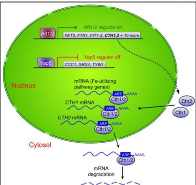
Fundamental for the transcriptional iron response network in yeast are iron-sulfur clusters (Li et al. 2012). When iron levels are high, Fe-S clusters are synthesized inside the mitochondria (Fig 1.2-1). These clusters are then transported to the cytosol by the mitochondrial transporter Atm1 (Fig 1.2-2), where they bind to glutathione (GSH), forming [2Fe-2S] bridged complexes with the monothiol glutaredoxins Grx3 and Grx4. These complexes, together with the cytosolic proteins Fra1 and Fra2 travel inside the nucleus and bind to Aft1/2 (Fig. 1.2-3), leading to the dissociation of these transcription factors from their target DNA (Fig. 1.2-4). At the same time the iron signal inside the nucleus leads to Yap5 activation by a yet unknown mechanism (Fig. 1.2-5) and consequently expression of its target genes (Fig. 1.2-6). Among them, CCC1 acts as an iron detoxifier by importing it inside the vacuole and lowering the cytosolic iron pool (Fig. 1.2-7), while Tyw1, an endoplasmic reticulum (ER)-bound protein, sequesters iron as protein-bound Fe-S clusters (Fig. 1.2-8). Yap5 also promotes the expression of Grx4, which maintains the Aft1/2 inactivation and is thought to act also as an iron detoxifier by directly binding to it. On the other hand, when the iron levels are scarce, the Fe-S clusters do not form and Aft1/2 are free to bind to their dependent genes, activating them. The net effect will be the import of iron inside the cell and the shutdown of the mechanisms that keep it inside the vacuole and bound to proteins as Fe-S clusters (Outten & Albetel 2013).
Figure 1.2 - Transcriptional iron regulation in yeast: when iron levels are high,
iron-sulfur clusters formed inside mitochondria travel to the nucleus bound to monothiol glutaredoxins Grx3 and 4. Inside the nucleus they bind to the transcription factors Aft1/2, leading to their dissociation from the respective target genes. Additionally, Yap5 is activated and consequently expression of its target genes, namely the iron detoxifier CCC1, GRX4 and ER-bound protein TYW1. In a scenario of iron deprivation, the Fe-S clusters are not formed and Aft1/2 are free to bind to their target genes, involved in several iron transport mechanisms (Outten & Albetel 2013)
Another layer of iron regulation is present in yeast, which relies on the mRNA-binding proteins (Fig. 1.3). Key to this post-transcriptional control are two mRNA-binding proteins Cth1 and Cth2 (Outten & Albetel 2013). This mechanism can be considered as a fine-tune of the iron metabolism when iron levels are low, as its impact is much smaller when compared to the transcriptional control of Aft1/2 and Yap5. In fact, the expression of Cth1 and Cth2 is directly dependent of the action of Aft1/2. These two proteins transport the respective target mRNAs to the cytosol and promote their degradation, by binding to AU-rich elements (AREs) present in the 3’ UTR (untranslated region) (Martínez-Pastor et al. 2013). The proteins under this control are involved in Fe-rich pathways, like respiration, tricarboxylic acid (TCA) cycle, haem biosynthesis, amino acid, sterol and
fatty acid metabolism, and mitochondrial Fe-S cluster biogenesis. Interestingly, both Cth1 and Cth2 have AREs in their own 3’ UTR, allowing a precise cross and auto-regulation of their expression (Fig 1.3) (Outten & Albetel 2013).
1.4 – Iron sensing and regulation in mammalian cells
In mammals, the vast majority of iron is present inside the RBCs as part of haem, the prosthetic group of haemoglobin (De Domenico et al. 2008). Additional iron storages are macrophages and the myoglobin of muscles, while excess iron in stored in the liver (Wang & Pantopoulos 2011). As mammals do not possess mechanisms for iron excretion, the control of iron levels depend on the regulation of dietary uptake in duodenum and macrophage recycling of erythrocytic iron. Iron is absorbed from the intestinal lumen to enterocytes by DMT1 (Divalent Metal Transporter 1) (Gunshin et al. 1997) or as haem-bound by haem transporters, and further from enterocytes to the bloodstream by ferroportin. Macrophages, which recycle iron by consuming senescent RBCs, also have ferroportin as the iron transporter to the bloodstream. Once found in the plasma, iron binds to the glycoprotein transferrin, which serves as a vehicle that transport iron to all the tissues. Transferrin-bound iron is then taken up by receptor-mediated endocytosis upon binding to transferrin receptor 1 and 2 (TfR1 and TfR2). While TfR1 is ubiquitously expressed, TfR2 is predominantly expressed in the liver. Once it is found in the cytosol, iron is transported into the mitochondria to be used for synthesis of haem and Fe-S clusters, while the excess is stored in ferritin, a cytosolic protein. The efflux of iron from enterocytes and macrophages is tightly regulated by the hepatic hormone hepcidin, which upon inflammation or high iron levels targets ferroportin to degradation. On the other hand, hepcidin expression is repressed by iron deficiency and increased erythropoiesis (Hentze et al. 2010).
Figure 1.3 - Post-transcriptional iron regulation in yeast: the two
mRNA-binding proteins Cth1 and Cth2, whose expression is activated by Aft1/2 upon iron deprivation, bind to AU-rich elements (AREs) in the 3’ UTR of the respective mRNAs and transport them to the cytosol where they will be degraded. These genes are involved in several iron utilizing pathways. Additionally, Cth1 and Cth2 themselves have AREs in their own 3’ UTR, allowing a precise regulation of their expression. (Outten & Albetel 2013)
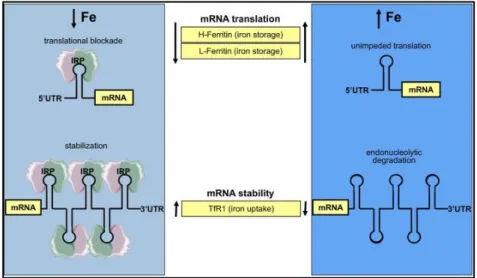
This systemic regulatory system is coordinated with an intracellular one, that makes use of mRNA-binding proteins. The two proteins involved in this system are iron regulatory proteins 1 and 2 (IRP1 and IRP2). While IRP2 acts only as a mRNA-binding protein, IRP1 also has aconitase activity (Wilkinson & Pantopoulos 2014). Aconitase is an enzyme of the TCA cycle that catalyses the stereo-specific isomerization of citrate to isocitrate via cis-aconitate (Beinert & Kennedy 1993). The dual function of this protein is due to the assembly
of a [4Fe-4S] cluster, which is absent in IRP2. When iron levels are low, these two proteins act in the respective targets by binding to highly conserved hairpin structures of 25-30 nucleotides, the iron responsive elements (IREs). The major difference between this system and the one present in yeast relies on the localization of the IREs in the target gene’s mRNA. Depending if the IREs is located in the 3’ or the 5’ UTR, the binding of the IRPs will lead to the stabilization or translational blocking of the target mRNA, respectively (Fig. 1.4) (Wilkinson & Pantopoulos 2014). When iron levels are low, both IRPs bind to the 5’ UTR of both subunits of ferritin (H-ferritin and L-ferritin), blocking its translation, and to the 3’ UTR of transferrin receptor 1, protecting the mRNA from endonucleolytic degradation. The combined effect of blocking iron storage inside ferritinand transport of iron by transferrin upon binding to TfR1 will restore the iron levels inside cells. In the opposite scenario, i.e., iron overload, IRP1 binds Fe-S cluster and acquires aconitase activity and consequently loses its mRNA binding activity, while IRP2 is degraded. The absence of IRP1 and IRP2 allow the translation of ferritin and consequent storage of iron excess, while the mRNA for TfR1 becomes vulnerable for endonucleolytic degradation. The drop in the number of transferrin receptors decreases the cellular iron transport, and consequently restores normal iron levels. More recently, IRE/IRP regulatory network was investigated on the genome-wide level by immunoprecipitation of ribonucleoprotein complexes containing IRP and bound mRNA followed by microarray analyses (Sanchez et al. 2011). In this way, further 35 novel target mRNAs that bind both IRP1 and IRP2 were identified, leading to a much more comprehensive understanding of the IRP regulon in mammalian cells.
1.5 – Iron in Plasmodium: what do we know?
For all the reasons described above, it is clear that iron possesses a central role for living organisms, and
Plasmodium is not an exception. Despite that, it is surprising how little is known concerning the
mechanisms employed by the parasite to deal with this resource. Using iron chelating compounds, it has been shown that iron is essential for the survival of the parasite across the life cycle: from gametocytes and blood stage forms (Ferrer et al. 2012) to liver stage forms (Stahel et al. 1988).
Figure 1.4 - Post-transcriptional regulation by IRPs/IREs in mammals: the
mRNA-binding proteins iron regulatory proteins 1 and 2 (IRP1 and IRP2) block or promote their target mRNAs translation by binding to iron responsive elements (IREs) in the 5’ or 3’ UTR respectively. Upon iron deprivation, they block the translation of both ferritin subunits and stabilize transferrin receptor 1 (TfR1), while upon iron excess they promote the unimpeded translation of the ferritin subunits and promote the endonucleolytic degradation of TfR1 mRNA. Figure from (Wilkinson & Pantopoulos 2014).
So far, two iron transporters have been identified in Plasmodium: ZIP domain-containing protein (ZIPCO) (Sahu et al. 2014) and Vacuolar Iron Transporter homologue (VIT) (Slavic et al. 2016). ZIPCO was shown to localise to the plasma membrane of rodent P. berghei parasites, and to be predominantly expressed during the liver stage development of parasites. Its gene deletion greatly impairs the parasite’s development in hepatocytes, but has no effect on the ability to infect mosquitoes and multiply in mouse blood (Sahu et al. 2014). VIT is a homologue of yeast’s CCC1 and plant Vacuolar Iron Transporters that is expressed in Plasmodium throughout the life cycle and provides an iron detoxification mechanism, by removing iron excess from cytosol (Slavic et al. 2016). As it happens with ZIPCO, VIT is not essential during the entire life cycle, which indicates the presence of compensatory mechanisms to transport and detoxify iron.
Haem biosynthesis is directly dependent on iron, and has been shown recently to play a role in the mechanism of action of artemisinin, the main drug used to treat the disease. During the intraerythrocytic developmental cycle the parasite degrades host haemoglobin, producing amino acids and releasing free haem, which is sequestered in the crystal hemozoin. The genetic down modulation of falcipains 2 and 3, two cysteine protease haemoglobinases, confers artemisinin resistance in rings, confirming the role of haemoglobin degradation in artemisinin’s mode of action (Xie et al. 2016). It seems that haem synthesized by the parasite during the ring stage and resultant from haemoglobin degradation in later stages, rather than free iron, is the main activator of artemisinin (Wang et al. 2015).
It is intriguing that even though degradation of haemoglobin leading to the release of haem plays such a central role in the survival of blood stage parasites, we still do not know what exactly is the source of iron that parasites use during their replication. Most of the release haem is converted into inert crystal hemozoin, preventing toxicity of excess haem. Evidence so far suggests that parasites cannot enzymatically degrade this haem, which would suggest that it could not serve as a source of iron (Sigala et al. 2012). Even less studied and known is the case of parasite iron metabolism at their liver stage of development.
No one can ignore the impact iron has in every aspect of Plasmodium biology, from its own survival and replication to all the interactions established with the host. The need for sensing and consequently adapting to iron levels must be of major importance to the parasite, and it wouldn’t be a surprise to observe a remodelling of gene expression in response to this resource, as it happens in yeast and mammalian cells. By using one of the most recent and advanced approaches to study genome-wide responses in organisms, carefully described in the next section, this work will try to provide new insights in the complex role of iron for Plasmodium parasites.
1.6 – Welcome to the omics’ era!
In the past two decades, the development of high-throughput sequencing technologies and bioinformatics tools to analyse the data generated had a major impact in science. The “omics” fields provided different layers for studying cellular and molecular systems: genes (genomics), mRNAs (transcriptomics), proteins (proteomics) and metabolites (metabolomics) can now be profoundly studied. By using one or more of these approaches it is possible to characterize an organism in a highly dynamic way. Genomes from several organisms are already fully sequenced (including P. falciparum (Gardner et al. 2002)), and many more will follow in the next few years, providing an invaluable resource for the studies of living organisms. It is also possible to address global responses to specific conditions, for example by using transcriptomics to compare the gene expression between a wild-type and a mutant organism, or proteomics to compare the protein expression between control and treated
condition (administration of a drug or toxic compound, different temperatures or pH levels, etc.). These kinds of studies are starting to become widely used in many areas of research, from evolutionary biology to biomedical sciences, and are of great importance to help solving fundamental questions like the genetic basis of adaptation and the evolution of gene expression profiles, among others.
1.6.1 – Our approach: RNA-seq! Whole transcriptome shotgun sequencing, a.k.a RNA-seq, is an approach that allows transcriptome profiling using deep-sequencing technologies (Chu & Corey 2012). The transcriptome is commonly defined as the complete set of transcripts and their abundance, in a cell, in a particular stage and condition (Wang et al. 2009). It is becoming widely used due to the advantages compared to DNA microarrays, namely the lower amount of material needed, lower background noise, higher
sensitivity, and the fact that it doesn’t need prior knowledge about the organism’s genome sequence (Wang et al. 2009).
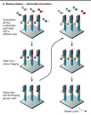
A typical RNA-seq protocols involves the following steps: total RNA isolation, ribosomal RNA depletion (as it represents about 95% of total RNA), cDNA synthesis (library preparation), ligation of adapters to each cDNA molecule to allow the binding to a solid support (cluster generation) and parallel sequencing of millions of locally amplified cDNA fragments via sequencing by synthesis (in this step, details vary according to the platform used) (Hoeijmakers et al. 2013). In our particular case, the platform used was Illumina. After the cDNA synthesis, cDNA molecules are bound to specific primers, which allows the binding to a solid support, where amplification occurs by bridge PCR. This is called solid-phase amplification (Fig. 1.5) (Metzker 2010). After this step, clonally amplified fragments are sequenced by cyclic reversible termination (CRT). In this phase, each nucleotide (A, T, C, G) has a specific fluorophore incorporated, as well as a terminating group. Upon binding of each nucleotide, mediated by a DNA polymerase, the incorporation is terminated and the signal in that position is recorded (Fig 1.6) (Metzker 2010). As
Figure 1.5. – Solid-phase amplification: library is loaded into a flow cell and
fragments hybridize to the surface. Each fragment is amplified by bridge PCR into a clonal cluster Figure from (Metzker 2010).
Figure 1.6. – Cyclic reversible termination: fluorescently
labelled nucleotides are added and the first base is incorporated. The flow cell is imaged and the emission from each cluster is recorded Figure from (Metzker 2010).
the four nucleotides are present during each sequencing cycle, natural competition between them minimizes incorporation bias. Also, the
separation between incorporation and recording of the signal increases the precision of the technique.
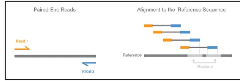
Another advance regarding this technology is the use of paired-end reads. This involves sequencing each end of the cDNA fragments in the library and aligning the forward and
reverse read as read pairs. Because the distance between each pair is known, alignment algorithms can use this information to map the reads more precisely (Fig. 1.7) (Illumina 2015).
There are several type of questions that might be addressed using RNA-seq, from the detection of alternative splicing events (Griffith et al. 2010), detection of non-protein coding RNAs (Broadbent et al. 2015), detection of fusion genes (Maher et al. 2009), among others. Despite that, the most common and straightforward application of RNA-seq is still the quantification of gene expression between samples, in order to find differentially expressed genes (Oshlack et al. 2010). As referenced above, several variables might be tested as factors affecting gene expression, from treatments with drugs to alterations in environmental conditions, or even compare the transcriptome of a wild-type and a mutant organism of the same species. Also, there are studies addressing transcriptomic changes in response to iron: it is described that yeast remodels its transcriptome in response to iron deprivation, switching from iron-dependent to iron-independent metabolic pathways by the activation of the iron independent transcription factor Aft1p (Shakoury-Elizeh et al. 2004) . In a subsequent paper, also using yeast in an iron deprivation condition, the authors identified 100 and 42 genes up and down-regulated, respectively. These represents around 2% of the genome, and most of them were associated with iron uptake and transport mechanisms (Jo et al. 2009). These works clearly show the potential of these approaches to study iron responses at a genome-wide level. Additionally, in a study from Moyerbrailean et. al, the authors analysed the transcriptomic profile by RNA-seq in response to 23 environmental perturbations (including changes in iron levels) in lymphoblastoid cell lines (Moyerbrailean et al. 2015), showing that these techniques will become common practice in several laboratories, due to the optimization of sequencing protocols and the decrease in cost.
These gene expression profiling studies are targets of great interest for studying pathogen’s responses to compounds, in order to find potential drugs. Indeed, this approach was already used in Plasmodium
falciparum, both using microarrays (Hu et al. 2010; Siwo et al. 2015) and RNA-seq (Shaw et al. 2015).
The fact that these parasites are easily maintained in continuous culture (Trager & Jensen 1976), facilitates in vitro drug testing.
1.7 – Plasmodium falciparum blood stage transcriptome: a unique gene regulation cascade
mechanism among eukaryotes
As described in the first section, the parasite goes through a complex sequence of events during the 48h blood stage, assuming three different stages with particular morphological and metabolic features: ring (0-18h), trophozoite (18-30h) and schizont (30-48h). In order to complete this cycle and consequently establish an infection in the human host, it must have a tight temporal regulation of transcription, as
Figure 1.7. – Paired-End Reads: both ends of the cDNA fragment are
sequenced, providing a read pair to be aligned to the reference sequence Figure from (Illumina 2015)
each stage is present within a specific window of time. This has been a fundamental question in the malaria parasite research, as it would deeply improve the knowledge of its biology and might open doors for the development of stage specific drugs or vaccines. Additionally, the gene expression regulatory elements themselves might be good targets for new antimalarial drugs, as the deregulation of the mechanisms that control the developmental cycle would prevent parasite growth and potentially eliminate it. The sequencing of P. falciparum genome was a major tool to elucidate the molecular mechanisms behind these developmental events. The 22.8 Mb genome encodes around 5400 genes and is one of the most AT-rich genomes among living organisms. It is organized in 14 nuclear linear chromosomes, a circular apicoplast genome and a linear mitochondrial genome (Gardner et al. 2002). While the nuclear genes’ expression is not coregulated, the plastid genes are organized in polycistronic units and highly coregulated (Bozdech et al. 2003).
Indeed, the first study of P. falciparum IDC transcriptome revealed amazing results. Using microarrays, Bozdech and Llinás deeply characterized the transcriptome during the entire IDC, by measuring mRNA levels of highly synchronized parasites during the 48 hours at a 1-hour time scale resolution (Bozdech et al. 2003). With these results, they observed that around 80% of the genes expressed have changes in transcript abundance across the IDC. More surprising is the fact that the parasite exhibits a continuous cascade of gene expression, with most of the genes being expressed only once during the IDC, with its maximum expression level correlating with the specific function of the product produced (Fig. 1.8 left) (Bozdech et al. 2003). During the first half of the IDC, equivalent to ring/early troph stages, genes involved in general cellular mechanisms are activated, i.e., transcription, translation, glycolysis and ribonucleotide synthesis (Fig. 1.8 left B-E). With the progression to the trophozoite stage comes the expression of genes involved in several metabolic pathways. This result is expected, as the trophozoite stage is the most metabolically active. The class of genes characteristic of this stage include DNA
Figure 1.8. – P. falciparum IDC transcriptome:
(left) - In panel A, each line represents a gene, with the correspondent level of expression across the IDC. On the left side of the figure is depicted the characteristic stage aligned to the genes with peak expression at that stage. Panels B to M depict specific biochemical processes, organized chronologically as they occur during IDC. (Bozdech et al. 2003); (right) – IDC of 3 strains of P. falciparum shows the conservation of the gene expression cascade (Llinas et al. 2006)
replication machinery, TCA cycle and deoxynucleotide synthesis (Fig. 1.8. left F-H), showing the coordinated transcription of genes involved in DNA synthesis and the precursors required for this process. Also, these findings clearly separate deoxynucleotide production as a trophozoite-process and ribonucleotide production as a ring-process. The shift to schizont stage induces genes involved in proteasome and plastid genome (Fig. 1.8. left I-J), indicating a role of protein degradation in the development of the parasite. On the other hand, the activation of plastid genome clearly shows the expression of parasite-specific mechanisms with the progression of IDC. In the final part of IDC, genes involved in host invasion are activated, as new merozoites are produced. These categories include merozoite invasion and actin myosin motility (Fig. 1.8. left K-L). The latter shows the implication of action and myosin in the remodelling of host cells, allowing the successful invasion. With the successful merozoite invasion and consequent formation of new rings, comes the expression of early ring transcripts (Fig. 1.8. left M).
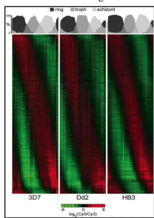
In a subsequent work, Llinás and Bozdech characterize the transcriptome of two additional P.
falciparum and were able to show that this pattern was conserved between them (Figure 1.8. right),
clearly placing it as a specific parasite phenomena (Llinas et al. 2006). Le Roch et al. observed similar results in their study of the transcriptome characterization of nine different stages of the parasite (including asexual IDC stages), like the high percentage of genes that are expressed only once during the IDC, the shift of functions from ring and trophozoite to schizont and the fact that almost half of the genes are cell cycle regulated (K G Le Roch et al. 2004).
The big question imposed by these results is how is this gene expression cascade across the IDC regulated. There are several evidences suggesting post-transcriptional and translation control outweighing transcriptional control: first, the reduced number of genes encoding transcription factors and transcription associated proteins in P. falciparum genome (Coulson et al. 2004) (despite the identification of the Apicomplexan AP2 family of DNA binding proteins as potential transcription factors controlling development) (Painter et al. 2012). Second, the significant delay observed between mRNA and protein abundance also imply mechanism of post-transcriptional and translational control (Karine G Le Roch et al. 2004; EM et al. 2014). However, Caro et al. recently observed that transcription and translation are closely coupled for most genes (Caro et al. 2014), giving strength to the debate. Third, several studies suggest that mRNA-binding proteins have a huge impact in gene expression regulation of the parasite: Reddy et al. performed a bioinformatic identification and annotation of 189 putative RNA-binding proteins of the parasite (Reddy et al. 2015). Additionally, in a very recent work, Bunnik et al. made a deep comparative genomics study complemented with experimental validation of the “mRNA-bound proteome”, being able to observe that P. falciparum has a higher number of genes encoding mRNA-binding proteins when compared to other organisms (Bunnik et al. 2016). Among these, PfAlba1 seems to have an important role in regulating time translation during IDC, as it binds to several mRNAs changing their steady state levels (Vembar et al. 2015). Finally, epigenetic mechanisms seem to play a role in transcriptional control of the parasite: Rovira-Graells et. al observed that the histone mark H3K9me3 (which promotes the formation of heterochromatin in higher eukaryotes) has a function in gene repression in the parasite (Rovira-Graells et al. 2012).
All the findings described above illustrate the importance of deep understanding of the regulation of the transcriptome across the IDC. However, it is also clear that these studies unveiled more questions than answers: what are the exact molecular pathways regulating gene expression during IDC? Does the parasite sense its surrounding environment (human host) in order to “decide” when it should start the development to the subsequent stage, or is this a stochastic event? Does it have any kind of quorum-sensing mechanism, proliferating adequately to the density of parasites in order to survive inside the human host without killing it? Also intriguing is the bridge to the host-pathogen battle for resources: are
there any mechanisms of sensing and consequent gene expression regulation in response to the host nutritional status? This topic will be particularly addressed in this thesis, making use of the tools provided by the boost of high-throughput sequencing with special emphasis in iron. Does the parasite have mechanisms of transcriptional and post-transcriptional regulation of gene expression in response to iron like yeast and mammalian cells? If it does, which genes are target of this regulation: iron transporters, detoxifiers or Fe-S cluster proteins? We hope to be able to start answering these (and many more) questions, which are of great relevance for the better understanding of the basic biology of the parasite, allowing for the development of broader (in terms of activity across the life cycle stage) and at the same time, more specific (in terms of targeting only the parasite and not the host) anti-malarial strategies.
2 – Thesis aims
As iron is a micronutrient with a major role in infection, being essential for both the pathogen and the host, the main objective of this work was to identify genes involved in iron sensing and regulation in
Plasmodium falciparum, the parasite responsible for the most severe form of Malaria in humans. To
address that, putative homologues of iron-responsive genes in model organisms were identified in P.
falciparum and it was tested how their expression levels were affected by changes in the iron
concentration of the parasite environment. However, since no homologues of the main iron-sensing proteins could be identified in Plasmodium, an RNA-seq experiment was performed in order to identify transcriptomic changes in P. falciparum exposed to iron excess and iron depletion condition. In this way, by identifying differentially expressed genes between the conditions tested through a genome-wide approach, we intend to identify new mechanisms of iron-sensing and regulation of the complex iron metabolic network in the malaria parasite.
3 – Methods
3.1 BLAST (Basic Local Alignment Search Tool) and Multiple Alignment
BLAST searches were performed in the NCBI website (https://blast.ncbi.nlm.nih.gov/Blast.cgi) using the protein blast (blastp) tool with default parameters. After the BLAST search, multiple alignments were performed using Clustal Omega (http://www.ebi.ac.uk/Tools/msa/clustalo/) with default parameters between the potential homologue in P. falciparum and the one used in the BLAST search. All protein sequences were provided in FASTA format (retrieved from Uniprot or PlasmoDB for S.
cerevisae and S. pombe or P. falciparum genes, respectively). In order to quantify the gene expression
by qPCR, primers were designed using the PrimerQuest tool in the IDT website (http://eu.idtdna.com/).
3.2 – Preparation of biological samples
3.2.1 – P. falciparum in vitro culture
P. falciparum 3D7 strain blood stage parasites were cultured using standard protocol for in vitro culture
(Trager & Jensen 1976). The parasites were kept in a culture flask in malaria complete medium (MCM) and blood diluted to 5% haematocrit. The incubator’s temperature was 37°C, the O2 and CO2 percentage
4 and 5, respectively.
Medium was changed every day or every two days, depending on the parasitemia (percentage of infected RBCs). Smears were made regularly to keep track of parasitemia and dilute the culture with MCM and blood if needed. Levels of parasitemia higher than 10% might lead to culture’s crash and consequent death of the parasites.