ARTIGO ORIGINAL
RESUMO
Introdução: Valores elevados de lipoproteína(a), relacionados com a progressão da aterosclerose, são frequentemente considerados marcadores de trombose. O perfil de lipoproteína(a) foi avaliado num grupo de doentes sem eventos cardiovasculares mas com elevado risco vascular, estabelecendo-se a correlação com outros fatores de risco cardiovascular e inferindo-se os resultados para doentes com alterações metabólicas e, pelo menos, dois fatores de risco vascular.
Material e Métodos: Este estudo observacional longitudinal incluiu 516 doentes com, pelo menos, dois fatores de risco cardiovascular e que frequentavam, regularmente e há pelo menos dois anos, a consulta ambulatória de metabolismo e risco vascular para prevenção primária. Os parâmetros sociodemográficos, clínicos e antropométricos foram recolhidos na primeira visita. A morfologia hepática foi avaliada por ultrassonografia em 509 doentes (98,6%). O risco vascular a 10 anos foi estimado através de tabelas de cálculo de risco de Framingham, doença cardiovascular e risco coronário sistemático.
Resultados: Foram encontradas correlações significativas entre os níveis de lipoproteína(a) e os fatores de risco vasculares analisados, assim como entre lipoproteína(a) e as escalas de risco de Framingham, doença cardiovascular e risco coronário sistemático. Os valores de lipoproteína(a) apresentaram-se mais elevados em doentes com esteatose.
Discussão: Os valores elevados de lipoproteína(a) estão diretamente associados com todos os marcadores de risco cardiovascular e com esteatose hepática não alcoólica.
Conclusão: Como tal, considerando a sua elevada acessibilidade e custo reduzido, o marcador lipoproteína(a) deverá ser integrado na avaliação de rotina de doentes com risco vascular.
Palavras-chave: Aterosclerose; Doenças Cardiovasculares; Lipoproteína(a); Portugal; Prevenção Primária
Lipoprotein(a) in the Evaluation of Cardiovascular Risk
in the Portuguese Population
Lipoproteína(a) na Avaliação do Risco Cardiovascular na
População Portuguesa
1. Faculty of Medicine. University of Porto. Porto. Portugal. 2. Starmedica Clinica. Paredes. Portugal.
Autor correspondente: Joaquim A. Meireles-Brandão. [email protected]
Recebido: 16 de janeiro de 2018 - Aceite: 08 de novembro de 2018 | Copyright © Ordem dos Médicos 2019
Joaquim A. MEIRELES-BRANDÃO1,2, Lúcia R. MEIRELES-BRANDÃO2, Rui COELHO1,
Francisco R. ROCHA-GONÇALVES1
Acta Med Port 2019 Mar;32(3):202-207 ▪ https://doi.org/10.20344/amp.10251
ABSTRACT
Introduction: High values of lipoprotein(a), related to atherosclerosis progression, are often considered a marker of thrombosis. We assessed the lipoprotein(a) profile in a group of patients with high vascular risk and no cardiovascular events, established its correlation with other cardiovascular risk factors and inferred the results for patients with metabolic disorders and, at least, two risk factors. Material and Methods: This longitudinal observational study included 516 patients, who had at least two cardiovascular risk factors and regularly attended, for at least two years, the outpatient consultations at a clinic of metabolism and vascular risk for primary prevention. Sociodemographic, clinical and anthropometric parameters were obtained at the baseline visit. Hepatic morphology was assessed in 509 patients (98.6%) by ultrasonography. The 10-year vascular risk was estimated using Framingham risk score, atherosclerotic cardiovascular disease and systematic coronary risk evaluation tables.
Results: Significant correlations were found between lipoprotein(a) levels and the addressed vascular risk factors, as well as between lipoprotein(a), and Framingham risk score, atherosclerotic cardiovascular disease and systematic coronary risk evaluation charts. Lipoprotein(a) values were also considerably higher in patients with steatosis.
Discussion: Increased lipoprotein(a) values were directly associated with all markers of cardiovascular risk and with non-alcoholic hepatic steatosis.
Conclusion: Due to its high availability and low cost, lipoprotein(a) should become part of the routine evaluation of patients at vascular risk.
Keywords: Atherosclerosis; Cardiovascular Diseases; Lipoprotein(a); Portugal; Primary Prevention
INTRODUCTION
Atherosclerosis remains the major cause of death and premature disability in developed societies.1 Current
pre-dictions estimate that by 2020 cardiovascular diseases (CVD),2 particularly atherosclerosis, will become a global
leading cause of death.3 Lipoprotein(a), or Lp(a), is identical
to the low-density lipoprotein cholesterol (LDLc), with the addition of apolipoprotein A (apoA), a highly glycosylated protein.4,5 Lp(a) is often considered a marker of thrombosis,
similarly to plasminogen, and a risk factor for CVD.6 The
cholesterol present in LDLc accounts for more than half of
plasma cholesterol, in most individuals. Approximately 70% of the circulating LDLc is cleared by LDL-receptor-mediated endocytosis in the liver. ApoA is synthesized in the liver and attached by disulphide linkage to apoB-100, a structural protein of Lp(a). Also, apoB is the main structural protein of chylomicrons.2 The human liver produces apoB-100,
where-as the intestine produces apoB-48. Clearance of Lp(a) oc-curs mainly through the liver, but the uptake pathway is still unknown.7 Nevertheless, Lp(a) is recognized as a strong
ARTIGO ORIGINAL
disease and might present other functions, including an im-portant role in the association between atherosclerosis and thrombosis. 8,9
Portugal is a Southern European country where CVD are the first cause of mortality and stroke incidence is higher compared to other European countries.10 In this context, we
assessed the levels of Lp(a) in Portuguese patients, who regularly attended outpatient consultations at the clinic of metabolism and vascular risk, over a two-year period. In ad-dition, those patients had metabolic disorders and, at least, two cardiovascular risk factors (CVRF), but no previous car-diovascular events. Thus, the aim of this study is to deter-mine the mean values of Lp(a) in the sample of patients and to infer the use of Lp(a) as an indicator of vascular risk in the Portuguese population with metabolic disorders and, at least, two CVRF.
MATERIAL AND METHODS
The longitudinal observational study included 516 pa-tients, as a random sample of a universe of 1677 patients from an outpatient setting at a clinic of metabolism and vas-cular risk. Inclusion criteria were defined for those patients without cardiovascular events having, at least, two family or personal CVRF and attending an outpatient consultation about metabolism and vascular risk for primary preven-tion, for at least two years (between 1995 and 2015), on a quarterly periodicity. All patients accepted and signed the informed consent form. The protocol used in this study was approved by the local Ethics Committee of the São João Hospital Center. All procedures conducted in this study comply with the Declaration of Helsinki.
Patients were assessed for sociodemographic data, including their professional status, in accordance with the 2010 classification, by the Portuguese National Statistics Institute (Instituto Nacional de Estatística, INE). Afterwards, clinical characterization of the study participants was deter-mined, at baseline, by anthropometric, biochemical and car-diovascular evaluation. The following tests were conducted: electrocardiogram (ECG), two-dimensional echocardiogram with ejection fraction calculation, and doppler ultrasonogra-phy of the supra-aortic trunks with evaluation of the carotid intima-media thickness (IMT). In addition, liver morphologi-cal changes were evaluated by ultrasonography. For clas-sification of alcohol consumption, the following criteria were used: never; moderate, if one or two drinks daily; excessive, if three drinks daily; alcoholism; and abstinence, if at least a year had passed since the last drink. Evaluation of vas-cular risk was performed considering the three most used scores: Framingham risk score (FRS), atherosclerotic car-diovascular disease (ASCVD) - AHA/ACC,11 and systematic
coronary risk evaluation (SCORE)-ESC.7
Results were summarized as mean, median and stand-ard deviation or count and percentage, for characterization of the study population. Normality distribution of data was assessed by the usual methodology used for validation (i.e., Kolmogorov-Smirnov test and Shapiro–Wilk test). For quan-titative comparison of two independent groups, we used the
t-test for independent samples or the Mann-Whitney
non-parametric test, depending on the assumptions’ validation by the statistical test. Spearman’s correlation coefficient (SCC) was used to assess the relationship between two quantitative variables, in case the normality assumption was not verified. Regarding the sample distribution by age groups, we used the Kruskal-Wallis (KW) non-parametric test, instead of ANOVA F-test, whenever the distributions within groups presented relevant deviations from normality. The KW test allowed a comparative analysis between three or more independent groups and a quantitative or ordinal variable (as the dependent variable). Bonferroni correction for multiplicity of testing was used to verify age groups con-trast [α’ = 0.005, for α (0.05)/k (10 tests)]. All statistical tests were 2-tailed considering a significance level of 5%. The statistical analysis was conducted using the software IBM®
SPSS® Statistics 19.
RESULTS
Sociodemographic parameters at first consultation
Most of the patients were women (56.6%) and Cauca-sian (98.6%). The patients’ median age was 46 years. Re-garding the educational background, 76.6% of the patients did not complete the third cycle; of those, 60.5% completed only the first cycle. Concerning the professional status, 72.5% of the patients were active and 13.2% were retired but still working. Among those actively employed patients, 40.5% had elementary occupations, 17.6% were skilled in-dustrial, construction or craft workers and 17.4% were per-sonal, protection or security service workers or sales work-ers.
Anthropometric and clinical evaluation
The median weight was 75.3 kg (range: 7.75 - 125.6 kg), while the median body mass index (BMI), calculated in kilogram per square meters, was 28.57 (range, 16.53 - 47.63 kg/m²). Amongst the study population, 25.0% had normal weight and 36.0% excess weight (BMI, 25 - 29 kg/ m²). Moreover, 26.2% of patients had grade I obesity (BMI 30 - 34 kg/m²), 9.9% grade II obesity (BMI 5 - 39 kg/m²), 2.5% grade III obesity (BMI 40 - 44 kg/m²) and 0.4% morbid obesity (BMI ≥ 45 kg/m²). The abdominal (waist-to-hip) cir-cumference (AC) had a median of 94 cm (range: 44 - 138 cm) and the average triceps skinfold (TSF) was 2.51 ± 1.13 cm (range: 0.3 - 5.0 cm; median: 2.4 cm).
Regarding cardiovascular evaluation, the average systolic blood pressure (SBP) was 145.52 ± 30.29 mmHg (range: 78 - 300 mmHg) with a median of 144 mmHg. The average diastolic blood pressure (DBP) was 87.91 mmHg (range: 42 - 148 mmHg) with a median of 90 mmHg. In addi-tion, the average heart rate was 88.98 ± 11.79 bpm (range, 50 - 170 bpm) with a median of 90 bpm. Overall, 50.6% of patients had normal cardiac auscultation.12
Concerning the lifestyle and patients’ habits, 66.0% had a moderate alcohol consumption, ranging from one (20.8%) to two daily drinks (45.2%), whereas 15.5% showed an ex-cessive alcohol consumption. Alcoholism was diagnosed in
ARTIGO ORIGINAL 7.0% of patients and abstinence in 0.4%. Moreover, 71.9% did not practice regular physical activity, while 75.4% were
non-smokers, 11.6% were ex-smokers and 13.0% were smokers.
In terms of the liver morphological evaluation, changes were detected through ultrasound in 509 patients (98.6%). Briefly, a pattern of hepatic steatosis was found in 435 pa-tients (85.4%), of which 208 (40.8%) had associated hepa-tomegaly. Only 5.6% of patients presented changes in liver function tests.
Vascular risk calculations
The 10-year vascular risk was estimated based on the most commonly used vascular risk scores. For the FRS (30 - 74 years), applied to 436 patients, we obtained an average of 36.57 ± 26.37 (median: 32.60; range: 1.10 - 99.5), while for ASCVD (40 - 79 years), applied to 338 patients, the average was 24.83 ± 20.61 (median: 20.61; range: 0.86 - 95.69). As for the SCORE low-risk algorithm (45 - 64 years), 202 patients were assessed, obtaining an average of 3.24 ± 3.81 (median: 2.23; range: 0.09 - 27.63).
Thus, both ASCVD and FRS scores revealed a high median value for the estimated vascular risk, at 10 years, whereas the SCORE low-risk algorithm indicated a moder-ated median value.
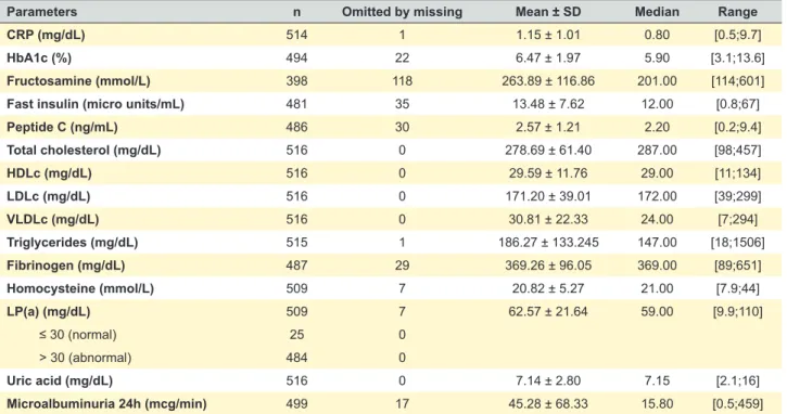
Laboratory values
Laboratory parameters assessed at the first consultation are described in Table 1. Briefly, a relevant increase of C-reactive protein (CRP), fibrinogen, homocysteine and Lp(a) were observed. HDLc showed a median value of 29.00 mg/ dl (min: 11.00 mg/dL), while a median value of 172.00 mg/dL
(max: 299 mg/dl) was obtained for LDLc. The average CRP was 1.15 ± 1.01 (min-max: 0.5 - 9.7 mg/dL), with a median value of 0.80 mg/dl. An average value of 7.14 ± 2.80 (min-max: 2.1 - 16 mg/dL) was found for uric acid. Urinary albu-min excretion (UAE) showed a median rate of 15.80 mcg/ min/24 h (min-max: 0.5 - 459 mcg/min/24h). Furthermore, the median value for homocysteine was 21.00 mmol/L (min-max: 7.0 - 44.0 mmol/L) and the average Lp(a) was 62.57 ± 21.64 (min-max: 9.9 - 110 mg/dL), with a median value of 59.00 mg/dL. Overall, normal laboratory values for Lp(2) (i.e., < 30 mg/dL) were found in 25 patients (4.9%), while abnormal values were detected in 484 patients (95.1%).
Association of independent variables and Lp(a)
Firstly, we confirmed that our study population did not follow a normal distribution, thus, only non-parametric mod-els were applied. As shown in Table 2, a high positive and significant correlation was detected between the Lp(a) and the following variables: IMT (rs = 0.575), LDLc (rs = 0.457) and homocysteine (rs = 0.565), and a negative and weak correlation with HDLc (rs = -0.111). Moderate, but also sig-nificant, correlations were found for CRP (rs = 0.354), ab-dominal circumference (rs = 0.335), Hb A1c (rs = 0.307), and weak correlations for BMI (rs = 0.276) and fast insulin (rs = 0.210).
By using the Mann-Whitney test, a significant relation-ship between Lp(a) and hepatic steatosis was obtained (p < 0.001). This association corresponded to 435 (85.4%) patients with hepatic steatosis (median of 65.00, range: 10.0 - 110.0; average: 66.29 ± 20.11) versus 74 (14.5%) patients without hepatic steatosis (median of 39.00, range: 9.9 - 110.0; average: 40.73 ± 16.98).
Table 1 - Laboratory parameters, at first consultation
Parameters n Omitted by missing Mean ± SD Median Range
CRP (mg/dL) 514 1 1.15 ± 1.01 0.80 [0.5;9.7]
HbA1c (%) 494 22 6.47 ± 1.97 5.90 [3.1;13.6]
Fructosamine (mmol/L) 398 118 263.89 ± 116.86 201.00 [114;601]
Fast insulin (micro units/mL) 481 35 13.48 ± 7.62 12.00 [0.8;67]
Peptide C (ng/mL) 486 30 2.57 ± 1.21 2.20 [0.2;9.4] Total cholesterol (mg/dL) 516 0 278.69 ± 61.40 287.00 [98;457] HDLc (mg/dL) 516 0 29.59 ± 11.76 29.00 [11;134] LDLc (mg/dL) 516 0 171.20 ± 39.01 172.00 [39;299] VLDLc (mg/dL) 516 0 30.81 ± 22.33 24.00 [7;294] Triglycerides (mg/dL) 515 1 186.27 ± 133.245 147.00 [18;1506] Fibrinogen (mg/dL) 487 29 369.26 ± 96.05 369.00 [89;651] Homocysteine (mmol/L) 509 7 20.82 ± 5.27 21.00 [7.9;44] LP(a) (mg/dL) 509 7 62.57 ± 21.64 59.00 [9.9;110] ≤ 30 (normal) 25 0 > 30 (abnormal) 484 0 Uric acid (mg/dL) 516 0 7.14 ± 2.80 7.15 [2.1;16] Microalbuminuria 24h (mcg/min) 499 17 45.28 ± 68.33 15.80 [0.5;459]
CRP: C-reactive protein; HbA1c: hemoglobin A1c; HDLc: high density lipoprotein cholesterol; LDLc: low density lipoprotein cholesterol; VLDLc: very-low-density lipoprotein cholesterol;
ARTIGO ORIGINAL
The association between Lp(a) and hepatic steatosis with hepatomegaly was also significant (p < 0.001). This analysis compared 208 (40.8%) patients with hepatomeg-aly (median of 77.90, range: 10.0 - 110.0; average: 74.90 ± 18.81) versus 301 (59.1%) patients with steatosis without hepatomegaly (median of 49.00, range: 9.9 - 105.0; aver-age 54.05 ± 19.25).
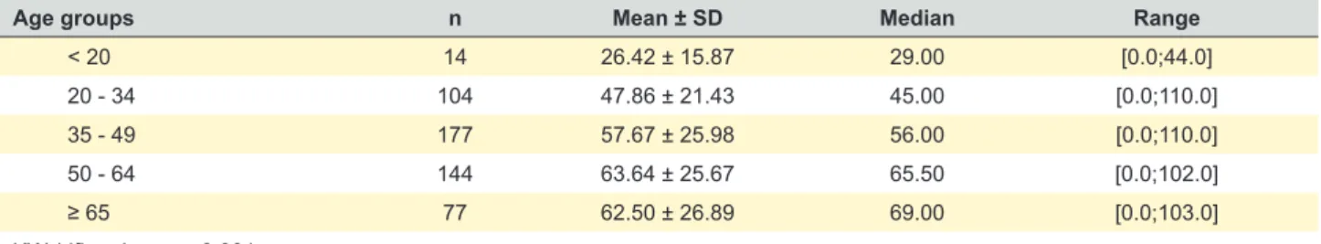
Table 3 describes the results that were significant (p < 0.001), regarding the distribution of Lp(a) by age group, with patients older than 50 years, achieving higher median values of Lp(a). Multiple comparisons between age groups (α’ = 0.005) allowed to determine that all groups presented significant differences (< 20 years versus other age groups, [p < 0.001]; 20 - 34 years vs other age groups [p < 0.001]; 35 - 49 years vs 50 - 64 years [p = 0.001], 35 - 49 years vs ≥ 65 years [p < 0.001]), except for age groups of 50 - 64 years
vs ≥ 65 years (p = 0.540).
A positive and significant correlation was found between Lp(a) and the vascular risk scores used in CVD stratification (p < 0.001), as shown in Table 4. Correlation between Lp(a) and cardiovascular risks was moderate, and high Lp(a) values were associated with high scores of those CV risks
stratification tables (i.e., FRS, ASCVD and SCORE).
DISCUSSION
The main finding of this study was that average values of Lp(a) were increased in patients at high vascular risk and were directly correlated with other CVRF. These data constitute valuable information for clinical evaluation, allow-ing to infer about the adequate orientations and therapeutic interventions, based on the patients’ personal and family history. Moreover, this observation can be employed to all three criteria presented for vascular risk calculation, includ-ing the SCORE low-risk algorithm, which is applicable to Portuguese patients.
A significant correlation was observed between Lp(a) and IMT, BMI, LDLc, homocysteine, CRP and abdominal circumference, as previously described.13,14 The negative,
weak significant correlation between Lp(a) and HDLc should also be highlighted. In addition, results regarding height were relevant if considering that the Portuguese population presents an average short height and individuals with pyk-nic morphology are more susceptible to CVD. Furthermore, the pro-inflammatory effect of Lp(a) is corroborated by its
Table 3 - Lp(a) distribution by age groups, at first consultation
Age groups n Mean ± SD Median Range
< 20 14 26.42 ± 15.87 29.00 [0.0;44.0] 20 - 34 104 47.86 ± 21.43 45.00 [0.0;110.0] 35 - 49 177 57.67 ± 25.98 56.00 [0.0;110.0] 50 - 64 144 63.64 ± 25.67 65.50 [0.0;102.0] ≥ 65 77 62.50 ± 26.89 69.00 [0.0;103.0] KW (df): value, p < 0.001
KW: non-parametric Kruskal-Wallis test; Lp(a): lipoprotein(a)
Table 4 - Correlation between Lp(a) and cardiovascular risk (10 years), at first consultation
Parameters Spearman correlation
rs
Lp(a) versus FRS (30 - 74 years) Initial 0.458*
Lp(a) versus ASCVD (40 - 79 years) Initial 0.414*
Lp(a) versus SCORE (low-risk algorithm, 45 - 64 years) Initial 0.391*
FRS: Framingham risk score; ASCVD: atherosclerotic cardiovascular disease; SCORE: systematic coronary risk evaluation. * p < 0.001
Table 2 - Correlation between Lp(a) and the other clinical and biochemical parameters, at first consultation
Parameters SCC p
Lp(a) versus HDLc rs = -0.111 0.012
Lp(a) versus LDLc rs = 0.457 < 0.001
Lp(a) versus Fast insulin rs = 0.210 < 0.001
Lp(a) versus Hb A1c rs = 0.307 < 0.001
Lp(a) versus Abdominal circumference (waist-to-hip) rs = 0.335 < 0.001
Lp(a) versus Height rs = 0.009 0.842
Lp(a) versus BMI rs = 0.276 < 0.001
Lp(a) versus CRP rs = 0.354 < 0.001
Lp(a) versus Homocysteine rs = 0.565 < 0.001
Lp(a) versus IMT rs = 0.575 < 0.001
SCC: Spearman correlation coefficient (rs); LP(a): lipoprotein(a); HDLc: high density lipoprotein cholesterol; LDLc: low density lipoprotein cholesterol; Hb A1c: hemoglobin A1c;
ARTIGO ORIGINAL correlation with the CRP.
15
We also highlight the significant relationship between Lp(a) and hepatic steatosis (p < 0.001). Lp(a) values were considerably higher in patients with steatosis. Further stud-ies are required to better understand the relationship in cases of non-alcoholic fatty liver disease (NAFLD) without secondary causes for steatosis, such as excessive alcohol consumption, virus infection or endocrine disorders. Although results among adult patients are variable, age has been significantly associated with increased Lp(a).16
Furthermore, besides that Lp(a) values increase with age, a significant difference between younger (20 - 39 years) and older (> 60 years) subjects has been described.17 Therefore,
we assessed the effect of age groups on the distribution of Lp(a), which showed that Lp(a) presented a significant as-sociation between age groups, except for the two groups of older patients (50 - 64 years vs ≥ 65 years; p = 0.540). Thus, an early perception of the vascular risk with an easy and adequate risk stratification in primary prevention should be provided.18 This information presupposes an early
reflec-tion to restructure the intervenreflec-tion procedure, so that pa-tients’ mortality and cardiovascular morbidity are reduced. Also, the positive correlation between Lp(a) and vascular risk scores suggest that Lp(a) plays a key role in vascular risk estimation. This correlation should be considered when planning or re-evaluating therapy interventions, in terms of primary prevention.18
Lp(a) presents some unique features that enable it to enrich the atherosclerotic plaques of cholesterol. Addition-ally, this lipoprotein has been shown to increase smooth muscle cell migration and proliferation, chemotactic activ-ity, endothelial adhesion molecule expression, foam cell formation and lipid-induced atherogenesis. Lp(a) particles accumulate in human atherosclerotic lesions in the same way as LDLc, but probably more easily, due to their greater affinity to the arterial wall than LDLc. For this reason, in-creased values of Lp(a) represent a CVRF and should be treated.19,20
Similarly to CVD, treatment of NAFLD is focused on re-ducing CVRF and resistance to insulin. Since well-estab-lished therapeutic options are still lacking, lifestyle modifi-cations and treatment of individual risk factors are recom-mended.21,22 Neverthless, albeit the known benefits of diet,
followed by physical activity and adequate control of modi-fiable risk factors, it hasn’t been demonstrated that those interventions have an impact on Lp(a) levels.23 Considering
the present results for the Portuguese patients, we can infer that, although the Mediterranean diet is an essential part of primary prevention,24,25 it should not be overrated compared
to other CVRF that have stronger associations with CVD and related consequences.26,27 Therefore, the increased
fo-cus on the Mediterranean diet may have contributed to the postponing of more incisive interventions and therapeutic orientations that, if initiated early, could be decisive in re-ducing the burden of cardiovascular disease.28,29
Overall, Lp(a) is associated with conventional CVRF, in-cluding high levels of LDLc, and were found to be increased in patients with hepatic steatosis and those with high vascu-lar risk. As such Lp(a) should be valued as a biomarker for an early initiation of therapy and intensive orientations for primary prevention of CVD.18,20,23,30
Finally, this study presents some limitations inherent to its design and observational nature. For instance, the inter-pretation of our results should account for possible within-subject variation and control variables (e.g., BMI, blood pressure and laboratory parameters, such as CRP or Hb Ac1), as well as omitted individuals (missing data), over the study period. Despite these limitations, this study provides us with unique data about the Lp(a) profile in Portuguese patients at high risk of vascular disease, with implications in current clinical practice.
CONCLUSION
In conclusion, Lp(a) is a strong indicator of vascular risk, directly correlated with all markers of cardiovascular risk and with non-alcoholic hepatic steatosis. Due to its high availability and low cost, Lp(a) should become part of rou-tine evaluation of at-risk patients in the Portugal.
ACKNOWLEDGMENTS
A grateful acknowledgement is due to Antonio Coca for helpful comments on this work.
PROTECTION OF HUMANS AND ANIMALS
The authors declare that the procedures were followed according to the regulations established by the Clinical Re-search and Ethics Committee and to the Helsinki Declara-tion of the World Medical AssociaDeclara-tion.
DATA CONFIDENTIALITY
The authors declare having followed the protocols in use at their working center regarding patients’ data publica-tion.
CONFLICTS OF INTEREST
All authors report no conflict of interest.
FUNDING SOURCES
This research received no specific grant from any fund-ing agency in the public, commercial, or not-for-profit sec-tors.
REFERENCES
1. Sidney S, Quesenberry CP, Jaffe MG, Sorel M, Nguyen-Huynh MN, Kushi LH, et alS. Recent trends in cardiovascular mortality in the United States and public health goals. JAMA Cardiol. 2016;1:594-9.
2. Boren J, Williams KJ. The central role of arterial retention of
cholesterol-rich apolipoprotein-B-containing lipoproteins in the pathogenesis of atherosclerosis: a triumph of simplicity. Curr Opin Lipidol. 2016.27:473-83.
ARTIGO ORIGINAL
medication use in adults aged 40 and over: United States, 2003-2012. NCHS Data Brief, 2014:1-8.
4. Tani M, Horvath KV, Lamarche B, Couture P, Burnett JR, Schaefer EJ, et al. High-density lipoprotein subpopulation profiles in lipoprotein lipase and hepatic lipase deficiency. Atherosclerosis. 2016;253:7-14. 5. Feinberg MW. No small task: therapeutic targeting of Lp(a) for
cardiovascular disease. Lancet. 2016;388:2211-2.
6. Norgren L, Hiatt WR, Dormandy JA, Nehler MR, Harris KA, Fowkes FG. Inter-Society Consensus for the Management of Peripheral Arterial Disease (TASC II). J Vasc Surg. 2007:45:S5-67.
7. Catapano AL, Graham I, De Backer G, Wiklund O, Chapman JM, Drexel H, et al. 2016 ESC/EAS Guidelines for the Management of Dyslipidaemias. Eur Heart J. 2016.37:2999-3058.
8. Hojo Y, Kumakura H, Kanai H, Iwasaki T, Ichikawa S, Kurabayashi M. Lipoprotein(a) is a risk factor for aortic and mitral valvular stenosis in peripheral arterial disease. Eur Heart J Cardiovasc Imaging. 2016;17:492-7.
9. Anuurad E, Boffa MB, Koschinsky ML, Berglund L. Lipoprotein(a): a unique risk factor for cardiovascular disease. Clin Lab Med. 2006;26:751-72.
10. Townsend N, Wilson L, Bhatnagar P, Wickramasinghe K, Rayner M, Nichols M. Cardiovascular disease in Europe: epidemiological update 2016. Eur Heart J. 2016;37:3232-45.
11. Goff DC, Lloyd-Jones DM, Bennett G, Coady S, D’Agostino RB, Gibbons R, et al. 2013 ACC/AHA guideline on the assessment of cardiovascular risk: a report of the American College of Cardiology/American Heart Association Task Force on Practice Guidelines. Circulation. 2014;129: S49-S73.
12. Mancia G, Fagard R, Narkiewicz K, Redón J, Zanchetti A, Böhm M, et al. 2013 ESH/ESC guidelines for the management of arterial hypertension: the Task Force for the Management of Arterial Hypertension of the European Society of Hypertension (ESH) and of the European Society of Cardiology (ESC). Eur Heart J. 2013;34:2159-219.
13. El-Gendi SS, Bakeet MY, El-Hamed EA, Ibrahim FK, Ahmed R. The value of lipoprotein (a), homocysteine, and Doppler of carotid and femoral arteries in assessment of atherosclerosis in asymptomatic cardiovascular risk patients. J Cardiol. 2008;52: 202-11.
14. Lloyd-Jones DM, Goff D, Stone NJ. Statins, risk assessment, and the
new American prevention guidelines. Lancet. 2014;383: 600-2. 15. Orso E, Schmitz G. Lipoprotein(a) and its role in inflammation,
atherosclerosis and malignancies. Clin Res Cardiol Suppl. 2017;12:S31-7.
16. Akita H, Matsubara M, Shibuya H, Fuda H, Chiba H. Effect of ageing on plasma lipoprotein(a) levels. Ann Clin Biochem. 2002;39:237-4. 17. Enkhmaa B, Anuurad E, Berglund L. Lipoprotein (a): impact by ethnicity
and environmental and medical conditions. J Lipid Res. 2016;57:1111-25.
18. Koschinsky ML, Boffa MB. Lipoprotein(a): an important cardiovascular risk factor and a clinical conundrum. Endocrinol Metab Clin North Am. 2014;43: 949-62.
19. Bucci M, Tana C, Giamberardino MA, Cipollone F. Lp(a) and cardiovascular risk: Investigating the hidden side of the moon. Nutr Metab Cardiovasc Dis. 2016;26:980-6.
20. Eckardstein AV. Will you, nill you, I will treat you: the taming of lipoprotein(a). Eur Heart J. 2017;38:1570-2.
21. Byrne CD, Targher G. NAFLD: a multisystem disease. J Hepatology. 2015;62:S47-64.
22. Patil R, Sood GK. Non-alcoholic fatty liver disease and cardiovascular risk. World J Gastrointest Pathophysiol. 2017;8:51-8.
23. Eckardstein AV. Lipoprotein(a). Eur Heart J. 2017;38:1530-41. 24. Estruch R, Salas-Salvado J. Towards an even healthier Mediterranean
diet. Nutr Metab Cardiovasc Dis. 2013;23:1163-6.
25. Appel LJ, Van Horn L. Did the PREDIMED trial test a Mediterranean diet? N Engl J Med. 2013;368:1353-4.
26. Boffa MB, Koschinsky ML. Lipoprotein (a): truly a direct prothrombotic factor in cardiovascular disease? J Lipid Res. 2016;57:745-57. 27. Koschinsky M, Boffa M. Lipoprotein(a) as a therapeutic target in
cardiovascular disease. Expert Opin Ther Targets. 2014;18: 747-57. 28. Yusuf S, Bosch J, Dagenais G, Zhu J, Xavier D, Liu L, et al. Cholesterol
lowering in intermediate-risk persons without cardiovascular disease. N Engl J Med. 2016;374: 2021-31.
29. Minder CM, Blumenthal RS, Blaha MJ. Statins for primary prevention of cardiovascular disease: the benefits outweigh the risks. Curr Opin Cardiol. 2013;28: 554-60.
30. Gencer B, Kronenberg F, Stroes ES, Mach F. Lipoprotein(a): the revenant. Eur Heart J. 2017;38:1553-60.