UNIVERSIDADE DE LISBOA Faculdade de Medicina Veterinária
EFFECTS OF METHADONE ON INTRAOCULAR PRESSURE IN DOGS AND CATS
MARIANA PINTO NUNES
CONSTITUIÇÃO DO JURI
Doutora Berta Maria Fernandes Ferreira São Braz
Doutora Esmeralda Sofia da Costa Delgado
Doutora Maria Luísa Mendes Jorge
ORIENTADORA Doutora Esmeralda Sofia da Costa Delgado
2018 Lisboa
UNIVERSIDADE DE LISBOA Faculdade de Medicina Veterinária
EFFECTS OF METHADONE ON INTRAOCULAR PRESSURE IN DOGS AND CATS
MARIANA PINTO NUNES
DISSERTAÇÃO DE MESTRADO INTEGRADO EM MEDICINA VETERINÁRIA
CONSTITUIÇÃO DO JURI
Doutora Berta Maria Fernandes Ferreira São Braz
Doutora Esmeralda Sofia da Costa Delgado
Doutora Maria Luísa Mendes Jorge
ORIENTADORA Doutora Esmeralda Sofia da Costa Delgado
2018 Lisboa
DECLARAÇÃO
Nome______________________________________________________________ Endereço eletrónico __________________________Telefone ________/________ Número do Bilhete de Identidade__________________________
Título: Dissertação ___________________________________________________________________ ___________________________________________________________________ Orientador(es) ___________________________________________________________________ Ano de conclusão____________ Designação do Mestrado ___________________________________________________________________ Nos exemplares das dissertações de mestrado entregues para a prestação de provas na Universidade e dos quais é obrigatoriamente enviado um exemplar para depósito legal na Biblioteca Nacional e pelo menos outro para a Biblioteca da FMV-ULISBOA deve constar uma das seguintes declarações:
3. DE ACORDO COM A LEGISLAÇÃO EM VIGOR, (indicar, caso tal seja
necessário, nº máximo de páginas, ilustrações, gráficos, etc.) NÃO É PERMITIDA A REPRODUÇÃO DE QUALQUER PARTE DESTA TESE/TRABALHO.
Faculdade de Medicina Veterinária da ULisboa, 30 de Novembro de 2018 (indicar a data da realização das provas públicas)
Acknowledgments
My words of acknowledgment are mostly towards my thesis advisor Professor Esmeralda Delgado for having her office door always open whenever I ran into a trouble spot or had a question about my research or writing. She consistently allowed this paper to be my own work but steered me in the right direction whenever she thought I needed it. Another thankful thought to Professor Telmo Nunes who helped me with the data analysis.
Finally, I must express my profound gratitude to my mother for all the care and help throughout all these years of hard work and for having sacrificed herself in behalf of my education. My grandparents for having raised me as a child and for making me smile even in their sickness, to my uncles who have always supported me in every decision I made.
I am also gratefully indebted to all the doctors, nurses and colleagues at the Teaching Hospital of The Faculty of Veterinary Medicine where I took my internship and for having helped me grow and become a better doctor every day.
Resumo
Objetivo: Este estudo teve como objetivo determinar os efeitos do uso de metadona como fármaco único de pré-medicação cirúrgica, na pressão intraocular, tanto em cães, como em gatos, submetidos a procedimentos cirúrgicos eletivos e/ou exames de diagnóstico complementares que necessitassem de sedação prévia.
Métodos: O grupo em estudo incluiu cães e gatos submetidos a sedação. Os valores de pressão intraocular foram medidos antes da administração intravenosa de metadona, na dose de 0,2 mg kg-1, e após 10 minutos (T10) e 20 minutos (T20) da mesma. As medições foram efetuadas por tonometria de ressalto (Icare ®, Helsínquia, Finlândia) com o animal posicionado em decúbito ventral, sem uso de colar isabelino e com a cabeça mantida numa posição relaxada ao nível do tórax. Os valores da variável pressão intraocular, obtidos nos três tempos, foram comparados utilizando o teste de análise de variância (ANOVA) para medidas repetidas, com o auxílio do software R® 3.3.3 software e na sua extensão R-Commander. As diferenças foram consideradas significativas quando p< 0.05.
Resultados: O grupo em estudo foi composto por 32 cães e 5 gatos com uma média de idades de 8,6 ± 3,3 e 6,02 ± 5,3 anos de idade, respetivamente. Os valores da média ± desvio padrão da pressão intraocular basais (T0) e após a administração de metadona (T10 e T20) foram, respetivamente: 17,1 ± 3,32 mm Hg, 16,9 ± 3,37 mm Hg e 16,3 ± 3,33 mm Hg. Na maioria dos indivíduos, os níveis de pressão intraocular diminuíram de forma menos marcada em T10 comparativamente com T20. Não se observou diferença estatisticamente significativa entre os três tempos em estudo (p=0,296).
Conclusões: Não se observaram variações estatisticamente significativas na pressão intraocular em cães e gatos após a administração de metadona como fármaco único de pré-medicação. Este fármaco pode ser uma boa opção na cirurgia intraocular ou na sedação de animais com diagnóstico prévio de glaucoma, uma vez que aparentemente não interfere com a pressão intraocular.
Abstract
Objective: The purpose of this study was to determine the effects of methadone as a solo-agent of anaesthetic premedication, on intraocular pressure (IOP) in dogs and cats undergoing both elective surgeries or diagnostic procedures.
Methods: The study group was composed of 32 and 5 cats. The baseline IOP (T0) of the subjects were registered before IV methadone at the 0.2 mg kg-1 dosage. IOP variations were registered ten (T10) and twenty (T20) minutes after the drug
administration. IOP values were measured with rebound tonometry (Icare ®, Helsinki, Finland), each animal being positioned in sternal recumbency, without e-collars and with the head maintained relaxed at the level of the thorax. All variables were
compared at each specific time point using a repeated-measures analysis of variance (ANOVA) with R® 3.3.3 software and the R-Commander extension. The differences were considered significant when p < 0.05.
Results: The study group was composed of 32 dogs with a mean age of 8,6 ± 3,3 years and 5 cats with a mean age of 6,02 ± 5,3 years. Ophthalmic exam was normal. The mean ± SD baseline (T0) and post-treatment (T10, T20) IOP values were
respectively: 17,1 ± 3,32 mm Hg, 16,9 ± 3,37 mm Hg and 16,3 ± 3,33 mm Hg. In the majority of the individuals, IOP levels decreased less significantly at T10 comparing to the mean values at T20. There were no statistically significant differences between baseline values and post-treatment values (p=0.296).
Conclusions: There were no statistically significant variations in IOP values in dogs and cats after the administration of methadone as a solo-agent of anaesthetic premedication. Methadone may be a good alternative as anaesthetic premedication in intraocular surgery or in sedation of glaucoma patients since it apparently does not interfere with IOP.
Table of Contents
Acknowledgments ... i Resumo ... iii Abstract ... v List of Figures ... ix List of Tables ... x List of Graphics ... xi Nomenclature ... xii Glossary ... xiiiChapter I – Traineeship report ... 1
Chapter II - Literature Review ... 3
1. Rationale for this study ... 3
2. Anatomy of the Eye's Outflow Structures... 3
2.2. Brief review of the eye’s outflow structures anatomy ... 3
2.3. Aqueous composition, production and drainage ... 5
3. Definition and Pathophysiology of Canine Glaucoma ... 8
3.4. Clinical signs ... 15
3.7. Diagnosis ... 20
4. Feline Glaucomas ... 24
5. Medical Treatment of Glaucoma... 25
5.1.1. Cholinergic agonists or Miotics (Pilocarpine, Demecarium bromide) ... 26
5.1.2. Drugs Acting on adrenoceptors ... 26
5.2. Carbonic Anhydrase Inhibitors (CAI) ... 27
5.3. Prostaglandin Analogues (PGA) ... 27
5.4. Osmotic agents (Mannitol, Hydroxyethyl Starch, Glycerin) ... 28
6. Neuroprotective agents... 28
7. Genetic therapy ... 29
8. Surgical Treatment ... 29
8.1. Visual Eyes ... 30
8.2. Blind Eyes ... 32
9. Drug effects on IOP ... 33
9.2. Anticholinergics ...34
9.3. Analgesics ...34
9.4. Dissociative agents / N-methyl-d-aspartate (NMDA) receptors antagonists ...36
9.5. Alpha-2 adrenergic agonists ...36
Chapter III – Effects of methadone on intraocular pressure in dogs and cats – a pilot study ...38
1. Objectives ...38
2. Materials and methods ...38
2.1. Animals Studied ...38
2.2. Inclusion criteria ...38
2.3. Experimental protocol ...38
3. Statistical data analysis...39
4. Results ...40
4.1. Sample analysis ...40
4.2. Results of the ophthalmic examination ...41
4.3. IOP measurement results ...41
5. Discussion ...45
5.1. Methadone as a premedication agent ...45
5.2. Methadone effects on IOP ...46
5.3. Limitations of the study ...48
Chapter IV – Bibliography ...49
List of Figures
Figure 1 - Structural components of the eye in a sagittal section and emphasizing the topography of the cornea, iris, ciliary body and sclera. Adapted from Uemura, 2015. ... 4 Figure 2 - Mechanisms for AH secretion ATPase – adenosine triphosphatase; NPE – nonpigmented epithelium; PE – pigmented epithelium. Adapted from Pizzirani, 2015. ... 7 Figure 3 – Measurement of IOP levels with the Icare ® tonometer at Faculty of
List of Tables
Table 1 – Aetiology of canine glaucomas. Adapted from Gellat, 2014... 11 Table 2 – Glaucoma commonly affected breeds of dogs. Adapted from Martín, 2017. ... 12 Table 3 - Morphological and clinical differences between episcleral and conjunctival vessels ... 17 Table 4 – Different clinical aspects of acute and chronic glaucomas. Adapted from Martín, 2017 ... 19 Table 5 – Differential diagnosis between acute uveitis, acute glaucoma, conjuntivitis and episcleritis. Adapted from Martín, 2017... 24 Table 6 – Recommended surgical procedures for visual and for blind eyes ... 30 Table 7 - Opiate receptor types and their associated effects, Adapted from KuKanich & Papich, 2018 ... 35 Table 8 – Summary of the general haemodynamic changes induced by drugs used in sedation in dogs and cats. NC=No change. Adapted from Duke-Novakovski, 2016 . 37 Table 9 - Variations of mean, difference between rows, total range and standard deviation in thirty-two dogs and five cats sedated with methadone ... 42 Table 10 - Variations of mean, difference between rows, total range and standard deviation in thirty-two dogs sedated with methadone. ... 43 Table 11 – Variations of mean, difference between rows, total range and standard deviation in five cats sedated with methadone. ... 44
List of Graphics
Graphic 1 - Absolute frequency distribution of the number of hours in each service department ... 1 Graphic 2 - Relative distribution of feline glaucoma causes (Dubielzig, 2010)... 24 Graphic 3 - Absolute frequency distribution of the animals’ procedures included in the study ... 41 Graphic 4 - Variations of mean and standard deviation in thirty-two dogs and five cats sedated with methadone. Obtained from R ® R-Commander. ... 42 Graphic 5 - Variations of mean and standard deviation in thirty-two dogs sedated with methadone. Obtained from R ® R-Commander. ... 43 Graphic 6 - Variations of mean and standard deviation in five cats sedated with
Nomenclature
Ach - Acetylcholine AH – Aqueous humour
CAI - Carbonic Anhydrase Inhibitors CB – Ciliary Body
CC – Ciliary cleft
ICA – Iridocorneal angle IOP – Intraocular pressure
OCT – Optical coherence tomography ONH – Optic nerve head
PGA - Prostaglandin analogues PL – Pectinate ligament
PLD – Pectinate ligament dysplasia POAG – Primary Open-Angle Glaucoma PACG – Primary Angle-Closure Glaucoma RGC – Retinal ganglion cells
RPE – Retinal pigmented epithelium TM – Trabecular meshwork
Glossary
Blepharospasm: spasm of the orbicularis oculi muscle causing eyelid closure.
Considered a sign of ocular pain.
Buphthalmia: pathologic enlargement of the globe due to chronically and notably
elevated intraocular pressure (IOP); pathognomonic change in glaucoma. Inherited as an autosomal recessive trait in New Zealand white rabbits which are used as an animal model of glaucoma. Especially notable in young animals because of the relative distensibility of their sclera.
Epiphora: an abnormal overflow of tears down the face, due to excess production
(secondary to painful ocular conditions) or reduced outflow through the lacrimal system.
Hyphema: red blood cells in the anterior chamber of the eye. Hypopyon: white blood cells in the anterior chamber of the eye.
Mydriasis: dilatation of the pupil. May be physiologic, due to reduced parasympathetic
and increased sympathetic tone to the iris sphincter and dilator muscles, or pathologic.
Miosis: constriction of the pupil. May be physiologic, due to increased parasympathetic
and decreased sympathetic tone to the iridal sphincter and dilator muscles, or pathologic.
Photophobia: abnormal visual intolerance to light. Expressed in animals by excessive
closing of the eyelids when exposed to light. However, since it is a symptom, it is impossible to differentiate from blepharospasm due to any painful eye disorder.
Chapter I – Traineeship report
The internship at the Teaching Hospital of The Faculty of Veterinary Medicine, University of Lisbon, under the supervision of Dr. Esmeralda Delgado, began on March 5th and ended on August 31st, having the total duration of six months.
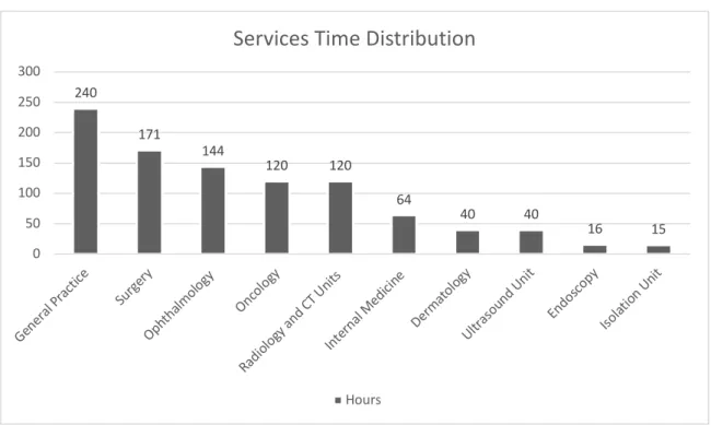
The rotation between several major departments provided countless opportunities to be in contact with a variety of clinical cases. The hospital runs a twenty-four hours permanent service with the clinical specialties of medical imaging (ultrasound, radiology and computed tomography scan units), surgery, endoscopy, internal medicine, ophthalmology, dermatology and oncology; and runs also a hospitalization and isolation units. The distribution time spent by the author in each speciality and unit is described in the graphic 1.
Graphic 1 - Absolute frequency distribution of the number of hours in each service department
The hospitalization shifts consisted of twelve hours during day or night shifts and the isolation unit held shifts of five hours each. The prescribed drugs in each patient chart were administrated by oral, intramuscular, subcutaneous or intravenous routes. Peripheral veins catheterization, collection of blood samples, glycemia measurements, blood arterial pressure monitoring and blood transfusions were some of the assignments. Nutritional support, palliative care and comfort of all the terminally ill patients was also provided.
240 171 144 120 120 64 40 40 16 15 0 50 100 150 200 250 300 Gene ral Pr actic e Surge ry Ophth almolo gy Onco logy Radio logy a nd CT Unit s Intern al Med icine Derma tolog y Ultra soun d Un it Endo scopy Isolat ion U nit
Services Time Distribution
At the surgery service, the student had the opportunity to calculate dosages, prepare and administer pre-anaesthetic drugs, place venous catheters for intravenous administrations and fluid therapy, prepare the anaesthesia induction, perform endotracheal tube placements and skin preparation of surgical sites. During surgery, the internee had the opportunity to help the main surgeon, be a circulating assistant or an anaesthetist assistant. As a surgeon’s assistant, the internee would also help the lead surgeon in tissue retraction and skin closure.
During both general practice and speciality consultations, the reception of the animal, the collection of the medical history or anamnesis and the performance of the physical exam would also be conducted by the student. Blood collection samples, vaccine administration or other drug preparations and placement of intravenous catheters were also practiced during consultations. The discussion of the case in terms of differential diagnosis, complementary diagnostic exams, therapeutic plan, follow-up plan and prognosis would occur after the consultation with the respective doctor in charge. This process also allowed the development of the clinician-to-owner communication skills, a component that is crucial to clinical practice and which is often undervalued.
At the imaging service, the positioning of the animals, selection of x-ray screening constants, preparation of contrast agents and the contribution to the interpretation and discussion of the imaging results were also the major assignments.
Chapter II - Literature Review 1. Rationale for this study
The effects of anaesthetic agents on the ocular physiology should be thoroughly understood by the ophthalmic surgeon so that the regulation of IOP is done properly in order to not be greatly affected. A good anaesthetic management should minimize any potential side effect and avoid an increase in IOP over the entire anaesthetic period.
Sedation may be of extreme value as an adjunct to manual restraint to ease the handling of veterinary patients and also reduce the anxiety associated with ophthalmic examination. Ocular pain when severe enough to enable a thorough ophthalmic examination may also be minimized with a good analgesia control. For these reasons, the need for understanding the effects, in particularly of methadone, in intraocular pressure on behalf of animals suffering from glaucoma or animals undergoing intraocular surgery is of paramount importance.
The objective of the study reported here was to determine the effects of methadone on IOP both in dogs and cats, admitted to the Teaching Hospital of The Faculty of Veterinary Medicine, University of Lisbon, for surgical correction of ocular and extraocular diseases. The author hypothesized that methadone administration, independent of other preanesthetic medication, would not be associated with a significant increase in IOP in both dogs and cats.
For purposes of a better understanding of all the topics related to this subject, a brief summary of the anatomy of the eye and its outflow structures, glaucoma and drug effects on IOP, are fully explored in this literature review.
2. Anatomy of the Eye's Outflow Structures
2.2. Brief review of the eye’s outflow structures anatomy 2.2.1. Ciliary body (CB)
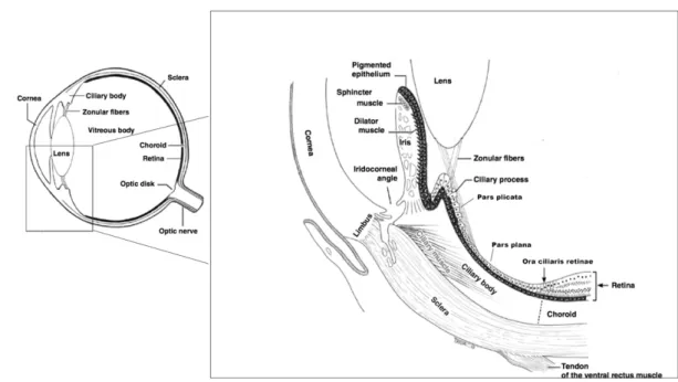
Represents the anterior extension of the choroid joining with the iris (Samuelson, 2013). The CB is divided into an anterior segment - pars plicata – and into a posterior flat segment - pars plana (fig. 1). The pars plicata comprises the ciliary muscle, the ciliary vessels and the ciliary processes. Altogether these structures are responsible for the suspension and accommodation of the lens, the production and outflow of the
aqueous humour, the maintenance of the blood-aqueous barrier and the synthesis of acid mucopolysaccharide components of the vitreous (Trattler, 2012).
Figure 1 - Structural components of the eye in a sagittal section and emphasizing the topography of the cornea, iris, ciliary body and sclera. Adapted from Uemura, 2015.
The CB muscle (figure 1) is composed of smooth muscle in mammals (Gellat, 2013) and its fibers are primarily oriented meridionally in the dog. The CB muscle contracts under parasympathetic stimulation of the oculomotor nerve which also innervates the pupillary sphincter muscle of the iris (Jeruma, 2015). Adrenergic nerve endings are also present to a lesser extent, located in the subepithelial portions of the CB (Gellat, 2013).
The vitreal surface of the CB is covered by an inner nonpigmented epithelium and an outer pigmented epithelium (Watté, 2014). This double layer continues posteriorly with the sensory retina at the ora ciliaris retinae and anteriorly with the posterior PE of the iris (Ofri, 2013). The PE, facing the sclera, is continuous with the retinal PE (fig. 1). These two epithelial layers continue anteriorly as the bilayered iris epithelium (Ofri, 2013).
The CB processes can be described as finger-like projections composed of a pigmented epithelium and nonpigmented epithelium bilayer epithelial, capillaries and stroma (Trattler, 2012).
The iridocorneal angle (ICA) is formed from the anterior recession of the CB, the base of the iris and the corneoscleral tunic and is also known as the filtration angle or anterior
chamber angle. The width of the ICA can vary depending on breed and age within each specie (Pizzirani, 2015).
The lenticular zonules are fibers arising from the peaks of the CB processes and are attached along the epithelial surface in a criss-cross pattern on the lens capsule, anterior and posterior to the equator. As the CB muscle contracts and relaxes, the tension on those fibers decrease and increase, respectively, allowing the process of accommodation (Lowe, 2014). The accommodation process aims to maintain a clear image of both near and far objects. Therefore, the adjustment of the lens position and curvature can properly lead to the optimal focal length, converging the received light beams at a single spot in the retina. Making the comparison to human’s level of accommodation, this capacity is known to be limited in dogs and cats (Watté, 2014). Contraction of the CB muscle not only provides accommodation but also increases the conventional aqueous outflow (Watté, 2014) by moving the lens forward and increasing the lens curvature by releasing tension on the zonular fibres and lens equator.
CB vassels are represented by two long posterior ciliary arteries, vortex veins and anterior ciliary arteries arising from branches of the ophthalmic artery (Gellat, 2013; Mitchell, 2014).
2.2.2. Pectinate ligaments
The pectinate ligaments (PL) consists of beams of iris tissue that span the ICA and connect the base of the iris to the cornea. The junction between the cornea and the PL can be seen at gonioscopy and it is seen as a pigmented line known as the Schwalbe’s line. These ligaments vary in number, pattern, length and thickness among different breeds and individuals and they are usually pigmented unless in sub-albinotic animals (Pizzirani, 2015).
2.2.3. Ciliary Cleft
The ciliary cleft (CC) represents the space posterior to the PL and juxtaposed to the sclera and filled with a sponge-like network of connective tissue beams, the trabecular meshwork (TM) (Watté, 2014; Hollingsworth, 2017). The TM and the CC are both contained within the ICA.
2.3. Aqueous composition, production and drainage
The aqueous humour (AH) corresponds to an optically clear fluid under regular conditions, which is produced by the CB and occupies both the anterior and the
posterior chambers. The cells responsible for its production are localized at the CB posterior nonpigmented epithelium, lining the ciliary processes.
Circulation of the AH has major purposes regarding the nourishment and the metabolic waste removal of avascular structures like the lens and the cornea; and it is of the upmost importance in maintaining physiological intraocular pressures (IOP), allowing an unaltered refractive status of the eye. In fact, the AH constitutes an important component of the eye’s optical system as it provides a transparent and colourless medium of approximately 1.33 refractive index (Gabelt, 2011), between the cornea and the lens.
The balance between aqueous production and aqueous outflow maintains the IOP physiologic range which may vary individually in consequence of daily circadian patterns and aging (Pizzirani, 2015).
Both in humans and dogs, it was identified higher normal IOP values measured in the morning and lower values in the evening. On the contrary, in cats, rabbits and nonhuman primates, is has been suggested the opposite (Miller, 2013). It seems also unanimous that both production and outflow of AH tend to decline with age, being the rate of the former higher in the majority of animals and humans (Miller, 2013).
The production of AH is conducted by both passive and active processes. The passive mechanisms do not contribute significantly to the formation of AH although they are able to generate a fluid reservoir within the CB. On the opposite, the active mechanisms are responsible for at least 80-90% of the AH formation (Pizzirani, 2015).
2.3.1. Passive production mechanisms
The passive production mechanisms can be described as two distinct processes of diffusion and ultrafiltration of plasma, both taking place in the vascularized CB stroma. Diffusion represents the passive passage of AH along with lipid-soluble molecules in favour of their concentration gradient, across the fenestrated CB vassels endothelia (Pizirrani, 2015). The ultrafiltration process occurs in consequence of an uprising in the hydrostatic pressure which forces the passage of water and water-soluble molecules through fenestrations in the CB endothelium (Miller, 2013).
2.3.2. Active secretion
The majority of AH production is catalysed by carbonic anhydrases which transform carbon dioxide and water into carbonic acid by the equation 1.
𝐶𝑂$+ 𝐻$𝑂 ↔ 𝐻$𝐶𝑂) ↔ 𝐻𝐶𝑂)* + 𝐻+
Equation 1 – Conversion of carbon dioxide and water into carbonic acid and its dissociation in bicarbonate and hydrogen ions.
The existence of sodium/potassium adenosine triphosphatase (ATPase) pumps (figure 2), that actively secrete sodium ions to the posterior chamber seem to drive negatively charged ions like bicarbonate. The entry of these solutes into the posterior chamber is accompanied by the diffusion of water, therefore composing the AH (Renwick, 2014).
Figure 2 - Mechanisms for AH secretion ATPase – adenosine triphosphatase; NPE – nonpigmented epithelium; PE – pigmented epithelium. Adapted from Pizzirani, 2015.
2.3.3. Trabecular Outflow (TO) or conventional/anterior outflow
TO is the major aqueous drainage system also known as the anterior or conventional outflow route. The AH passes from the posterior chamber, through the pupil, and into the anterior chamber (Miller, 2013). At this level, the AH is filtered through the TM achieving the angular aqueous plexus, composed of a series of collecting vessels that are anatomic and functional similar to the Schlemm’s canal in primates (Pizzirani, 2015). The angular aqueous plexus eventually may drain the AH both anteriorly to the episcleral and conjunctival veins, or posteriorly into the scleral venous plexus and the choroidal vortex venous system (Miller, 2013).
2.3.4. Uveoscleral outflow (UO) or unconventional/posterior outflow
Occurs when the AH has entered the TM and escapes passively the TO through the CB muscle interstitium reaching the suprachoroidal space (Pizzirani, 2015).
3. Definition and Pathophysiology of Canine Glaucoma
During the decade of the 1950s, the term glaucoma was often referred to as a sign rather than a disease per se, and its definition comprised a group of diseases with an abnormal elevation of the IOP regardless of their nature, both in human and in veterinary medicine. Therefore, since 1980s, the plural form of the name, glaucomas, started to be adopted given the variety of factors that can influence the IOP in normal individuals (Gellat, 2014).
In humans, the most recent studies concluded that in addition to the two major forms of human glaucomas, Primary Open-Angle Glaucoma (POAG) and Primary Angle-Closure Glaucoma (PACG), there are some POAG individuals with normal IOP measurements and this exception created a new POAG category, referred as normotensive glaucoma. Although normotensive glaucoma does not meet the 21-mmHg cut-off criterion for ‘elevated IOP’ in humans, IOP is probably still too high in these eyes (Tan & Kaufmann, 2014).
In all of the human glaucomas forms, including normotensive glaucoma, the progressive optic neuropathy with visual field loss, the thinning of the retinal nerve fibre layer and the excavation of the optic nerve head are present (Saeedi, Ramulu & Friedman, 2014). For this reason, the IOP increase which has been historically a common definition for the disease in small animals, is not mentioned in the accepted definition used by physician ophthalmologists. The fact that even a controlled normalized IOP does not alter the progression of the disease in both humans and dogs, suggests that additional pathophysiologic mechanisms must be involved (Pizzirani, 2015).
Some of the risk factors leading to the development of some glaucomas forms are:
a. Constant IOP raise
The reduction in choroidal vascular perfusion impairs both microcirculation and the axoplasmic flow in the retinal ganglion cell axons leading to outer retinal necrosis (Martín, 2017).
b. Age
Age-related abnormalities within the TM may increase the outflow resistance. These changes can include the thickening of basal and trabecular membranes, partial loss of endothelial cells and the accumulation of materials such as pigment granules and collagen (Pizzirani, 2015).
c. Familiar history
The genetic inheritance seems to be an important risk factor both in humans and dogs with some causative genes being already identified (Komaromy, 2015).
d. Breed and presence of goniodysgenesis
Goniodysgenesis corresponds to a defect in the development of the ICA which has its width decreased due to the adjacent pectinate ligaments dysplasia (PLD). These ligaments undergo a process of consolidation, forming a broad sheet structure which may be excessively extensive but can also, in some cases, be perforated by intermittent flow holes (Renwick, 2014). It is believed that PLD has genetic causes considering the high incidence in certain breeds (Pizzirani, 2015) but it has not been directly related to an increased resistance to the outflow unless the condition progresses and affects over 180º of the ICA (Gellat, 2014). It is also important to differentiate PLD from inflammatory associated anterior synechiae (Gellat, 2014).
e. Systemic blood hypotension
Systemic hypotension can dysregulate ocular perfusion by primary tissue ischemia and is sufficient to induce permanent damage in RGC if hypoxia is superior to a six hours duration period (Pizzirani, 2015).
Additional factors that can raise IOP in healthy animals:
• Positioning and degree of restraint: any increase in venous pressure reduces venous drainage and AH outflow from the eye, increasing the IOP (Clark, 2014). The use of collars, neck leads or excessive restraint may cause this jugular occlusion.
• Pressure on the ocular globe or forced lid opening: exophthalmic or brachycephalic breeds are more prone to this occurrence (Renwick, 2014) • Diurnal variation: in dogs and humans, the IOP is slightly increased in the
• It is also undeniable that other factors including anatomy variations of the anterior segment and endogenous compounds such as neurotransmitters, hormones, prostaglandins among many others, can influence these mechanisms and alter the IOP baseline (Miller, 2013).
3.2. Classification of canine glaucomas
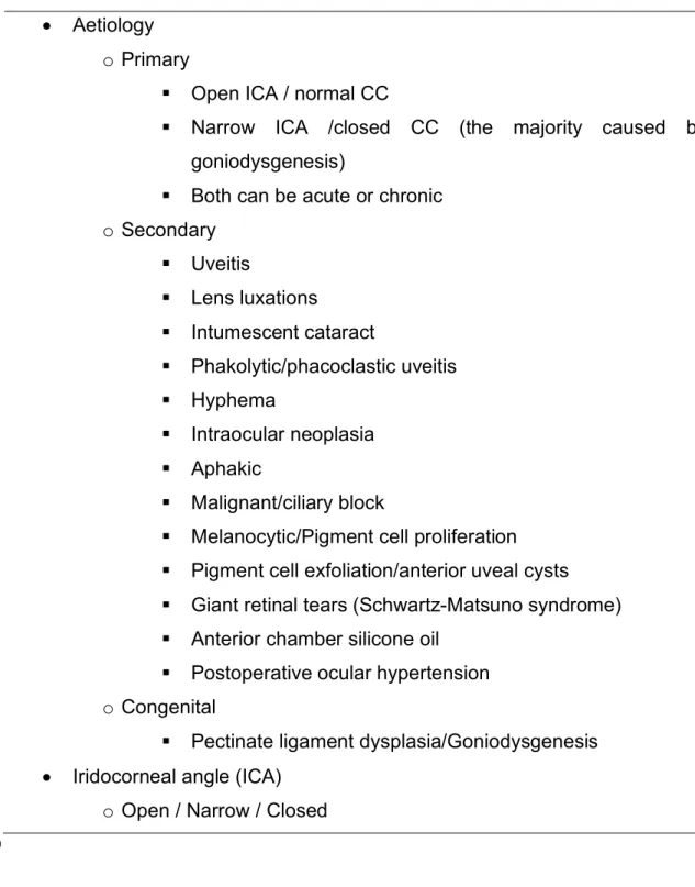
Canine glaucomas can be classified according to their aetiology, the gonioscopic appearance of the filtration angle (ICA and CC) and the staging of the course of the disease (table 1).
Table 1 – Aetiology of canine glaucomas. Adapted from Gellat, 2014.
• Aetiology o Primary
§ Open ICA / normal CC
§ Narrow ICA /closed CC (the majority caused by goniodysgenesis)
§ Both can be acute or chronic o Secondary § Uveitis § Lens luxations § Intumescent cataract § Phakolytic/phacoclastic uveitis § Hyphema § Intraocular neoplasia § Aphakic § Malignant/ciliary block
§ Melanocytic/Pigment cell proliferation § Pigment cell exfoliation/anterior uveal cysts § Giant retinal tears (Schwartz-Matsuno syndrome) § Anterior chamber silicone oil
§ Postoperative ocular hypertension o Congenital
§ Pectinate ligament dysplasia/Goniodysgenesis • Iridocorneal angle (ICA)
• Ciliary cleft (CC)
o Open / Narrow / Collapsed • Staging
o Acute/ Chronic
3.2.1. Congenital Glaucoma
This severe and rare form of glaucoma develops due to an abnormal formation of the anterior segment including the TM, immediately after birth or within the first six months of life (Martin, 2017). The first clinical signs consist of rapid onset of buphthalmia, inability to close the palpebral fissure and development of exposure corneal disease (Gellat, 2014).
3.2.2. Primary Glaucoma
Primary glaucomas are defined as a group of disorders in which occur characteristic changes to the optic nerve without any proven association with other ocular or systemic disorder (Miller & Bentley, 2015). They emerge in approximately 20% of the total dog glaucomas and they are thought to have a genetic inherited aetiology, associated with breed predisposition and present themselves as bilateral even though they can be asymmetrical (Martín, 2017). They can be classified according to the gonioscopy appearance of the drainage angle in POAG and PCAG.
3.2.2.1. Primary Open-Angle Glaucoma (POAG)
The drainage ICA appears normal and indistinguishable from a healthy eye. Recent evidences of an increase in altered glycosaminoglycans (GAGs) forming the TM and ultimately leading to a deposition of elastin membranes and type IV collagen are considered to contribute to the resistance of AH flow, causing IOP to rise (Martín, 2017). An enzyme defect allowing the accumulation of extracellular matrix material in the TM is also questioned (Miller, 2013). This type of glaucoma accounts for about 3% of the total dog glaucomas (Martín, 2017).
3.2.2.2. Primary Angle-Closure Glaucoma (PACG)
PACG is the most common form of canine primary glaucoma and is a complex trait with multiple genetic and possibly environmental risk factors (Komáromy, 2015). According to Martín, 2017, approximately 80% of the PACG’s in dogs are due to PLD, an abnormality already explained in page 8. Besides the majority of
goniodysgenesis-related glaucoma, anomalous conformation of both a narrow CC and anterior chamber, anterior displacement of the lens and iris plateau may also contribute to PACG (Martín, 2017). The iris plateau abnormality has been described as an anatomic variant of the iris structure in which the iris periphery angulates sharply forward from its insertion point and then again angulates sharply and centrally backward (Diniz Filho, 2008). PACG represents itself as a bilateral disorder although it might be initially unilateral. The raise in IOPs is marked and rapid, affecting middle-aged to older dogs of certain breeds (see table 2) (Miller, 2013).
Female dogs can possibly have a shorter axial globe length and a narrower ICA opening and for these reasons they are twice as likely to be affected as male dogs (Komáromy, 2015).
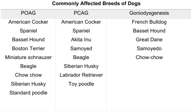
Table 2 – Glaucoma commonly affected breeds of dogs. Adapted from Martín, 2017.
Commonly Affected Breeds of Dogs
POAG PCAG Goniodysgenesis
American Cocker Spaniel Basset Hound Boston Terrier Miniature schnauzer Beagle Chow chow Siberian Husky Standard poodle American Cocker Spaniel Akita Inu Samoyed Beagle Siberian Husky Labrador Retriever Toy poodle French Bulldog Basset Hound Great Dane Samoyedo Chow-chow 3.2.3. Secondary Glaucomas
Secondary glaucomas are common both in dogs and cats but particularly on the latter where 95 to 98% of the feline glaucomas can be categorized as secondary (Pumphrey, 2015).
3.2.3.1. Uveitis
Responsible for up to 45% of secondary glaucomas in dogs and both uveitis and glaucoma may occur at the same time or glaucoma can develop months to years after
the uveal tract induces glaucoma are predominantly linked to the deposition of inflammatory debris, such as blood components and fibrin, obstructing the pupil, ICA or TM (Pumphrey, 2015; Renwick, 2014). Those deposits can form adhesions between the iris and the lens (posterior synechiae) preventing AH flow through the pupil and being withheld behind the iris which bulges forward. The formation of what is known as “iris bombé” occurs when these adhesions are present at 360 degrees. Peripheral anterior synechiae can also develop associated with iris bombé, luxation of the lens by pushing the iris forward or intumescent cataracts (Renwick, 2014)
The CC is also composed of uveal tissue and any swelling of the TM structures can therefore cause an additional block of the AH outflow.
Uveitis and glaucoma should be well differentiated by tonometry because they may both be presented as a red and painful eye (Miller, 2013).
3.2.3.2. Cataracts and cataract surgery
The leakage of antigenic lens proteins into the AH is induced in mature or hypermature cataracts associated with lens rupture, inducing a secondary glaucoma by the inflammation it creates (Renwick, 2014). This lens induced uveitis aetiology is not clear but the presence of keratic precipitates and inflammatory cell clumps are often present (Pumphrey, 2015).
3.2.3.3. Lens instability and lens luxation
Lens luxation can occur primary or secondary to a glaucoma. The primary lens luxation (PLL) or subluxation can induce glaucoma, especially if the luxated lens enters the anterior chamber causing a pupillary block and impairing the AH outflow (Miller, 2013). In the second scenario, the primary glaucoma in a buphthalmic globe, stretches and subsequent tears off the zonular fibres which support the lens, resulting of its subluxation or even complete luxation (Miller, 2013).
3.2.3.4. Neoplasia
There are multiple ways in which an intraocular neoplasia can induce glaucoma. The direct mass effect or the accumulation of exfoliated neoplastic in the ICA or TM may impair normal aqueous drainage (Pumphrey, 2015).
Primary uveal melanoma, primary ciliary body adenoma/adenocarcinoma and lymphoma are the most common types of neoplasms (Renwick, 2014). Metastatic
neoplasms account for about 4% of the total ocular neoplasia in dogs and the involvement may be bilateral (Pumphrey, 2015)
In the Comparative Ocular Pathology Laboratory of Wisconsin (COPLOW) collection, of the 5722 cases of neoplasia, 1516 had glaucoma as part of the syndrome and with the following definitive histopathological type: 57% melanoma, 21% iridociliary epithelial tumours, 5% metastatic tumours and 4.5% lymphoma.
The pathogenesis of PACG due to neoplasms can be explained by the mechanical displacement of the iris and lens by the mass effect in neoplasms of the CB, choroid, or retina, which leads to pupillary blockage (Dubielzig, 2010).
3.2.3.5. Hyphema
The presence of blood in the anterior chamber, which may also appear along with hypopyon or fibrin, can occlude the pupil, the ICA or the TM by the deposition of blood component cells and debris (Pumphrey, 2015). Lower IOP in cases of trauma and severe intraocular haemorrhage may be present due to CB damage or scleral rupture leading to a marked decrease in aqueous production (Renwick, 2014).
3.3. Pathophysiology
The retina is embryologically derived from the optic cup, which is a double layer of neural ectoderm, and it is divided in a neurosensory or inner layer which is non-pigmented and in the retinal non-pigmented epithelium (RPE), which forms the outer layer (McLellan, 2014).
The optic nerve head (ONH), also known as the optic disc or optic papilla, is the beginning of the myelination of the retinal ganglion cell (RGC) axons as they converge to exit the globe through the lamina cribosa of the sclera. The continuous mechanical compression constricts and obstructs the axoplasmatic flow and allows the apoptosis of the nerve cells to initiate. The impairment of the physiological movement of molecules impairs the transport of neurotrophins, which are essential for the development and maintenance of the neurons function and promotes the accumulation of oxygen free radicals and excitotoxins like glutamate (Martín, 2017). Glutamate in high concentrations induces the entrance of sodium, chloride and water into the cells causing oedema and cell membrane rupture (Pizzirani, 2015).
The high levels of calcium outside the axons seems also to generate more free radicals which activates in their turn, proteases, kinases and phospholipases. All of these
events lead to an optic neuropathy in which the RGC undergo apoptosis and an irreversible blindness occurs (Martín, 2017; Pizzirani, 2015).
3.4. Clinical signs
Clinical signs of glaucoma are directly related to the level and duration of the elevation in the IOP (Gellat, 2014) and are similar regardless of the cause of the elevation (Miller, 2013). Most owners will not be able to recognize the disease early in its course until the IOP approaches 40 mmHg or until there is noticeable loss of vision (Martín, 2018).
3.5. Early to mid-stage glaucoma clinical signs 3.5.1. Pain
In general, animals tend to disguise evident signs of pain, therefore a good anamnesis should be accurately obtained. Complaints of lack of interest in playing or climbing on the couch, starting to hide in unusual locations, having reduced tolerance for disturbances, having loss of appetite or spending too much time sleeping can be the first indicators of lethargy and pain. On the other hand, other animals can exhibit continuous blepharospasm, epiphora, photophobia and protrusion of the nictitating membrane (Martín, 2017), particularly when the IOP is above 40-50 mmHg where the discomfort may be so severe as to cause vocalization (Renwick, 2014). Blepharospasm can also be present in both eyes even when only one is affected. This fact might be explained due to a generalized migraine sensation, in similarity with what is reported in human glaucomas (Miller & Bentley, 2015).
3.5.2. Pupil alterations
Mydriasis, the dilation of the pupil is often present in acute glaucomas with IOPs above 40-50 mmHg, causing pressure high enough to induce paresis or paralysis of the iris sphincter muscle (Martín, 2017). If glaucoma is associated with uveitis in the same eye, the pupil may be normal or even constricted (Martín, 2017; Renwick, 2014). The direct pupillary light reflex is reduced or absent which does not equal a total loss of vision. In this situation, the potential vision must rather be assessed with the testing of the menace response, dazzle reflex or the consensual pupillaru light reflex in the unaffected eye, illuminating only the affected eye (Renwick, 2014).
Peripheral anterior synechia can be formed rapidly and the ischemia originated also in the iris sphincter muscle can be responsible for sectorial or diffuse atrophy of the iris (Miller & Bentley, 2015).
3.5.3. Corneal Oedema
The normal optical clarity of the cornea depends primarily of its state of dehydration. This state is achieved owing to the active transport mechanism of the cornea endothelium, which transports solutes from the cornea into the aqueous humour, against the IOP and causing the movement of water out of the corneal stroma by diffusion (Renwick, 2014; Maggs, 2013). Although the increase in IOP alone is not sufficient to disrupt this mechanism, persistently elevated IOP drives fluid across the corneal endothelium, creating oedema of both epithelium and stroma. (Miller & Bentley, 2015). In conclusion, corneal oedema results when excess fluid accumulates within the stroma and forces the collagen lamellae apart, leading to loss of transparency (Maggs, 2013).
The ability to blink and distribute the tear film can be compromised with the globe enlargement which can result in the formation of irregular areas in the epithelium or even ulceration mainly in the centre of the cornea (Renwick, 2014).
3.5.4. Episcleral congestion
The increase in IOP reduces the flow through the CB to the vortex veins leading to the flow to pass via anastomosis through the episcleral veins and capillaries to a lesser extent (Miller, 2013). The engorgement of superficial conjunctival vessels indicates an ocular surface disease and for this reason it is important to be able to distinguish them from the episcleral vascular vessels with the features presented at table 3.
Intermittent episcleral injection can sometimes be the only early sign in patients whose IOP remains normal on the physical exam (Miller, 2013).
Table 3 - Morphological and clinical differences between episcleral and conjunctival vessels
Episcleral vessels Conjunctival vessels
• Dark red
• Larger calibre and run at right angles to the limbus
• Do not move in association with movement of the overlying lids and
conjunctiva
• Blanch themselves poorly and slowly after the application of topical 1% adrenaline or 10% phenylephrine
• Bright pink to red
• Cover larger portions of the sclera • Typically branched
• Blanch themselves after the application of topical 1% adrenaline
or 10% phenylephrine within 1 or 2 minutes
Episcleral congestion can also be present, apart from glaucoma in cases of: • Uveitis / endophthalmitis / panophthalmitis
• Episcleritis (often localized in dogs)
• Retrobulbar space-occupying lesion (e.g. abscesses, neoplasia) • Horner’s syndrome
• Hyperviscosity syndromes • Excitement
3.6. Chronic glaucoma clinical signs
Chronic glaucoma can develop after an uncontrolled or misdiagnosed of an acute-onset episode or it may progress from an insidious first episode. In chronic cases there also may be present some of the clinical signs seen in acute glaucomas although less markedly. The following chronic related signs indicate poor prognostic for vision.
3.6.1. Buphthalmia / Buphthalmos
Pathologic enlargement of the ocular globe due to chronically and notably elevated intraocular pressure and a pathognomonic change in glaucoma. It is often responsible for irreversible blindness (Martín, 2017), although limited vision may be retained for a while in puppies and sharpeis (Miller, 2013). The differential diagnosis with exophthalmos must be made, as this sign occurs in a normal sized eye that is being pushed forward by a retrobulbar abscess or neoplasia that is occupying space (Renwick, 2014). This condition leads often to exposure keratitis, corneal ulceration, subluxation of the lens and occasionally scleral ectasia (Miller & Bentley, 2015). Lens
subluxation must also be carefully differentiated from PLL which also leads to secondary glaucoma (Miller & Bentley, 2015).
3.6.2. Luxation or subluxation of the lens
The fact that the lens starts to occupy a position away from the visual axis develops an aphakic crescent which becomes visible (Renwick, 2014). Movements of both the lens (phacodonesis) or the iris (iridodonesis) may also be seen, as well as an abnormally shallow or deep anterior chamber (Martín, 2017; Miller, 2013).
Lens opacities can also develop in chronic glaucomas due to poor lens nutrition and/or the accumulation of toxic products such as glutamate (Renwick, 2014).
3.6.3. Haab Striae / Descemet’s Streaks
The pressure induced by the stretching of the globe can cause breaks in the Descemet membrane which can be detected as blue-grey streaks across the cornea (Miller & Bentley, 2015). Other grey opacities within the cornea may develop in cases of scar tissue formation following ulceration or secondary to exposure and subsequent keratinization of the cornea epithelium (Renwick, 2014).
3.6.4. Equatorial staphyloma
The stretching of the sclera can lead to its thinning at the equator of the globe and the formation of staphylomas (Renwick, 2014). This scleral defect is filled directly by uveal tissue and, after a few hours, by blood clots and exudate (Stades et al, 2007).
3.6.5. Phthisis Bulbi
Atrophy of the eye that may occur in advanced cases of glaucoma, when the CB no longer produces AH. This condition can also be a sequela of some therapies aimed at destroying AH production or to a lesser degree after gonio-implantation with excessive filtration (Miller & Bentley, 2015). When phthisis bulbi is present at the time of examination, the IOP values are typically normal or even reduced (Renwick, 2014).
3.6.6. Optic nerve atrophy and Retinal Degeneration
The optic disc represents the location where the optic nerve enters the ocular globe and all the retinal nerve fibres converge. The optic nerve axons are supported in a fenestrated scaffold within approximately a third of the inner sclera called lamina
the lamina cribosa connective tissues causes the cupping of the optic disc and compromises both the axonal flow and the blood supply, leading ultimately to the optic nerve axonal death (Plummer, 2013). The optic disc is presented with signs of cupping when it bows posteriorly through the lamina cribrosa (Miller, 2013).
The retinal thinning and atrophy follows the induced pressure of the choroidal vasculature which supplies the outer layers of the retina (Renwick, 2014). This results in a tapetal hyper-reflexion with an increased reflection of light back from the tapetum (lay within the choroid).
Some clinical findings are more prone to be observed in acute cases while others are more prevalent in chronic glaucoma cases as seen in table 4.
Table 4 – Different clinical aspects of acute and chronic glaucomas. Adapted from Martín, 2017 Acute
Glaucoma
Chronic Glaucoma
IOP >30
mmHg
High, normal or low
Episceral congestion
Present Present
Diffuse corneal oedema
Present May be present. Haab striae. Exposure keratitis
Ocular globe size Normal Normal or buftalmic
Pupil Midriasis Midriasis or normal
Lens position Normal Normal, subluxated or
luxated. Cataracts
Optic nerve Normal Atrophy
Retina Normal Vascular attenuation
Tapetal hyper-reflectivity
3.7. Diagnosis
In addition to the clinical signals described, the first diagnostic methods that are fundamental to confirm the clinician suspicion can both be used to evaluate intraocular pressure or/and the ICA.
3.7.1. Tonometry
The measurement of IOP is of the utmost importance in diagnosis and monitoring ocular conditions in which it is predicted a disturbance of IOP regulation. As already mentioned, IOP above 25 mmHg is suspicious for glaucoma in dogs, and above 30 mmHg in cats (Martín, 2017).
The direct form of measuring IOP can be performed through paracentesis of the anterior chamber which due to its invasiveness it is highly impractical in clinical practice (Spiessen et al, 2015).
All of the currently available tonometers do not actually measure IOP directly but a physical property of the cornea and use it to estimate true IOP (Miller & Bentley, 2015).
• Indentation tonometry – the Schiotz tonometer measures the degree of corneal deformity by applying a standard force with a metal rod in a topically anesthetized cornea (Heinrich, 2014). The greater the distance of the rod indents into the cornea, the lower is the patient’s IOP (Maggs, 2013). The cornea must be in a horizontal position and without excessive pressure on the neck, to obtain readings. This technique is the most difficult to use correctly, having the largest number of potential errors and its use is limited in patients with severely ulcerated corneas or with other pathologic defects (Miller, 2015; Heinrich, 2014).
• Applanation tonometry: the indirect IOP measured with these tonometers (e.g. Tono-Pen XL®, Tono-Pen Vet® or Avia®) results from the force required to flatten the corneal surface. The readings with these devices can be altered with corneal drying (after application of a topical anaesthetic) or increased tear film viscosity (from mucoid ocular discharge or artificial tears) (Miller & Bentley, 2015).
the cornea surface it returns or rebounds to the instrument. The instrument assesses the probe deceleration (rebound) which allows the conclusion that eyes with a higher IOP cause a more rapid deceleration of the probe and shorter return time to the instrument than those with a lower IOP (Gellat, 2013). An altered ocular surface tension as seen with the application of topical medications (including topical anaesthetic) can affect the results with rebound tonometry. For this reason, this technique should be performed before the application of such medications (Maggs, 2013).
With all the methods described, it is extremely important to perform a careful restraint of the patient to prevent an IOP overestimation, avoiding an excessive compression both of the neck where the jugular veins are located and the ocular globe (Martín, 2017).
It is also advisable to obtain more than one measurement and if possible at different periods of the day.
3.7.2. Gonioscopy
Although the gonioscopy evaluation requires considerable practice, the technique allows the clinician to estimate the severity of the obstruction of the drainage angle, identifying the type of glaucoma present and the likely location of the impediment to the outflow. Since this procedure compresses the cornea it should never be performed before tonometry evaluation (Miller & Bentley, 2015). To perform gonioscopy, a goniolens is required (Barkan, Koeppe, Goldmann) and its application should be made upon the anaesthetised cornea without compressing it excessively (Martín, 2017). This technique may be useful in the diagnosis of abnormalities such as PLD or closure of the CC and pectinate ligament, infiltration by neoplastic tissue, inflammatory deposits or even abnormal pigment deposition and fibrovascular tissue ingrowth (Renwick, 2014).
A grading system is used to compare objectively not only eyes within the same patient and between different patients but also the same eye in distinct moments of assessment. The Shaffer scale modified by Ekesten and Narfstrom scores the width of the iridocorneal angle in the grades of 0 (closed), 1 (narrowed), 2 (slightly narrowed), 3 (open) and 4 (wide open) (Martín, 2017).
The European College of Veterinary Ophthalmologists (ECVO) adopted other nomenclature system based on the PLD occlusion, assuming that it is always
associated with an open ICA, an assumption that is not widely accepted (Pizzirani, 2015). In this system the grade of occlusion is graded ascending from A (slight occlusion) to C (almost complete occlusion).
3.7.3. Ophthalmoscopy
The cupping of the optic disc can be accessed by direct or indirect ophthalmoscopy but early detection of optic disc changes is extremely difficult because of the large amount of myelin over the disc surface in dogs (Miller & Bentley, 2015).
In cats, the optic disc cupping can be difficult to identify because of the physiological lack of myelin in their ONH, therefore a dark to pale grey appearance, peri-pigmentation around the ONH, a hyperreflective halo and the fact that blood vessels can no longer be observed crossing the rim of the ONH may cause suspicion (McLellan, 2015).
Other advanced techniques can give the clinician a more detailed description of anatomical structures, as to mention optical coherence tomography (OCT) and high frequency ultrasounds (Martín, 2017).
High frequency ultrasounds are only capable of studying the anterior segment of the eye due to its poor penetration strength but do it with extreme accuracy, all the anatomic details of the ICA aperture evaluating the cornea, sclera, CB and iris can be accessed, having a particular utility for the diagnose of primary glaucomas or intraocular neoplasia (Martín, 2017).
The OCT technique uses near-infrared light that allows the acquisition of cross-sectional sets and 3D images, approaching it to in vivo “histopathology” with distinct layers visible as alternating bright and dark signals (Miller & Bentley, 2015). The anterior segment structures can be detailed without the need of direct contact between the transducer and the surface of the eye, minimizing tissue distortion and trauma (Dennis, Johnson & McLellan, 2014).
3.7.4. Genetic Testing
As already reported, primary glaucoma, both POAG and PACG, have an important genetic predisposition although the studies within each breed are relatively scarce. The studies conducted in POAG dogs reveal that they can be related to mutations in the genes ADAMTS10, ADAMTS17 and Fibrillin-1 (FBN1) causing modifications in the soft tissue composition and leading to an impaired microfibrils formation (Martín, 2017).
The ADAMTS10 codifies a type of metalloprotein expressed in the tissues that compose the TM and the mutation in this gene was identified in Beagles with POAG. Although far from being completely understood, it seems that the inheritance of this trace is recessive in Beagles (Komaromy, 2015). The ADAMTS17 mutation were associated with PLL in the Jack Russell Terrier, Bull Terrier and Lancashire Heelers, therefore, inducing a secondary glaucoma. Ultimately, the Fibrillin-1 is an important component of the lenticular zonules and is secreted by the NPE of the CB (Martín, 2017).
3.8. Differential Diagnosis
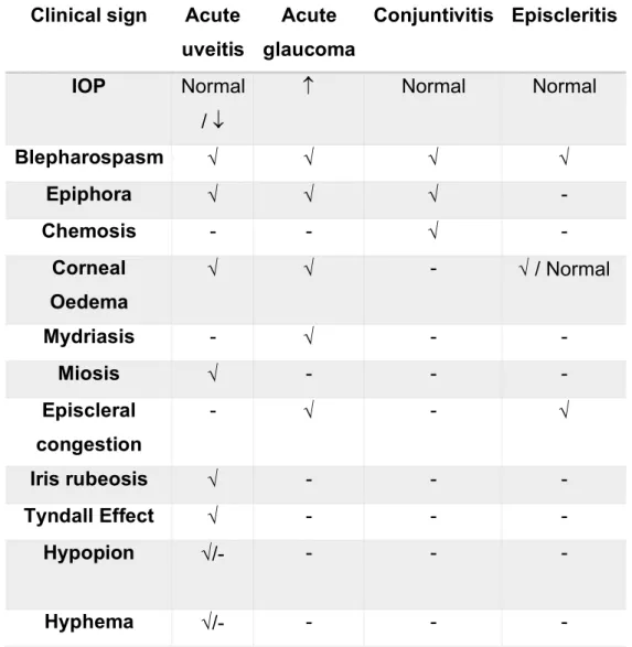
Although the IOP remains the primary clinical sign that distinguishes glaucoma, confusion can be made with the diagnosis of other ocular pathologies mainly in cases of uveitis, conjunctivitis and episcleritis. The following table helps to clarify the diagnosis process.
Table 5 – Differential diagnosis between acute uveitis, acute glaucoma, conjuntivitis and episcleritis. Adapted from Martín, 2017. Clinical sign Acute
uveitis Acute glaucoma Conjuntivitis Episcleritis IOP Normal / ¯ Normal Normal Blepharospasm Ö Ö Ö Ö Epiphora Ö Ö Ö - Chemosis - - Ö - Corneal Oedema Ö Ö - Ö / Normal Mydriasis - Ö - - Miosis Ö - - - Episcleral congestion - Ö - Ö Iris rubeosis Ö - - - Tyndall Effect Ö - - - Hypopion Ö/- - - - Hyphema Ö/- - - -
4. Feline Glaucomas
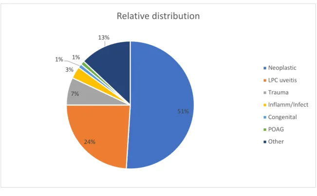
The majority of feline glaucomas are secondary to other ocular or systemic disease, primary glaucomas are rare (McLellan, 2015). According to the COPLOW, 29% of the 3212 feline enucleated globes entries were due to glaucoma, from 1983 to 2008 (Dubielzig, 2010). The relative distribution of the causes of feline glaucoma, identified by COPLOW follows in the graphic 2.
Graphic 2 - Relative distribution of feline glaucoma causes (Dubielzig, 2010)
4.1. Primary Glaucoma
Compared with dogs, goniodysgenesis appears to be quite rare in cats although PLD has been reported in Burmese cats (Renwick, 2014). POAG affects usually middle-aged to older adult cats (mean age around 10 years), unilaterally or bilaterally but most frequently asymmetric (McLellan, 2015). Inherited POAG is described in Siamese cats but PACG is extremely rare to non-existent (Miller, 2013).
4.2. Secondary Glaucoma
The majority of the total of glaucoma cases are due to neoplasia and uveitis. Diffuse iris melanoma and uveal lymphoma are the most common tumours (Renwick, 2014).
51% 24% 7% 3% 1% 1% 13%
Relative distribution
Neoplastic LPC uveitis Trauma Inflamm/Infect Congenital POAG OtherExudative uveitis in cases of feline infectious peritonitis (FIP), toxoplasmosis, feline immunodeficiency virus (FIV) or feline leukaemia virus (FeLV) can also obstruct the CC by deposition of the lymphocytic / plasmocytic cellular component (Martín, 2017). Other causes such as intraocular haemorrhage and hyphema due to systemic blood hypertension or trauma can also lead to glaucoma.
Primary lens luxation seems to have a low incidence in comparison with dogs and is usually associated with the underlying uveitis (Renwick, 2014).
In older cats, glaucoma may develop due to a shallow anterior chamber where there is an anterior displacement of the iris and lens by an expanded vitreous, creating a posterior misdirection of the AH rather than anteriorly (McLellan, 2015).
4.3. Clinical Signs
Cats often present clinical signs that are less severe because of the slow and insidious rise in IOP that tends to mask ocular pain although buphthalmia can be quite extreme (McLellan, 2015). Mydriasis, exposure keratitis and retinal degeneration are the most common signs and the loss of vision is slower than in dogs (Miller, 2013). In cases of uveitis, the signs are also very mild and a slit-lamp biomicroscopy should be performed to rule it out (Renwick, 2014).
A subtle anisocoria may be present especially in the eyes suffering from AH misdirection syndrome (Miller, 2013)
5. Medical Treatment of Glaucoma
The main criteria concerning the medical treatment of glaucoma is preserving the optic nerve and the RGC function for as long as possible, keeping the patient visual and without pain. Reducing IOP by enhancing the AH drainage or inhibiting its production, remains the mainstay of the medical management (Alario, 2015). The prophylactic use of neuroprotective and neurotrophic agents is also an important keystone.
The animal presented with a new diagnose of glaucoma should be first assessed in terms of the acuteness or chronicity of the disease or, in other words, if the eye still has the potential for vision or if it is irreversibly blind (Miller, 2013).
Acute glaucoma with more than 40-50 mmHg of IOP is usually associated with a very recent onset that requires emergency treatment (Martín, 2017).
The long-term control of glaucoma is usually not attainable regarding only medical therapy. The urgency of combined medical and surgical therapies is often required,
since the persistence of high IOP during 24 to 72 hours results in irreversible vision loss (Miller, 2013).
The majority of studies were conducted in population of research beagles affected with normotensive or POAG and these do not represent the typical PACG type of glaucoma that represents the vast majority of canine glaucomas (Alario, 2015).
5.1. Topical ocular hypotensive drugs
Topical hypotensive drugs are largely more effective than systemic therapy and with lower side effects. The major classes commonly used are presented below:
5.1.1. Cholinergic agonists or Miotics (Pilocarpine, Demecarium bromide)
The stimulation of acetylcholine (Ach) receptors of the parasympathetic nervous system in the eye leads to the CB muscle contraction and miosis, widening the conventional outflow pathway via the trabecular meshwork. Their major indication is for POAG since when goniodysgenesis happens or there is a CC collapse these agents will not prove effective (Renwick, 2014).
The direct-acting agents (pilocarpine and carbachol) activates the Ach receptors directly while indirect-acting agents (demecarium bromide) acts as an anticholinesterase increasing the concentration and time exposure of Ach to its receptors (Alario, 2015).
The miosis induced may lead to the formation of posterior synechiae and even iris bombé in cases where concomitant uveitis is present (Alario, 2015).
5.1.2. Drugs Acting on adrenoceptors
5.1.2.1. Nonspecific adrenergic agonists (Epinephrine and Dipivefrin)
Although not completely understood, these agents seem to decrease AH production due to decreased blood flow to the CB and they also seem to promote the conventional outflow by binding to β-receptors in the TM (Alario, 2015). These agents are not commonly used because of the strong mydriasis they promote (Renwick, 2014).
5.1.2.2. α2-Adrenergic Agonists (Apraclonidine, Brimonidine)
The activation of α2 receptors inhibits norepinephrine release and therefore the sympathetic stimulation of AH production at the CB epithelium is blocked (Alario,
mainly bradycardia that ought to be monitored for about one-hour post administration (Renwick, 2014). Brimonidine seems to have little effect in canine patients (Alario, 2015).
5.1.2.3. β-Adrenergic Antagonists (β-Blockers) (Timolol, Betaxolol)
β-Blockers reduce AH production firstly by blocking β2-receptors on the CB epithelium which also inhibits norepinephrine stimulation and secondly because of the block of sodium potassium ATPase which are responsible for the active transport and ultrafiltration process (Alario, 2015). Their effect is also limited and they are often combined with a carbonic anhydrase inhibitor. Timolol is the agent of first choice although bradycardia and bronchospasms are some of the side effects reported. Betaxolol has less side effects because of its β1 blocker specificity (Renwick, 2014).
5.2. Carbonic Anhydrase Inhibitors (CAI)
Carbonic anhydrase is the enzyme involved in the formation of carbonic acid from carbon dioxide and water and for this direct reason their effect can be the reduce of IOP levels by up to 50% (Martín, 2017). They are used in long term medical management and are available both in topical (dorzolamide, brinzolamide) and systemic formulations (acetazolamide, methazolamide). The systemic CAI have important side effects such as potassium depletion, metabolic acidosis, diuresis, anorexia and gastrointestinal disturbances (vomiting and diarrhoea) and for all of these the topical CAI are preferred (Renwick, 2014). Acetazolamide may be used intravenously in emergency cases. Dorzolamide and brinzolamide are the topical CAI most frequently prescribed and they both have a similar maximum effect peak at around 6 hours after treatment, suggesting that an 8 hours interval of administration is the optimal regime (Alario, 2015).
5.3. Prostaglandin Analogues (PGA)
These synthetic derivatives of prostaglandin F2α increase the uveoscleral outflow by mechanisms not completely understood but which are thought to be related to the hydrolysis of the extracellular matrix within the muscle fibres of the CB by the activation of metalloproteinases (Martín, 2017). Latanoprost and travoprost are the most commonly used. Depending on the study, once a day application of latanoprost reduced IOP by 22% to 40% of normotensive dogs of various breeds and by 50% in laboratory Beagles with POAG (Alario, 2015). PGA appear to be ineffective in cats