Faculdade de Ciˆ
encias
Departamento de F´ısica
Development of an EMG
controlled hand exoskeleton:
towards an application for
post-stroke rehabilitation
Ana Rita Bairros Moital
Disserta¸c˜
ao supervisionada pelo Dr. Hugo Alexandre
Ferreira e pela Dr. Sanja Dogramadzi
Mestrado em Engenharia Biom´
edica e Biof´ısica
Perfil de Engenharia Cl´ınica e Instrumenta¸c˜
ao M´
edica
”`A minha maravilhosa fam´ılia, amigos e querido namorado.” ”To my wonderful family, friends and beloved boyfriend.”
Os n´umeros de popula¸c˜ao idosa, de acordo com as Na¸c˜oes Unidas, trip-licaram nos ´ultimos 50 anos e voltar˜ao a triplicar nos pr´oximos 50. Em consequˆencia deste crescimento, as patologias relacionadas com a idade, tais como o acidente vascular cerebral (AVC), ter˜ao uma maior taxa de ocor-rˆencia na popula¸c˜ao mundial. O AVC caracteriza-se por uma interrup¸c˜ao no fluxo sangu´ıneo cerebral, resultando na morte celular nas zonas do c´ ere-bro afetadas pela ineficiente irriga¸c˜ao. As les˜oes cerebrais provocadas pelo AVC podem conduzir ao aparecimento de problemas diversos e cr´onicos, desde insuficiˆencia motora a complica¸c˜oes cognitivas. A n´ıvel das patologias motoras, estas podem traduzir-se em fraqueza muscular, falta de vigor e controlo muscular ou at´e mesmo total paralisia. V´arios estudos tˆem vindo a demonstrar que a repeti¸c˜ao intensiva de exerc´ıcios de reabilita¸c˜ao ´e ´util na restaura¸c˜ao de algumas funcionalidades motoras num per´ıodo de recuper-a¸c˜ao p´os AVC, mesmo em doentes com paralisias aparentemente cr´onicas. Neste sentido, o uso de dispositivos rob´oticos que permitam a execu¸c˜ao con-trolada, bem definida, repetitiva e consistente de determinados exerc´ıcios de treino, tem sido muito apelativa. Em ´ultima an´alise, a terapia auxil-iada por dispositivos rob´oticos poder´a permitir ao paciente submeter-se a um processo de reabilita¸c˜ao bastante intensivo, realizado no conforto do seu lar, sem requerer a presen¸ca de um profissional de sa´ude e baseado nas suas necessidades individuais.
Nas ´ultimas d´ecadas tem havido um crescente n´umero de projetos de investiga¸c˜ao relacionados com a tem´atica da reabilita¸c˜ao auxiliada por sis-temas rob´oticos, tendo sido desenvolvidos alguns dispositivos que permi-tiriam a sua utiliza¸c˜ao em processos de reabilita¸c˜ao dos membros superiores. Mas, atualmente, o n´umero de sistemas dispon´ıveis no mercado ´e bastante reduzido. Al´em disso, os sistemas que se encontram dispon´ıveis possuem algumas caracter´ısticas que dificultam a sua utiliza¸c˜ao pela generalidade da popula¸c˜ao, tais como as suas grandes dimens˜oes, impedindo o f´acil trans-porte e por consequˆencia limitanto geograficamente a sua utiliza¸c˜ao, e os custos elevados associados `a aquisi¸c˜ao dos equipamentos.
Este projeto foi ent˜ao desenvolvido com o objetivo de criar um sistema rob´otico que poderia ser utilizado pela generalidade da popula¸c˜ao, para
re-abilita¸c˜ao ap´os ocorrˆencia de um AVC. Para tal, foi utilizado um exosqueleto da m˜ao, leve e de baixo custo, em desenvolvimento no Bristol Robotics Lab-oratory, e utilizados os sinais electromiogr´aficos (EMG) do utilizador como mecanismo de controlo do sistema. A partir destes sinais, ser´a poss´ıvel dis-tinguir determinados movimentos da m˜ao, e atrav´es do uso do exosqueleto exercitar a m˜ao cujas capacidades motoras se encontram diminu´ıdas. Numa primeira fase de reabilita¸c˜ao, dada a comum atrofia motora que se verifica em pacientes ap´os a ocorrˆencia deste tipo de patologia, e uma vez que nor-malmente apenas um dos membros se encontra afetado, um treino bilateral poder´a ser mais adequado, adquirindo os sinais EMG do membro n˜ao afe-tado e reproduzindo esses movimentos na m˜ao paralisada atrav´es do uso do exosqueleto. Numa fase mais avan¸cada da terapia, quando j´a existe algum controlo motor mas a amplitude dos movimentos n˜ao ´e a desejada, os sinais EMG poder˜ao ser adquiridos do pr´oprio membro afetado, respondendo o exosqueleto ao esfor¸co do paciente para realizar determinadas tarefas, fun-cionando como um auxiliar de movimento. Este dispositivo permitiria assim a execu¸c˜ao intensiva de exerc´ıcios de reabilita¸c˜ao sem a necessidade da pre-sen¸ca de um profissional de sa´ude, oferecendo um processo personalizado uma vez que o sistema responderia ao pr´oprio esfor¸co do paciente e tendo em conta as suas necessidades pessoais.
Durante este projeto foi recrutado um sujeito saud´avel de 22 anos, do sexo feminino, e os sinais EMG foram adquiridos a partir do antebra¸co do membro contr´ario ao que utilizava o exosqueleto. O exosqueleto utilizado ´e, at´e `a data de elabora¸c˜ao deste projeto, apenas constitu´ıdo pelos dedos indicador, m´edio e polegar, mas este ´ultimo n˜ao foi utilizado no decurso deste trabalho. Foram utilizados cabos de 0.38 mm de diˆametro conectados a motores na parte traseira do exosqueleto, funcionando como tend˜oes. Para distin¸c˜ao entre a extens˜ao e flex˜ao do dedo indicador e dedo m´edio, e ainda para reconhecimento da execu¸c˜ao de atividade da vida di´aria (do inglˆes Activities of Daily Living, ADL) tais como, segurar numa garrafa, numa caneta ou numa escova de dentes, foram utilizadas t´ecnicas de reconheci-mento de padr˜oes em sinais, implementadas em Matlab®. Os sinais EMG foram adquiridos com recurso ao BITalino Plugged, uma placa de aquisi¸c˜ao dedicada a biosinais, desenvolvida e comercializada pela empresa BITalino, e utilizados trˆes sensores EMG. O BITalino Plugged utiliza tecnologia wire-less de comunica¸c˜ao (Bluetooth 2.0) e foi definida a mais alta frequˆencia de aquisi¸c˜ao permitida (1000 Hz). Seis el´etrodos descart´aveis foram colocados no antebra¸co do sujeito, nas zonas superficiais correspondentes aos m´ uscu-los Flexor Carpi Radialis, Flexor Carpi Ulnaris e Extensor Digitorum, e um outro colocado na zona da articula¸c˜ao do cotovelo funcionando como ponto de referˆencia dada a proximidade com o osso e a n˜ao existˆencia de m´ uscu-los nessa regi˜ao. A localiza¸c˜ao dos el´etrodos foi escolhida tendo em conta estudos anat´omicos e por inspe¸c˜ao visual da movimenta¸c˜ao superficial dos m´usculos aquando da execu¸c˜ao dos movimentos em estudo. Os seis el´etrodos
Recorrendo `a Pattern Recognition Toolbox para Matlab®, uma toolbox gratuita de processamento de sinais e reconhecimento de padr˜oes, foram testados quatro classificadores, e a sua efic´acia foi calculada, primeiramente offline atrav´es de valida¸c˜ao cruzada leave-one-out, tendo sido posteriormente efetuadas algumas experiˆencias em tempo real. Foram escolhidas quatro car-acter´ısticas dos sinais de entre um conjunto de oito estudadas: comprimento da forma das ondas (Waveform Length), ra´ız quadrada do valor quadr´atico m´edio (Root Mean Square), frequˆencia m´edia (Mean Frequency) e medi-ana das frequˆencias (Median Frequency). A metodologia e resultados deste projeto podem ser divididos em trˆes fases tendo em conta os movimentos em estudo. Inicialmente, apenas a extens˜ao e flex˜ao do dedo indicador e a posi¸c˜ao de repouso, foram estudadas. Neste caso, foi poss´ıvel obter uma efic´acia de classifica¸c˜ao offline de 100%, utilizando o classificador Partial Least Discriminant e janelas de an´alise de 0.25 s. Foram elaboradas exper-iˆencias em tempo real tendo sido poss´ıvel reproduzir a extens˜ao e flex˜ao do dedo indicador na m˜ao direita (que utilizava o exosqueleto) atrav´es da clas-sifica¸c˜ao dos sinais do antebra¸co esquerdo quando estes movimentos eram efetuados. De seguida, foi adicionado ao estudo a extens˜ao e flex˜ao do dedo m´edio, tendo sido poss´ıvel obter uma efic´acia de classifica¸c˜ao offline de 95%, com o classificador Naive Bayes e janelas de an´alise do mesmo tamanho que as utilizadas no estudo anterior (0.25 s). Aquando das exper-imenta¸c˜oes em tempo real verificaram-se muitas falhas na classifica¸c˜ao dos sinais adquiridos podendo estes resultados ser explicados principalmente pela dificuldade em reproduzir os movimentos em estudo exatamente da mesma forma durante todo o processo de aquisi¸c˜ao de dados e teste. Por exemplo, verificou-se grande dificuldade em estender o medo m´edio sem movimentar qualquer outro dedo da m˜ao, produzindo sinais ligeiramente diferentes em cada repeti¸c˜ao do movimento dada a movimenta¸c˜ao simultˆanea de v´arios de-dos, dificultando assim uma classifica¸c˜ao eficaz. Ainda a similaridade entre os movimentos estudados ao n´ıvel do movimento muscular e dos sinais obti-dos `a superf´ıcie da pele, dificultou o processamento dos sinais e constru¸c˜ao dos classificadores, uma vez que os dedos indicador e m´edio s˜ao movimenta-dos por m´usculos adjacentes profundos o que, `a superf´ıcie da pele ´e dif´ıcil de discriminar. Por ´ultimo, e tal como referido, foram estudadas algumas ADL, tendo sido obtidas efic´acias de classifica¸c˜ao offline acima de 95% tamb´em atrav´es do classificador Naive Bayes. Neste estudo foram utilizadas janelas de an´alise de 1 s uma vez que estes movimentos n˜ao s˜ao t˜ao simples como a extens˜ao ou flex˜ao dos dedos mas sim uma combina¸c˜ao de movimentos dos dedos. Apesar dos resultados promissores obtidos, `a semelhan¸ca do estudo anterior, as experiˆencias em tempo real evidenciaram a grande fragilidade dos classificadores ocorrendo muitas falhas na classifica¸c˜ao, especialmente na distin¸c˜ao do movimento de segurar a caneta. Estes resultados podem ser
ex-plicados pelo peso da caneta uma vez que o simples movimento de segurar a caneta n˜ao requer esfor¸co muscular suficiente para que os sinais EMG sejam suficientemente distintos para que este movimento n˜ao seja confundido com, por exemplo, a posi¸c˜ao de repouso. De um modo geral, verificou-se ainda que aquando da distin¸c˜ao entre movimentos mais semelhantes e portanto envolvendo processos mais complexos, os classificadores mais elaborados, como o Partial Least Discriminant, possu´ıam resultados menos satisfat´orios sendo reflexo do overfitting produzido pelos mesmos dadas as semelhan¸cas das caracter´ısticas dos sinais obtidos.
Foi ent˜ao poss´ıvel concluir que a utiliza¸c˜ao de sinais EMG recolhidos `a superf´ıcie da pele torna muito dif´ıcil o processo de distin¸c˜ao de movimen-tos finos da m˜ao, mas que tal processo n˜ao ´e imposs´ıvel e que com alguns ajustes poder´a ser alcan¸cado com sucesso. A utiliza¸c˜ao de mais sensores EMG e um estudo mais intensivo da coloca¸c˜ao dos el´etrodos poder´a auxiliar neste processo, assim como a utiliza¸c˜ao de outros m´etodos de classifica¸c˜ao. Ap´os ser alcan¸cada a eficiente classifica¸c˜ao dos movimentos individuais dos dedos da m˜ao poder˜ao ser realizadas experiˆencias com outros indiv´ıduos e desenvolvido o exosqueleto para toda a m˜ao e n˜ao apenas para dois dedos.
The number of older persons has tripled over the last 50 years, and will more than triple over the next 50 years, according to the United Nations. With this growth comes increased incidence of age-related pathologies in-cluding stroke. Rehabilitation after stroke is of crucial importance since patients often have severe motor impairments that affect their daily activi-ties. It has been found that repeated exercises are useful in restoring some degree of motor performance post-stroke, even in individuals with persistent motor impairment. So, in the past decades, a number of robotic systems have been proposed for stroke rehabilitation but very few are available in the market. Additionally, because of their high costs they are not widely accessible.
With the goal to create a system that could be used as a rehabilita-tion device controlled by the user, this report presents the development of a light-weight hand exoskeleton controlled by patient’s surface electromyogra-phy (sEMG) signals. This system could empower the patient to intensively exercise the hand and control its movement with his/her muscles. This would personalize the rehabilitation process since the system would respond to the patient’s own effort to move. sEMG signals were acquired from the forearm of a healthy subject and pattern recognition techniques were used to distinguish extension and flexion of the index and middle fingers, as well as discretization of some Activities of Daily Living (ADL).
Offline individualization of index finger was accomplish with 100% curacy. Middle finger movements, as well as distinction of ADL was ac-complished, offline, with more than 95% of accuracy. Regarding real time experiments, only distinction of the index finger was achieved with enough accuracy to implement with the exoskeleton.
The results obtained showed great potential of the system but some mod-ification still need to be done in order to create a completely reliable system.
Agradecimentos
Gostaria de agradecer ao Bristol Robotics Laboratory por me ter recebido e providenciado todas as condi¸c˜oes de trabalho necess´arias para que fosse poss´ıvel terminar com sucesso o meu mestrado. Agrade¸co por todo o bom e amig´avel ambiente vivido no laborat´orio e pelas pessoas que tive o prazer de conhecer.
Estou muito grata aos meus orientadores, Dr. Sanja Dorgramadzi e Dr. Hugo Alexandre Ferreira, pelo seu apoio, orienta¸c˜ao e paciˆencia. Gostaria de mencionar a Antonia Tzemanaki e agradecer-lhe pela ajuda providenciada no decorrer deste projecto. Ao Peter, Cora, Gloria, Marta e Saul, obrigada pelo apoio e pelas relaxantes pausas de almo¸co.
`
A minha fam´ılia, quero expressar a minha gratitude, especialmente aos meus pais, por me terem permitido esta maravilhosa experiˆencia e por me terem apoiado sempre no decorrer da mesma, sempre com um sorriso e muito amor. `A minha irm˜a, obrigada pelo encorajamento e por me ajudar a ver sempre o lado positivo de todas as situa¸c˜oes.
Um especial agradecimento ao meu maravilhoso e querido namorado Jo˜ao Pedro Pinto que, com toda a sua paciˆencia e compreens˜ao, me apoia e en-coraja a ser sempre uma pessoa melhor e a dar o meu m´aximo em todas as situa¸c˜oes. Foi e ´e um pilar na minha vida e por tudo isso lhe agrade¸co.
N˜ao menos importante foi o apoio das minhas amigas de infˆancia Ana e Inˆes Perdiz e Ana Ismael. Pela sua amizade e alegria serei para sempre grata.
Finalmente, quero agredecer a alguns colegas de curso que, apesar de terem vivido em cidades e at´e pa´ıses diferentes neste ´ultimo ano, foram um grande sistema de apoio para mim ao longo de toda esta experiˆencia no ensino superior. Carina Mendes, Andreia Gaspar e Diogo Duarte, um grande obrigada aos trˆes. Foi um prazer partilhar estes 5 anos convosco.
I would like to express my special appreciation to the Bristol Robotics Laboratory for receiving me and for providing all the work conditions that I needed to successfully finish my master. For the good and friendly environ-ment provided and for all the people I had the pleasure to share these past months.
I am grateful to my supervisors, Dr. Sanja Dorgramadzi and Dr. Hugo Alexandre Ferreira, for their support, guidance and patience. I would also like to mention Antonia Tzemanaki and thank her for the help provided during this project. To Peter, Cora, Gloria, Marta and Saul, thank you for the support and relaxing lunch breaks.
To my family, I would like to express my gratitude, specially to my par-ents, for allowing me this wonderful opportunity and for supporting me all the way through it, always with a big smile and a lot of love. To my sister, thank you for the encouragement and for helping me to always see the bright side of things.
A really special thank you to my wonderful and beloved boyfriend Jo˜ao Pedro Pinto that, with all his patience and comprehension, supported me and encouraged me to always be a better person and give my best at all times. He was, and is, a pillar of my life and I thank him for it.
No less important was the support from my childhood friends, Ana and Inˆes Perdiz and Ana Ismael. For their friendship and cheerfulness I will always be grateful.
Finally, I want to thank some of my portuguese colleges that, although living in different cities and even in different countries in the last year, were a big support system for me throughout this all experience in academic life. Carina Mendes, Andreia Gaspar and Diogo Duarte, a really big thank you to all of you. It was a pleasure to have shared these 5 years with you.
Contents
Resumo i Abstract v Agradecimentos vi Acknowledgements vii List of Figures xList of Abbreviations xii
1 Introduction 1
1.1 Stroke . . . 2
1.1.1 Epidemiology . . . 2
1.1.2 Statistics and Costs . . . 5
1.1.3 Motor Impairment . . . 5
1.2 State of the Art: Rehabilitation . . . 6
1.2.1 Conventional treatment techniques . . . 6
1.2.2 Robotic Systems . . . 8
1.3 Hand Anatomy and Biomechanics . . . 16
1.3.1 Bones and Joints . . . 17
1.3.2 Muscles . . . 18
1.4 Electromyography . . . 19
1.4.1 Signal Characteristics . . . 20
1.4.2 Signal Processing: Pattern-Recognition . . . 21
2 Project Overview 25 2.1 Aims and Objectives . . . 25
2.2 Hand Exoskeleton . . . 26
3 Methodology and Results 31
3.1 Signal Acquisition and Processing . . . 31
3.1.1 Index Finger Extension and Flexion . . . 34
3.1.2 Middle and Index Finger Extension and Flexion . . . 34
3.1.3 Activities of Daily Living . . . 35
3.2 Real Time Experiments . . . 36
3.2.1 Index Finger Extension and Flexion . . . 37
3.2.2 Middle and Index Finger Extension and Flexion . . . 39
3.2.3 Activities of Daily Living . . . 40
4 Discussion and Conclusions 43 4.1 Future Work . . . 45
Bibliography 47 Appendices 55 A Bitalino Plugged Data Sheet . . . 56
B EMG Sensor Data Sheet . . . 57
C HS-322HD Servo Motor Datasheet . . . 59
D MG996R Servo Motor Datasheet . . . 60
E REHAB 2015 Conference Paper . . . 62
F Codes . . . 67
F.1 Data Acquisition . . . 67
F.2 Building Classifiers and Accuracy Calculation . . . 72
List of Figures
1.1 Population aged 60 or over . . . 1
1.2 Representation of the types of stroke . . . 3
1.3 Pattern of recovery after stroke . . . 7
1.4 Examples of hand therapeutic devices . . . 9
1.5 Classification of hand exoskeletons according to the various criteria. . . 10
1.6 Hand-Object-Hand (H-O-H) system . . . 11
1.7 MIT-Manus, InMotion ARMand InMotion HANDrobotic systems . . . 12
1.8 Bi-Manu-Track system . . . 13
1.9 Braccio di Ferro system . . . 13
1.10 Hand Spring Operated Movement Enhancer (HandSOME) system . . . 14
1.11 HANDEXOS index finger module . . . 15
1.12 Tong et al. hand robotic system . . . 15
1.13 Amadeo® system . . . 16
1.14 Bones and joints of a human hand . . . 17
1.15 Motor Unit representation. . . 20
1.16 Electromyography (EMG) Spectrum and noise influence. . . . 20
1.17 Stages of EMG signal processing for pattern recognition. . . . 21
2.1 Exoskeleton’s DIP, PIP and MCP joint mechanism. . . 26
2.2 MCP joint new mechanism. . . 27
2.3 3D model of the exoskeleton attached to the motors. . . 27
2.4 BITalino Plugged system, EMG sensor and cables. . . 29
2.5 Schematic of electrodes positioning for EMG acquisition. . . . 29
3.1 Feature Calculation for extension and flexion of the index finger. 32 3.2 Individual finger movements and Activities of Daily Living studied. . . 33
3.3 Variation of classifier’s accuracy with increase in window sizes for index finger extension and flexion distinction. . . 34
3.4 Variation of classifier’s accuracy with increase in window sizes for index and middle fingers extension and flexion distinction. 35 3.5 Variation of classifier’s accuracy with increase in window sizes
for ADLs distinction. . . 36 3.6 Real time experiment data for index finger extension and
flex-ion classificatflex-ion. . . 38 3.7 Real time experiment with the index finger exoskeleton. . . . 39 3.8 Real time experiment data for index and middle fingers
ex-tension and flexion classification. . . 40 3.9 Real time experiment data for Activities of Daily Living (ADL)
List of Abbreviations
AAROM Active Assistive Range of Motion ADL Activities of Daily Living
AROM Active Range of Motion CMC Carpometacarpal
CPM Continuous Passive Motion CVA Cerebrovascular Accident DI Dorsal Interossei
DIP Distal Interphalangeal DOF Degrees of Freedom EMG Electromyography
FES Functional Electrical Stimulation FIM Functional Independence Measure iEMG Intramuscular Electromyography IP Interphalangeal
MCA Minimal Crosstalk Area MCP Metacarpophalangeal
MIT Massachusetts Institute of Technology MUAPs Motor Unit Action Potentials
OT Occupational Therapy PI Palmar Interossei
PIP Proximal Interphalangeal PROM Passive Range of Motion PT Physical Therapy
ROM Range of Motion
sEMG Surface Electromyography UK United Kingdom
Introduction
According to the United Nations, population ageing is unprecedented, without parallel in the history of humanity. The number of older persons has tripled over the last 50 years, and it will more than triple again over the next 50 years (Figure 1.1). It is a global phenomenon affecting every country and region of the world [1].
Figure 1.1: Population aged 60 or over: world and development regions, 1950-2050 [1].
Population age-ing is profound, hav-ing major consequences and implications for all facets of human life. In the so-cial sphere, popula-tion ageing affects health and health care, family compo-sition and living ar-rangements. This increase in the older population is the re-sult of the demo-graphic transition from high to low levels of fertility and mortal-ity [1].
With this growth comes increased incidence of age-related pathologies including stroke, the largest cause of complex disability where half of all survivors have some kind of impairment. Although age is not the single risk factor for this disease, it is a very important factor [2]. According to the American Heart Association, the chance of having a stroke approximately doubles for each decade of life after age 55 [3]. So, with all this phenomenon
1.1. Stroke
happening across the globe, it is very important to find news ways of im-proving quality of life of those who unfortunately suffer from any disability related to this type of disease.
With an increased need for more intensive, affordable, personalized and home performed rehabilitation techniques and, in order for patients to re-gain faster and efficiently the lost capabilities, researchers across the world have been proposing and testing the use of robots for helping and improving recovery after stroke [4–8]. From 1997 to 2007, there was an increase from 33% to 80% in the number of submitted articles to the International Confer-ence on Rehabilitation Robotics, which shows a sharp increase in research in this area, as mentioned [9]. This strong and sustained growth of activity in recent years is due to a significant shift away from assistive technology for people with disabilities (conceptually, ”smart” versions of a crutch) towards robotic therapy, which uses the technology to support and enhance clini-cians’ productivity and effectiveness as they try to facilitate the individual’s recovery [10].
Rehabilitation has a specific characteristic that allows robots to be spe-cially useful: repetitiveness; but also a characteristic that brings a lot of challenges to their use: customization to each patient’s needs and capa-bilities. Although a lot of progress have been made in this area, there is still a long way to go in order to achieve the ideal conditions for the use of these technologies by everyone who need them, in everyday life and with the desired outcomes from the use of new equipments. So, this has been the main motivation for my work to design and implement a lightweight and affordable exoskeleton that could be used for rehabilitation of patients after stroke and that could provide a personalized therapy according to the per-son’s recovery process over time. In the following sections an introductory explanation about stroke, their costs and statistics are going to be presented, as well as a state-of-the-art review, the methodologies used in this project, the results obtained in it and the conclusions that we can infer from them.
1.1
Stroke
1.1.1 Epidemiology
Stroke, also known as Cerebrovascular Accident (CVA), is a medical con-dition characterized by an interruption of blood supply to part of the brain, resulting in cell death. Due to its life-threatening nature, urgent treatment is essential and the sooner a person receives treatment, the lesser damage is likely to happen. There are two types of stroke (Figure 1.2):
Ischemic, where the blood supply is stopped due to a blood clot. There are mainly three reasons why this might happen:
– Thrombosis: obstruction of a blood vessel by a blood clot form-ing locally;
– Embolism: obstruction due to an embolus from elsewhere in the body;
– Systemic hypoperfusion: general decrease in blood supply, for example, in cases of shock.
Hemorrhagic, where a weakened blood vessel supplying the brain bursts [11].
Figure 1.2: Representation of the two main types of stroke: ischemic (left) and hemorragic (right) [12].
There are some distinct symptoms of stroke and the acronym FAST (Face-Arms-Speech-Time) can be used to remember these sudden signs:
Face: One-sided facial weakness. The person may not be able to smile or their mouth and eye may drop.
Arms: Inability of lifting one, or both, arms and keeping them there due to arm weakness or numbness. Often the affected limb is on the side of the body opposite from where the stroke occurred in the brain. Speech: Slurred or garbled speech or even total inability to speak.
1.1. Stroke
Time: If any of the above signs occur, it is time to call the emergency number as soon as possible [11].
There are a number of risk factors associated with stroke, such as: Age: Stroke is most common among the elderly and the chance of
having a stroke approximately doubles for each decade of life after age 55.
Heredity: If a direct family member has had a stroke, the risk of having one is greater. Strokes can be symptoms of genetic disorders. Gender: Woman have more strokes than man and more woman are
killed by this disease than men. Use of birth control pills, pregnancy, oral contraceptive use and post-menopausal hormone therapy may pose special stroke risks for women.
Prior incidents: The risk of stroke for someone who has already had a CVA is at least 10 times higher than a person who never had one. High Blood Pressure: High blood pressure is the leading cause of
stroke and the most important controllable risk factor for this disease. Smoking: In recent years, studies have shown cigarette smoking to be an important risk factor for stroke. The nicotine and carbon monox-ide in cigarette smoke damage the cardiovascular system in numerous ways.
Diabetes Mellitus: Diabetes is an independent risk factor for stroke. Although, in most cases, diabetes is treatable the presence of the dis-ease still incrdis-eases the risk of stroke. Furthermore, many people with diabetes also have high blood pressure, high blood cholesterol and are overweight, which are also risk factors associated with CVA.
High blood cholesterol: People with high blood cholesterol have an increased risk for stroke.
Poor diet, physical inactivity and obesity: These risk factors are associated with the heart and blood diseases mentioned above [3]. The injury to the brain caused by a stroke can lead to widespread and long-lasting problems, from motor to cognitive issues. The long-term effect of stroke is determined by the site and size of the initial stroke lesion and by the extent of subsequent recovery.
1.1.2 Statistics and Costs
According to the Stroke Association [2], stroke occurs approximately 152,000 times a year in the United Kingdom (UK) and is the fourth sin-gle largest cause of death in the UK and second in the world. In Portugal, according to the National Institute of Medical Emergency (INEM - Instituto Nacional de Emergˆencia M´edica) [13], since 2010, the number of strokes per year was always higher then 2,870. Although stroke incidence rates fell 19% from 1990 to 2010 in the UK, it is still, as mentioned before, the largest cause of complex disability and half of all survivors have some kind of incapacity. Stroke causes a greater range of disabilities than any other condition, and 77% of stroke survivors have upper limb/arm weakness [2].
Over a third (37%) of stroke survivors in England, Wales and Northern Ireland are discharged from hospital requiring help with Activities of Daily Living (ADL)1 [15], and 43% of all survivors wanted more therapy support once discharged home [16].
The average cost of care (acute and rehabilitation) per stroke patient is currently £23,315. The cost of a single treatment of thrombolysis is approx-imately £480 and a weekly stay in a residential care home is £523 [2].
Due to the great financial impact of this disease in survivors’ families, it is imperative to find new and affordable ways of helping patients return to normal life as much and as soon as possible, by regaining and relearning the skills of everyday living.
1.1.3 Motor Impairment
The most widely recognized impairment caused by stroke is motor im-pairment [17], which is the partial or total loss of function of a body part, usually a limb or limbs. This may result in muscle weakness, poor stamina, lack of muscle control, or total paralysis [18]. Other common impairments include those of speech and language, swallowing, vision, sensation, and cognition [17].
In the early stage of stroke, the normal resting tone on the more affected side is diminished, sometimes totally flaccid, and the muscles are unable to produce adequate force for even small movements. Individuals with stroke frequently experience decreased tactile sensation and diminished proprio-ception. Without sensation, there is poor feedback of the activity being performed, leading to poor task coordination. This combination of altered sensation and decreased motor control of the hand and arm impacts the ability to perform normal daily tasks. Individuals frequently resort to re-peatedly using the lesser involved limb to compensate for the ”weak” hand in functional activities such as picking up, holding, and manipulating items. 1Basic self-care tasks, similar to the kinds of skills that people usually learn in early
1.2. State of the Art: Rehabilitation
Lack of movement, increases in flexor tone, and strength imbalances between antagonistic muscle groups result in a stereotypical flexed hand and wrist. It is known that when soft tissue are subjected to prolonged changes in length and position, physiologic and anatomical changes occur in the tissue, resetting it to a shortened position [8].
The implications for stroke survivors are significant, with the most com-mon functional consequence involving the development of joint contractures. Muscle and soft tissue shortening, increased tissue stiffness, and involuntary activation of flexors at rest, all impair the ability to extend the fingers. Some individuals with stroke will develop spasticity and exaggerated reflex activity, which can also contribute to the movement impairment [19]. Most stroke survivors regain the ability to flex the fingers voluntarily, but recovery of voluntary extension is limited. Active movement is also impaired by the inability to activate extensors and abnormal co-contraction of flexors during voluntary extension tasks. Inter-joint coordination and finger fractionation can also be impaired [8].
1.2
State of the Art: Rehabilitation
Recovery is a complex process that occurs through a combination of spon-taneous and learning-dependent processes, including restitution (restoring the functionality of damaged neural tissue), substitution (reorganization of partly-spared neural pathways to relearn lost functions), and compensation (improvement of the disparity between the impaired skills of a patient and the demands of their environment) [17].
In this chapter a review of previous studies on rehabilitation techniques after stroke and their results will be presented. In the first section, conven-tional therapeutic techniques will be addressed and in the second section robotic aided approaches will be introduced.
1.2.1 Conventional treatment techniques
Rehabilitation involves working on the ability to produce strong move-ments or the ability to perform tasks using normal patterns. For most peo-ple with stroke, Physical Therapy (PT) and Occupational Therapy (OT) are two of the cornerstones of the rehabilitation process. PT focuses on joint range of motion and strength by performing exercises and re-learning functional tasks such as bed mobility, transferring, walking and other gross motor functions. Physiotherapists can also work with patients to improve awareness and use of the hemiplegic side. OT is involved in training to help relearn ADL such as eating, drinking, dressing, bathing, cooking, reading, writing, and toileting. Stroke rehabilitation should be started as quickly as possible and can last anywhere from a few days to over a year. Most return
of function is seen in the first few months, and then improvement falls off (Figure 1.3) [17].
Figure 1.3: Hypothetical pattern of recovery after stroke with timing of intervention strategies [17].
Stroke rehabilitation typically entails a cyclical process involving: (1) as-sessment, to identify and quantify the patient’s needs; (2) goal setting, to define realistic and attainable goals for improvement; (3) intervention, to assist in the achievement of goals; and (4) reassessment, to assess progress against agreed goals [17].
After suffering a stroke, patients undergo intensive and specified physio-therapy, relying on their therapist for guidance, help and encouragement.
The use of botulinum toxin is used clinically to decrease tone. But solely decreasing tone does not seem to change active function of the hand and arm [20]; there needs to be active effort on the part of the subject to have an influence on activity [21].
Functional Electrical Stimulation (FES) has also been used for stroke rehabilitation. In this technique a shock is delivered to the patient’s mus-cle which activates the nerves making the musmus-cle move. Electrodes can be placed on the wrist extensor muscles of the forearm, for example. The pa-tient relaxes the hand, then contracts the wrist extensor muscle to cause movement. This movement triggers an electric shock to the wrist extensor muscle, which causes greater movement of the hand than the patient could make. Electrical stimulation can be used on all parts of the body, including the shoulders and legs [22]. Some studies suggest that this technique en-hances the rehabilitation process [23–27], while other suggest that there are
1.2. State of the Art: Rehabilitation
no significant changes in patients after the use of FES as a rehabilitation technique [28].
There has been a definite trend in using ”task-specific training” that in-corporates context-specific training and complex tasks involving many De-grees of Freedom (DOF). The goal is to maximally promote skill acquisition, strength, speed, coordination, timing, and modulation of effort [8].
Although poststroke patients require one-on-one manual interaction with the physical therapist, unfortunately, present demands and budget restric-tions do not allow this intensive rehabilitation [5]. Despite that, there are a number of exercises that patients can do by themselves at home in order to improve the results from the treatment. There are no clear guidelines for best levels of practice but the principle that increased intensive training is helpful is widely accepted [17]. So, using the strong arm, patients can move the weak or paralyzed hand in order to stimulate the impaired limb. This is called Passive Range of Motion (PROM). On the other hand, instead of relying totally on the strong limb, Active Assistive Range of Motion (AAROM) can be performed. In this case, the weak limb is only assisted through the movement but some kind of effort is also done by the impaired hand. AAROM can only be performed in cases where the affected limb is not completely paralyzed and it is helpful in strengthening a limb that does not yet have full range of motion. When a patient has full range of mo-tion without the need of assistance, Active Range of Momo-tion (AROM) techniques can be performed. In this case, the patient may not have enough strength to add resistance to exercises and so it helps in joint flexibility, strengthening, and increased muscular endurance. There are also specific strengthening or resistance training that the patient could do, as well as stretching, weight bearing and balance exercises [29].
These techniques are more effective when patients are supervised by a caregiver in order to encourage the patients to push themselves, prevent posture errors or inappropriate movements. Hence, there is an urge for new technologies that improve the efficacy and effectiveness of poststroke rehabilitation [5].
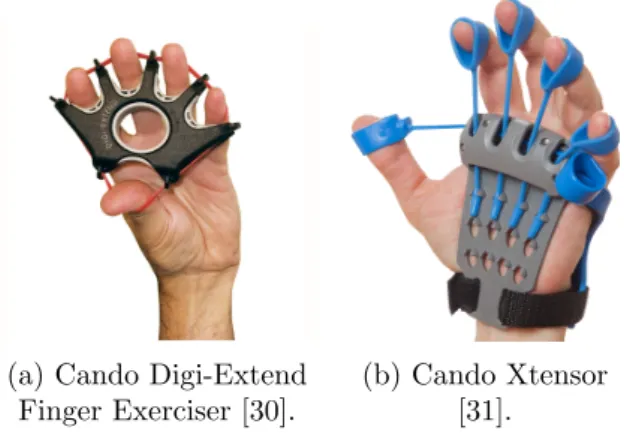
It is possible for patients to acquire some simple instruments that help them with this kind of exercises, like therapeutic balls, hand grips or finger ladders, or some more complex and a little more expensive devices like the Cando Digi-Extend Finger Exerciser (Figure 1.4a) or the Cando Xtensor (Figure 1.4b).
1.2.2 Robotic Systems
Studies based on repetitive training of simple flexion and extension fin-ger movements have reported improvements in hand function after stroke [32, 33]. Robotic devices, because of their programmable force-producing ability, can replicate some features of a therapist’s manual assistance,
al-(a) Cando Digi-Extend Finger Exerciser [30].
(b) Cando Xtensor [31].
Figure 1.4: Examples of hand therapeutic devices
lowing patients to semiautonomously practice their movement training [34]. The robot can allow completion of movements throughout the Range of Motion (ROM) and can also prevent inappropriate movements [8].
Robotic hand devices that can be used independently by patients in both acute and postacute settings can be a valuable adjunct to conventional ap-proaches that focus on compensation, whereas wearable devices can be in-tegrated directly into task-specific training [8].
Over the years, a number of robotic devices have been proposed for upper limb rehabilitation after stroke. It is possible to divide them into different groups according to diverse criteria, e.g. lateral specificity, part of the limb on which the therapy is focused, mechanical characteristics or control strat-egy (Table 1.1).
Due to the aim of this project, we will focus on robotic systems for hand and wrist rehabilitation.
As presented in the table above, we can divide robotic systems according to their mechanical characteristics in exoskeletons and operational machines or end-effectors. An end-effector is the device at the end of a robotic arm, designed to interact with the environment. It is the last link of the robot and it may consist of a gripper or a tool [35]. An exoskeleton is an outer frame-work worn by a person and powered by a system of motors that delivers at least part of the energy for limb movement [36]. Exoskeletons, in particular hand exoskeletons, can also be classified according to various criteria such as actuator type, power transmission method, DOF, intention sensing method, and control method (Figure 1.5) [37].
1.2. State of the Art: Rehabilitation
Table 1.1: Upper limb robotic systems classification [5].
Classification Characteristics
According to lateral specificity
- Unilateral Training - Bilateral Training According to the part of the upper
limb on which the therapy is focused
- Shoulder movement - Elbow movement - Wrist movement - Hand movement According to their mechanical
characteristics
- Exoskeleton
- Operational machines/end-effectors
According to the control strategy
- Passive movement in which the robotic device moves the patient’s arm.
- Active nonassist mode in which the subject executes the exercise and the robot provides no help.
- Active assist mode in which the subject attempts to move, and the robot provides assistance when there are some voluntary but inadequate movements.
- Resistive mode when the subject is required to perform an exercise against an antagonist force provided by the robot. - Bimanual exercise in which active movement of the unaffected arm is mirrored by simultaneous active/passive/assistive movement of the affected arm by means of the robotic device.
Figure 1.5: Classification of hand exoskeletons according to various criteria [37].
rehabili-tation will be presented.
Hand-Object-Hand (H-O-H)
Figure 1.6: Hand-Object-Hand (H-O-H) system. Top-view of the
force transducer between the distal parts of the handles [38]. In 1993, Lum et al. designed and
pre-sented the Hand-Object-Hand system (H-O-H), which was one of the first bi-lateral training device to be introduced in stroke rehabilitation (Figure 1.6). In this system, both hands are placed in two rigid handles that constrain the pa-tient’s movements to flexion and exten-sion of the wrist, that is, to a single DOF per hand. The two handles are connect at their distal parts by two ma-chine screws also attached to both sides of a force transducer. The robotic aid consists of a computer-controlled motor mounted underneath one of the handles. The system is capable of partially, or fully, substitute one hand, in two tasks: rhythmically moving the object from left to right and vice versa (i.e., parallel
movements of the hands) and squeezing. Although the H-O-H system has been advocated for post-stroke upper limb training it is not commercially available [38, 39].
MIT-Manus and InMotion ARM
At the Massachusetts Institute of Technology (MIT) and at the Burke Rehabilitation Hospital in New York, a device called MIT-Manus was devel-oped and presented by Krebs et al., in 1994. Until 2004 more than 250 stroke patients used this device daily in their rehabilitation process (Figure 1.7a). This device has two active DOF and is configured for safe, stable and com-pliant operation in close physical contact with humans, which is achieved using backdrivable hardware and impedance control. Each active module can move, guide or perturb movements of a patient’s limb and can record motions and mechanical quantities such as the position, velocity, and forces applied. The robot control system is an impedance controller that modu-lates the way the robot reacts to mechanical perturbation from a patient or clinician and ensures a gentle compliant behavior [40].
Over the years, the system has been improved and its clinical version, called InMotion ARM (Figure 1.7b), is provided by Interactive Motion Technologies®. Clinical studies showed a 31 point change in the Functional
1.2. State of the Art: Rehabilitation
Independence Measure (FIM) 2 scores of patients from admission to dis-charge. The studies also showed the maintenance of the improvements from robot therapy on a 3 year follow up evaluation [42]. This is the most thor-oughly researched device for upper extremity neurorehabilitation, having been used in more than 800 patients and been submitted to large multi-site randomized controlled clinical trials.
Besides this device, Interactive Motion Technologies® also offers two other robotic systems for upper limb rehabilitation: InMotion WRIST and InMotion HAND. InMotion HAND (Figure 1.7c) is an add module to be used with the InMotion ARM robot and it provides strength, sensorimotor, sensory and continuous passive motion training for grasp and release [43,44].
(a) (b)
(c)
Figure 1.7: (a) MIT-Manus [45], (b) InMotion ARM[46] and InMotion HAND[44] robotic systems.
Bi-Manu-Track
A research group in Berlin, Germany, lead by Stefan Hesse, developed in 2003 the Bi-Manu-Track (Figue 1.8). It is a computerized motor-driven arm trainer that allows bilateral training of two movement patterns: forearm pronation and supination and wrist flexion and extension.
2Uniform system of measurement for disability based on the International Classification
of Impairment, Disabilities and Handicaps; measures the level of a patient’s disability and indicates how much assistance is required for the individual to carry out activities of daily living [41].
Figure 1.8: Bi-Manu-Track system [47].
The Bi-Manu-Track supports three computer-controlled modes of practice. In the passive-passive mode the robot controls both arms. In the active-passive mode the less impaired upper limb actively moves the handle while the robot guides the most impaired upper limb. In the active-active mode both arms perform actively by overcoming an initial isometric resistance. Movements can be ei-ther mirror-symmetric (in-phase) or parallel (antiphase). Amplitude, speed, and resis-tances can be set individually. Some studies have been made with this device and an improvement in patient’s condition was observed. The Bi-Manu-Track is commercially available at Reha-Stim, Berlin, Germany [39, 48].
Braccio di Ferro
Figure 1.9: Braccio di Ferro system [39].
Casadio et al. from the University of Genova, presented the Braccio di Ferro in 2006 (Figure 1.9). Although it has been primarily designed for unilateral use it can be used as a bilateral training de-vice. It is a planar manipulandum with 2 DOF operated by two motors and the system generates four types of forces: an assistive field, a resistive elastic field, a rigid ”wall”, and a viscous field. In two studies, the Braccio di Ferro was used by stroke patients in training cy-cles consisted of five sessions over 2 to 3 weeks, each session lasting no more than 45 minutes. All patients improved their performance: movements became faster, smoother, more precise, and required de-creasing levels of assistive force.
How-ever, no clinical outcomes have been reported for the device and the system is not commercially available [39, 49].
HandSOME
The Hand Spring Operated Movement Enhancer (HandSOME) (Figure 1.10), developed by Brokaw et al. in 2010, is a 0.22 kg passively operated device for giving an extension moment to the finger joints so that it
com-1.2. State of the Art: Rehabilitation
pensates for the finger flexor hypertonia caused by a stroke. It is designed to follow the normal kinematic trajectory of the hand during pinch-pad grasp-ing. A four bar linkage mechanism was designed for the thumb and finger parts to coordinate the natural grasping motion. The attachment point of the spring can be changed to adjust the torque profile [37]. The design uses bungee cords as springs to assist with finger and thumb extension, and with assistance profiles that emulate the torque vs extension angle profiles for passive movement. Changes in the spring location and used stiffness, allows the therapist to vary the assistance profile and magnitude. According to the authors, the ROM testing and functional use testing showed large im-provement with HandSOME. All three subjects that tested the system had increased maximum ROM with the HandSOME device, although it is not commercially available. The device weight was the only complaint presented by the users since it was difficult to lift with shoulder weakness. [50].
Figure 1.10: Hand Spring Operated Movement Enhancer (HandSOME) system [50].
HANDEXOS
The hand exoskeleton developed by Chiri et al. in 2009, HANDEXOS, (Figure 1.11), is composed of an external backing element applicable on the dorsum of the wearer’s hand, and shell-like elements applicable on each phalanx and connected each other by translational and rotational joints. It has 5 independent modules for the fingers. Each module is composed of 3 links for the phalanges, where the center of rotation of each connection is matched with the corresponding joint of the human finger. The flexion and extension of the Metacarpophalangeal (MCP) joint is driven by a slider-crank-like mechanism, while the Proximal Interphalangeal (PIP) and Distal Interphalangeal (DIP) joints are driven by Bowden cable transmissions. The 3 joints of each finger are underactuated because they are driven using a single actuator unit. For the finger module, 3 force sensors are mounted
on the surface of the inner side of each of the three palmar shells to sense the interaction force. The linear slider for MCP rotation is equipped with strain gauges to measure the force transmitted by the driving cable [37]. The entire mechanical design of HANDEXOS is patented [51].
Figure 1.11: Overview of the HANDEXOS index finger module [52].
Tong et al. [53]
Figure 1.12: Tong et al. hand robotic system [53]. In 2010, Tong et al. presented a hand
exoskeleton which consists of 5 finger as-semblies where each finger has 1 active DOF actuated by a linear actuator, caus-ing coupled movement of the MCP and PIP joints (Figure 1.12).
The device has 4 modes of operation: 1) Continuous Passive Motion (CPM) - the device imposes the movement in the pa-tient’s hand, whom those not need to make any effort; 2)Electromyography (EMG)-triggered motion - the device starts flex-ion or extensflex-ion motflex-ion when the corre-sponding EMG signal exceeds a certain threshold; 3) continuous EMG-driven mo-tion - the movement continues as long as the user’s effort exists; 4) freerunning -the device selects flexion or extension of the device according to a comparison of the EMG signals from the two muscles that represent flexion and extension [37].
1.3. Hand Anatomy and Biomechanics
AMADEO®
Amadeo®(Figure 1.13) is an effective instrument, provided by Tyromo-tion GmbH, for supporting a patient in rehabilitaTyromo-tion. Depending on the degree of neurological damage, the patient can be treated either merely pas-sively or actively. His/her therapist can define a personalized program: 1) CPM Therapy – the passive hand is stimulated; 2) Assistive Therapy – ac-tive training at the individual limit of performance; 3) Interacac-tive Therapy – active training in a virtual environment based on the goal-oriented tasks. The patented mechanism of Amadeo® mimics the natural grasping move-ment and imprints it on the patient’s hand. In patients with a limited scope of movement of individual fingers or the entire hand, the targeted exercises performed with the therapy unit help to improve motor and sensory func-tions. As a result of the therapy program that is tailored to their individual needs, patients quickly regain more quality of living. This device also en-ables the measurement of the isometric force and of the scope of movement for the upper extremities having a integrated real-time biofeedback [54, 55].
Figure 1.13: Amadeo® system [54].
A number of other robotic systems have been presented over the years [56–64] and others are available in the market, but, not many of them are light-weighted, affordable and/or portable. Thus the rehabilitation process is always dependent on the existence of clinical facilities that can provide after stroke care near the patient’s home, or means of transport that enable the patient to travel to the nearest rehabilitation center, and funds or finan-cial support. The system developed combines some of the best features of the equipment presented above such as being light-weighted, portable and functioning depending on the patient’s effort to move.
1.3
Hand Anatomy and Biomechanics
The human hand is one of the most complex parts of the human body. It gives powerful grip but also allows manipulation of small objects with great precision. The complex arrangement of muscles, tendons, ligaments
and bones, gave humans a big evolutionary advantage to other creatures on the planet. Being one of the most used parts of the body in everyday tasks, even the smallest lack or difficulty in mobility brings enormous challenges to any person. This is the main reason why, rehabilitation after stroke is so important.
But, in order to chose and implement the best and safer approach for this problem, it is necessary to understand the anatomy and biomechanics of the human hand. Therefore the systematic knowledge helps achieving proper functions for rehabilitation and assistance. Thus, in this section, an overview of the biomechanics of the human hand will be presented including the bones, joints and the more relevant muscles for finger movement. 1.3.1 Bones and Joints
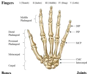
The bones of the human hand can be divided in two groups: carpus and digits. The eight carpal bones compose the carpus, or wrist, and are arranged in proximal and distal rows of four. Between the two is the inter-carpal articulation. These small bones give flexibility to the wrist.
The five digits are named as follows from the radial to the ulnar side: thumb (I), index finger (II), middle finger (III), ring finger (IV) and little finger (V). Each digit has one metacarpal and three phalanges (proximal, middle, and distal) except for the first (thumb), which has only two (proxi-mal and distal) [65], as we can see in Figure 1.14. Each metacarpal consists of a base, shaft, and head. The proximal bases of the metacarpals articulate with the carpal bones, and the distal heads of the metacarpals articulate with the proximal phalanges forming the knuckles. The first metacarpal (of the thumb) is the thickest and shortest of these bones.
Figure 1.14: Bones and joints of a human hand.
There are 19 bones and 14 joints distal to the carpals. Each finger articu-lates proximally with a par-ticular carpal bone at the Carpometacarpal (CMC) joint. The CMC joint of the thumb is a sellar joint, exhibit-ing two degrees of freedom: flexion and extension, and abduction and adduction. The CMC joints of the sec-ond, third and forth fingers are classified as plane joints with one degree of freedom, while the fifth CMC joint is often classified as a
semi-1.3. Hand Anatomy and Biomechanics
saddle joint with conjunc-tional rotation. The next joint of each finger links the metacarpal bone to the proximal phalanx at the MCP joint. MCP joints are classified as el-lipsoidal or condylar joints with two degrees of freedom, which again permit flexion, extension, abduction, and adduction movements. In MCP joints, the metacarpal heads fit into shallow cavities at the base of the proximal phalanges. The PIP and DIP joints are found between the phalanges of the fingers; the thumb has only one Interphalangeal (IP) joint. They are both bicondylar joints with subsequently greater congruency between the bony surfaces, and have one degree of freedom. The transverse diameters of the IP joints are greater than their antero-posterior diameters and the thick collateral ligaments are tight in all positions during flexion, contrary to those in the MCP joint. Although the IP joints are frequently modeled and assumed as having single axis of rotation for simplicity, in fact they do not remain constant during flexion and extension [37].
The different shapes of the finger joints result in varying DOF at each joint. Also, the orientation of the thumb and the unique configuration of its CMC joint provide this digit with a large range of motion and greater flexibility. The MCP joints are flexed approximately 45°, the PIP joints are flexed between 30° and 45°, and the DIP joints are flexed between 10° and 20° at the resting posture. Flexion of the MCP joints is approximately 90°, and the little finger is the most flexible (at about 95°), while the index finger is the least flexible (at about 70°). The extension varies widely among individuals. For PIP and DIP joints, flexion of about 110° and 90° occurs. Extension beyond the zero position is regularly observed and depends largely on the ligamentous laxity [37].
1.3.2 Muscles
As mentioned before, the human hand is a very complex system, and its movements are accomplished by the coordinated action of groups of muscles. The majority of muscles responsible for hand movements are the extrinsic muscles that originate from the arm and forearm. They are responsible for flexion and extension of the digits. On the other side, the intrinsic muscles are located entirely within the hand, and they permit the independent action of each digit.
There are nine extrinsic muscles and three muscles among them - the flexor digitorum superficialis, the flexor digitorum profundus, and the flexor pollicis longus - contribute to finger flexion. Five extrinsic muscles con-tribute to the extension of the fingers, while one extrinsic muscle (abductor pollicis longus) contributes to the abduction of the thumb. The Dorsal Interossei (DI) and Palmar Interossei (PI) are groups of muscles arising be-tween the metacarpals and attached to the base of the proximal phalanges or to the extensor assembly. The interossei flex the MCP joint and extend
the PIP and DIP joints. They are also effective abductors and adductors, and produce some rotations of the MCP joint. Because of this interaction between the extrinsic and intrinsic musculature, the actions of the PIP and DIP joints are functionally coupled [37].
1.4
Electromyography
The human body, like any other complex system, has its own ways to communicate in order to function in the most balanced way possible. These pathways of communication can be chemical, electrical, mechanical, acousti-cal or optiacousti-cal. All these kinds of signals are known as biosignals and can be continuously measured and monitored. Bioelectrical signals are frequently used in diagnosis, monitoring of patients, rehabilitation and biomedical re-search. There are different examples of bioelectrical signals in the human body like those recorded using Electroencephalography (EEG), Electrocar-diography (ECG), Electromyography (EMG), Mechanomyography (MMG), Electrooculography (EOG), Galvanic skin response (GSR) or Magnetoen-cephalography (MEG) [66].
EMG signals correspond to the electrical potential generated by nerve cells that control muscle cells (from skeletal muscles) when they are electri-cally or neurologielectri-cally activated [67, 68].
EMG can be performed in two different ways: Surface Electromyography (sEMG) and Intramuscular Electromyography (iEMG). iEMG is an inva-sive technique that includes the placement of a needle electrode, or a needle containing two fine-wire electrodes, through the skin into the muscle tissue. Due to the invasive nature of this technique, sEMG is most commonly used. In this case, instead of analyzing only a few fibers, the signal acquired re-lates to the activation of the muscle in a much higher level of complexity. Which means that the acquisition is not as precise as the iEMG in terms of individual identification of the muscles evolved in each movement [69, 70].
One of the most important characteristics of sEMG that has to be taken into account while analyzing the acquired signals, is that the signal is made up of superimposed Motor Unit Action Potentials (MUAPs) from several motor units3 (Figure 1.15) [67]. So, the sEMG signal is not a recording of a single nerve but from several neurons that coordinate the contraction and relaxation of the muscle region where the electrode is placed.
3A motor unit is made up of a motor neuron and the skeletal muscle fibers innervated
by that motor neuron’s axonal terminals. Groups of motor units often work together to coordinate the contractions of a single muscle [71].
1.4. Electromyography
Figure 1.15: Motor Unit representation [72].
EMG may be used in different areas and for various purposes. Since it provides information about muscle health it is used for diagnosis, but it can also be used for monitorization since it is possible to relate EMG signals to movement. Furthermore, EMG signals are also used as a control signal for prosthetic devices (prosthetic hands, arms, lower limbs) or exoskeletons. 1.4.1 Signal Characteristics
The sEMG signal’s amplitude lies in between 1-10 mV and, therefore, as a weak signal, amplification must be considered. The signal lies in the frequency range from 0-500 Hz and is most dominant in between 50-150 Hz. sEMG signal is highly influenced by noise so, noise filtration is an important part of sEMG signal processing (Figure 1.16).
Ambient noise can be caused by electromagnetic radiation sources, like radio transmission devices, fluorescent lights and power line interference from electrical wires. These interferences are almost impossible to avoid from external means. This particular noise exists in the frequency range of 50-60 Hz. Noise can also be generated from motion. The two main sources of this noise are instability of electrode-skin interface and movement of the electrode cable, and it lies mostly in the range of 0-20 Hz. It can be eliminated by proper set of EMG equipment and circuitry [73].
Crosstalk is another source of noise in EMG recordings. This phe-nomenon occurs when the EMG signal from one muscle interferes with the signal of another muscle, leading to incorrect interpretation of the signal in-formation. This source of noise can be minimized by choosing carefully elec-trode’s size and inter-electrode distances. Minimal Crosstalk Area (MCA) is defined as a surface where crosstalk versus co-contraction of muscles is minimal [69].
Many factors contribute to the difficulty of extracting sufficient informa-tion from the sEMG for dexterous, multifuncinforma-tional control. The most ob-vious and important is the lack of physiologically appropriate musculature from which to estimate the intended motion. This is especially problematic in individuals with high-level limb deficiency, because little or no muscle remains that would produce force in the absent joints [74].
1.4.2 Signal Processing: Pattern-Recognition
Most of the approaches to EMG pattern recognition have the fundamen-tal processing stages shown in Figure 1.17 [74].
Figure 1.17: Stages of Electromyography (EMG) signal processing for pattern recognition.
The first stage of the process (signal preprocessing) consists on filtering, amplification, noise cancellation and Analog-to-Digital conversion. After that, it is necessary to divide the signal in smaller windows in order to an-alyze the signal along time since an instantaneous value of a EMG signal is
1.4. Electromyography
not an useful input for pattern-recognition techniques, due to its random na-ture. Usually, for pattern-recognition purposes, windows have a percentage of overlap, which means that the same data will be used more than once but this is proven to increase the accuracy of the classification process. Then, for each window of data, the features are extracted. Features are measures of the signal that can be of two types: time domain (measured as a function of time) or frequency domain (measured using the spectrum of the signal). Below is a list of eight features (6 time domain and 2 frequency domain) which will be evaluated further in this report.
Time Domain
Integrated EMG (IEMG)
IEM G =
N
X
n=1
|xn| (1.1)
Mean Absolute Value (MAV)
M AV = 1 N N X n=1 |xn| (1.2)
Simple Square Integral (SSI)
SSI =
N
X
n=1
|xn|2 (1.3)
Variance of EMG (VAR)
V AR = 1 N − 1 N X n=1 x2n (1.4)
Root Mean Square (RMS)
RM S = v u u t 1 N N X n=1 x2n (1.5) Waveform Lenght (WL) W L = N −1 X n=1 |xn+1− xn| (1.6)
Frequency Domain Mean Frequency (MNF) M N F = M X fjPj ,M X Pj (1.7) Median Frequency (MDF) M DF = 1 2 M X Pj (1.8)
xndenotes the nth sEMG signal sample, N is the length of the signal, fj
the signal frequency spectrum and Pj the sEMG power spectrum.
Choosing the right set of features may not be trivial, but it is a crucial step towards an effective signal classification. There has to be a balance between complexity and overall performance in order to use sEMG has a control signal for prostheses or any other type of system. A limited number of features should be chosen and the arrangement of features should be taken into account. The ideal criterion for classification should be the minimization of the probability of misclassification, but generally simpler criteria based on class separability are chosen. Furthermore, the exhaustive search among all possible subsets of features is often impractical, and some nonexhaustive and sequential methods are used [75].
With the right set of features extracted from the signal, a data set is created and the classifier is built. There are also different types of classi-fiers that can be used such as: K-Nearest Neighbors; Naive Bayes; Partial Least Squares Discriminant; Fisher Linear Discriminant; Kernel Matched Subspace Detector; Generalized Likelihood Ratio Test; Distance Likelihood Ratio Test; Artificial Neural Networks; or Support Vector Machines.
sEMG signal patterns differ among individuals. Moreover, electrical impedance of the skin; electrode locations; time variations caused by fa-tigue, sweat, and so on, differ from user to user and from time to time [75]. So, for a good and effective use of sEMG as a control mechanism, it is impor-tant to create a specific data set for each individual and, if possible, before each new use.
Project Overview
In this chapter the aims and objectives of this project will be presented and an overview of the system and its functioning will be introduced. In order to clarify its operation, it will be divided in 2 parts: hand exoskeleton and acquisition system, and both will be explored in detail.
2.1
Aims and Objectives
This project aims to develop a system that could be used by patients after a stroke in order to improve their recovery process, training at home without needing the supervision of a caregiver but still experiencing a personalized rehabilitation.
In order to fulfill all the requirements above mentioned, the system needs to be lightweight, portable and adaptable to different people, but also not too expensive, easy to use and, of course, reliable, safe and effective.
One of the key features of this system is the personalization of the re-habilitation process. This will be achieved by using sEMG signal from the patient’s forearm and promoting the movement in the limb wearing the ex-oskeleton according to the movement that is being performed on the first one. This will provide the user control over the system and the rehabilita-tion process, not needing to rely on another person to perform simple but repetitive rehabilitation exercises. The use of sEMG will not only allow sim-ple opening and closing of the two fingers at once but also individual finger movements.
In the first stages of rehabilitation, due to the common muscle atrophy and synergies (co-activation of both extensors and flexors muscles), and since only one side of the body is usually more affected than the other, the sEMG signals can be recorded in the non-impaired limb, and the movement can be imposed in the opposite one. When the patient is able to individually move the fingers but the ROM is not the best, the sEMG can be acquired on the
2.2. Hand Exoskeleton
same arm that is wearing the exoskeleton.
2.2
Hand Exoskeleton
In this section, the design and mechanism of the exoskeleton used in this project will be detailed. This system is a redesigned version of the ex-oskeleton built by Tzemanaki et al [76]. The first design of the exex-oskeleton, built by Burton et al. [77], was created for rehabilitation purposes but, since then, three new versions were produced to be used as a intuitive interface be-tween surgeons’ hands and minimally invasive surgery instruments, as part of a Robot-Assisted Minimally Invasive Surgical (RA-MIS) system. Never-theless, the characteristics of the last created system, such as being suited for a large number of hand sizes (fits comfortably around the 5th to 95th percentile of hand lengths and breadths [76]) or being lightweight (154 gr), make it as well suitable for use as a rehabilitation device. That would allow patients to diminish the costs associated with their therapy and, possibly, enhance their recovery after suffering a stroke. Thus, a motorized version of the exoskeleton was designed and tendons were implemented through the fingers by creating holes for the tendons to go through.
The index and middle fingers of the exoskeleton have 4 DOFs: flex-ion/extension for DIP, PIP and MCP joints and also abduction/adduction for MCP joint. Because MCP joint is more complex, a ball and socket mech-anism was first used (Figure 2.1 - right) and then changed for two individual joints instead (Figure 2.2). Whereas, for the other joints, each segment op-erate around miniature bearings placed on either side of the joint (Figure 2.1 - left). The use of a lead screw adjustment mechanism allows for the bearings to be positioned with their axes aligned with the joints’ natural axis of rotation [76].
Figure 2.1: Exoskeleton’s DIP and PIP joint mechanisms (left) and the MCP joint mechanism with a ball joint (right) [76].
Figure 2.2: Exoskeleton’s MCP joint mechanism with two individual joints. In order to actuate each finger, a 0.38 mm diameter cable made of 49 intertwined strands of stainless steel was used, and passed through the holes created on the exoskeleton’s finger, to simulate a tendon. This cable was then connected, via a 41.94 mm diameter pulley, to a servo motor which was controlled by an Arduino Duemilanove Board. The dimension of the pulley was calculated by Hamid [78] for the purposes of their project. Figure 2.3 shows a 3D model of the all system with the two fingers of the exoskeleton, their cables/tendons attached to the corresponding motors and the Arduino board. The position of the motors was chosen so that the pulley would be aligned with the corresponding cable allowing its proper curling.
Figure 2.3: 3D model of the two digit exoskeleton attached to the correspondent motors.
Two different servo motors were used: a Hitec HS-322HD Standard Deluxe servo (Appendix C) and a MG996R High Torque Metal Gear Dual Ball Bearing servo (Appendix D). The use of different motors was due to the availability of this material in the lab when the system was built, but since their relevant characteristics were similar, as is going to be presented next, no other material was purchased.
![Figure 1.1: Population aged 60 or over: world and development regions, 1950-2050 [1].](https://thumb-eu.123doks.com/thumbv2/123dok_br/19186895.947985/15.892.193.465.608.851/figure-population-aged-world-development-regions.webp)
![Figure 1.2: Representation of the two main types of stroke: ischemic (left) and hemorragic (right) [12].](https://thumb-eu.123doks.com/thumbv2/123dok_br/19186895.947985/17.892.212.601.436.760/figure-representation-main-types-stroke-ischemic-hemorragic-right.webp)
![Figure 1.3: Hypothetical pattern of recovery after stroke with timing of intervention strategies [17].](https://thumb-eu.123doks.com/thumbv2/123dok_br/19186895.947985/21.892.216.612.249.582/figure-hypothetical-pattern-recovery-stroke-timing-intervention-strategies.webp)
![Figure 1.7: (a) MIT-Manus [45], (b) InMotion ARM[46] and InMotion HAND [44] robotic systems.](https://thumb-eu.123doks.com/thumbv2/123dok_br/19186895.947985/26.892.230.733.435.812/figure-mit-manus-inmotion-inmotion-hand-robotic-systems.webp)
![Figure 1.10: Hand Spring Operated Movement Enhancer (HandSOME) system [50].](https://thumb-eu.123doks.com/thumbv2/123dok_br/19186895.947985/28.892.364.599.494.713/figure-hand-spring-operated-movement-enhancer-handsome.webp)
![Figure 1.11: Overview of the HANDEXOS index finger module [52].](https://thumb-eu.123doks.com/thumbv2/123dok_br/19186895.947985/29.892.261.561.296.529/figure-overview-handexos-index-finger-module.webp)
![Figure 1.13: Amadeo ® system [54].](https://thumb-eu.123doks.com/thumbv2/123dok_br/19186895.947985/30.892.221.744.552.707/figure-amadeo-system.webp)
![Figure 1.16: EMG Spectrum and noise influence on this spectrum [73].](https://thumb-eu.123doks.com/thumbv2/123dok_br/19186895.947985/34.892.298.638.782.1040/figure-emg-spectrum-noise-influence-spectrum.webp)