Felipe Nunes BritoI, Fabiel Spani VendraminII, Cinthia Távora de Albuquerque LopesIII, Mayra Pauline
Ribeiro CostaIV, Otávio Mitio OhashiV, José Guilherme Soares MaiaVI, Lydia Masako FerreiraVII, Joyce
Kelly do Rosário da SilvaVIII, Moysés dos Santos MirandaIX
Proliferation of human adipose tissue-derived stem cells
stimulated by oil rich in thymol of
Lippia origanoides
1Abstract
Purpose: To evaluate the effects of this thymol-rich oil in the proliferation of human adipose
tissue-derived stem cells.
Methods: Stem cells were isolated from human adipose tissue by liposuction. After the first
passage, cells were cultivated in triplicate for three days in control medium and medium supplemented with three oil samples (1.0 μg/mL, 5.0 μg/mL, and 25.0 μg/mL). Cells were analyzed by the MTT assay at passage 1 (P1), and cell proliferation of control and 1 μg/mL groups was determined with a hemocytometer at P2 and P3.
Results: Viability of the essential oil-treated cells was significantly higher than the control
group at P1 (p = 0.0008). The treatment with the oil, at a concentration of 1 µg/mL, led to increases of 24.8% at P1 and 43.0% at P3 in the rate of cell proliferation compared with control cells.
Conclusion: Supplementing culture medium with essential oil of Lippia origanoides increased
cell proliferation, especially at later passages.
Key words: Stem Cells. Adipose Tissue. Cell Proliferation. Thymol.
IMD, School of Medicine, Universidade Federal University do Pará (UFPA), Belem-PA, Brazil. Conception of the study,
interpretation of data, technical procedures, manuscript preparation.
IIAssistant Professor, Surgery Division, UFPA, Belem-PA, Brazil. Interpretation of data, critical revision. IIIMSc, UFPA, Belem-PA, Brazil. Technical procedures, interpretation of data.
IVGraduate student, UFPA, Belem-PA, Brazil. Technical procedures, interpretation of data.
VPhD, Full Professor, Biomedicine Division, UFPA, Belem-PA, Brazil. Analysis and interpretation of data, critical revision. VIPhD, Full Professor, Chemistry Division, Universidade Federal do Oeste do Pará (UFOPA), Santarem-PA, Brazil. Analysis
and interpretation of data, critical revision.
VIIPhD, Head and Full Professor, Plastic Surgery Division, Universidade Federal de São Paulo (UNIFESP), Brazil. Analysis
and interpretation of data, critical revision.
VIIIPhD, Assistant Professor, Biotechnology Division, UFPA, Belem-PA, Brazil. Conception of the study, statistical analysis,
interpretation of data, critical revision.
IXPhD, Assistant Professor, Biotechnology Division, UFPA, Belem-PA, Brazil. Conception of the study, analysis and inter
activity of this essential oil is attributed to the presence of phenol compounds such as thymol, carvacrol, and derivatives13.
Several biological activities have been reported for thymol. This phenolic compound plays an important role in the scavenging of free radicals such as DPPH (1,1-diphenyl-2-picrylhydrazyl) and ABTS (2,2′-azino-bis-3-ethylbenzthiazoline-6-sulphonic acid). In addition, at lower concentrations, it showed protective effects to cells against damage induced by the H2O2 radicals
14,15. Thus, the aim
of this study was to assess the proliferation of human ADSCs in culture medium treated with natural antioxidants present in the L.
origanoides Kunthessential oil.
■
Methods
The project was approved by the Ethics in Research Committee for Human Beings, Hospital Universitário João de Barros Barreto, Belem-PA, Brazil (Number of process: 487.533).
Adipose tissue was collected from the infra-umbilical region of three patients (27, 29, and 33 years of age; average, 29 years), without comorbidities, treated with liposuction in the private hospital. All patients signed a Free and Informed Consent Form.
Liposuction
Patients were operated on under peridural anesthesia. To collect the lipoaspirate, asepsis and antisepsis were performed in the infra-umbilical region, followed by infiltration with 0.9% saline solution with 1: 500,000 adrenalines at the demarcated site. The sample was aspirated through a syringe with a 3.5-mm diameter cannula and decanted by gravity for 30 minutes. A total of 10 mL of adipose tissue was collected with partial removal of the bloody contents.
■
Introduction
The isolation of mesenchymal stem cell (MSC) from human adipose tissue obtained from liposuction was described in 20011.
Liposuction is one of most common esthetic surgical procedures worldwide, and it generates an average of 100–3000 mL of adipose tissue that is routinely discarded, along with its incorporated stem cells2. In addition to being
easily obtained, adipose-derived stem cells (ADSCs) also have significant therapeutic use3,
as an acute myocardial infarction4 and bone
regeneration5. However, according to cellular
therapy protocols used in clinical trials, more than one million stem cells are necessary per treatment, necessitating in vitro expansion6.
Repeated passaging of cells in vitro incurs the risk of replicative senescence, which primarily occurs due to oxidation caused by free radicals and can be identified by telomere shortening and fibroblast flattening with a concurrent increase in nuclear/nucleolar size that results in stalled cell proliferation7. These
phenomena limit in vitro cell expansion and might hinder their therapeutic applications8.
Antioxidants are compounds that directly or indirectly inhibit production of reactive oxygen species9 (ROS). In recent
years, there is an increasing interest in finding antioxidant phytochemicals, because they can inhibit the propagation of free radical reactions10. Many essential oils have been
reported as natural alternative for controlling of oxidative stress such as the essentials oils from Lippia species11.
Lippia origanoides Kunth (Verbenaceae)
Isolation and cultivation of MSCs from human adipose tissue
The lipoaspirate tissue (2mL) was washed with 2 mL of phosphate-buffered saline (PBS; Sigma Aldrich, St. Louis, Missouri, USA) and incubated with 200 U/mL collagenase II for 60 min at 37ºC. After incubation, the collagenase was neutralized by adding complete cultivation medium (Dulbecco’s Modified Eagle’s Medium (DMEM; Sigma Aldrich) supplemented with 10% fetal bovine serum (FBS; Sigma Aldrich), 3.7 g/L NaHCO3 (Sigma Aldrich), and 5 µg/
mL of gentamicin (Sigma Aldrich). Following centrifugation, the pellet was resuspended twice with DMEM and plated. After 24 hours, any cells still in suspension were removed and the cultivation medium was renewed. The remaining adherent cells, representing MSCs, were kept at 37°C in 5% CO2 with high humidity,
and the cultivation medium was changed every three days until the third passage.
Cellular and histological characterization of cell differentiation
Histology with tissue-specific dyes was used to characterize the ability of MSCs to differentiate into bone tissue, adipose tissue, and cartilage tissue.
To induce osteoblast differentiation, cells at 70% confluence were treated with osteogenic induction medium (DMEM, 0.1 µM dexamethasone (Sigma Aldrich), 50µM ascorbic acid-2-phosphate (Sigma Aldrich), and 10-2M β-glycerophosphate (Mallinckrodt Baker,
Phillipsburg, New Jersey, USA)) and cultivated for 21 days.
To induce adipocyte differentiation, nearly confluent cells were treated with adipogenic induction medium (DMEM, 1 µM dexamethasone, and 10µM insulin (Sigma Aldrich)) and cultivated for 14 days.
To induce chondrocyte differentiation,
viable cells/mL, and after 2 hours, cells were treated with chondrocytic induction medium (DMEM, 6.25 µg/mL insulin, 50μM ascorbic acid-2-phosphate, and 10 µg/mL TGF-β1 (Cell Signaling Technology, Beverly, Massachusetts, USA) and cultivated for 21 days.
After cultivation, the differentiation medium was removed, and the cells were washed once with PBS. Next, cell monolayers were fixed with paraformaldehyde (Electron Microscopy Sciences) for 30 minutes and then washed twice with PBS. To detect osteoblasts, cells were stained with Alizarin Red S (Life Technologies, Carlsbad, California, USA) for 3 minutes. To detect adipocytes, cells were stained with Oil Red O (Life Technologies) for 30 minutes. To detect chondroblasts, cells were stained with Alcian Blue (Life Technologies) for 30 minutes. After staining, cells were washed three times with distilled water and analyzed by an optical microscope (911873 AXIOLAB, Ziss, Germany) – osteoblasts and chondroblasts - and by an inverted microscope (TE–DH100W, Nikon, Japan) – adipocytes.
Plant collection and extraction and analysis of essential oil
following conditions: DB-5ms (30 m x 0.25 mm; 0.25 mm film thickness) fused-silica capillary column; programmed temperature, 60–240°C (3ºC/min); injector temperature, 250 ºC; carrier gas, helium, adjusted to a linear velocity of 32 cm/s (measured at 100°C); injection type, split (1 μL), from 1:1000 hexane solution; split flow was adjusted to yield a 20:1 ratio; septum sweep was a constant 10 ml/min; EIMS, electron energy, 70 eV; temperature of the ion source and connection parts, 200°C. The quantitative data regarding the volatile constituents were obtained by peak area normalization using a FOCUS GC/FID operated under similar conditions of the GC–MS, except the carrier gas, which was nitrogen. The retention index was calculated for all the volatiles constituents using a homologous series of n-alkanes (C8-C32, Sigma-Aldrich)17.
For cell treatments, the essential oil was initially solubilized in dimethyl sulfoxide (DMSO; Sigma-Aldrich) at concentration 10.0 μg/mL. This stock solution was diluted to concentrations of 25.0 μg/mL, 5.0 μg/mL and 1.0 μg/mL in DMEM.
MTT assay of cellular growth rate
The 3-(4,5-dimethylthiazol-2-yl)-2,5-diphenyltetrazolium bromide (MTT) assay was performed during the first cellular passage (P1) to determine cell viability in the treatment and control groups. Cells were plated at a concentration of 3x10³ cells/well in a
96-well plate (TPP, Trasadingen, Schaffhausen, Switzerland) and incubated with different essential oil concentrations (1.0 μg/mL, 5.0 μg/ mL, and 25.0 μg/mL)in triplicate. After the third day of cell cultivation, the culture medium was replaced with 100 μL of 5 mg/mL MTT solution in 900 μL in DMEM (without FBS). After 3 hours incubation at 37ºC, the MTT solution was removed and 100 μL of DMSO was added to each well and the plate was shaken for 10
minutes. The percentage of cellular growth rate (GR %) was based on absorbance at 490
nm (A490) measured in spectrophotometer UV/
VIS and calculated according to the following equation.
Cell counting
At P2 and P3, 4x10⁴ cells/mL were
plated in two 35-mm plates, one with only DMEM and the other with 1.0 μg/mL essential oil (the concentration with the highest rate of cell proliferation). After 72 hours, cells were re-suspended with trypsin and 1 µl of cell were placed and counted in a Neubauer Chamber. All the cells within each medium square were counted using x4 objective and multiplied for a correction factor of 104 (cell/ml).
Statistical analysis
Statistical analyses were performed using the GraphPad Prism version 5.0 (GraphPad Software, California, USA). A one-way ANOVA test was performed to assess the rate of cell proliferation using values obtained from the MTT assay followed by Tukey’s post hoc test. Values of p ≤ 0.05 were considered significant.
■
Results
Characterization of tissues
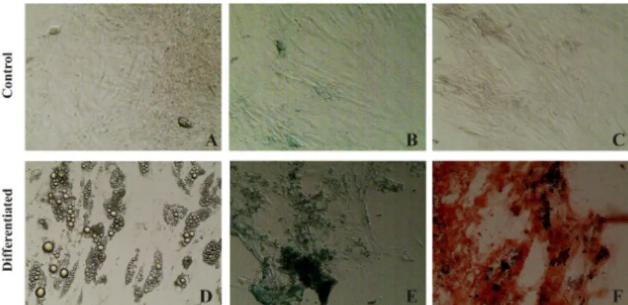
differentiated from ASCs are shown in Figure 1. Figure 1A-C exhibit the control of each
histological characterization. Numerous
Figure 1 - (A-C) ASCs cultured control. (D) Adipogenic differentiation of the ASCs. Fat vesicles detected by
staining with Oil Red O after 14 days of induction. (E) Chondrogenic differentiation of ASCs stained with Alcian
Blue after 21 days of induction. (F) Osteogenic differentiation of ASCs stained with Alizarin Red S after 21 days
of induction.
The proliferation results obtained by the MTT assay are shown in Figure 2. The absorbance values passed the Normality Test (p = 0.722) and the Equality of Variance Test (p = 0.24) in all treatments. Compared with the control, cell proliferation was higher in the three experimental groups treated with essential oil from L. origanoides (p = 0.0008). Cell proliferation did not significantly differ between the three experimental groups.
Figure 2 – Averages and standard deviation for
absorbance related to cell proliferation (MTT assay) of stem cells derived from human adipose tissue cultivated with essential oil of Lippia origanoides in the 1st passage. *indicates a significant difference (p < 0.05) in relation to the control group.
The proliferation rates cells treated
mL of essential oil were 24.8%, 14.7%, and 16.3% greater than control cells, respectively (Figure 3). At P2, control cells and cells treated with 1 μg/mL of essential oil were plated at a concentration of 4x10⁴ cells/mL. After 72 h of
cultivation and subsequent trypsin treatment, both groups had a concentration of 18x104
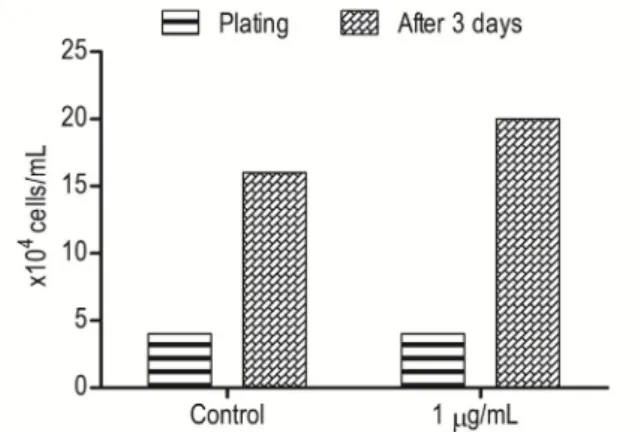
cells/mL (Figure 4). At P3, the cells were again plated at 4x10⁴ cells/mL However, in this case
the concentration of the essential oil-treated cells was approximately 43% greater than that of the control cells after cultivation and enzymatic digestion (20x104 cells/mL vs. 14x104
cells/mL) (Figure 5).
Figure 3 – Growth rate of stem cells derived from
Figure 4 – Effect of the essential oil of Lippia origanoides (1 μg/mL) on multiplication of stem cells derived from human adipose tissue in the 2nd passage after 3 days of cultivation.
Figure 5 - Effect of the essential oil of Lippia origanoides (1 μg/mL) on multiplication of stem cells derived from human adipose tissue in the 3rd passage after 3 days of cultivation.
The dried aerial parts of L. origanoides provide oil yield of 3.3%. Individualcomponents were identified by comparison of both mass spectrum and GC retention data with authentic compounds, which were previously analyzed and stored in the data system. Seventeen volatile components were identified in the oil, comprising nearly 100% of its composition (Table 1). Thymol (88.2%) was the main constituent, followed by p-cymene (6.4%), its biogenetic precursor.
Table 1 - Chemical composition of the essential
oil of Lippia origanoides.
Compound RIa RIb Oilc (%)
α-Thujene 916 924 0.2
α-Pinene 922 932 0.3
β-Pinene 966 974 0.1
α-Phellandrene 999 1002 0.1
p-Cymene 1014 1020 6.4
E-β-Ocimene 1034 1044 1.1
Terpinolene 1074 1086 0.1
Linalool 1088 1095 0.5
Umbellulone 1158 1167 0.1
Terpinen-4-ol 1168 1174 0.3
Thymol, methyl ether 1217 1232 0.1
Thymol 1279 1289 88.2
Carvacrol 1285 1298 1.0
α-Copaene 1360 1374 0.1
β-Caryophyllene 1402 1417 0.3
α-Humulene 1438 1452 0.7
δ-Amorphene 1500 1511 0.1
Monoterpene hydrocarbons 8.3
Oxygenated monoterpene 90.2
Sesquiterpene hydrocarbons 1.2
Total 99.7
aRetention index calculated using the homologous series of n-alcanes; bRetention index in the literature (ADAMS, 2007); cPercentage of compounds obtained by integrating the area in
the flame ionization detector.
■
Discussion
Essential oils are one among the most valuable plant products used in medicine and complementary treatment strategies. However, this is the first study, which reports the use of essential oil from L. origanoides as a supplement in human stem cell culture.
exhibit different mechanisms of action because the presence of a phenolic ring in its structure enhance the antioxidant activity due to its ability for scavenging free radicals, donation of hydrogen atoms or electron, or chelation of metal cations16. We have hypothesized
that the cell proliferation was due to Thymol’s cytoprotective effect, as another monoterpene (1,8-Cineole, Camphor, α-Pinene and β-pinene) presents in oil of Salvia lavandulifolia Vahl showed a protective effect in cultured human astrocytoma too18.
Tavakolinejad et al.19 cultivated human
adipose tissue-derived stem cells in medium containing platelet-rich plasma (PRP) instead of FBS or L. origanoides. Cellular growth rates in the work of them (40%) were similar to this study at P3 (40 vs. 43%). However, even automated methods for obtaining PRP can result in variable product quality. On the other hand, essential oil from L. origanoides function as antioxidants, allowing extended cell cultivation
in vitro without probable cell damage.
Cells become senescent with increasing passage number, thus reducing their growth8.
Previous studies have found stem cells derived from senescent human adipose tissue at P2 based on a β-galactosidase assay. By P5, almost 20% of cells were derived from senescent tissue20. In our study, the antioxidant function
of essential oil from Lippia origanoides may have inhibited the oxidation of free radicals produced in vitro, enabling cell growth even at P3. Future studies should be done to verify the maximum passage number that benefits from essential oil-treatment, as well as examine other basic cell characteristics such as cell senescence and telomere shortening.
■
Conclusions
Supplementing culture medium with essential oil of Lippia origanoides increased the proliferation of stem cells derived from human adipose tissue in vitro, especially at later cell
to become a cheap and efficient mean for optimally expanding adult stem cells in vitro for cell therapy.
■
References
1. Zuk PA, Zhu M, Mizuno H, Huang J, Futrell JW, Katz AJ, Benhaim P, Lorenz HP, Hedrick MH. Multilineage cells from human adipose tissue: implications for cell-based therapies. Tissue Eng. 2001;7:211-28. PMID: 11304456. 2. Zhu T, Liu T, Song K, Fan X, Ma X, Cui Z.
Adipose-derived stem cell: a better stem cell than BMSC. Cell Biochem Funct. 2008;26:664-75. doi: 10.1002/cbf.1488. 3. Nakamuta JS, Danoviz ME, Marques FL, dos
Santos L, Becker C, Gonçalves GA, Vassallo PF, Schettert IT, Tucci PJ, Krieger JE. Cell therapy attenuates cardiac dysfunction post myocardial infarction: effect of timing, routes of injection and a fibrin scaffold. PLoS One. 2009;4:1-10. doi: 10.1371/journal. pone.0006005.
4. Houtgraaf JH, den Dekker WK, van Dalen BM, Springeling T, de Jong R, van Geuns RJ, Geleijnse ML, Fernandez-Aviles F, Zijlsta F, Serruys PW, Duckers HJ. First experience in humans using adipose tissue-derived regenerative cells in the treatment of patients with ST-segment elevation myocardial infarction. J Am Coll Cardiol. 2012; 59:539-40. doi: 10.1016/j.jacc.2011.09.065.
5. Li W, Liu Y, Zhang P, Tang Y, Zhou M, Jiang W, Zhang X, Wu G, Zhou Y. Tissue-engineered bone immobilized with human adipose stem cells-derived exosomes promotes bone regeneration. ACS Appl Mater Interfaces. 2018;10:5240-54. doi: 10.1021/ acsami.7b17620.
6. Cheng AKL, Reuveny S, Oh SKW. Application of human mesenchymal and pluripotent stem cell microcarrier cultures in cellular therapy: achievements and future direction. Biotechnol Adv. 2013;31:1032-46. doi: 10.1016/j.biotechadv.2013.03.006.
7. Parrinello S, Samper E, Krtolia A, Goldstein J, Melov S, Campisi J. Oxygen sensitivity severely limits the replicative lifespan of murine fibroblasts. Nat Cell Biol. 2003;5:741-7. doi: 10.1038/ncb1024.
12. doi: 10.1371/journal.pone.0002213. 9. Khlebnikov AI, Schepetkin IA, Domina
NG, Kirpotina LN, Quinn MT. Improved quantitative structure–activity relationship models to predict antioxidant activity of flavonoids in chemical, enzymatic, and cellular systems. Bioorg Med Chem.
2007;15:1749–70. doi: 10.1016/j.
bmc.2006.11.037.
10. Carocho M, Ferreira ISFR. A review on antioxidants, prooxidants and related controversy: Natural and synthetic compounds, screening and analysis methodologies and future perspectives. Food Chem Toxicol. 2013;51:15–25. doi: 10.1016/j.fct.2012.09.021.
11. Damasceno EIT, Silva JKR, Andrade EHA, Sousa PJC, Maia JGS. Antioxidant capacity and larvicidal activity of essential oil and extracts from Lippia grandis. Rev Bras Farmacogn. 2011;21:78-85. doi: 10.1371/ journal.pone.0175598.
12. Maia JGS, Zoghbi MGB, Andrade EHA. Plantas aromáticas da Amazônia e seus óleos essenciais. Belém (Pará): Museu Paraense Emílio Goeldi; c2001.
13. Ribeiro AC, Andrade EHA, Salimena FRG, Maia JGS. Circadian and seasonal study of the cinnamate chemotype from Lippia origanoides Kunth. Biochem Syst Ecol. 2014;55:249–59. doi: 10.1016/j. bse.2014.03.014.
14. da Silva APS, Silva NF, Andrade EHA, Setzer TGWN, Maia JGS, da Silva JKR. Tyrosinase inhibitory activity, molecular docking studies and antioxidant potential of chemotypes of Lippia origanoides (Verbenaceae) essential oils. PLoS One. 2017;12:1-17. doi: 10.1371/ journal.pone.0175598.
15. Llana-Ruiz-Cabello M, Gutiérrez-Praena D, Puerto M, Pichardo S, Jos A, Cameán AM. In vitro pro-oxidant/antioxidant role of carvacrol, thymol and their mixture in the intestinal Caco-2 cell line. Toxicol In Vitro. 2015;29:647-56. doi: 10.1016/j. tiv.2015.02.006.
16. Adams, RP. Identification of essential oil components by gas chromatography/mass spectrometry. 4ed. Carol Stream (Illinois): Allured Publishing Corporation; 2007. 17. Da Silva JKR, Pinto LC, Burbano RMR,
Montenegro RC, Guimarães EF, Andrade EHA, Maia JGS. Essential oils of Amazon Piper species and their cytotoxic, antifungal,
antioxidant and anti-cholinesterase
activities. Ind Crops Prod. 2014;58:55-60. doi: 10.1016/j.indcrop.2014.04.006.
18. Martínez MP, González-Burgos E, Carretero ME, Gómez-Serranillos MP. Influence of phenological stage on chemical composition and antioxidant activity of Salvia lavandulifolia Vahl. essential oils. Ind Crops Prod. 2014;53:71-7. doi: 10.1016/j. indcrop.2013.12.024.
19. Tavakolinejad S, Khosravi M, Mashkani B, Bideskan AE, Mossavi NS, Parizadeh MR, Alamdari DH. The effect of human platelet-rich plasma on adipose-derived stem cell proliferation and osteogenic differentiation. Iran Biomed J. 2014;18:151-7. doi: 10.6091/ ibj.1301.2014.
20. Estrada JC, Benguría A, Dopazo A, Roche E, Carrera-Quintanar L, Pérez RA, Enríquez JA, Torres R, Ramírez JC, Samper E, Bernad A. Human mesenchymal stem cell-replicative senescence and oxidative stress are closely linked to aneuploidy. Cell Death Dis. 2013;691:1-13. doi: 10.1038/ cddis.2013.211.
Correspondence:
Felipe Nunes Brito
Rua Borges Lagoa, 71/114
040380-030 São Paulo – SP Brasil Tel.: (55 11)98181-5928
felipnunes@hotmail.com
Received: Jan 10, 2018 Review: Mar 08, 2018 Accepted: Apr 11, 2018
Conflict of interest: none Financial source: CNPq
1Research performed at In Vitro Fertilization