UNIVERSIDADE DA BEIRA INTERIOR
Ciências
Biosynthesis of a G-Quadruplex—forming
sequence and its stabilization by ligands
Josué Leandro de Oliveira e Carvalho
Dissertação para obtenção do Grau de Mestre em
Biotecnologia
(2º ciclo de estudos)
Orientador: Prof.ª Doutora Carla Cruz
Co-orientador: Prof. Doutor Eurico Cabrita
“If I have seen further it is by standing on the shoulders of giants”
Sir Issac Newton
To my parents,
for their never-ending love and support in all my efforts.
Acknowledgments
First of all, I would like to express my profound gratitude to my supervisor, Professor Carla Cruz, for all the support, guidance and patience throughout this past year. I value the freedom that was given to me in order to explore my research and to be involved in a variety of projects, which I believe will be very rewarding in the future.
I would like to thank Professor Eurico Cabrita for being constantly involved in this work, supporting the project with useful ideas and expertise.
To Health Sciences Research Centre for providing the conditions and equipment for the development of this research project.
I am also thankful and grateful to Patrícia Pereira, for all the precious support and assistance which allowed me to overcome the challenges of this research. I thank you for all the knowledge that you passed on to me.
To Eduarda Coutinho who did the sequencing experiments, thank you for all the valuable advices and availability during this last year, you were a pleasure to work with.
To my lab colleagues, thanks for all the moments of distraction and joy that you gave me, as well as the knowledge that we shared. To João for providing the synthetic molecules, thank you.
I express the most sincere gratitude and appreciation to my parents, for all the support during my whole life and for giving me the foundations which made me the person I am today. I love you both. Also, my brother Carlitos and my sister Filó, you’re the best.
Resumo
Além da forma B Watson e Crick do ADN duplex, os G-quadruplexes são estruturas de ADN de quatro cadeias, formadas in vivo pela auto-associação de sequências ricas em guaninas. Estas podem ser formadas por uma, duas ou quatro cadeias distintas de ADN e apresentar uma diversidade de topologias, definidas pela orientação da cadeia, tamanho dos loops e a sequência. G-quadruplexes podem ser encontrados nos telómeros, regiões de troca das imunoglobulinas e nas regiões dos promotores génicos. A localização biologicamente relevante no genoma faz com que estas estruturas altamente ordenadas sejam um alvo atrativo do desenho de fármacos e o desenvolvimento de ligandos altamente específicos que ligam e estabilizam o G-quadruplex com ação terapêutica. Neste trabalho, descreve-se a biossíntese da nova sequência de ADN rica em guaninas e formadora de G-quadruplex 58Sγ3, utilizando amplificação por plasmídeo. A recuperação e purificação do oligonucleótido 58Sγ3 é efetuada por cromatografia de exclusão molecular. A formação de G-quadruplex é promovida e a sua topologia é determinada por dicroísmo circular. A estabilização da estrutura do G-quadruplex com ligandos derivados de quinolina e naftaleno é estudada utilizando ensaios de estabilização térmica no dicroísmo circular, G4-FID e PCR-stop. Os resultados sugerem que 58Sγ3 adota uma estrutura G-quadruplex paralela em tampão 500 mM KCl e que os ligandos de naftaleno ligam e estabilizam a estrutura do G-quadruplex. Os ligandos demonstraram também ser específicos do G-quadruplex em relação ao ADN duplex além de inibir a Taq ADN polimerase. Este trabalho fornece evidência da formação de G-quadruplex nas regiões de troca das imunoglobulinas. Além disso, sugere que os derivados de naftaleno atuam como ligandos do G-quadruplex e que podem ser potencialmente utilizados para inibir a transcrição de genes em células tumorais.
Palavras-chave
G-quadruplex; ADN plasmídico; biossíntese; cromatografia de exclusão molecular; ligandos de quadruplex; dicroísmo circular; PCR-stop; G4-FID
Resumo Alargado
Durante muitos anos, o ADN foi caracterizado como uma “molécula passiva” cujas funções seriam unicamente o armazenamento de material genético no núcleo das células. A descoberta de Watson e Crick em 1953 abriu novas perspetivas para que fosse descoberto o segredo da vida por biólogos e geneticistas. As suas publicações revelaram a estrutura química do ADN como uma hélice dupla e explicaram como a informação genética era passada de geração em geração, como havia sido postulado um século antes por Mendel. Desde então, diversas estruturas alternativas de ADN derivadas do modelo de dupla hélice foram propostas e o seu papel biológico tem sido alvo de intensa investigação.
G-quadruplex é uma estrutura altamente ordenada que resulta da capacidade de sequências de ADN ricas em guaninas se organizarem espontaneamente em estruturas tetraméricas de quatro cadeias. A estrutura do G-quadruplex é constituída por diversas tétradas de guaninas empilhadas verticalmente, denominadas de quarteto, o núcleo da formação do G-quadruplex. Cada G-quarteto é formado pelo arranjo planar de quatro guaninas ligadas por pontes de hidrogénio Hoogsteen. A formação de G-quadruplexes é observada em solução em condições fisiológicas, sendo a sua formação e estabilização dependente de catiões monovalentes, especialmente potássio e sódio, sendo o potássio considerado mais relevante biologicamente dada a sua elevada concentração intracelular, quando comparado com os outros iões. Os G-quadruplexes podem ser formados por uma (intramolecular), duas ou quatro (intermolecular) cadeias distintas de ADN e apresentar diversas topologias, definidas pela orientação das cadeias (paralelo ou antiparalelo), tamanho e sequência dos loops. Estudos estruturais demonstraram esse mesmo polimorfismo estrutural, dependente das condições experimentais, a presença e coexistência de iões metálicos, a concentração do ião e condições de aglomerado molecular. Sequências capazes de formar G-quadruplex podem ser encontradas nos telómeros, nas regiões de promotores génicos (notavelmente nos oncogenes), nas regiões de troca das imunoglobulinas, assim como noutros locais menos comuns. Neste contexto, foi já proposto que o G-quadruplex esteja envolvido em diversos processos biológicos importantes como a transcrição, tradução, replicação e recombinação de ADN, além da manutenção da estabilidade do genoma. Deste modo, o G-quadruplex tem sido extensivamente estudado como um potencial alvo terapêutico e o desenvolvimento de pequenas moléculas, altamente específicas, que ligam e estabilizam a sua estrutura foi intensificado.
Diversos ligandos do G-quadruplex foram já desenvolvidos como agentes anticancerígenos, dada a sua habilidade para modular a atividade transcricional dos oncogenes e a atividade de enzimas relacionadas com o cancro tais como a telomerase. O ligando mais estudado é o TMPyP4, uma porfirina catiónica capaz de inibir a atividade da telomerase e a proliferação de diversas células tumorais, além de reprimir a transcrição de oncogenes tais como c-myc, c-kit, VEGF e KRAS.
Contudo as cargas positivas do TMPyP4 promoveram interações não-especificas como o ADN de dupla-cadeia e a inibição da proliferação de células saudáveis foi verificada. Por este motivo, o desenvolvimento de novos agentes de estabilização do G-quadruplex é da maior importância. Entre estes, ligandos aromáticos acíclicos com largos anéis capazes de emparelhar com as tétradas, funcionalizados com braços protonáveis capazes de interagir com as cadeias de ADN. Também o desenvolvimento de novas matrizes para o desenho destas moléculas terapêuticas é da maior importância, pelo que a identificação e produção de novas sequências do genoma, capazes de formar G-quadruplex, deve ser estimulada.
Neste trabalho, foi realizada a biossíntese de uma nova sequência capaz de formar G-quadruplex, denominada 58Sγ3. Esta é uma sequência de 58 pares de bases, encontrada num fragmento da região de troca Sγ3 de uma imunoglobulina murina, região esta que consiste em repetições degeneradas de guaninas, com um tamanho entre 2 e 10 mil bases e é necessária para o processo de recombinação dos anticorpos. A sequência 58Sγ3 foi produzida por uma estratégia que envolveu a clonagem da sequência no plasmídeo pVAX1-lacZ, a sua amplificação por replicação autónoma em Escherichia coli DH5α e consequente recuperação por lise alcalina. O isolamento e purificação da 58Sγ3 foram efetuados por digestão enzimática do plasmídeo, seguido de separação e purificação do fragmento de restrição por cromatografia de exclusão molecular. Esta estratégia provou ser mais rentável quando comparada ao processo caro e laborioso de síntese química de ADN, contudo mais morosa e com rendimentos baixos.
Após a purificação e isolamento da sequência 58Sγ3, a formação de G-quadruplex e a sua topologia foram estudados por dicroísmo circular. Esta técnica é utilizada para detetar a formação de G-quadruplexes e identificar as topologias paralela e antiparalela adotadas. Os resultados do dicroísmo circular sugerem que a sequência 58Sγ3 adota uma conformação paralela em solução de 500 mM KCl. Estudos com a porfirina TMPyP4, conhecida por induzir a formação de G-quadruplex, indicaram o mesmo resultado. Seguidamente, quatro ligandos derivados de naftaleno e quinolina foram testados para o reconhecimento e estabilização da estrutura do G-quadruplex. Foram efetuados ensaios de estabilização térmica no dicroísmo circular, nos quais se determinaram as temperaturas de fusão do G-quadruplex com e sem ligando. Recorrendo ao ensaio de PCR-stop também foi testada a indução e estabilização do G-quadruplex em sistemas biológicos, além da capacidade dos ligandos de inibir a enzima Taq ADN polimerase. O mesmo ensaio foi efetuado com uma sequência mutada por forma a descriminar se os ligandos reconheciam efetivamente a estrutura do G-quadruplex, ou se a inibição da enzima Taq era devida a interações não específicas. Por último, a afinidade e especificidade dos ligandos para o G-quadruplex em relação ao ADN dupla-cadeia foi avaliada pelo ensaio G4-FID, que usa um fluoróforo cuja emissão de fluorescência é multiplicada quando ligado ao ADN, mas quando desligado por competição com um ligando, perde-a por completo. Os três ligandos derivados de naftaleno demonstraram resultados promissores na estabilização do G-quadruplex da sequência 58Sγ3, sendo por isso moléculas promissoras para o desenvolvimento de agentes terapêuticos.
Abstract
In addition to the Watson and Crick B-form duplex DNA, G-quadruplexes are four-stranded DNA structures formed in vivo by the self-assembly of guanine-rich sequences. These can be formed by one, two or four separate strands of DNA and present a diversity of topologies, defined by the strand orientation, loop size and sequence. G-quadruplexes can be found in telomeres, immunoglobulin switch regions and gene promoter regions. The biological relevant location on the genome makes these high-order structures an attractive target for drug design and the development of highly specific ligands that bind and stabilize G-quadruplex with therapeutic activity. Herein, the biosynthesis of a novel G-rich quadruplex-forming DNA sequence 58Sγ3 is described by plasmid amplification. The recovery and purification of 58Sγ3 oligonucleotide using size-exclusion chromatography is presented. The G-quadruplex formation is promoted and its topology is determined by circular dichroism. The stabilization of the G-quadruplex structure with quinoline and naphthalene-based derivatives is studied using melting analysis, G4-FID and PCR-stop assays. The results suggest that 58Sγ3 folds into a parallel-stranded G-quadruplex structure in 500 mM KCl buffer and that naphthalene-based ligands bind and stabilize the G-quadruplex structure. The ligands are also found to be G-quadruplex-specific over duplex DNA and inhibit Taq DNA polymerase. This work provides evidence for G-quadruplex formation within the immunoglobulin switch regions. Furthermore, it is suggested that the novel ligands here reported act as potent specific G-quadruplex binders and may also potentially be used to inhibit genes transcription in tumor cells.
Keywords
G-quadruplex; plasmid DNA; biosynthesis; size-exclusion chromatography; quadruplex-ligands; circular dichroism; PCR-stop assay; G4-FID assay.
Table of Contents
CHAPTER 1 - INTRODUCTION ... 1
1.1 G-quadruplex ... 2
1.1.1 G-quadruplex sequence ... 4
1.1.2 G-quadruplex structure ... 5
1.1.3 G-quadruplex structural polymorphism ... 6
1.1.4 Metal ion coordination ... 11
1.2 Biological relevance of G-quadruplexes ... 12
1.2.1 Telomeres... 13
1.2.2 Gene promoters ... 15
1.2.3 Immunoglobulin switch regions ... 18
1.3 G-quadruplex as a therapeutic target ... 19
1.4 Methods for studying G-quadruplex structure and ligand-binding ... 22
1.4.1 Methods for G-quadruplex structure determination ... 22
1.4.2 Methods for studying G-quadruplex—interactions ... 24
1.5 Plasmid technology ... 28
CHAPTER 2 – AIMS OF WORK ... 31
CHAPTER 3 – MATERIALS AND METHODS ... 33
3.1 Materials ... 33
3.2 Methods ... 33
3.2.1 Molecular cloning ... 33
3.2.1.1 Preparation of competent E. coli DH5α cells... 33
3.2.1.2 pVAX1-lacZ production in E. coli DH5α ... 33
3.2.1.3 Plasmid purification using NZYTech NZYMiniprep kit ... 34
3.2.1.4 pVAX1-lacZ digestion ... 34
3.2.1.5 Oligonucleotides preparation ... 34
3.2.1.6 Ligation ... 35
3.2.1.7 Transformation ... 35
3.2.1.9 Preparation of E. coli DH5α cell banks for pVAX-G4 ... 36
3.2.2 pVAX-G4 plasmid production and insert purification... 37
3.2.2.1 pVAX-G4 production ... 37
3.2.2.2 Plasmid recovery and purification ... 37
3.2.2.3 Agarose gel electrophoresis ... 38
3.2.2.4 pVAX-G4 digestion ... 38
3.2.2.5 Size-exclusion Chromatography (SEC) ... 38
3.2.3 G-quadruplex characterization and interaction studies ... 38
3.2.3.1 Circular dichroism (CD) studies ... 38
3.2.3.2 CD melting ... 39
3.2.3.3 PCR stop assay ... 39
3.2.3.4 Fluorescence intercalator displacement assay (G4-FID) ... 40
CHAPTER 4 – RESULTS AND DISCUSSION ... 41
4.1 Initial studies and G-quadruplex sequence selection ... 41
4.2 Construction of plasmid pVAX-G4 ... 43
4.3 Biosynthesis of 58Sγ3 and purification ... 45
4.4 58Sγ3 G-quadruplex formation and structure assessment ... 49
4.5 Binding and stabilization of 58Sγ3 quadruplex with ligands ... 51
4.6 Ligands block Taq DNA polymerase in a concentration-dependent manner ... 55
4.7 Ligands affinity towards G-quadruplex over duplex DNA ... 58
CHAPTER 5 – CONCLUSIONS AND FUTURE PERSPECTIVES ... 61
List of Figures
Figure 1 - Molecular modelling representations of the three major nucleic acid duplex
conformations ... 1
Figure 2 - Non-duplex DNA structures formed by repeating sequences ... 2
Figure 3 - The structure of the G-quartet, showing the hydrogen bonding arrangement between the four coplanar guanine bases ... 3
Figure 4 – Schematic representation of the G-quartet and the G-quadruplex structure ... 5
Figure 5 - Schematic illustration of the four major groups of loops ... 6
Figure 6 - Guanines in anti and syn glycosidic conformations ... 7
Figure 7 - Four types of G-quartet cores ... 7
Figure 8 - Schematic representation of a G-quartet core grooves ... 8
Figure 9 - Schematic structure of unimolecular G-quadruplexes ... 9
Figure 10 - Schematic structure of bimolecular G-quadruplexes ... 9
Figure 11 - Schematic structure of a trimolecular G-quadruplex ... 10
Figure 12 - Schematic structure of tetramolecular G-quadruplexes ... 10
Figure 13 – Structural polymorphism of G-quadruplex DNA ... 11
Figure 14 - Schematic view of the shelterin complex and the various associated proteins .... 13
Figure 15 – Schematic illustration of the telomeric G-quadruplex therapeutic strategy hypothesis ... 14
Figure 16 - Schematic illustration of the promoter G-quadruplex therapeutic strategy hypothesis ... 16
Figure 17 - Recombination events in class switch recombination ... 18
Figure 18 – Structures of the compounds TMPyP4, telomestatin and BRACO-19 ... 20
Figure 19 – Schematic representation of three G-quartet—ligand binding modes ... 24
Figure 20 – Schematic representation of the G4-FID basis ... 26
Figure 21 - Schematic representation of the PCR-stop assay ... 28
Figure 22 – Schematic diagram of pVAX1/lacZ ... 35
Figure 23 – Schematic representation of the molecular cloning process for the construction of pVAX-G4. ... 35
Figure 24 - Representation of a 96-well plate and disposition of wells in a G4-FID assay. ... 40
Figure 25 - Schematic diagram of pPH600 ... 42
Figure 26 – 2% Agarose gel electrophoresis for the confirmation of annealing procedure ... 44
Figure 27 - Schematic diagram of the constructed plasmid pVAX-G4 ... 45
Figure 28 – Transformed E. coli DH5α growth curve ... 46
Figure 29 – Analysis of the purity and quality of pVAX-G4 by 1% agarose gel electrophoresis . 46 Figure 30 – Assessment of the effectiveness of the enzymatic double digestion ... 47
Figure 31 - Example of the chromatographic profiles obtained in the purification of 58Sγ3 at 1 mL/min and 0.75 mL/min ... 48 Figure 32 - Example of the chromatographic profiles obtained in the purification of 58Sγ3 at 0.5 mL/min ... 49 Figure 33 - CD titration spectra of 58Sγ3 G-quadruplex at 25 °C with increasing concentrations of KCl ... 50 Figure 34 - CD titration spectra of 58Sγ3 G-quadruplex at 25 °C with increasing concentrations of TMPyP4 ... 51 Figure 35 - Chemical structures of the ligands used in this work L1, L2, L3, L4 and TMPyP4. 52 Figure 36 - CD titration spectrum of 58Sγ3 G-quadruplex at 25 °C in 30 mM phosphate buffer (500 mM KCl) with increasing concentrations of (a) L1, (b) L2, (c) L3 and (d) L4 ... 52
Figure 37 - CD melting curves of 58Sγ3 G-quadruplex in the absence and in the presence of 8 molar equivalents of ligands ... 53 Figure 38 – CD spectra of the comparison between the folded (25 °C) and unfolded (95 °C) 58Sγ3 G-quadruplex. ... 54 Figure 39 - PCR-stop assay and the effect of the ligands on the 58 bp double-stranded PCR product ... 55 Figure 40 – Fluorescence quantification of the PCR-stop assay gel bands ... 56 Figure 41 – Effect of the ligands on the 58 bp double-stranded PCR product with a control mutated 58Sy3mu oligonucleotide... 57 Figure 42 - TO displacement plot with the percentage of displacement of TO for each ligand with G-quadruplex and duplex DNA ... 58
Lists of Tables
Table 1 - Primers used for pVAX-G4 sequencing ... 36 Table 2 - Primers used for pPH600 sequencing ... 42 Table 3 - QGRS sequences found and the respective length and G-Scores. ... 43 Table 4 - Sequences of forward and reverse oligonucleotides used for the construction of the 58Sγ3 insert. ... 44 Table 5 - Thermal Stability of 58Sγ3 with Ligands Measured by CD Melting Experiments. ... 54 Table 6 - Effect of the ligands on 58Sy3 measured by the PCR-stop assay. ... 57
List of Acronyms
°C Degrees Celsius
ATP Adenosine triphosphate bcl2 B cell lymphoma 2
BLAST Basic Local Alignment Search Tool
bp Base pair
BRAF v-Raf murine sarcoma viral oncogene homolog B C Constant region
CD Circular dichroism DMS Dimethyl sulfate DNA Deoxyribonucleic acid ds Double-stranded
DSC Differential scanning calorimetry DTT Dithiothreitol
E Efficiency of energy transfer EDTA Ethylenediaminetetraacetic acid FAM 6-carboxyfluorescein
FRET Fluorescence resonance energy transfer G Gibbs free energy
g/L Gram per liter
G4-FID G-quadruplex fluorescent intercalator displacement gDNA Genomic DNA
GMP Guanosine monophosphate
H Enthalpy
Hif-1α Hypoxia-inducible factor 1α hPOT1 Human protection of telomeres 1 hTERC Human telomerase RNA component hTERT Human telomerase reverse transcriptase IC50 Half maximal inhibitory concentration
ICD Induced Circular Dichroism Ig Immunoglobulins
ITC Isothermal titration calorimetry KB Binding constant
kb Kilobases
KRAS Kirsten rat sarcoma viral oncogene homolog
ln Linear M Molar m/v Mass/Volume mL Milliliter mM Millimolar n Stoichiometry
NCBI National Center for Biotechnology Information NHE Nuclease hypersensitive element
nm Nanometer
oc Open-circular OD Optical density
PCR Polymerase chain reaction PDB Protein Data Bank
PDGF-A Platelet-derived growth factor α polypeptide QGRS Quadruplex forming G-rich sequences
RAP1 Repressor activator protein 1 RAP1 Ras-related protein 1
RNA Ribonucleic acid RT-PCR Real-time PCR
S Heavy chain switch regions
S Entropy
sc Super-coiled
SDS Sodium dodecyl sulfate
SEC Size-exclusion chromatography ss Single-stranded
TAE Tris-acetate-EDTA
TAMRA 6-carboxytetramethylrhodamine TEM Transmission electron microscopy TIN2 TRF1-interacting nuclear factor 2 Tm Melting temperature
TO Thiazole Orange TPP1 Tripeptidyl peptidase 1
TRAP Telomeric repeat amplification protocol TRF1 Telomere repeat binding factor 1 TRF2 Telomere repeat binding factor 2 Tris Tris(hydroxymethyl)aminomethane VDJ Variable region
VEGF Vascular endothelial growth factor βTBP Telomere end-binding protein β θ Ellipticity
μg Microgram
μL Microliter
Chapter 1
Introduction
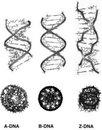
DNA is the primary genetic material of all cellular organisms and DNA viruses. Its sequence carries all the genetic information needed to build and maintain an organism. In April 1953, James Watson and Francis Crick published the world-renowned Nature paper proposing a structure model for the right-handed double helix DNA [1]. Watson and Crick suggested that DNA had two helical chains coiled around the same axis running through the complementary base pairs between each chain – adenine (A) with thymine (T) and guanine (G) with cytosine (C) using two or three hydrogen bonds, respectively [1,2]. This structure we know as B-form of DNA is the basic dominant form of DNA in vivo. However, DNA molecule is highly polymorphic and can adopt many forms depending on its sequence and environmental conditions [3]. The three major double helical forms are A-, B- and Z-DNA, all made of antiparallel strands (Figure 1).
Figure 1 - Molecular modelling representations of the three major nucleic acid duplex conformations. Below: the orthogonal representations. Adapted from [4].
Other non-helical secondary structures exist such as triplexes, bent DNA, cruciforms, nodule DNA, slipped structures (hairpin), sticky DNA and G-quadruplexes (also referred to as tetraplexes, G4 DNA or simply quadruplexes) (Figure 2) [5]. While cruciform and triplex DNA retain the AT and GC Watson-Crick base pairing, G-quadruplex DNA does not involve any GC
base pairs and requires the complementary DNA strands to unwind in order to allow the G-rich strands to fold into four-stranded structures [3]. The formation of this non-canonical DNA structures depends on sequence, topology (supercoiling), binding of proteins, DNA modifications, temperature, dehydration and ionic strength [5]. Evidence points to the important biological roles and implications of such diverse and dynamic structures [5].
1.1 G-quadruplex
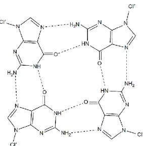
The guanine quadruplex structure is the most studied non-canonical DNA conformation since the earliest physical studies of nucleic acids dated from the 1960’s [5]. The first observation of the self-assembly of guanylic acid, also known as guanosine monophosphate (GMP), was made in the twentieth century by Ivar Bang (1910). Bang observed the formation of a clear gel at high GMP concentrations (25 mg/mL) and pH 5 [6]. Later on in 1962, Gellert, Lipsett and Davies proposed the core of the guanine quadruplex (Figure 3).
Through X-ray diffraction studies of fibers formed from 3’- and 5’-GMP gels, they found that tetrameric guanine residues arranged in vertically stacked hydrogen-bonded guanine tetrads (later termed as G-quartet or G-tetrad) [7]. Gellert et al. also reported the high stability of the structural arrangement provided by the hydrogen-bonding of the four guanine bases, with two hydrogen bonds between each pair involving the N1, N7, O6 and N2 atoms [8]. In 1978, Miles and Frazier showed that guanine tetrads stability depended on a central positive ion that interacts with the oxygen atoms of the guanines [9]. A decade later, new findings pointed to the formation of these four-stranded structures in guanine-rich sequences of immunoglobulin switch regions and in telomeric regions at the end of eukaryotic chromosomes [10,11]. These sequences present discrete runs of guanine tracts (G-tracts), which form compact structural arrangements, termed G-quadruplexes, instead of the continuous helices proposed years before by Watson and Crick [8]. Sen and Gilbert also postulated that the four-stranded structures formed in the immunoglobulin switch regions may have a role in meiosis [11]. In 2001, Schaffitzel et al. found strong evidence for the existence of quadruplex structures in vivo by staining the ciliate Stylonychia lemnae macronuclei with high affinity quadruplex-specific antibodies Sty49 [12]. More recently, G-quadruplexes were found in the mammalian cell nuclei by visualization using immunofluorescent antibodies evidencing its existence in vivo [13]. Guanine-rich sequences were also identified in oncogene promoter regions [14–16]. The technological advance in terms of nucleotide sequencing and conclusion of the human genome mapping in 2001 and the use of bioinformatics approaches led to the identification of new sequences with putative quadruplex formation in the DNA and even at RNA level (over 300,000 potential quadruplex-forming sequences) [8,17]. These guanine-rich sequences seem to be implicated in the function and regulation of cellular pathways at the transcriptional and translational levels, which generated much interest and investigation [2]. To date, several studies have been conducted to explore the native state of the quadruplex structures under physiological conditions.
Figure 3 - The structure of the G-quartet, showing the hydrogen bonding arrangement between the four coplanar guanine bases. Taken from [8].
1.1.1 G-quadruplex sequence
The availability of the complete human genome, as well as several other organisms genomes, provided the needed data in which the existence and identity of guanine-rich sequences could be systematically searched [8,18]. Potential quadruplex-forming sequences can be described by the following sequence motif:
Gm Xn Gm Xo Gm Xp Gm,
where m is the number of guanine residues in each G-tract, usually directly involved in G-tetrad formation [19]. Xn, Xo and Xp can be any combination of residues, including guanines, and are
responsible for the loops formation. The G-tracts form the core of the quadruplex, while the loops, positioned on the exterior, help to maintain the overall structure intact. This sequence motif does not assume that all G-tracts are of equal length, and if one of the short G-tracts is longer than the others, the adjacent guanines will be located in the loops [19]. Both G-tracts and loops are usually limited to 3 ≤ m ≤ 5 and 1 ≤ X ≤ 7, respectively, but deviations to the rule are arguable [8,20]. Using this motif as a quadruplex probe, two distinct research groups conducted bioinformatics analysis using the ENSEMBL genome browser and both found approximatively 375,000 potential quadruplex sequences in the human genome, despite of the statistical and analytical approaches used being different [20,21]. These results indicate that such sequences are highly significant and non-random. Of these ≈375,000 potential quadruplex sequences, ≈223,000 are found in intergenic regions, ≈151,000 within genes and just ≈14,000 within exons [8]. However, the question if these sequences are capable of forming quadruplexes cannot be answered since the experimental data available on structure and stability is insufficient. The search of sequence similarities and distribution of those sequences by clusters with similar tertiary folds is one way to address this problem [21].
A number of informatics resources available online to search databases of quadruplex motifs and to find quadruplex-forming regions within a determined DNA sequence were developed over the years. Generally these search programs use the motif described above as a quadruplex probe with the same limits described. Of the several tools developed, only a few are still available and online. One of those programs is QGRS Mapper, developed by Paramjeet Bagga and his collaborators in 2006 [18]. The main goal of QGRS Mapper is to identify the presence of putative quadruplex forming G-rich sequences (QGRS) in any NCBI nucleotide sequence identified or user-provided sequence [18]. Using a scoring method called G-score, the program then returns the likelihood of the evaluated sequence to form a stable G-quadruplex. Higher scores denote better candidates for quadruplex-formation. The maximum possible G-score, using the default highest QGRS length of 30, is 105 [18]. The scoring method uses three principles based on previous studies: i) shorter loops are more common than longer ones; ii) G-quadruplex loops tend to be approximately equal in size; iii) sequences with greater number of G-quartets form more stable quadruplexes [18].
1.1.2 G-quadruplex structure
As mentioned before, Gellert, Lipsett and Davies introduced the core motif of all G-quadruplex structures, the G-quartet. Each quartet is composed of four guanines, arranged in a rotationally symmetric manner, connected by Hoogsteen hydrogen-bonds in which each guanine base forms two hydrogen bonds with its neighbors, from N1-O6 and N2-N7 (Figure 4) [2,8].
Figure 4 – Schematic representation of the G-quartet and the G-quadruplex structure. Left: hydrogen bond pattern in a G-quartet. A monovalent cation occupies the central position. Right: Schematic diagram of the vertically stacked G-quartets in the G-quadruplex structure. Taken from [20].
The fact that the G-quartets have two hydrogen bonds per base, contrarily to 1 or 1.5 bonds per base in duplex DNA Watson-Crick base pairing (A-T and G-C, respectively), suggests that the G-quadruplex structure is more stable form and presents higher melting temperatures than duplex DNA [22]. The association of guanines N7 atom in the assembly of G-quartets protects them from dimethyl sulfate (DMS) methylation. This unique feature is used to chemically discriminate G-quadruplex formation from other DNA structures (DMS methylation protection assay) [23]. These square planar structures are stabilized by monovalent cations such as potassium (K+) and sodium (Na+) and to a lesser extent ammonium (NH
4+), which interact with
the central electronegative carbonyl O6 atoms of the quartet core [2]. The most stable G-quadruplex structure is formed by the intercalation of K+ between two adjacent G-quartets
[24]. In fact, the G-quadruplex structure formed in the presence of K+ is considered to be
biologically more relevant due its higher intracellular concentration (≈140 mM) [25]. X-ray crystallography and nuclear magnetic resonance (NMR) studies of short oligonucleotide quadruplex structures provided a reliable set of hydrogen bond distances and angles. The hydrogen-bonds distance between nitrogen-oxygen in G-quartets ranges from 2.7-3.0 Å [8]. These G-quartets have large π-surfaces, hence they tend to stack on each other by π-π stacking (2-4 quartets) and enable cations to intercalate between them [2,26]. The stacked G-quartets overlap at a distance of 3.3 Å and are joined together by the sugar-phosphate backbone [27]. The bases that do not participate in the assembly of the G-quartets form loops with different lengths and sequence so that a variety of topologies can be formed (Figure 5).
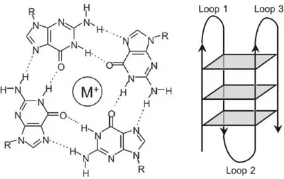
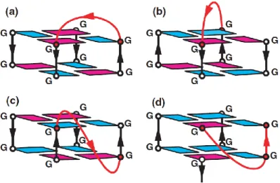
Figure 5 - Schematic illustration of the four major groups of loops. (a) Edge-wise, (b) diagonal, (c) double-chain reversal or propeller and (d) V-shaped loops. The loops connect individual strands bridging two G-tetrad planes. Color coding for illustration is as follows: anti guanines in blue and syn guanines in magenta. G-rich strands in black and connecting loops in red. Taken from [28].
The loop base composition and length determines the structural conformation and stability [28]. Depending on the size and sequence, the loops can be classified into four major groups: i) Edge-wise or lateral loops which connect two adjacent anti-parallel strands, generally composed of two or more residues (Figure 5a); ii) Diagonal loops which connect two opposing anti-parallel strands, generally composed of three or more residues (Figure 5b); iii) Double-chain reversal or propeller loops which connect adjacent parallel strands, and can be as small as one and as large as six or more residues (Figure 5c); iv) V-shaped loops which connect two corners of a G-quartet with a missing support column (Figure 5d) [28].
1.1.3 G-quadruplex structural polymorphism
G-quadruplexes display a wide variety of topologies, owing to the strand stoichiometry, polarity and orientation (parallel or antiparallel), additionally to the loop size and sequence as stated above [19]. The identity of the cation also contributes to the structural polymorphism of G-quadruplex structures. Regarding the strand stoichiometry, G-G-quadruplexes may be unimolecular, bimolecular and tetramolecular, whether it’s formed by one, two or four separate strands of DNA or RNA, respectively. In principle, three strand arrangements are possible but have yet to be validated. Both the bimolecular and tetramolecular structures are intermolecular quadruplexes while the unimolecular form is a intramolecular quadruplex [19]. Guanine-rich sequences with potential to fold into intramolecular quadruplexes are comprised of four consecutive runs of guanines, separated by three loop regions of different lengths and sequences, while bimolecular quadruplexes are formed by sequences with two G-tracts [19]. On its turn, tetramolecular quadruplexes form from single-repeat G-tracts containing sequences [29]. Since there is directionality in the strands, generally described from 5’ end to 3’ end, the different strands that constitute the quadruplex may have different polarities. The adjacent strands can be parallel or antiparallel depending on the conformation of the guanine glycosidic torsion angles (Figure 6).
Figure 6 - Guanines in anti and syn glycosidic conformations. Taken from [26].
Parallel G-quadruplexes have all the guanines in anti conformations, being all the strands parallel to each other (Figure 7a); antiparallel G-quadruplexes have both syn and anti guanines, being at least one of the strands antiparallel to the others (Figure 7b-d) [26]. There are four types of G-quartet core configuration: i) All the strands in the same direction with all the guanines in anti or syn conformation (parallel G-quartet core, Figure 7a); ii) Three strands in one direction and the fourth in the opposite direction, where the stacked G-quartets adopt
anti-anti-anti-syn and syn-syn-syn-anti alignments ((3+1) G-quartet core, also called
hybrid/mixed core, Figure 7b); iii) Two adjacent strands in the same direction and the two other in opposite direction, where the stacked G-quartets adopt a syn-syn-anti-anti alignment (type 1 antiparallel G-quartet core, Figure 7c); iv) Alternating antiparallel strands with the diagonally opposite legs in the same direction, where the stacked G-quartets adopt an
anti-syn-anti-syn alignment (type 2 antiparallel G-quartet core, Figure 7d) [26,30].
Figure 7 - Four types of G-quartet cores. (a) Parallel G-quartet core, (b) (3+1) G-quartet core, (c) antiparallel G-quartet core (up–up–down–down) and (d) antiparallel G-quartet core (up–down–up–down). Taken from [30].
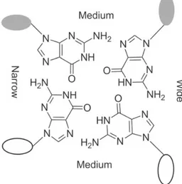
One feature that arises from the guanine glycosidic torsion angles and differs significantly from duplex DNA is the number of grooves [16,26]. While duplex B-DNA has one minor and one major groove, in G-quartets there are four grooves with three different dimensions: wide, medium and narrow (Figure 8). A medium groove is formed by two adjacent guanines in an anti or a syn configuration; a narrow groove is formed by an anti and a syn guanine if the syn guanine is perpendicular to the anti guanine; finally, a wide groove is formed if the anti guanine is
perpendicular to the syn guanine [16,26]. Some restrictions apply to adjacent guanines involved in the same G-quartet. If they are on parallel strands, they must have the same glycosidic torsion angles, and contrariwise if they are on antiparallel strands they must have opposite glycosidic torsion angles [16].
Figure 8 - Schematic representation of a G-quartet core grooves. Three types of grooves can be found: narrow, medium and wide. Taken from [26].
Putting together all the possible variations mentioned above along with the loop types, several different topologies were already determined and described in the literature.
A. Unimolecular G-quadruplex
G-quadruplexes formed by one strand display a variety of structures, as it can adopt three types of loops, namely the diagonal loop, the lateral loop, and the external loop [26]. Among them, the chair-type, with a folding pattern of antiparallel-stranded quadruplex and three lateral loop types (Figure 9a). The second model structure is the basket-type, which is also an antiparallel-stranded quadruplex and has one central diagonal loop and two edgewise loops (Figure 9b). Another model is the propeller-type, which is a parallel-stranded quadruplex with three double-chain reversal loops (Figure 9c). Furthermore, there’s hybrid-types 1 and 2, a mixture of antiparallel/parallel-stranded structures formed by two lateral loops and one propeller loop with different arrangements (Figure 9d-e) [30]. The structural polymorphism was demonstrated by Webba da Silva who predicts that, with different loop length and sequence, unimolecular quadruplexes could give rise to 26 different topologies [31]. Recently, a NMR study was able to determine a new folding of the sequence d[G3ATG3ACACAG4ACG3] into a
intramolecular antiparallel (3+1) G-quadruplex exhibiting three stacked G-quartets connected with the three types of loops possible: propeller, diagonal and edgewise loops of different lengths [32].
Figure 9 - Schematic structure of unimolecular G-quadruplexes. (a) Chair-type form G-quadruplex formed by variant human telomeric sequence d[A(GGGCTA)3GGG] in K+ solution; (b) Basket-type form observed
for d[A(GGGTTA)3GGG] in Na+ solution; (c) Propeller-type form observed for d[A(GGGTTA)3GGG] in a K+
-containing crystal; (d) (3 + 1) Form 1 observed for d[TA(GGGTTA)3GGG] in K+ solution; (e) (3 + 1) Form 2
observed for d[TA(GGGTTA)3GGGTT] in K+ solution. Adapted from [30].
B. Bimolecular G-quadruplex
The association of two G-rich DNA strands can also display a variety of topologies. The association leads to the formation of lateral-looped (Figure 10a-b), diagonal-looped (10c) and external-looped G-quadruplexes (Figure 10d), whether these contain two lateral (in one or opposite directions), two diagonal or two double-chain reversal loops, respectively [30]. The first three structures are antiparallel-stranded while the latter is parallel-stranded. There is also an unusual (3+1) mixed type bimolecular quadruplex observed for the three-repeat human telomeric sequence d[G3(T2AG3)2T] and the single-repeat human telomeric sequence
d[TAGGGT] in Na+ solution [26,30].
C. Trimolecular G-quadruplex
Despite being rare, in 2012 the Mergny group demonstrated the formation of an unprecedented trimolecular quadruplex structure [29]. Although it was a guided assembly of the G-quadruplex, this work proves the putative formation of three stranded quadruplexes under some special conditions. The structure contained some distinctive features as the fact that it had two duplex tails established in the upper and lower ends of the structure (Figure 11). Three single T1, T2 and T3 strands were used as guide strands to position the G-rich tracts in Li+
solution, and then the structure formation was induced by adding Na+ [29].
Figure 11 - Schematic structure of a trimolecular G-quadruplex. Two duplex tails are established at the upper and lower ends of the structure. T1: d[TGAGATGTACTATGAGGGGGTGTCATGGTAGAAGT]; T2: d[GGGGGTCATAGTACATCTCA]; T3: d[ACTTCTACCATGACAGGGGGTTTGGGGG]. Taken from [26].
D. Tetramolecular G-quadruplex
On its turn, the formation of tetramolecular quadruplexes seems much easier (Figure 12a). The crystal structure of hexanucleotide d[TG4T] in Na+ solution exhibit all four strands in a
parallel-stranded structure, with all guanines in anti configuration, medium-size grooves and inexistent loops [26]. However, due to the glycosidic torsion angles of the guanines involved in the G-quartets formation and the different orientations of each independent strand, more complex and unusual structures are possible. For example, the tetranucleotide d[GGGT] was shown to adopt an interlocked dimeric G-quadruplex structure containing two parallel tetramolecular quadruplexes connected by an extra G-quartet (Figure 12b) [26].
Figure 12 - Schematic structure of tetramolecular quadruplexes. (a) Tetrameric parallel-stranded G-quadruplex observed for the single-repeat human telomeric sequences d[TTAGGG] and d[TTAGGGT] in K+
The overall structures, their sequences and Protein Data Bank (PDB) accession numbers of crystallography and NMR determined structures are listed in Figure 13.
Figure 13 – Structural polymorphism of G-quadruplex DNA. Taken from [34].
1.1.4 Metal ion coordination
The stability and conformation of quadruplexes depend on cations. As stated before, the G-quartets central atoms O6 participate in the coordination of the cations. This feature is itself a difference from the duplex DNA, which is stabilized by magnesium (Mg2+) ion while the
G-quartet is not stabilized by such ion. In addition to the stabilization role of the cations, these ions help reducing the electronegative charges repulsion of each G-quartet [8,28]. The structural features of the G-quartets is optimal for the coordination of metal cations within its central cavities. G-quadruplex formation is induced by the presence of K+, Na+ and Rb+ while
cations such as Li+ and Cs+ have a limited ability to induce the formation. However,
G-quadruplexes prefer K+ over Na+, reflecting the much greater energetic penalty for Na+
dehydration [28]. Also, K+ produces more stable quadruplexes than Na+. This fact is due to the
size of the cation, K+ ion is larger than Na+ and forms a much more compact unit upon stacking
between two stacked G-quartets [19]. All together, these facts suggest that the formation of G-quadruplexes depends on the size and desolvation energy cost of the cation. From studies of the ions ability to induce gel formation in guanosine nucleotides it was possible to deduce the following order: Sr2+>K+>>Na+>Rb+~Ba2+>NH
4+>Ca2+> Mg2+~Cs+>>Li+ [16,35].
One interesting behavior of G-quadruplexes is the topology transition observed under different ionic conditions. The same sequence can adopt different G-quadruplex conformations in K+ and
Na+. For example, based on NMR and crystallographic studies of the human telomeric DNA
d[AGGG(TTAGGG)3], Na+ seems to favor an antiparallel conformation while K+ induces a parallel
or hybrid form [36,37]. Interestingly, RNA-quadruplexes formation is generally independent of the cation identity and bimolecular quadruplexes are not affected by the nature of the cation equally to the unimolecular quadruplexes [38,39].
1.2 Biological relevance of G-quadruplexes
Guanine-rich sequences with putative G-quadruplex formation are widely dispersed in eukaryotic and prokaryotic genomes. A number of critical regions in the eukaryotic genome have been reported to adopt G-quadruplex structures. These include promoter regions of oncogenes such as c-myc and c-kit, both short micro-satellite and longer mini-satellite repeats, ribosomal DNAs, as well as telomeres in eukaryotes and immunoglobulin heavy chain switch regions of higher vertebrates [16]. G-rich sequences were also found in the retinoblastoma susceptibility gene and upstream of the insulin gene [16]. These guanine-rich sequences have the potential to influence the gene metabolism processes, as well as key biological processes such as DNA replication and recombination, transcriptional regulation and genome stability [26]. The visualization of G-quadruplex structures in vivo was already reported. Besides the work with the ciliate Stylonychia lemnae described above, recent work reported by Biffi et al. described a monoclonal single chain antibody BG4 with high affinity and specificity for intramolecular G-quadruplex structures [13]. This work reported crucial evidence for the formation of G-quadruplex DNA within mammalian cells genomes, in a cellular context, the cell-cycle dependence of the structures (the occurrence of G-quadruplexes was maxed during the S phase) and provided an important basis to help understand the biological roles of G-quadruplex structures [13]. Additional works using monoclonal antibodies followed, strengthening the evidence of the G-quadruplex formation in vivo [40,41].
The existence of natural proteins that recognize G-quadruplexes in vivo, provided important insights in the location of G-quadruplex structures in the genome and reinforces the biological relevance of such sequences [28]. Such proteins bind, promote or disrupt the quadruplex formation, and over 30 proteins have been reported so far [2]. For instance, both telomere end-binding protein (βTBP) in Oxytricha and repressor activator protein 1 (RAP1) in
Saccharomyces cerevisiae promote intermolecular G-quadruplex formation [28]. The MutSα
protein, which is involved in mismatch repair, targets G-quadruplex DNA and promotes synapsis of transcriptionally activated immunoglobulin switch regions [28]. In addition, POT1 binding to DNA disrupts G-quadruplex formation at the telomeric G-rich overhangs, promoting telomerase activity [28]. Furthermore, the Escherichia coli RecQ protein can unwind G-quadruplex DNA and is essential for maintaining genomic stability of many organisms, being the RecQ DNA helicase family conserved from E. coli to humans [23,28]. Genome integrity is essential in order to maintain the normal cellular function, and malfunctioning DNA replication or repair processes can lead to genetic instability and disease [13]. In humans, the helicase-homologue protein ATRX was shown to bind G-rich sequences in both telomeres and euchromatin. It was proposed that such protein was able to modify the epigenetic state of G-quadruplex sites and to resolve its structure [13].
1.2.1 Telomeres
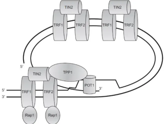
The telomere is a specialized nucleoprotein complex (termed shelterin) existent at the ends of all eukaryotic chromosomes [8,28]. The human telomere consists of tandem repeats of the motif d[TTAGGG] and is associated with a variety of telomeric proteins and other DNA-repair and damage response proteins [42]. The telomere function is to stabilize the termini of linear eukaryotic chromosomes, forming special T-loop-like structures, and protecting the chromosomes from unwanted recombination and degradation, while providing sites for recombination events and transcriptional silencing [28,42]. Telomeres are thought to play a critical role in cellular aging and cancer. Human telomeric DNA is typically 5-15 kilobases (kb) long duplex DNA with a single-stranded 3’ overhang of 150-250 bases at the 3’ extreme end. These G-rich single-stranded overhangs are attractive sites for potential G-quadruplex formation. The length of the duplex portion of the telomeres decreases progressively after each cell division cycle in somatic cells as a consequence of the end-replication effect. On the other hand, the single-stranded 3’ overhangs can be elongated by telomerase, an enzyme with reverse transcriptase activity, which is expressed in the majority of cancer cells (80-85%) and primary tumors, thus maintaining the telomere-length homeostasis [8,28,42]. Telomerase is a ribonucleoprotein which contains an RNA template (subunit hTERC) from which its reverse transcriptase subunit hTERT copies and adds TTAGGG repeats to the single-stranded 3’ overhangs of the telomeres [8,42]. In normal somatic cells, when telomeres reach critical short lengths (the Hayflick limit of about 40 cell divisions), cells enter irreversible p53 and Rb-dependent replicative senescence, and ultimately apoptosis. In cancer cells, where telomerase is expressed, it maintains the telomere length acting as a tumor promoter and helping the cells bypassing apoptosis and achieving cellular immortality [42]. The 3’ single-stranded overhang is protected and maintained by several copies of hPOT1 (protection of telomeres 1), a single-stranded binding protein which interacts with other proteins of the shelterin complex to regulate telomerase-mediated telomere elongation. The shelterin complex proteins include the telomere repeat binding factors (TRF1 and TRF2), which are responsible for the T-loop formation; the associate-proteins (RAP1, TPP1, and TIN2) on its turn, are responsible for mediating the interaction between TRF1 and TRF2, as well as POT1 and TPP1 (Figure 14).
T-loop formation involves the folding back of the single-stranded overhang and pairing with a complementary portion of the telomeric double-stranded region [8]. The disruption of telomeric DNA-hPOT1 association leads to quadruplex formation, deprotects telomeres and initiates DNA damage-response mediated cell death [28,42]. Thus, the development of small molecules that bind and stabilize the single strand G-quadruplex, competing with hPOT1 and initiating this response, is a viable and promising anti-cancer therapeutic strategy (Figure 15). Also, the formation of a stable G-quadruplex structure inhibits the activity of telomerase [19]. Therefore, much effort has been placed in the structural characterization of the telomeric G-quadruplex topologies in order to find scaffolds for anti-cancer drug development [28].
Figure 15 – Schematic illustration of the telomeric G-quadruplex therapeutic strategy hypothesis. The biological roles of telomeric DNA and telomerase are shown; G-quadruplex formation can inhibit telomerase activity. Taken from [34].
The human telomeric G-quadruplex structure is highly polymorphic and to date several structure models were proposed [8]. In 1992, the Patel group proposed a model for the single-repeat d[TTAGGGT) human telomere sequence which formed a parallel-stranded G-quadruplex in K+ solution [43]. The structure was formed by three G-quartets with all guanines in anti
conformation. Later on, the two-repeat human telomere sequence G-quadruplex model was proposed and the crystal structure was identified. The crystallography analysis demonstrated that the sequence d[TAGGGTTAGGGT] folds into a bimolecular quadruplex in K+ solution, with
all the strands in a parallel orientation and connected by propeller-type loops [36]. Following NMR studies of the same sequence confirmed the G-quadruplex topology [44]. Other NMR studies, involving the three-repeat human telomere sequence 5’-GGGTTAGGGTTAGGGT-3’ demonstrated the formation of a peculiar G-quadruplex structure. This structure contained three strands in the same direction and one in the opposite direction (3+1 G-quadruplex) [45]. Such structure could occur in vivo in the T-loop region where the single-stranded overhang pairs
with a complementary portion of the telomeric double-stranded region. Such results supported the biological and therapeutic implications of the G-quadruplex formation in the telomeres, and the use of small molecules for its stabilization [45]. The cation-dependent structural polymorphism was demonstrated by studies performed with the four-repeat human telomere sequence d[AG3(T2AG3)3]. In a solution containing Na+, the sequence folds into a antiparallel
G-quadruplex structure with a central diagonal loop and two edgewise loops, as determined by NMR [37]. However, in K+ solution, a X-ray crystal structure of the same sequence
demonstrated the folding of a intramolecular G-quadruplex composed of three G-quartets, with all four strands in parallel joined by three propeller-type loops [36]. Because the intracellular concentration of K+ (≈140 mM) is higher than that of Na+ (≈10 mM), the propeller-type
G-quadruplex seems to be more physiological relevant. Further studies demonstrated the existence of other G-quadruplex conformations in the human telomeres such as hybrid-type mixed parallel/anti-parallel and basket-type quadruplexes [46–48].
1.2.2 Gene promoters
Apart from telomeric sequences, there are other biological relevant G-rich sequences that have the ability to form G-quadruplex structures, such as the gene promoter regions. The first evidence of the formation of unusual DNA conformations in gene promoters was found by Larsen and Weintraub who demonstrated that chicken β-globin promoter adopted high order structures
in vivo [49]. Since then, the use of bioinformatics approaches led to the discovery of putative
quadruplex-forming sequences in the regulatory and promoter regions of oncogenes such as
c-myc, c-myb, c-Fos, c-kit, KRAS, vascular endothelial growth factor (VEGF), platelet-derived
growth factor α polypeptide (PDGF-A), Rb, RET, hypoxia-inducible factor 1α (Hif-1α), B cell lymphoma 2 (bcl2), and hTERT [50]. These proto-oncogenes are involved in growth and proliferation processes and their proximal promoter regions contain several G and C-rich regions. The formation of G-quadruplexes in these regions is believed to modulate the genes function and transcriptional activity. Moreover, these genes are important in cell signaling and are involved in a variety of cancers [50]. Therefore, the study of such G-quadruplex structures and the development of targeted-drugs, capable of interacting with the specific G-quadruplex and modulate the transcriptional activity of the associated gene, poses itself as a promising therapeutic strategy. This therapeutic approach is presented in Figure 16 in a broad perspective and represents the formation of G-quadruplex structures in gene promoters in a process that affects the transcriptional activity.
Figure 16 - Schematic illustration of the promoter quadruplex therapeutic strategy hypothesis. G-quadruplex forming region in the upstream (promoter) region (in green) of genes (in red). Taken from [42].
Some important oncogene promoter quadruplexes are described in more detail below.
A. c-myc
The proto-oncogene c-myc encodes a multifunctional transcription factor thought to regulate 10–15% of all cellular genes and is involved in processes such as cell cycle regulation, apoptosis, metabolism, cellular differentiation and cell adhesion [23]. The overexpression of c-myc leads to cellular proliferation and inhibition of differentiation and is associated with a variety of human cancers, such as colon, breast, small-cell lung, osteosarcomas, glioblastomas, and myeloid leukemia [51]. The deregulation of the c-myc transcription factor is a consequence of gene amplification, translocations, altered ploidy or enhanced transcription owing to upstream signaling abnormalities [50]. Because of this, transcriptional modulation of the c-myc oncogene has been an attractive target for anti-cancer drug development. The regulation of c-myc transcription is complex and involves multiple promoters (P0, P1, P2, and P3) and seven nuclease hypersensitive elements (NHEs). The nuclear hypersensitivity element III1 (NHE III1),
located -142 to -145 base pairs (bp) upstream of the P1 promoter of c-myc, is a G-rich strand that controls 80-90% of c-myc transcription and contains a 27 bp sequence (Pu27) with propensity to fold into a stable G-quadruplex structure [52]. Pu27 is capable of engaging in a slow equilibrium between B-form duplex DNA, single-stranded DNA and G-quadruplex DNA [51]. This 27 bp sequence contains six G-tracts with different lengths and can form a variety of intramolecular G-quadruplex structures depending on the G-tracts combinations [23]. Is worth to notice that for a quadruplex to form, the two complementary sequences must first separate to allow the G-rich strand to fold in G-quadruplex [53]. The first observation of a G-quadruplex formed by c-myc promoter in K+ solution was made by the Simonsson group [14]. The
G-quadruplex consisted of three G-quartets bound by two lateral loops and a central diagonal loop. Since Pu27 forms a dynamic mixture of four G-quadruplex loop isomers, smaller sequences derived from Pu27 started being used, in order to determine the physiologically more relevant quadruplex within this region [23]. Two different sequences, Myc-2345 and Myc-1245 (numbers
correspond to the G-tract position), were analyzed by NMR and found to form intramolecular propeller-type G-quadruplexes in K+ solution. The core of the structures was formed by four
parallel strands, with all guanines in anti conformation and three propeller-type loops [23]. Another example is Myc22-G14T/G23T, in which two guanines have been mutated to thymines, and forms a parallel-stranded G-quadruplex with propeller-type loops [54].
B. c-kit
The c-kit proto-oncogene encodes a 145-160 kDa membrane bound receptor tyrosine kinase that constitutes a cell signaling system and can stimulate cell proliferation, differentiation, migration and survival [50]. The overexpression or activating mutations of c-kit may lead to aberrant function and oncogenic cellular transformations, being involved in a variety of human cancers such as mast cell tumors, germ cell tumors, ovarian carcinomas, malignant melanomas, gastrointestinal stromal tumors, small-cell lung cancer, neuroblastoma, and breast carcinoma [23]. Upstream of the c-kit transcription initiation site there’s a G-rich strand which is essential for the promoter activity. c-kit quadruplexes are therefore attractive targets for anti-cancer drug development. Two G-quadruplex structures have been identified in the G-rich promoter region of c-kit, c-kit87up and c-kit21. c-kit87up is a 22 nucleotide sequence, located 87 base pairs upstream of the transcription initiation site, and was found to form a single G-quadruplex structure in K+ [55]. The G-quadruplex structure formed was very peculiar as an isolated guanine
was involved in the G-quartet core formation and it presented four distinct loops, two propeller-type, a single-residue loop and a five-residue loop [55]. This emphasizes the importance of the sequence as non-G-tract residues can participate in the G-quadruplex core formation. On its turn, c-kit21 was found to adopt a variety of conformations and mutations studies need to be employed in order to form a single G-quadruplex [23].
C. bcl-2
The human bcl-2 gene encodes a 25 kDa mitochondrial membrane protein that blocks programmed cell death. The overexpression of bcl-2 gene occurs in a variety of human cancers, such as B-cell and T-cell lymphomas, breast, cervical, non-small-cell lung, prostate, and colorectal cancers, which reduces the rate of cell death and also interferes with the therapeutic action by resisting apoptosis induced by chemotherapy [56]. Therefore, bcl-2 gene has been considered as an important target for developing anti-cancer compounds. The human bcl-2 gene is regulated by two promoters P1 and P2. A 39 bp G-rich sequence (bcl-2Pu39) located in the bcl-2 P1 promoter plays a significant role in the regulation of bcl-2 transcription [23]. The sequence contains six G-tracts (5’-AGGGGCGGGCGCGGGAGGAAGGGGGCGGGAGCGGGGCTG-3’) with different sizes and was shown to adopt a mixed parallel/antiparallel G-quadruplex intramolecular G-quadruplex conformation [57]. Moreover, the structure contains two edgewise loops and one propeller-type loop and the middle four G-tracts are the ones that generate the predominant G-quadruplex structure (MidG4), since like c-myc this sequence can adopt multiple intramolecular G-quadruplexes (5’G4, MidG4, and 3’G4) [23,57].
1.2.3 Immunoglobulin switch regions
Other important genomic regions have the ability to form G-quadruplex structures. Of interest are the regions encoding immunoglobulin heavy chain switch (S) regions of higher vertebrates. These regions are critical for class switch recombination processes of B lymphocytes. B lymphocytes, or B cells, are responsible for the production of different isotypes of immunoglobulins (antibodies) for diverse pathogens [58]. The process that enables the production of immunoglobulins (Ig) to change from one isotype to another, termed class switch recombination, is a region-specific recombination process that bring an expressed variable (VDJ) region to a new constant (C) region during the differentiation of B lymphocytes to plasma cells [11,59]. The isotype switching from IgM to IgG3 and IgA is presented in Figure 17 as an example.
The VDJ regions form a domain that recognize the antigen, while C region determines how the antigen is removed from the body [58]. During this recombination process numerous kb of DNA between the constant and variable regions are deleted. These processes are essential and if impaired, it can result in immunodeficiency [58]. The S regions of 2 to 10 kb in length, lie upstream of the constant-region genes and contain repetitions of highly degenerate G-rich consensus sequences which are 20-50 bp long [11,58]. Several repetitive motifs occur such as GGGGT, GAGCT and the conserved sequence (G)GGGGAGCTGGGG which is found in Sγ1, Sγ2b and Sγ3 S regions. Two different studies using the same Ig switch region sequences demonstrated the formation of stable G-quadruplex structures, suggesting a role of such structures in recombination events [11,15]. More recently, Maizels group reported the
Figure 17 - Recombination events in class switch recombination. During isotype switching, a portion of the DNA is looped out as switch regions recombine. A different constant regions is placed downstream of VDJ region. After recombination, the constant region directly downstream of the VDJ region will encode the immunoglobulin isotype produced on the surface of the B cell. Taken from [59].
formation of a novel G-quadruplex structure called G-loops by using plasmid vectors containing inserts from murine switch regions [60]. For instance, pPH600, a plasmid containing a 604 bp fragment of the murine Sγ3 switch region, upon in vitro transcription of the G-rich regions formed G-loops observed by transmission electron microscopy (TEM) [60]. However, to my best knowledge, no studies regarding the determination of the G-quadruplex structure were performed so far.
1.3 G-quadruplex as a therapeutic target
Due to the biological relevant location of G-quadruplexes throughout the genome and the implication in key processes such as maintenance of chromosomal ends, transcription, translation, DNA replication and recombination, G-quadruplex is a promising and viable therapeutic target. This therapeutic strategy involves the development of selective drug-like small ligands that strongly bind and stabilize the G-quadruplex structures. Over the past years, there has been a remarkable effort on developing G-quadruplex ligands, especially to target human telomeres and oncogene promoters in order to block the action of telomerase and the oncogene transcription, respectively [8,42]. This concept was first validated through the demonstration that the compound 2,6-diamidodianthraquinone was capable of inhibiting the activity of telomerase by interacting with and stabilizing quadruplex structures [28]. G-quadruplex structure presents a large π-surface due to the guanine tetrads, approximately twice as large as that found in duplex DNA. Therefore, the majority of quadruplex-ligands have a large aromatic core, with a large π-surface, in order to maximize the π-π interactions [53]. Compounds containing polyaromatic heterocyclic ring systems, such as anthraquinones, acridines, naphthalenes, perylenes and porphyrins, capable of π-stacking interactions, are good candidates [28]. Another feature of these ligands, relates to the fact that G-quadruplexes, like all nucleic acids, have a high negative charge due to the backbone negative phosphate groups. Hence, cationic ligands will generally bind more strongly to the G-quadruplex structure [53]. On the other hand, the inclusion of sidechain specific functional groups, such as protonable side arms, once they participate in the recognition of G-quadruplex via actions in the grooves and enhance the interactions with the structure [53,61]. One important issue in the development of such small ligands, is their specificity for G-quadruplex over duplex DNA. This is a major problem since G-quadruplexes are far outnumbered by the duplex DNA in the cells [2]. The compounds synthesized must promote cell death in cancer cells, while ensuring that the toxicity to normal healthy cells is low.
Until now, some compounds with experimental evidence for G-quadruplex binding and therapeutic activity have been developed such as BRACO19 [62], telomestatin [63], and TMPyP4 [64] (Figure 18).
Figure 18 – Structures of the compounds TMPyP4, telomestatin and BRACO-19. Taken from [2].
BRACO19 is a 3,6,9-tri-substituted acridine that appears to directly target the telomeres and displayed the capacity to inhibit the catalytic function of telomerase in human cancer cells and destabilize the telomere complex [65]. BRACO19 induced tumor regression within 7-10 days of the initiation of treatment of prostate cancer cells. Its major limitations are lack of membrane permeability and small therapeutic window [65]. Telomestatin, a natural occurring macrocycle isolated from Streptomyces anulatus, is the tightest known G-quadruplex binder and telomerase inhibitor [65]. Telomestatin interacts preferentially with intramolecular G-quadruplex structures and has a 70-fold selectivity over duplex DNA. Telomestatin displayed promising anticancer activity in human cancer cells, within 3–5 weeks at the minimal effective concentration inhibited telomerase activity, reduced telomere length and caused apoptotic cell death in multiple myeloma cell lines [65]. TMPyP4, is a cationic porphyrin designed with attractive properties such as a fused planar ring system, positive charge and appropriate size to stack with the G-tetrads [65]. It has shown to inhibit human telomerase in HeLa cell extracts and to downregulate c-myc. However, TMPyP4 has only limited selectivity for G-quadruplex over duplex DNA [65]. Naphthalene-based ligands were also reported as having the necessary features to bind planar substrates such as nucleotides, mainly through stacking interactions [66]. Potent G-quadruplex binders containing naphthalene rings were reported by Neidle et al., which were found to promote a dose dependent cell arrest of mutated c-kit cell lines [8,67]. However, some non-specific interactions were reported as well [67]. Despite the number of compounds already designed and synthesized, only one reached phase II clinical trials (Quarfloxin/CX-3543; (ClinicalTrials.gov identifier: NCT00780663), probably due to the specificity and selectivity requirements [68]. Quarfloxin is a fluoroquinolone which targets majorly c-myc promoter and ribosomal quadruplexes, with high specificity and selectivity, and demonstrated potent and tumor-selective activity in vitro and in vivo [65]. G-quadruplex sequences on its own can act as therapeutic agents as shown by Paula Bates group, who developed the G-quadruplex aptamer AS1411 which targets nucleolin, an overexpressed protein
![Figure 2 - Non-duplex DNA structures formed by repeating sequences. Adapted from [5].](https://thumb-eu.123doks.com/thumbv2/123dok_br/18068051.864251/28.892.288.563.246.793/figure-non-duplex-structures-formed-repeating-sequences-adapted.webp)
![Figure 6 - Guanines in anti and syn glycosidic conformations. Taken from [26].](https://thumb-eu.123doks.com/thumbv2/123dok_br/18068051.864251/33.892.256.679.122.401/figure-guanines-anti-syn-glycosidic-conformations-taken.webp)
![Figure 9 - Schematic structure of unimolecular G-quadruplexes. (a) Chair-type form G-quadruplex formed by variant human telomeric sequence d[A(GGGCTA) 3 GGG] in K + solution; (b) Basket-type form observed for d[A(GGGTTA) 3 GGG] in Na + solution; (c) Pr](https://thumb-eu.123doks.com/thumbv2/123dok_br/18068051.864251/35.892.230.692.117.478/schematic-structure-unimolecular-quadruplexes-quadruplex-telomeric-sequence-solution.webp)
![Figure 13 – Structural polymorphism of G-quadruplex DNA. Taken from [34].](https://thumb-eu.123doks.com/thumbv2/123dok_br/18068051.864251/37.892.151.778.165.487/figure-structural-polymorphism-g-quadruplex-dna-taken.webp)