Bothrops moojeni
myotoxin-II, a Lys49-phospholipase A
2
homologue:
An example of function versatility of snake venom proteins
☆
Rodrigo G. Stábeli
a, Saulo F. Amui
b, Carolina D. Sant'Ana
b, Matheus G. Pires
a, Auro Nomizo
b,
Marta C. Monteiro
c, Pedro R.T. Romão
d, Renata Guerra-Sá
e, Carlos A. Vieira
f, José R. Giglio
f,
Marcos R.M. Fontes
g, Andreimar M. Soares
b,⁎
a
Instituto de Pesquisas em Patologias Tropicais, IPEPATRO, Universidade Federal de Rondonia, UNIR-RO, Brazil b
Departamento de Análises Clínicas, Toxicológicas e Bromatológicas, Faculdade de Ciências Farmacêuticas de Ribeirão Preto, Universidade de São Paulo, USP, Ribeirão Preto-SP, Brazil
cUniversidade Estadual do Centro-Oeste/UNICENTRO, Guarapuava-PR, Brazil
dLaboratório de Imunoparasitologia, UNISUL, Tubarão-SC, Brazil
eLaboratório de Bioquímica e Biologia Molecular-Departamento de Ciências Biológicas, UFOP, Ouro Preto-MG, Brazil
fDepartamento de Bioquímica e Imunologia, FMRP, USP, Ribeirão Preto-SP, Brazil
gDepartamento de Física e Biofísica, IB, UNESP, Botucatu-SP, Brazil
Received 22 September 2005; received in revised form 22 November 2005; accepted 23 November 2005 Available online 24 January 2006
Abstract
MjTX-II, a myotoxic phospholipase A2(PLA2) homologue fromBothrops moojenivenom, was functionally and structurally characterized. The MjTX-II characterization included: (i) functional characterization (antitumoral, antimicrobial and antiparasitic effects); (ii) effects of structural modifications by 4-bromophenacyl bromide (BPB), cyanogen bromide (CNBr), acetic anhydride and 2-nitrobenzenesulphonyl fluoride (NBSF); (iii) enzymatic characterization: inhibition by low molecular weight heparin and EDTA; and (iv) molecular characterization: cDNA sequence and molecular structure prediction. The results demonstrated that MjTX-II displayed antimicrobial activity by growth inhibition againstEscherichia coliand Candida albicans, antitumoral activity against Erlich ascitic tumor (EAT), human breast adenocarcinoma (SK-BR-3) and human T leukemia cells (JURKAT) and antiparasitic effects against Schistosoma mansoni and Leishmania spp., which makes MjTX-II a promising molecular model for future therapeutic applications, as well as other multifunctional homologous Lys49-PLA2s or even derived peptides. This work provides useful insights into the structural determinants of the action of Lys49-PLA2homologues and, together with additional strategies, supports the concept of the presence of others“bioactive sites”distinct from the catalytic site in snake venom myotoxic PLA2s.
© 2005 Elsevier Inc. All rights reserved.
Keywords: Bothrops; cDNA cloning; Chemical modification; Myotoxin; Microbial; Parasiticidal and antitumoral activity; Phospholipase A2; Snake venom
www.elsevier.com/locate/cbpc
Abbreviations:Basp,B. aspermyotoxins; BnSP,B. neuwiedimyotoxins from São Paulo State; bp, base pair; BPB, 4-bromophenacyl bromide; BthTX,B.
jararacussubothropstoxins; CFU, colony-forming units; CK, creatine kinase; CNBr, cyanogen bromide; EDTA, ethylenediaminetetraacetic acid; MjTX,B. moojeni
myotoxins; MjTX-II, myotoxin-II fromBothrops moojenivenom; MTT, 3-(4,5-dimethylthiazol-2-yl)-2,5-diphenyltetrazolium bromide; NBSF, 2-nitrobenzenesul-phonyl fluoride; PLA2, phospholipase A2; PrTX,B. pirajaipiratoxin; RT-PCR, reverse transcriptase–polymerase chain reaction.
☆
This paper is part of a special issue of CBP dedicated to“The Face of Latin American Comparative Biochemistry and Physiology”organized by Marcelo Hermes-Lima (Brazil) and co-edited by Carlos Navas (Brazil), Tania Zenteno-Savín (Mexico) and the editors of CBP. This issue is in honour of Cicero Hermes-Lima and the late Peter W. Hochachka, teacher, friend and devoted supporter of Latin American science.
⁎Corresponding author. Fax: +55 16 3602 4878.
E-mail address:[email protected] (A.M. Soares).
1. Introduction
Phospholipases A2(PLA2s; EC 3.1.1.4) are widely
distrib-uted enzymes (Nevalainen et al., 2004a,b) of high medical-scientific interest due to their involvement in several inflam-matory human diseases and envenomation by snake and bee venoms. PLA2s constitute a superfamily of different enzymes
belonging to four groups on the basis of their source, amino acid sequence and biochemical characteristics (Six and Dennis, 2000; Murakami and Kudo, 2002). PLA2s from classes I, II and
III have low Mr (13,000–18,000), are Ca2+-dependent and
structurally different from class IV PLA2s. Those from class IV
are intracellular, cytosolic, Ca2+-dependent and have high Mr
(85,000). Recently discovered enzymes, either secreted (sPLA2s) or cytosolic (cPLA2s), were identified and did not
clearly fit in any of the above classes. New classes were then proposed (Six and Dennis, 2000; Murakami and Kudo, 2002, 2004).
PLA2s from snake venoms have been classified as groups I
and II on the basis of their primary structure and disulfide bridge pattern (Balsinde et al., 1999; Six and Dennis, 2000). In addition to their primary catalytic role, snake venom PLA2s
show other important toxic/pharmacological effects including myonecrotic, neurotoxic, cardiotoxic, hemolytic, hemorrhagic, hypotensive, anticoagulant, platelet aggregation inhibition and edema-inducing activities (Gutiérrez and Lomonte, 1997; Ownby, 1998; Ownby et al., 1999; Soares et al., 2004a).
Muscular necrosis is a serious consequence of Bothrops
snakebites that may lead to permanent loss of tissue or function and to require amputation of the affected member. Myonecrosis may be due to an indirect action as consequence of vessel degeneration and ischemia caused by hemorrhagic metallopro-teases or by a direct effect of myotoxic PLA2s homologues on
plasma membranes of muscle cells (Gutiérrez and Lomonte, 1997; Ownby, 1998; Ownby et al., 1999; Gutiérrez, 2002; Soares et al., 2004a).
During the last 15–20 years, there has been an increasing interest in the study of the myonecrotic venom components, resulting in the isolation and structural/functional characteriza-tion of several myotoxic PLA2 homologues from Bothrops
venoms (Soares et al., 2004a). Myotoxins isolated from these venoms belong to group IIA of PLA2s and may be subdivided
into two subgroups: (i) Asp49 myotoxins with relatively low to moderate enzymatic activity and (ii) Lys49 myotoxins with practically no hydrolytic activity on artificial substrates (Gutiérrez and Lomonte, 1997; Ownby, 1998; Ownby et al., 1999; Soares et al., 2004a).
Myotoxin-I and -II homologues purified from Bothrops moojeni (MjTX-I and -II) are Lys49-PLA2 and have been
isolated and characterized (Lomonte et al., 1990; Soares et al., 1998, 2000a,b, 2004a). The structures of native MjTX-I and -II and of the complex MjTX-II-stearic acid have been recently solved (de Azevedo et al., 1997; Marchi-Salvador et al., 2005a; Watanabe et al., 2005). In order to study the function versatility, MjTX-II was functionally and structurally characterized. This characterization included: (i) parasiticidal, antimicrobial and antitumor effects; (ii) effects of chemical modifications by
4-bromophenacyl bromide (BPB), cyanogen bromide (CNBr), acetic anhydride and 2-nitrobenzenesulphonyl fluoride (NBSF); (iii) inhibition by low molecular weight heparin, EDTA and PLA2-antibodies; and (iv) cDNA sequence and molecular
structure of this myotoxin.
2. Materials and methods
2.1. Purification procedure
B. moojenivenom was purchased from Bioagents Serpentar-ium (Batatais-SP) and fractionated on CM-Sepharose column at pH 8.0 as previously described (Soares et al., 1998). Homogeneity was demonstrated by 12% SDS-PAGE, basic PAGE at 10%, isoelectric focusing, N-terminal amino acid sequencing, RP-HPLC and mass spectrometry (Soares et al., 2000a,b, 2003). Molecular weight of the MjTX-II was also determined by MALDI mass spectroscopy. Far UV circular dichroism spectra (190–250 nm) were measured with a JASCO 810 (JASCO Inc., Tokyo, Japan) using 1 mm path length cuvettes and protein concentrations of 250 mg/mL for native or modified myotoxic PLA2s. In all cases, a total of three spectra
were collected, averaged and corrected by subtraction of a buffer blank.
2.2. Chemical modification
Modification of histidine residues with 4-bromophenacyl bromide (BPB) was carried out as previously described (Soares et al., 2000a,b, 2001a,b) using 0.1 M ammonium bicarbonate buffer. Three milligrams of toxin were dissolved in 1 mL of 0.1 M ammonium bicarbonate containing 0.7 mM EDTA (pH 8.0) and 150 mL of BPB (0.8 mg/mL), and the mixture incubated for 24 h at 25 °C.
Modification of Lys residues with acetic anhydride (AA) was performed at a protein/reagent molar ratio of 1:50 (Soares et al., 2000a,b, 2001a,b). MjTX-II (3 mg) was dissolved in 1.5 mL of 0.2 M ammonium bicarbonate buffer at pH 8.0 plus 10 mL of AA and the mixture was incubated for 1 h at 25 °C.
Tyrosine residues were modified by treatment with 2-nitrobenzenesulphonyl fluoride (NBSF) as previously described (Soares et al., 2000a,b, 2001a,b). For that, 1 mmol of MjTX-II (9 mmol of Tyr) was dissolved in 14 mL of 0.1 M Tris–HCl (pH 8.0) and incubated with 9 mmol of NBSF for 20 h at 25 °C.
Modification of Trp residues was performed according to
Soares et al. (2000a,b, 2001a,b). 9 mg of MjTX-II was dissolved in 4 mL 50% acetic acid containing 1 mg of o -nitrophenylsulphenyl chloride (NPSC) and incubated for 1 h at 25 °C. In all cases, excess reagent was removed by ultrafiltration through an Amicon YM-3 membrane and washed with water or 0.05 M ammonium bicarbonate (pH 8.0), followed by lyophilization.
2.3. Treatment of myotoxin-II with cyanogen bromide
over the methionine residues was added (Soares et al., 2000a,b, 2001a,b). The cleavage reaction developed under nitrogen for 24 h at room temperature. Control and cleaved myotoxin were applied on a Sephadex G-25 column (100 × 2.0 cm), which was equilibrated and eluted with 0.05 M, pH 8.0 ammonium bicarbonate.
2.4. Myotoxic activity
Groups of five male Swiss mice (18–22 g) were injected in the right gastrocnemius muscle with MjTX-II/50μL PBS. After
1, 3 and 6 h, blood was collected from tail vessels in heparinized tubes and centrifuged for separation of the plasma. The amount of creatine kinase (CK) was then determined using 4μL plasma
incubated for 3 min at 37 °C with 1.0 mL of the reagent according to the kinetic CK-UV protocol from Sigma Chem. Co. Activity was expressed in U/L, 1 unit corresponding to the production of 1 μmol NADH/min (Soares et al., 2000a,b, 2001a,b).
2.5. Edema-inducing activity
Groups of five male Swiss mice (18–22 g) were injected in the subplantar region with different doses of MjTX-II/50 μL
PBS. After 0.5 h, the paw edema was measured with a low pressure spring caliper (Mitutoyo-Japan) (Soares et al., 2000a,b, 2001a,b). Zero time values were then subtracted and the differences expressed in median % ± S.D.
2.6. Disruption of liposomes
Negatively charged liposomes (phosphatidylcholine, 63μmol;
dicethylphosphate, 18 μmol; and cholesterol, 9 μmol) were
obtained from Sigma Chem. Co. (Missouri, USA). The assay followed the procedure ofSoares et al. (2000a,b, 2001a,b)on microplates, incubating 20μL of the liposome suspension and
20μL of MjTX-II solutions (in PBS) for 30 min at 37 °C or
4 °C.
2.7. Cancer cell lines culture
Human breast (SK-BR-3) and acute T cell leukemia (Jurkat) cancer cell lines were maintained on RPMI 1640 medium supplemented with 2 mM L-glutamine, 1.5 g/L sodium
bicarbonate, 4.5 g/L glucose, 10 mM HEPES, 1.0 mM sodium pyruvate, 10% fetal bovine serum, 100 U/mL penicillin and 100 μg/mL streptomycin. All cell culture reagents were
purchased from Gibco. All cell lines were maintained at 37 °C in 5% CO2and 95% air with more than 95% humidity.
2.8. Tumor cytotoxic activity
Tumor cytotoxic activity of PLA2s was assayed with
3-(4,5-dimethylthiazol-2-yl)-2,5-diphenyltetrazolium bromide (MTT) staining as described by Mosmann (1983). Tumor cells cultivated in appropriate flasks and maintained in continuously exponential growth were detached with 0.05% trypsin, 0.02%
EDTA in calcium-free phosphate-buffered saline (PBS) and washed three times with RPMI medium at 500×g/15 min/10 °C. Cells were disposed in 96-well plates at a density of 1 × 10⁵cells/well. After 24 h, the medium was removed and fresh
medium, with or without different concentrations of indicated compounds (PLA2or methotrexate, 1–0.01 mg/mL), was added
to the wells and incubated for 24 h (Roberto et al., 2004). Cytotoxic rate was calculated as follows: % of cytotoxicity of compounds = 1−Abs drug treated / Abs control × l00.
In some experiments, cytotoxic activity was determined on Erlich ascitic tumor (EAT) cells grown in the peritoneal cavity of Swiss mice (Chwetzoff et al., 1989a). EAT cells were suspended in Tyrode-Ringer buffer (4 × 10⁶ cells in the final volume of 1 mL) and incubated with several concentrations of PLA2(2–0.01 mg/mL) for 60 min. One hundred microlitres of
Tryphan Blue solution (1% in saline) were then added and the dead stained cells, as well as unstained cells, were indepen-dently counted using a haemocytometer.
2.9. Microbicidal activity
Escherichia coli (ATCC 29648) and Candida albicans
(ATCC 24433) were dispersed in 0.01 M sodium phosphate pH 7.4 buffer containing 1% peptone. These bacteria, harvested from fresh agar plates and adjusted to 4 × 10⁵colony-forming
units (CFU)/mL, were utilized as a target for determining bactericidal activity. For that, 4 × 10⁵cells were incubated with varying amounts of MjTX-II, for 30 min at 37 °C, in PBS plus 1% peptone. Surviving bacteria were counted by the dilution plate technique as previously described (Soares et al., 2000a,b, 2001a,b).
2.10. Leishmania strains and culture conditions
Leishmania amazonensis(MPRO/BR/72/M1841-LV-79),L. braziliensis(MHOM/BR/75/M2904),L. major(LV-39, clone 5-Rho-SU/59/P) andL. donovani(clone LV9-3 from MHOM/ET / 67/HU3) strains were assayed on this study. Promastigote forms of allLeishmaniaspecies were grown in M199 medium (Gibco) supplemented with 40 mM Hepes (pH 7.4), 0.1 mM adenine, 7.7 mM haemin, 10% (v/v) heat-inactivated fetal calf serum, 50 U/mL penicillin and 50μg/mL streptomycin. The culture of
parasites was incubated at 26 °C, keeping them at densities ranging between 5 × 10⁵and 3 × 10⁷parasites/mL (Kapler et al., 1990). Viability was evaluated from motility and cell density was determined using a haemocytometer.
2.11. Cytotoxic effect of the MjTX-II on Leishmania viability
The direct cytotoxic effect of purified MjTX-II against
Leishmania species was measured. Parasites (3 × 10⁶/well)
were incubated in M199 medium supplemented with 10% heat-inactivated fetal calf serum (FCS) in the presence or absence of myotoxin (0.1–100 μg/mL) for 4 h, then pulsed
with 0.5 μCi/well [3H]thymidine, and the incorporation of
2.12. Effect of the MjTX-II on Schistosoma mansoni
Adult Balb/c mice were divided into 7 groups with 10 animals each. Six groups were inoculated subcutaneously with
S. mansoniusing 60 cercariae/mouse and the remaining group was maintained without infection. Among the six infected groups, four were treated with MjTX-II, as described: (i) two groups received doses of 100 μg on days 0 and 20 after
inoculation of the cercariae and (i) other two groups received 50 μg of MjTX-II also on days 0 and 20. At the end of the
8th and 10th weeks after infection, all mice were submitted to fecal examination using the Kato-Katz technique.
2.13. Other activities
Platelet aggregation was measured turbidimetrically using washed rabbit platelets and a Whole Blood Lumi-Aggregometer as previously described (Andrião-Escarso et al., 2002). Phospholipase activity was evaluated using egg yolk phospho-lipids as substrate (Andrião-Escarso et al., 2000, 2002). The recalcification time test of citrated sheep platelet poor plasma was used to determine the anticoagulant effect (Soares et al., 2001a,b). Lethality induced by native and modified MjTX-II was evaluated by i.p. injection (Soares et al., 2000a,b, 2001a,b). Neurotoxic activity was assayed as previously described (Rodrigues et al., 2004).
2.14. Activity inhibition
Inhibition by low molecular weight heparin (Fragmin®, 25,000 IE/mL, Mr 5000) was evaluated after incubation for
30 min at 37 °C, molar ratio heparin/toxin = 2:1. EDTA was used at 1 mM.
2.15. cDNA cloning, sequencing and computer sequence analysis
The terminal primer was constructed on basis of the N-terminal sequence of MjTX-II with some degenerated positions (5′TTAAGCTTAGCCTGYTNGARYTGGGG3′) and of com-plementary C-terminal sequences of all Lys49 PLA2s
homo-logues (5′TAGAATTCGA GGGTTTTTTCCCGGCC3′—
EcoRI restriction site underlined) (GIBCO-BRL). The GIBCO-BRL procedure was performed, using TRIZOL for the extraction of total RNA fromB. moojenivenom glands. The first cDNA strand was synthetized starting from 5μg total RNA
in the presence of Um-MLV reverse transcriptase and oligo(dT) for 1 h at 37 °C. Forty cycles were performed, each one consisting of: one denaturation step, 1 min at 95 °C; one primer annealing step, 2 min at 42 °C; and one extension, 1.5 min at 72 °C. The RT-PCR product was identified, purified and cloned on Ready-To-Go TM pUC18 SmaI/BAP (Pharmacia-Biotech). The size of the insert (cDNA) was evaluated on a 1% (w/w) agarose gel after digestion of pUC18 plasmid withEcoRI and
HindIII enzymes. The dideoxy chain termination method in both strands was used with the aid of an automatic DNA sequencer (ALF Express, Pharmacia-Biotech). Nucleotide and
amino acid sequences were aligned with those taken from the Gene Bank (National Center for Biotechnology Information, USA) and Swiss-Prot. Identification and comparison of these sequences were carried out through BLAST (Basic Local Alignment Search Tool-NCBI). The cDNA sequence was recorded in the GenBank under no. AF145759 and SwissProt under no. Q9I834.
2.16. Crystal structure analysis of MjTX-II
The MjTX-II crystal structure was previously determined to a resolution of 1.8 Å (Watanabe et al., 2005). This structure was analysed such that each amino acid residue type was ranked according to the solvent exposed area of the side chains as calculated using WHATIF program. Chemical modification of highly exposed residues was assumed to be more probable than buried residues and this analysis allows possible target residues identification for the several modifications. These possible target sites were subsequently localized in the three-dimension-al structure and displayed using RIBBONS program (Carson, 1997).
3. Results and discussion
Structure–function relationships of several snake venom myotoxins, which are PLA2s homologues have been
extensive-ly investigated (Kini, 1997, 2003). Aiming at the elucidation of the action mechanism of these enzymes, several attempts have been described such as chemical modifications of specific amino acids (Soares and Giglio, 2003), site directed mutagen-esis (Chioato and Ward, 2003), X-ray crystallographic techni-ques (Murakami and Arni, 2003), NMR, spectrofluorimetry, complex formation with inhibitors and natural or artificial substrates (Kini, 1997; Ward et al., 1998a,b).
The Lys49 myotoxin inactivation upon artificial substrates, belonging to class II of Crotalinae snake PLA2s (Maraganore et al., 1984; Maraganore and Heinrikson, 1986), suggested the presence of a“toxic”site distinct from the catalytic site of these proteins. Several Lys49 myotoxins have been structurally characterized (Arni and Ward, 1996; Ward et al., 1998b; Magro et al., 2003; Soares et al., 2004a; Watanabe et al., 2005) and attempts to define the toxic and pharmacological effects performing region(s) have been reported (Ownby et al., 1999; Soares and Giglio, 2003; Soares et al., 2004a).
MjTX-II induces muscle fibres myonecrosis, evidenced by light intravital microscopy, and dose-dependent CK liberation (Fig. 1A); this myotoxin is cytotoxic to muscle cells (Fig. 1B), induces blockage of the neuromuscular junction (Fig. 1C) and formation of time depending edema in mice (Fig. 1D). MjTX-II also disrupts negatively charged liposomes in a dose–
temperature-dependent manner (Fig. 1E) and shows toxicity by i.p. route at ∼8 mg/kg. MjTX-II does not display anticoagulant or PLA2activity upon egg yolk.
PLA2s are multifunctional proteins able to participate as
arthritis, as bactericidal agents in lachrymal glands and other tissues, as a new class of HIV inhibitors by blocking the host cell invasion and as potential antimalarial agents (Soares et al., 2004a).
MjTX-II also displayed antimicrobial activity inhibiting growing of E. coli and C. albicans cell lines when incubated for 30 min (Fig. 2A) and antitumoral activity against some human and mice cell lines (Fig. 2B). In addition to its lethal effect on fungi, bacteria and tumor cells, MjTX-II is effective as parasiticidal agent against Leishmania sp. (Fig. 2C) and S. mansoni(Table 1). Its anti-S. mansoni activity was character-ized and further studies are in progress. AgainstS. mansoni, the toxin promotes dissociation of the couple, oviposition inhibi-tory effect and death of the parasite. A quantitative reduction of
S. mansonieggs/gram of feces was observed in mice treated with MjTX-II (50 or 100 μg) on the 0th and 20th day after
infection (Table 1). This is another promising future therapeutic
application of these multifunctional proteins or even for a derived peptide.
The antitumor, parasiticidal and microbicidal activities of MjTX-II were independent on its catalytic activity, since this Lys49 myotoxin is catalytically inactive. Some snake venom PLA2s show antitumor activity over different cell lines, as
Crotalus durissus terrificus crotoxin on murine erythroleuke-mia cells in vitro and for patients with solid tumors (Corin et al., 1993; Cura et al., 2002),Bothrops asperbasic myotoxic PLA2
(myotoxin-III) on mouse adrenal tumor cells (Bultrón et al., 1993) andNaja naja najaacidic PLA2s on Ehrlich ascites tumor
cells (Rudrammaji and Gowda, 1998). We propose that the cytotoxic activity on tumor cell lineages observed in this work is associated with apoptosis induction. This hypothesis was based on the fact that PLA2enzymes have been proposed to play a role
mediating apoptosis in various models, including tumor cell lines (Cummings et al., 2000).Paramo et al. (1998)showed that
0 20 40 60 80 100 120 0
20 40 60 80 100
C
MjTX-II (25µg) Control
Twitch-Tension (%)
Time (min)
0 1 2 3 4 5 6 7 0 1 2 3 4 5
0 20 40 60 80 100 D
Edema (%)
Time (h)
0 20 40 60 80 100
E
37°C 4°C
Peroxidase Release (%)
0 20 40 60 80 100 120 0
400 800 1200 1600 2000
A
CK (U/L)
MjTX-II ( g)
MjTX-II ( g) MjTX-II ( g)
0 10 20 30 40 50
0 20 40 60 80 100 B
Cytotoxicity (%)
Fig. 1. Biological activities induced byB. moojeniMjTX-II. (A) Myotoxic activity ofB. moojeniMjTX-II (20–120μg) in mice. Plasma creatine kinase (CK) increases
after the intramuscular injection of toxin. (B) The cytotoxic effect of MjTX-II (5–50μg) on the C2C12 myoblast/myotubes cells was estimated by the release of lactate
B. aspermyotoxin-II (Lys49) and -III (Asp49) were lethal for a variety of bacteria. Since Basp-II (Lys49) is catalytically inactive, the bactericidal effect is likely to be independent on this activity. Its 115–129 C-terminal peptide reproduced the bactericidal effect of the whole molecule.
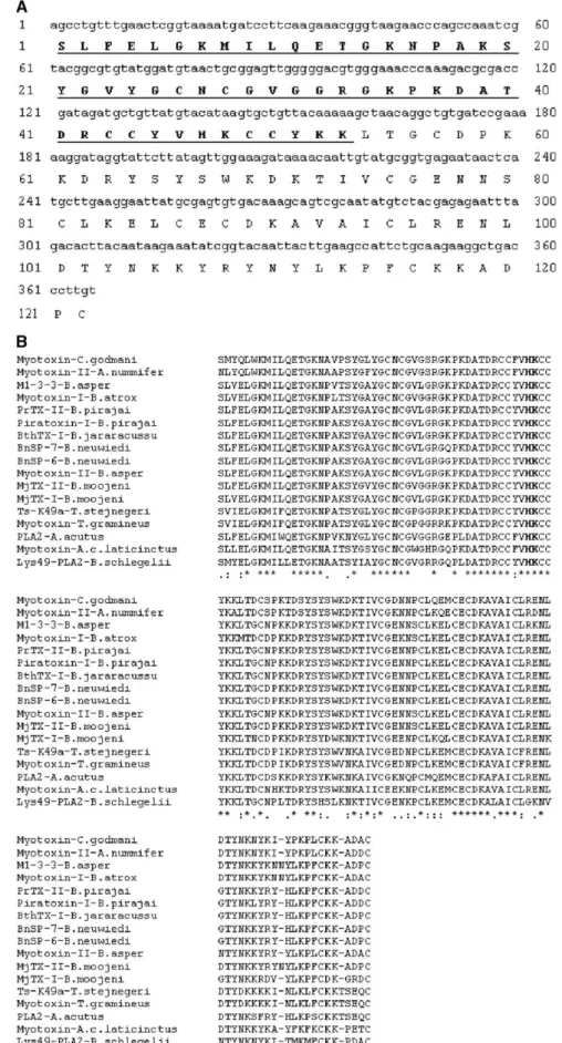
The sequence of MjTX-II obtained from a 366pb cDNA fragment is able to codify for a mature 122 amino acid residues protein, confirmed by direct sequencing of the first N-terminal 53 residues (Fig. 3A). The complete sequence was used for the refinement of the crystal structure at 1.8 Å resolution (Watanabe et al., 2005).
The primary structure of MjTX-II shows high homology with other Lys49 snake venom myotoxins (Fig. 3B), with an insertion of bases which codify for an Asn, at position 120, not usually found in all other PLA2s. The amino acids involved with
the catalytic site of Lys49 PLA2s are also conserved in MjTX-II,
Asn28, Gly30, Val31, Leu32, Gly33, His48, Lys49 and Asp99. Inspection of the primary structures of the myotoxic Lys49 isoforms MjTX-I and MjTX-II revealed 20 substituted residues, including: Asn60↔Gly60, Asp76↔Ser76, Glu77↔Asn77,
Lys110↔Leu110, Gly111↔Asp111, Arg117↔Tyr117,
Asp126↔Lys126 and Asp131↔Pro131. These 20 mentioned
0 50 100 150 200 250
0 20 40 60 80 100 A
Antibacterial
Antifungal
Inhibition of CFU (%)
0 10 20 30 40 50
0 20 40 60 80 100 C
75 25
5
Anti-Leishmania
Activity (%)
L. amazonensis L. braziliensis L. donovani L. major
0 20 40 60 80 100 B
100 50
10
Antitumoral Activity (%)
JURKAT
SK-BR-3
EAT
EAT+MTX
MjTX-II ( g)
MjTX-II ( g)
MjTX-II ( g)
Fig. 2. Functional biological applications induced byB. moojeniMjTX-II. (A) Microbicide activity induced by different concentrations of MjTX-II uponE. coli(
▪
)andC. albicans(□). (B) Antitumoral activity of MjTX-II upon different cell lines. Different concentrations (10, 50 and 100μg) of myotoxin were incubated with
human breast (SK-BR-3), acute T cell leukemia (JURKAT) and Erlich ascitic tumors (EAT) cells lines. Methotrexate (MTX, 100μg) was used positive control. (C)
Cellular viability ofL. amazonensis,L. braziliensis,L. donovaniandL. majorafter treatment with MjTX-II (5, 25 and 75μg). Cytotocixity was expressed as
changes might be involved with the differences reported for the neuromuscular and myotoxic effects on these toxins. MjTX-II induced post-synaptic blockage on mice synaptosome prepara-tions, while MjTX-I did not show any effect. Both myotoxins showed the“inverted pre-synaptic” site (Ward et al., 1998a), Asn86/Lys93; however, no pre-synaptic effect was observed on neuromuscular preparations.
MjTX-II showed toxicity 10% to 15% more effectively than MjTX-I and this difference might be explained by the higher basicity of the C-terminal region of MjTX-II. Inspection of this region, supposed to be responsible for part of the myotoxic effect ofB. asperLys49 Basp-II, shows that MjTX-I displays a net charge +3 in this region at pH7.4 compared to +6 in MjTX-II and +6 or +7 in other Lys49 myotoxins (Soares et al., 2004a).
Chemical treatment of proteins may result in modification of multiple residues due either to the occurrence of a given amino acid at various positions in the protein or to non specific chemical reactions. Experiments were therefore performed to evaluate the number and specificity of residues that were modified by each chemical treatment.
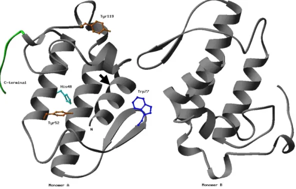
Amino acid analysis comparisons of native and modified MjTX-II showed that single His and two Tyr residues were modified by BPB and NBSF, respectively. The dimeric MjTX-II crystal structure is shown as a ribbon diagram with the side chains of residues, which underwent chemical modifications (Tyr52, Tyr119, His48 and Trp77) in a ball-stick representation for a monomer (Fig. 4).
Basic PAGE electrophoresis demonstrated that acetylation of Lys residues strongly reduced the toxins basicity since they did not migrate in the gel after treatment with acetic anhydride. Only 50% of the Trp residues were modified by NPSC, sinceA365nm of the NPS-toxin was about 50% when compared with NPS-Trp. Identification of the N-terminal residue of the fractions from the Sephadex G-25 column after treatment of MjTX-II with CNBr revealed Ser-1/Asp-1 for the shorter chains and Ile-1/Ile-1 for the longer chains (Ile-9 in the protein), respectively. Circular dichroism spectra of the native and modified MjTX-II demonstrate defined minima at 211 and 222 nm, indicating a strong a-helical contribution to the CD signal, which agrees with the high á-helical content. The majority of the chemical modifications did not result in significant alterations of the spectra, which suggests that the secondary structure of the proteins remains largely un-changed. However, cyanogen bromide treatment resulted in a wavelength shift of the first minimum to 207 nm with a decreased minimum value at 222 nm, which indicates that
secondary structural modification is associated with cleavage of the N-terminal octapeptide.
Chemical modification of Met residues by carboxymethyla-tion, as well as of His with BPB, Trp with NPSC, Tyr with NBSF and Lys by acetylation, affected differentially its pharmacological or toxic activities (Table 2). The neuromuscu-lar activity was almost fully abolished by these modifications. Liposome disrupting activity was not significantly affected by these modifications. Cleavage of the N-terminal octapeptide from MjTX-II largely reduced its lethal, myotoxic, neurotoxic and edema-inducing activities, but only partially the liposome disrupting and cytotoxic effects.
Carboxymethylation of MjTX-II Met-8 reduced its myotoxic and lethal effects. Met-8 is highly conserved in snake venom PLA2s. Carboxymethylation of Met-8 generates a variant with
2% of the enzymatic activity and less than 3% of the lethal activity of native Pa-11 from Pseudechis australis venom (Takasaki et al., 1990); it had about 5% of the original ability to block directly and indirectly stimulated mouse synaptosome preparations.
The highly conserved His48 in PLA2s participates in
catalysis (Scott et al., 1992). Alkylation of this residue induces loss of the hydrolytic activity on phospholipids and reduction of the toxic and pharmacological effects of PLA2s (Soares and Giglio, 2003).
The first report of a PLA2chemically modified with His48
was bovine pancreatic PLA2 (Volwerk et al., 1974). This
enzyme, when alkylated with p-bromophenachyl bromide (BPB), looses completely its enzymatic activity. Alkylation of MjTX-II His48 with BPB reduced its myotoxic, lethal and neurotoxic effects, and, in a smaller level, the edema-inducing and liposome disruption effects, thus suggesting a dissociation or partial overlapping of the pharmacological and toxic effects. The cytotoxic and bactericidal activities are not affected, thus corroborating the hypothesis that the functional site for these effects is the C-terminal domain and not the catalytic site. The exact mechanism by which BPB disturbs the toxic and pharmacological properties of the Lys49-PLA2s myotoxins
remains to be determined. An acidic Asp49-PLA2 from
Bothrops jararacussu was crystallized (Takeda et al., 2004) and the structure was solved (Magro et al., 2005) showing tertiary and quaternary structural changes. The Lys49-PLA2
BthTX-I chemically modified by BPB was recently crystallized by us (Marchi-Salvador et al., in preparation).
Treatment ofB. moojeniand other similar venoms with BPB is a strategy to attenuate their toxic effects, making possible their better utilization for the production of anti-Bothrops or anti-PLA2 serum, since higher doses of antigens could be
administered without causing extensive damage to the inocu-lated animals (Soares et al., 2004b).
The recurrence of nine invariant Tyr residues in almost all venom phospholipases A2suggests that they are responsible for
some important function in the molecule (Soares and Giglio, 2003; Soares et al., 2004a). p-Nitrobenzenesulfonyl fluoride (NBSF) was found to be a specific, mild reagent for the modification of tyrosine residues in proteins. Tyr residues are partially responsible for the myotoxic and neurotoxic effects of
Table 1
Results from the feces examinations between the 8th and 10th weeks after infection withS. mansoni, in the mice treated with MjTX-II and untreated mice Weeks Untreated Treatment (days/dose)
0 20
50μg 100μg 50μg 100μg
8th 245 94 48 16 0
Fig. 3. Primary sequence ofB. moojeniMjTX-II. (A) The cDNA and deduced amino acid sequences ofB. moojeniMjTX-II. The sequence of the toxin was confirmed by the direct sequencing of the first 52 amino acid residues underlined (Soares et al., 1998). (B) Alignment of amino acid sequence ofB. moojeniMjTX-II and other PLA2s-like toxins. Gaps were introduced to maximize the sequence homology. Alignments were made according to the half-cystine residues (bovine pancreatic PLA2
MjTX-II and, in a lesser extension, for other effects. The two Tyr residues (Tyr52 and Tyr119) with the highest exposed surface areas in the MjTX-II crystal structure are probably those which were chemically modified and therefore participant of the myotoxic and neurotoxic activities of this toxin.
Modification of Trp reflected only in the neuromuscular effect caused by MjTX-II, and therefore this residue is likely to have a relevant participation in this activity and/or this chemical modification can interfere in the stability of the interaction between the monomers of this dimeric toxin. Trp77 is known to be useful to maintain this homodimeric interaction. Acetylation of Lys residues demonstrated that basic character is important for the toxic effects (myotoxic, cytotoxic and lethal activities). Recently,Angulo et al. (2005) demonstrated that the cytolytic activity of various Lys49-PLA2homologues in vitro, as well as
theirs myotoxic activity in vivo are reduced, although not abolished, at pH 5.0, when compared with pH 7.2. This suggests that the switch from the dimeric to the monomeric forms of
these myotoxins may lead to a reduced ability to disrupt plasma membranes.
Cleavage of the N-terminal octapeptide of MjTX-II seems to be very relevant for all pharmacological effects, corroborating the hypothesis that interaction between the N-terminal region and the membrane is the first step of recognition, which precedes PLA2's action. Some snake venom PLA2s have been
treated with cyanogen bromide to cleave the NH2-terminal
region (Chwetzoff et al., 1989a,b), since Met-8 is highly conserved in many of these enzymes (Ward et al., 1998a,b; Ownby et al., 1999). This region is also antigenically important in some snake PLA2s, since removal of the N-terminal
octapeptide affects its reactivity against monoclonal and polyclonal antibodies (Díaz et al., 1994; Angulo et al., 2001).
Due to this functional diversity, these structurally similar proteins arose the interest of many researchers as molecular models for studies of structure–function relationships. One of the employed strategies for the relationship between catalytic activity and toxic/pharmacological effects of PLA2s elucidation
is based on the evaluation of these parameters variations, following chemical modifications of specific amino acid residues (His, Met, Lys, Tyr, Trp and others) of the enzyme. Using this approach, a dissociation of pharmacological effects and enzymatic activity has been observed. Overall, specific amino acid modification studies, although failing to pinpoint the region of the PLA2molecule responsible for pharmacological
activities, complies with the separate sites determining catalytic and pharmacological activities hypothesis (Soares and Giglio, 2003).
Bactericidal, neurotoxic and cytotoxic effects of MjTX-II are fully abolished with heparin incubation. It is known that heparin binds to the C-terminal (115–129) ofB. aspermyotoxin-II, and
Fig. 4. Crystal structure ofB. moojeniMjTX-II. The dimeric MjTX-II crystal structure (Watanabe et al., 2005) is shown as a ribbon diagram with the side chains of residues which underwent chemical modifications (Tyr52, Tyr119, His48 and Trp77) in a ball-stick representation (Carson, 1997). The C-terminal region is shown in green and the position of the N-terminal octapeptide cleavage with CNBr indicated by arrow.
Table 2
Biological effects ofB. moojeninative (N) or modified (M) MjTX-II Myotoxin LD50
(mg/kg)
Myotoxicity (%)
Edema (%)
Cytotoxicity (%)
that this region is related to its cytotoxic, myotoxic and bactericidal effects, with a probable contribution of Lys36 and Lys38 residues (Lomonte et al., 2003; Soares et al., 2004a). Incubation of MjTX-II with heparin completely inhibited the cytotoxic and bactericidal activities (Table 2), but only partially the myotoxic, edema-inducing and lethal effects. Incubation with EDTA did not significantly reduce any of the above effects (results not shown). Since homology between MjTX-II and Basp-II is near ∼98% in these regions, the C-terminal region obviously also plays a key role in the membrane damaging effect of MjTX-II, including neurotoxic, cytotoxic and bactericidal activities.
Snake venom PLA2s are multifunctional proteins with
promising biotechnological applications. Isolation and func-tional characterization of these enzymes will provide better insights of their mechanism of action as well as an interesting tool to study structure–activity relationships and open the possibility to use these proteins as molecular models for treatment of diseases in the future.
Acknowledgements
The authors express their gratitude to Fundação de Amparo à Pesquisa do Estado de São Paulo (FAPESP), Conselho Nacional de Desenvolvimento Científico e Tecnológico (CNPq) and Fundação para o Desenvolvimento da UNESP (FUNDUNESP) for financial support. Thanks are also due to Prof. Dr. R.K. Arni (UNESP-SJRP-Brazil) and R.J. Ward (FFCLRP-USP-Brazil) for their collaboration and discussion of the structural analysis of the toxin, to Dr. Yamileth Angulo, Prof. Dr. J.M. Gutiérrez and Prof. Dr. B. Lomonte (ICP-Costa Rica) for their collaboration and discussion of our results, to Dr. Caroline Borja Ribeiro and Prof. Dr. L. Rodrigues-Simioni for the neurotoxicity experiments (UNICAMP-Brazil), to Javier Nunez for the hystopathological analysis (ICP-Costa Rica), and Danilo Menaldo (TT-USP) and O.A.B. Cunha (FMRP-USP) for general technical assistance.
References
Andrião-Escarso, S.H., Soares, A.M., Rodrigues, V.M., Angulo, Y., Díaz-Oreiro, C., Lomonte, B., Gutiérrez, J.M., Giglio, J.R., 2000. Myotoxic phospholipases A2 in Bothrops snake venoms: effects of chemical
modifications on the enzymatic and pharmacological properties ofBothrops
toxins fromBothrops jararacussu. Biochimie 82, 755–763.
Andrião-Escarso, S.H., Soares, A.M., Fontes, M.R., Fuly, A.L., Correa, F.M., Rosa, J.C., Greene, L.J., Giglio, J.R., 2002. Structural and functional characterization of an acidic platelet aggregation inhibitor and hypotensive phospholipase A2 from Bothrops jararacussu snake venom. Biochem.
Pharmacol. 64, 723–732.
Angulo, Y., Núñez, C.E., Lizano, S., Soares, A.M., Lomonte, B., 2001. Immunochemical properties of the N-terminal helix of myotoxin II, a lysine-49 phospholipase A2 from Bothrops asper snake venom. Toxicon 39,
879–887.
Angulo, Y., Gutierrez, J.M., Soares, A.M., Cho, W., Lomonte, B., 2005. Myotoxic and cytolytic activities of dimeric Lys49 phospholipase A2
homologues are reduced, but not abolished, by a pH-induced dissociation. Toxicon 46, 291–296.
Arni, R.K., Ward, R.J., 1996. Phospholipase A2—a structural review. Toxicon
34, 827–841.
de Azevedo Jr., W.F., Ward, R.J., Lombardi, F.R., Giglio, J.R., Soares, A.M., Fontes, M.R.M., Arni, R.K., 1997. Crystal structure of myotoxin-II: a myotoxic phospholipase A2 homologue from Bothrops moojeni venom.
Prot. Peptide Letters 4, 329–334.
Balsinde, J., Balboa, M.A., Insel, P.A., Dennis, E.A., 1999. Regulation and inhibition of phospholipase A2. Annu. Rev. Pharmacol. Toxicol. 39, 175–189.
Bultrón, E., Thelestam, M., Gutiérrez, J.M., 1993. Effects on cultured mammalian cells of myotoxin III, a phospholipase A2 isolated from
Bothrops asper (Terciopelo) venom. Biochim. Biophys. Acta 1179,
253–259.
Carson, M., 1997. Ribbons. Methods Enzymol. 277, 493–505.
Chioato, L., Ward, R.J., 2003. Mapping structural determinants of biological activities in snake venom phospholipases A2by sequence analysis and site
directed mutagenesis. Toxicon 42, 869–883.
Chwetzoff, S., Tsunasawa, S., Sakiyama, F., Menez, A., 1989a. Nigexine, a phospholipase A2from cobra venom with cytotoxic properties not related to
esterase activity. Purification, amino acid sequence, and biological properties. J. Biol. Chem. 264, 13289–13297.
Chwetzoff, S., Couderc, J., Frachon, P., Menez, A., 1989b. Evidence that the anti-coagulant and lethal properties of a basic phospholipase A2from snake
venom are unrelated. FEBS Lett. 248, 1–4.
Corin, R.E., Viskatis, L.J., Vidal, J.C., Etcheverry, M.A., 1993. Cytotoxicity of crotoxin on murine erythroleukemia cells in vitro. Invest. New Drugs 11, 11–15.
Cura, J.E., Blanzaco, D.P., Brisson, C., Cura, M.A., Cabrol, R., Larrateguy, L., Mendez, C., Sechi, J.C., Silveira, J.S., Theiller, E., de Roodt, A.R., Vidal, J. C., 2002. Phase I and pharmacokinetics study of crotoxin (cytotoxic PLA2,
NSC-624244) in patients with advanced cancer. Clin. Cancer Res. 8, 1033–1041.
Cummings, B.S., McHowat, J.G., Schnellmann, R.G., 2000. Phospholipase A2s
in cell injury and death. J. Pharmacol. Exp. Ther. 294, 793–799. Díaz, C., Alape, A., Lomonte, B., Olamendi, T., Gutiérrez, J.M., 1994. Cleavage
of the NH2-terminal octapeptide of Bothrops asper myotoxic lysine-49
phospholipase A2, reduces its membrane-destabilizing effect. Arch.
Biochem. Biophys. 312, 336–339.
Gutiérrez, J.M., Lomonte, B., 1997. Phospholipase A2 myotoxins from
Bothrops snake venoms. In: Kini, R.M. (Ed.), Venom Phospholipase A2
Enzymes: Structure, Function and Mechanism. John Wiley and Sons, Chichester, pp. 321–352.
Gutiérrez, J.M., 2002. Understanding snake venoms: 50 years of research in Latin America. Rev. Biol. Trop. 50, 377–394.
Kapler, G.M., Coburn, C.M., Beverley, S.M., 1990. Stable transfection of the human parasiteLeishmania majordelineates a 30-kilobase region sufficient for extrachromosomal replication and expression. Mol. Cell. Biol. 10, 1084–1094.
Kini, R.M., 1997. Phospholipases A2: a complex multifuncional protein puzzle.
In: Kini, R.M. (Ed.), Venom Phospholipase A2 Enzymes: Structure,
Function and Mechanism. John Wiley and Sons, Chichester, pp. 1–28. Kini, R.M., 2003. Excitement ahead: structure, function and mechanism of
snake venom phospholipase A2enzymes. Toxicon 42, 827–840.
Lomonte, B., Gutierrez, J.M., Furtado, M.F., Otero, R., Rosso, J.P., Vargas, O., Carmona, E., Rovira, M.E., 1990. Isolation of basic myotoxins from
Bothrops moojeni and Bothrops atrox snake venoms. Toxicon 28,
1137–1146.
Lomonte, B., Angulo, Y., Calderón, L., 2003. An overview of lysine-49 phospholipase A2myotoxins from crotalid snake venoms and their structural
determinants of myotoxic action. Toxicon 42, 885–901.
Magro, A.J., Soares, A.M., Giglio, J.R., Fontes, M.R.M., 2003. Crystal structures of BnSP-7 and BnSP-6, two Lys49-phospholipases A2: quaternary
structure and inhibition mechanism insights. Biochem. Biophys. Res. Commun. 311, 713–720.
Magro, A.J., Takeda, A.A.S., Soares, A.M., Fontes, M.R.M., 2005. Crystal structure of BthA-I complexed with p-bromophenacyl bromide: possible implications with the lack of pharmacological activities. Acta Crystallogr. D61, 1670–1677.
Maraganore, J.M., Heinrikson, R.L., 1986. The lysine-49 phospholipase A2
Maraganore, J.M., Merutka, G., Cho, W., Welches, W., Kezdy, F.J., Heinrikson, R.L., 1984. A new class of phospholipases A2 with lysine in place of
aspartate 49. J. Biol. Chem. 259, 13839–13843.
Marchi-Salvador, D.P., Silveira, L.B., Soares, A.M., Fontes, M.R.M., 2005a. Crystallization and preliminary X-ray diffraction analysis of myotoxin I, a Lys49-phospholipase A2 fromBothrops moojeni. Acta Crystallogr. F61,
882–884.
Marchi-Salvador, D.P., Amui, S.F., Soares, A.M., Fontes, M.R.M., in preparation. Crystallization and preliminary X-ray diffraction analysis of a Lys49-PLA2complexed withp-bromophenacyl bromide. in preparation.
Mosmann, T., 1983. Rapid colorimetric assay for cellular growth and survival: application to proliferation and cytotoxicity assays. J. Immunol. Methods 65, 55–63.
Murakami, M.T., Arni, R.K., 2003. A structure based model for liposome disruption and the role of catalytic activity in myotoxic phospholipase A2s.
Toxicon 42, 903–913.
Murakami, M., Kudo, I., 2002. Phospholipase A2. J. Biochem. ( Tokyo ) 131,
285–292.
Murakami, M., Kudo, I., 2004. Secretory phospholipase A2. Biol. Pharm. Bull.
27, 1158–1164.
Ownby, C.L., 1998. Structure, function and biophysical aspects of the myotoxins from snake venoms. J. Toxicol., Toxin Rev. 17, 213–238. Ownby, C.L., Selistre-de-Araújo, H.S., White, S.P., Fletcher, J.E., 1999. Lysine
49 phospholipase A2proteins. Toxicon 37, 411–445.
Nevalainen, T.J., Quinn, R.J., Hooper, J.N., 2004a. Phospholipase A2 in porifera. Comp. Biochem. Physiol. B 137, 413–420.
Nevalainen, T.J., Peuravuori, H.J., Quinn, R.J., Llewellyn, L.E., Benzie, J.A., Fenner, P.J., Winkel, K.D., 2004b. Phospholipase A2 in cnidaria. Comp. Biochem. Physiol. B 139, 731–735.
Paramo, L., Lomonte, B., Pizarro-Cerdá, J., Bengoechea, J.A., Gorvel, J.P., Moreno, E., 1998. Bactericidal activity of Lys49 and Asp49 myotoxic phospholipases A2 from Bothrops asper snake venom: synthetic Lys49
myotoxin II (115–129)-peptide identifies its bactericidal region. Eur. J. Biochem. 253, 452–461.
Roberto, P.G., Kashima, S., Marcussi, S., Pereira, J.O., Astolfi-Filho, S., Nomizo, A., Giglio, J.R., Fontes, M.R., Soares, A.M., Franca, S.C., 2004. Cloning and identification of a complete cDNA coding for a bactericidal and antitumoral acidic phospholipase A2from Bothrops jararacussuvenom.
Protein J. 23, 273–285.
Rodrigues, V.M., Marcussi, S., Cambraia, R.S., de Araujo, A.L., Malta-Neto, N. R., Hamaguchi, A., Ferro, E.A., Homsi-Brandeburgo, M.I., Giglio, J.R., Soares, A.M., 2004. Bactericidal and neurotoxic activities of two myotoxic phospholipases A2 from Bothrops neuwiedi pauloensis snake venom.
Toxicon 44, 305–314.
Rudrammaji, L.M., Gowda, T.V., 1998. Purification and characterization of three acidic, cytotoxic phospholipases A2 from Indian cobra (Naja naja naja) venom. Toxicon 36, 921–932.
Scott, D.L., Achari, A., Vidal, J.C., Sigler, P.B., 1992. Crystallographic and biochemical studies of the (inactive) Lys-49 phospholipase A2 from the
venom of Agkistrodon piscivorus piscivorus. J. Biol. Chem. 267, 22645–22657.
Six, D.A., Dennis, E.A., 2000. The expanding superfamily of phospholipase A2
enzymes: classification and characterization. Biochim. Biophys. Acta 1488, 1–19.
Soares, A.M., Giglio, J.R., 2003. Chemical modifications of phospholipases A2
from snake venoms: effects on catalytic and pharmacological properties. Toxicon 15;42 (8), 855–868.
Soares, A.M., Rodrigues, V.M., Homsi-Brandeburgo, M.I., Toyama, M.H., Lombardi, F.R., Arni, R.K., Giglio, J.R., 1998. A rapid procedure for the isolation of the Lys-49 myotoxin II from Bothrops moojeni (caissaca) venom: biochemical characterization, crystallization, myotoxic and edema-togenic activity. Toxicon 36, 503–514.
Soares, A.M., Andrião-Escarso, S.H., Angulo, Y., Lomonte, B., Gutiérrez, J.M., Marangoni, S., Toyama, M.H., Arni, R.K., Giglio, J.R., 2000a. Structural and functional characterization of a myotoxin I from Bothrops moojeni
(caissaca) snake venom. Arch. Biochem. Biophys. 373, 7–15.
Soares, A.M., Guerra-Sá, R., Borja-Oliveira, C., Rodrigues, V., Rodrigues-Simioni, L., Rodrigues, V., Fontes, M.R.M., Giglio, J.R., 2000b. Molecular cloning and functional characterization of BnSP-7, a myotoxin Lys-49 phospholipase A2homologue, fromBothrops neuwiedi pauloensisvenom.
Arch. Biochem. Biophys. 378, 201–209.
Soares, A.M., Mancin, A.C., Cecchini, A.L., Arantes, E.C., Franca, S.C., Gutiérrez, J.M., Giglio, J.R., 2001a. Effects of chemical modifications of crotoxin B, the phospholipase A2subunit of crotoxin fromCrotalus durissus
terrificussnake venom, on its enzymatic and pharmacological activities. Int.
J. Biochem. Cell Biol. 33, 877–888.
Soares, A.M., Andriao-Escarso, S.H., Bortoleto, R.K., Rodrigues-Simioni, L., Arni, R.K., Ward, R.J., Gutiérrez, J.M., Giglio, J.R., 2001b. Dissociation of enzymatic and pharmacological properties of piratoxins-I and-III, two myotoxic phospholipases A2 fromBothrops pirajaisnake venom. Arch.
Biochem. Biophys. 387, 188–196.
Soares, A.M., Marcussi, S., Stabeli, R.G., Franca, S.C., Giglio, J.R., Ward, R.J., Arantes, E.C., 2003. Structural and functional analysis of BmjMIP, a phospholipase A2myotoxin inhibitor protein fromBothrops moojenisnake
plasma. Biochem. Biophys. Res. Commun. 302, 193–200.
Soares, A.M., Fontes, M.R.M., Giglio, J.R., 2004a. Phospholipases A2
myotoxins fromBothropssnake venoms: structure–function relationship. Curr. Org. Chem. 8, 1677–1690.
Soares, A.M., Sestito, W.P., Marcussi, S., Stáblei, R.G., Andrião-Escarso, S.H., Cunha, O.A.B., Vieira, C.A., Giglio, J.R., 2004b. Alkylation of myotoxic phospholipases A2inBothrops moojenivenom: a promising approach to an
enhanced antivenom production. Int. J. Biochem. Cell Biol. 36, 258–270. Takasaki, C., Sugama, A., Yanagita, A., Tamiya, N., Rowan, E.G., Harvey, A.L.,
1990. Effects of chemical modifications of PA-11, a phospholipase A2from
the venom of Australian king brown snake, on its biological activities. Toxicon 28, 107–117.
Takeda, A.A., dos Santos, J.I., Marcussi, S., Silveira, L.B., Soares, A.M., Fontes, M.R., 2004. Crystallization and preliminary X-ray diffraction analysis of an acidic phospholipase A2complexed withp-bromophenacyl
bromide and alpha-tocopherol inhibitors at 1.9- and 1.45A resolution. Biochim. Biophys. Acta 1699, 281–284.
Volwerk, J.J., Pieterson, W.A., de Haas, G.H., 1974. Histidine at the active site of phospholipase A2. Biochemistry 13, 1446–1454.
Ward, R.J., Rodrigues Alves, A., Ruggiero Neto, J., Arni, R.K., Casari, G., 1998a. A sequence space analysis of phospholipase A2. Protein Eng. 11,
285–294.
Ward, R.J., de Azevedo, W.F., Arni, R.K., 1998b. At the interface: crystal structures of phospholipases A2. Toxicon 36, 1623–1633.
Watanabe, L., Soares, A.M., Ward, R.J., Fontes, M.R., Arni, R.K., 2005. Structural insights for fatty acid binding in a Lys49-phospholipase A2: